- Advanced Laser Processing Research Team, RIKEN Center for Advanced Photonics, Wako, Japan
Laser-induced near-field effect, which confines the laser energy in a nano scale region to be enhanced, allows the laser fabrication with a resolution much smaller than the wavelength. Owing to such a high fabrication resolution, the laser-induced near-field fabrication has been attracting much attention as a tool for the surface nanostructuring. In this report, we introduce a novel method based on the laser-induced near-field reduction using a femtosecond laser by which gold nanocluster arrays are formed on substrates with the assistance of self-assembled silica microspheres. In the laser near-field reduction, the incident laser is focused in the vicinity of the backside of the silica microspheres to initiate synthesis of gold nanoparticles, followed by creation of gold nanoclusters by continuous growth of the gold nanoparticles along the silica microsphere surfaces. In addition, laser-treated MoS2 quantum dots are mixed in the gold precursor to increase the reduction efficiency for the formation of spherical gold nanoclusters. The gold nanocluster arrays provide potential applications for plasmonic devices.
1 Introduction
Near-field induced by microsphere-assisted laser processing is an attractive method to fabricate nanostructures with a high fabrication resolution far beyond the optical diffraction limit. The laser beam can be tightly focused by the microsphere when its diameter is close to the laser wavelength [1]. The near-field is usually confined in much smaller space than the laser wavelength where the electric field is locally enhanced. Based on the near-field effect, the materials can be accurately processed in the near-field with pulsed (nanosecond or shorter pulse) laser irradiation [2]. Till now, the laser near-field ablation utilizing the monolayer of silica microspheres is used for the fabrication of periodic nanostructures for surface nanopatterning. For example, Khan et al. used a contact particle lens array combined with laser scanning to control the wettability of silicon substrate [3]. The ablation depth depended on the laser processing parameters such as number of laser shots, laser pulse energy and laser wavelength [4]. However, there’s few works on the femtosecond laser near-field processing to build up three-dimensional (3D) nanostructures by an additive scheme. Recently, we reported that based on femtosecond laser near-field processing, the silver superstructure array could be fabricated by photonic reduction, by which the plasmon peak of array could be tuned by adjusting the period of superstructure. The fabricated silver superstructure array was used for the trace-detection of fluorescent toxic substance (perfluorooctanoic acid) by surface-enhanced Raman scattering (SERS) [5]. The gold superstructure array would generate different plasmon peak to provide better SERS performance for detection of such fluorescent substances. However, the reduction efficiency was too low to build up 3D gold nanostructure by femtosecond laser irradiation.
In this report, we present the laser near-field reduction to build up 3D gold nanoclusters with porosity, which are composed of gold nanoparticles. An array of the gold nanoclusters is fabricated in hydrogen tetrachloroaurate solution with the assistance of self-assembled silica nanospheres. To increase the reduction efficiency and thereby to create spherical gold nanoclusters, laser-treated MoS2 quantum dots are introduced in the gold precursor. We further found addition of citrate and polyvinylpyrrolidone (PVP) enables the anisotropic growth of gold nanoplates on the silica microshperes to create an array of nanoclusters composed of gold nanoplates. The nanocluster array provides potential applications for plasmonic devices.
2 Methods
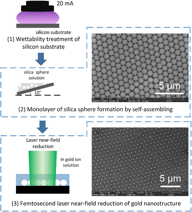
Trisodium citrate (Junsei Chemical Co. Ltd), PVP (K-90, NACALAI TESQUE, INC), hydrogen tetrachloroaurate tetrahydrate (NACALAI TESQUE, INC) and molybdenum disulfide (SIGMA-ALDRICH) were used for preparation of an aqueous gold ion precursor for laser near-field reduction. Typically, 43 mM C6H5Na3O7 and 20 mM HAuCl4 in 2 ml deionized water were mixed with 80 mg PVP in a glass vessel. First, surface of silicon substrate (n-type, 12.8–15.7 Ω•cm) was treated to improve wettability by plasma etching with a pressure of 20 Pa and a discharge current of 20 mA (PIB-20, Vacuum Device) [Figure 1(1)]. Monolayer of silica spheres of 1 μm in diameter (SIGMA-ALDRICH) was self-assembled on the treated silicon substrate [Figure 1(2)] which was then immersed in the gold ion precursor for laser near-field reduction. In order to form the monolayer of silica spheres, the substrate was tilted at 10° which illustrated in the previous works [6, 7]. A second harmonic (515 nm) converted from femtosecond (fs) laser with a pulse width of 223 fs and a wavelength of 1030 nm by a lithium triborate (LBO) crystal was loosely focused by a convex lens to achieve a spot diameter of 1 mm on the sample surface [Figure 1(3)]. The laser pulse energy and the repetition rate were set at 1 nJ and 1 MHz, respectively. After the laser near-field reduction, the sample was treated with hydrofluoric acid (HF) to completely etch away silica spheres, resulting in formation of gold nanodot or hollow nanocluster arrays. The fabricated gold nanostructure array was observed by an environmental scanning electron microscope (SEM).

FIGURE 1. Fabrication procedure of the femtosecond laser near-field reduction for formation of periodic gold nanostructures with SEM micrographs of samples at (2) self-assembling of monolayer of silica sphere array and (3) formation of 2D periodic gold nanostrictures.
3 Results and Discussion
According to Mie theory, the sphere will focus the laser when its diameter is slightly larger than wavelength, otherwise the laser will be scattered. Since the diameter (1 μm) of silica microspheres in experiments is larger than the laser wavelength (515 nm), the laser beam can be focused on the backside of silica microspheres to enhance the electric field in near-field. The photo-induced reduction is confined in the vicinity of backside of silica microspheres because the two-photon absorption takes place for the near-field reduction due to enhanced laser intensity:
Minor reduction by hydrated electrons is also induced by femtosecond laser [8]:
According to Eqs. 1, 2, the gold nanoparticles are synthesized at the backside of silica microspheres, forming a nanodot array as shown in a right side photo of Figure 1(3). The formed gold nanodots act as the nucleus for the continuous reduction, resulting in the growth of gold nanoparticles along the surface of silica microspheres to create gold nanoclusters [9]:
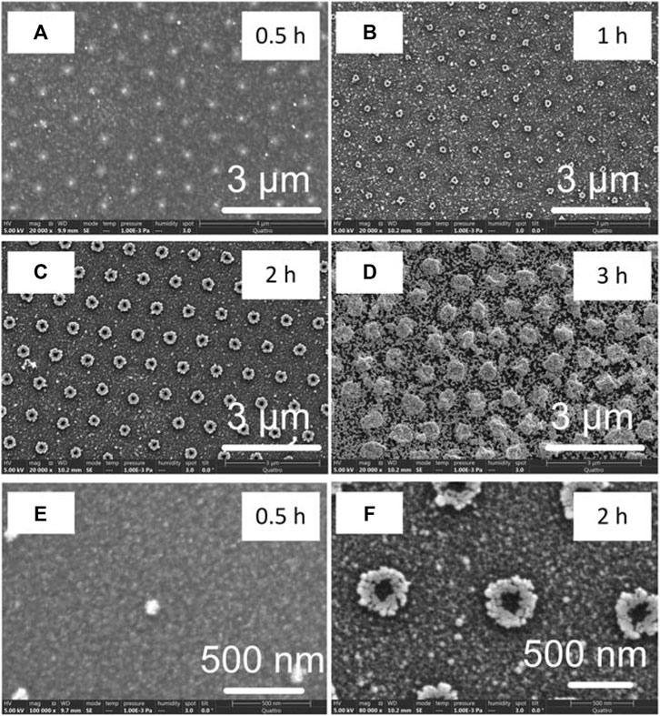
Geometry of created gold nanostructures can be controlled by the reduction time. Figure 2 shows gold nanostructure array with different geometries created at the reduction time from 0.5 h to 3 h. The smallest nanodot with a diameter of ∼104 nm is generated at 0.5 h (Figure 2A), which grows to nanobowl with a diameter of ∼800 nm as the reduction time increases to 3h (Figure 2D). Figure 1 E and F show the magnified SEM images of nanodot and nanobowl array. Because the gold nanoparticles are generated at the backside of the silica microsphere, the center of nanodot and nanobowl has a void where the silica microspheres contact to the silicon substrate. As the reduction time increases, the gold nanoparticles grow along the surface of silica microsphere to form the nanobowls. According to Eq. 3, the gold nanoparticles are reduced and aggregated in the near-field. Since the nanobowl is composed of gold nanoparticles, it has a porous structure. The growth mechanism of gold nanostructure induced by femtosecond laser was shown in Supplementary Figure S1. Due to near-field effect, the gold nanoparticles are synthesized at the backside of silica spheres. The synthesized gold nanoparticles enhance the electric field of incident laser beam due to localized surface plasmon resonance to further grow the gold nanoparticles along the surface of silica sphere. HF etching after entire growth of the gold nanoparticles along the surface of silica sphere eventually leaves hollow nanoclusters. However, the reduction efficiency is not sufficiently high for the creation of gold microsphere because PVP greatly suppresses the reduction of gold ions [10]. To increase the efficiency, MoS2 quantum dots are added to the gold precursor.

FIGURE 2. (A)—(D) SEM images of periodic gold periodic nanostructures formed at the reduction times of 0.5 h, 1 h, 2 h and 3 h, respectively (E) and (F) High magnification SEM images of (A) and (C), respectively.
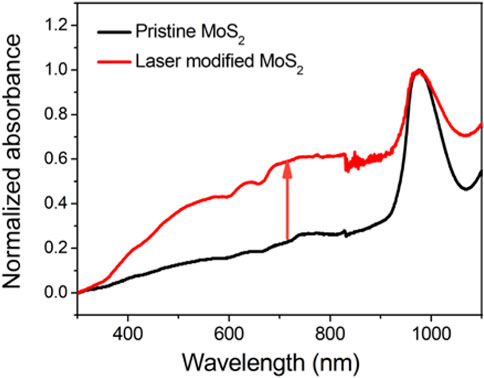
It is well known that laser-modified MoS2 drives the reduction of gold ions to gold nanoparticles, resulting from the unbound sulfur formed on MoS2 surface induced by femtosecond laser [11, 12]. The unsaturated active S atoms with dangling bonds have the chemical activity to directly reduce the Au3+ ions. 200 μL MoS2 quantum dots (4 wt%) were added into the aforementioned gold ion precursor to increase the efficiency of femtosecond laser-induced near-field reduction. To confirm the effectiveness of laser-modified MoS2, we compared the absorption of pristine MoS2 and the laser-modified MoS2 as shown in Figure 3. In laser modification, the MoS2 quantum dots solution was irradiated by 515 nm fs laser for 2 h. The laser fluence and repetition rate were set as same as the conditions for the fabrication of gold nanodot array. After modification, the absorption of MoS2 in the visible region is increased because of a strong surface plasmon resonance (SPR). This increment of SPR in the visible region is attributed to the vacancy-induced free electrons in MoS2, forming the redox pair with AuCl4- to allow the spontaneous reduction of gold ions to nanoparticles [13].
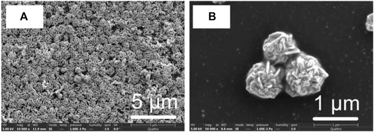
Owing to the chemical activity of laser-treated MoS2, the reduction efficiency is highly increased. As shown in Figure 4A, an array of periodic gold spheres with a porous structures is generated. For comparison, we also show the gold nanoparticles covering silica microsphere before hydrofluoric acid etching in Supplementary Figure S2. The diameter of gold spheres is about one um since the one um silica spheres are used as the template for the femtosecond laser-induced near-field reduction. Therefore, the period of the gold spheres is dependent on the diameter of silicon spheres. In addition, the gold spheres shown in Figure 4B are composed of nanoplates, demonstrating the growth of gold nanomaterials with a specific crystal orientation. In the precursor, citrate and PVP are typical capping agents to control the synthesis of gold nanoparticles. The citrate is an ionic capping agent, which allows the crystal growth in 111) plane. Additionally, PVP (∼3.6 wt%) selectively blocks the side faces of gold nanoplates in a high concentration to obtain the nanoplates composing nanoclusters [14, 15]. The nanocluster array consisting of an anisotropic structure has the potential applications for the plasmonic devices due to modulation of SPR [5, 16].

FIGURE 4. (A) SEM image of gold nanocluster array formed at a reduction time of 1 h with the assistance of the laser-treated MoS2 quantum dots (B) SEM image of gold nanocluster composed of gold nanoplates.
4 Conclusion
In this report, we presented a method of silica microsphere-assisted femtosecond laser near-field reduction for formation of gold nanocluster arrays. Owing to the microsphere, the laser was focused in the vicinity of backside of microspheres to reduce the gold ions. Since the reduction was confined in the near-field, the diameter of gold nanoparticle was about 100 nm (one-fifth of laser wavelength). The period of formed periodic structure was determined by the diameter of silica microsphere. To increase the reduction efficiency, laser-treated MoS2 quantum dots were used to enhance the SPR in the visible regions. In addition, we found the PVP and citrate contributed to the synthesis of nanoplate, enabling creation of a unique feature of nanocluster. The nanocluster array will be of use for the plasmonic device in future work.
Data Availability Statement
The original contributions presented in the study are included in the article/Supplementary Material, further inquiries can be directed to the corresponding authors.
Author Contributions
SB conducted the experiments. KO prepared the materials. SB, and KS wrote the manuscript.
Funding
This work was supported by a KAKENHI Grant-in-Aid (No. JP21K14444) from the Japan Society for the Promotion of Science (JSPS).
Conflict of Interest
The authors declare that the research was conducted in the absence of any commercial or financial relationships that could be construed as a potential conflict of interest.
Publisher’s Note
All claims expressed in this article are solely those of the authors and do not necessarily represent those of their affiliated organizations, or those of the publisher, the editors and the reviewers. Any product that may be evaluated in this article, or claim that may be made by its manufacturer, is not guaranteed or endorsed by the publisher.
Acknowledgments
The authors would like to thank Daishi Inoue and Mari Kikuya from the Materials Characterization Support Team at RIKEN for their assistance in performing SEM analyses.
Supplementary Material
The Supplementary Material for this article can be found online at: https://www.frontiersin.org/articles/10.3389/fphy.2022.917006/full#supplementary-material
References
1. Boneberg J, Leiderer P, Leiderer P. Optical Near-Field Imaging and Nanostructuring by Means of Laser Ablation. Opto-Electron Sci (2021) 1:210003. doi:10.29026/oes.2022.210003
2. Plech A, Leiderer P, Boneberg J. Femtosecond Laser Near Field Ablation. Laser Photon Rev (2009) 3:435–51. doi:10.1002/lpor.200810044
3. Khan A, Wang Z, Sheikh MA, Whitehead DJ, Li L. Laser Micro/Nano Patterning of Hydrophobic Surface by Contact Particle Lens Array. Appl Surf Sci (2011) 258:774–9. doi:10.1016/j.apsusc.2011.08.089
4. Wu Y, Ji L, Lin Z, Li Q, Jiang Y. Substrate Effect of Laser Surface Sub-micro Patterning by Means of Self-Assembly SiO2 Microsphere Array. Appl Surf Sci (2015) 357:832–7. doi:10.1016/j.apsusc.2015.09.066
5. Bai S, Hu A, Hu Y, Ma Y, Obata K, Sugioka K. Plasmonic Superstructure Arrays Fabricated by Laser Near-Field Reduction for Wide-Range SERS Analysis of Fluorescent Materials. Nanomaterials (2022) 12:970. doi:10.3390/nano12060970
6. Denkov N, Velev O, Kralchevski P, Ivanov I, Yoshimura H, Nagayama K. Mechanism of Formation of Two-Dimensional Crystals from Latex Particles on Substrates. Langmuir (1992) 8:3183–90. doi:10.1021/la00048a054
7. Micheletto R, Fukuda H, Ohtsu M. A Simple Method for the Production of a Two-Dimensional, Ordered Array of Small Latex Particles. Langmuir (1995) 11:3333–6. doi:10.1021/la00009a012
8. Rodrigues CJ, Bobb JA, John MG, Fisenko SP, El-Shall MS, Tibbetts KM. Nucleation and Growth of Gold Nanoparticles Initiated by Nanosecond and Femtosecond Laser Irradiation of Aqueous [AuCl4]−. Phys Chem Chem Phys (2018) 20:28465–75. doi:10.1039/c8cp05774e
9. Eustis S, Hsu H-Y, El-Sayed MA. Gold Nanoparticle Formation from Photochemical Reduction of Au3+ by Continuous Excitation in Colloidal Solutions. A Proposed Molecular Mechanism. J Phys Chem B (2005) 109:4811–5. doi:10.1021/jp0441588
10. Shen XS, Wang GZ, Hong X, Zhu W. Nanospheres of Silver Nanoparticles: Agglomeration, Surface Morphology Control and Application as SERS Substrates. Phys Chem Chem Phys (2009) 11:7450–4. doi:10.1039/b904712c
11. Zuo P, Jiang L, Li X, Li B, Xu Y, Shi X, Shape-Controllable Gold Nanoparticle-MoS2 Hybrids Prepared by Tuning Edge-Active Sites and Surface Structures of MoS2 via Temporally Shaped Femtosecond Pulses. ACS Appl Mater Inter (2017) 9:7447–55. doi:10.1021/acsami.6b14805
12. Rani R, Yoshimura A, Das S, Sahoo MR, Kundu A, Sahu KK, Sculpting Artificial Edges in Monolayer MoS2 for Controlled Formation of Surface-Enhanced Raman Hotspots. ACS Nano (2020) 14:6258–68. doi:10.1021/acsnano.0c02418
13. Li J, Xu X, Huang B, Lou Z, Li B. Light-Induced In Situ Formation of a Nonmetallic Plasmonic MoS2/MoO3-X Heterostructure with Efficient Charge Transfer for CO2 Reduction and SERS Detection. ACS Appl Mater Inter (2021) 13:10047–53. doi:10.1021/acsami.0c21401
14. Ujihara M. Solution-Phase Synthesis of Branched Metallic Nanoparticles for Plasmonic Applications. J Oleo Sci (2018) 67:689–96. doi:10.5650/jos.ess17262
15. Li N, Zhang Q, Quinlivan S, Goebl J, Gan Y, Yin Y. H2O2-Aided Seed-Mediated Synthesis of Silver Nanoplates with Improved Yield and Efficiency. ChemPhysChem (2012) 13:2526–30. doi:10.1002/cphc.201101018
Keywords: laser reduction, microsphere, nanodot, near-field, nanocluster
Citation: Bai S, Obata K and Sugioka K (2022) Femtosecond Laser Near-Field Reduction for Fabrication of 3D Gold Nanocluster Array Assisted by MoS2 Quantum Dots. Front. Phys. 10:917006. doi: 10.3389/fphy.2022.917006
Received: 10 April 2022; Accepted: 12 May 2022;
Published: 26 May 2022.
Edited by:
Xingzhan Wei, Chinese Academy of Sciences, ChinaReviewed by:
Jatish Kumar, Indian Institute of Science Education and Research, Tirupati, IndiaAlpan Bek, Middle East Technical University, Turkey
Liangping Xia, Yangtze Normal University, China
Copyright © 2022 Bai, Obata and Sugioka. This is an open-access article distributed under the terms of the Creative Commons Attribution License (CC BY). The use, distribution or reproduction in other forums is permitted, provided the original author(s) and the copyright owner(s) are credited and that the original publication in this journal is cited, in accordance with accepted academic practice. No use, distribution or reproduction is permitted which does not comply with these terms.
*Correspondence: Shi Bai, c2hpLmJhaUByaWtlbi5qcA==; Koji Sugioka, a3N1Z2lva2FAcmlrZW4uanA=
 Shi Bai
 Kotaro Obata