 Isabelle Russier-Antoine1
 Christian Jonin2Emmanuel Benichou1Franck Bertorelle3
 Pierre-François Brevet1*
- 1Institut Lumière Matière, Université de Lyon, UMR 5306 CNRS, Université Claude Bernard Lyon 1, Villeurbanne, France
- 2Laboratoire Charles Coulomb, Université de Montpellier, UMR CNRS 5221, Montpellier, France
- 3Laboratoire Unité en Sciences Biologiques et Biotechnologies, UMR CNRS 6286, Université de Nantes, Nantes, France
In this work, the first hyperpolarizability of Aux@Ag100-x core-shell nanoparticles with x the gold molar fraction in percent and the gold core of which is 12 nm in diameter, is determined and compared to that of their corresponding laser annealed nanoparticles using hyper Rayleigh scattering experiments. Laser annealing transforms the initially composite Aux@Ag100-x core-shell nanoparticles into alloyed AuxAg100-x homogeneous nanoparticles, providing a reference for comparison. It is observed that the evolution with the relative molar ratio between gold and silver of the first hyperpolarizability magnitude of both the Aux@Ag100-x core-shell and the alloyed AuxAg100-x nanoparticles is driven by the SPR resonance enhancement occurring at the harmonic wavelength due to red shifting of the SPR band away from the harmonic wavelength. Furthermore, the first hyperpolarizability magnitude of the Aux@Ag100-x core-shell nanoparticles is found to be about three orders of magnitude larger than that of the annealed alloyed AuxAg100-x nanoparticles. This feature may be attributed to the existence of the two nonlinearities, namely, the surface nonlinearity due to the surrounding medium–silver layer interface and the silver–gold metal - metal interface constructively contributing due to their close localization. The core-shell morphology is thus highly beneficial in view of applications as compared to the alloyed one.
Introduction
Nanoparticles have attracted a significant attention recently as they have found applications in a broad range of fields, from drug vector supports in biotechnology or substrate in sensing to highly efficient imaging probes (Myroshnychenko et al., 2008; Pérez-Juste et al., 2005). Nanoparticles have optical, structural and chemical properties that are altered as compared to their bulk material counterparts. This difference principally stems from the reduced size resulting in a large surface-to-volume ratio. Therefore, it is of fundamental importance to correctly unravel the nanoparticle properties and notably their interaction with light. Optical properties of metal nanoparticles particularly, especially made of silver or gold, have been of particular interest because of the intense features observed in the UV-visible region of the spectrum in extinction spectra due to the collective excitation of the conduction band electrons known as the surface plasmon resonance (SPR) (Amendola et al., 2017; Maier, 2007). This resonance strongly depends on the nanoparticles size, shape or morphology thereby attracting a great deal of attention. Besides, it induces large local electromagnetic field enhancements leading to the possibility to readily observe nonlinear optical processes besides the standard linear optical ones.
Second Harmonic Generation (SHG), the nonlinear optical phenomenon involving the conversion of two photons at a fundamental frequency ω into a single photon at the harmonic frequency 2ω is a well-established technique to convert frequency in laser physics and to investigate the structure and the dynamics of interfaces between two centrosymmetric materials (Shen, 2003; Shen, 1986). Indeed, within the electric dipole approximation, SHG is forbidden in bulk media having inversion symmetry while it does not vanish at interfaces where the inversion symmetry is broken. This property has been extensively used in the past to investigate at the molecular level a wide range of interfaces, for instance air–liquid, solid–liquid or liquid–liquid interfaces (Hicks et al., 1986; Brevet et al., 1996). All these studies have essentially been performed at planar interfaces where the experimental geometry is rather simple. A different approach has to be devised though to extend the technique to non-planar interfaces, and more particularly to spherical surfaces with a radius of curvature much smaller than the wavelength of light like that of nanoparticles. Such an approach is the scattering configuration of SHG in a liquid phase where instantaneous orientational fluctuations break the centrosymmetry of the medium. This configuration is known as Hyper Rayleigh Scattering (HRS) and as a result, a large number of reports have appeared investigating plasmonic nanoparticles (Vance et al., 1998; Johnson et al., 2002; Galletto et al., 1999; Hao et al., 2002). It is important here to point that in this configuration, the symmetry is not broken by the presence of a substrate and therefore the true response of the nanoparticles is accessed.
Gold and silver nanoparticles, but also dielectric nanoparticles, have thus received a lot of attention with the determination of their first hyperpolarizability, namely, their cross-section for the SHG phenomenon, rather large absolute values being obtained and supporting their use in the above-mentioned applications. Also, in addressing the exact origin of their response, the competition between their shape and their size have been unraveled, synthesis providing nowadays a large range of shapes indeed, from sphere and rods to cubes and prisms and multifaceted nanoparticles like decahedra (Nappa et al., 2005; Russier-Antoine et al., 2007; Duboisset and Brevet, 2019). Centrosymmetric material nanoparticles like gold and silver exhibit a surface origin for their nonlinearity whereas non centrosymmetric material ones exhibit a volume response, provided their size is not too small to be able to an neglect surface contributions. More recently, in the search for ever more efficient morphologies, mixed metal like gold-silver alloys or two-material systems like metal-dielectric core-shell systems have been investigated (Russier-Antoine et al., 2008; Kim et al., 2002).
In this respect, the metal-metal core-shell nanoparticles present an interesting perspective as they present two interfaces, the outer one between the nanoparticle itself and the surrounding medium, possibly mediated by a surfactant layer introduced for suspension stability purposes and the inner one between the core and the shell. In the present work, we therefore report a study where gold core–silver shell nanoparticles are investigated using HRS. Their first hyperpolarizability is compared to both pure gold and pure silver nanoparticles. The size of those nanoparticles remains smaller than 25 nm in diameter, a size sufficiently small to avoid the appearance of retardation effects, namely, features due to the spatial phase of the fields over the size of the nanoparticles. Finally, laser annealing is also performed to compare the resulting alloyed nanoparticles first hyperpolarizability to that of the core-shell morphology.
Methods
Particles synthesis
Tetrachloroauric acid (HAuCl4. 3H20), cetyltrimetylammonium bromide (CTAB), silver nitrate (AgNO3) and ascorbic acid were purchased and used as received. Milli-Q water with a 18.2 MΩ resistivity was used in all the preparations. Gold nanoparticles were prepared using a seed mediated method. This allows obtaining particles of the desired size with good mono-dispersity. Briefly, the seed solution was prepared by adding a NaBH4 ice-cold solution into a mixture of HAuCl4 and CTAB followed by rapid inversion mixing for 2 min. In order to synthesize 12 nm diameter nanoparticles, the growth solution was done by adding 2 mL of HAuCl4 with a concentration of 2.5 × 10⁻² M into 200 mL of a 50 mM CTAB solution followed by 1 mL of ascorbic acid at a concentration of 0.3 M under agitation. Then, 2 mL of the seed solution was added under constant stirring and the resulting solution was stirred for another 3 h at 60°C. A concentrated solution was thus obtained by centrifugation to removed excess CTAB and residual smaller nanoparticles. The gold concentration was determined by spectrophotometric method using Rhodamine B/gold complex as described in reference (Macnulty and Woollard, 1955). To obtain the gold core-silver shell nanoparticles, hereafter indicate as Au@Ag, the same protocol was used for all syntheses. Briefly, a desired amount of 12 nm gold nanoparticles was added to a 0.5 mM CTAB solution, followed by the addition of AgNO3. Next, a 2.5 × 10⁻² M ascorbic acid solution, namely, 4 times the amount of AgNO3, was added dropwise. The solution was then stirred at 25°C for 4 h with the addition of 10 µL of 5M NaOH solution at half-time. In all syntheses, the final volume of solutions was 20 mL and the total metal concentration, i.e., gold plus silver metal ions, equal to 2.5 × 10⁻⁴ M.
Particle characterization
TEM images of the gold core solution were recorded as well as the core-shell solutions. From the TEM images, the particles diameter of the core solution was determined to be indeed 12 nm in diameter as expected with a width of the size distribution of less than 15%. The diameter of the core-shell solutions was calculated assuming that all the silver reacted with the gold core. Figure 1 displays a TEM image of the 12 nm diameter gold core nanoparticles and a close view of a single Au50@Ag50 core-shell nanoparticle, where 50 is the gold molar ratio in percent.
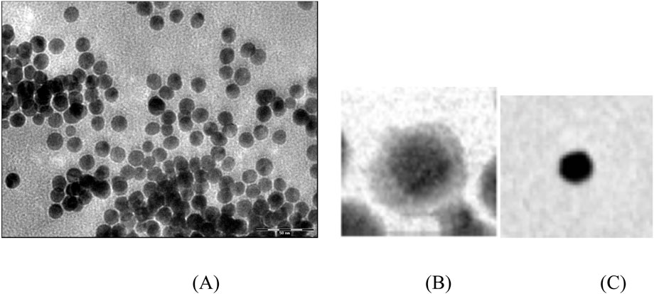
Figure 1. Transmission Electron Microscopy (TEM) image of (A) the 12 nm diameter gold core nanoparticles, (B) a close view of a single Au50@Ag50 nanoparticle with a 12 nm gold core and (C) a single annealed Au50Ag50 nanoparticle.
UV-Visible Absorption spectroscopy was performed as well using a compact spectrophotometer (Ocean Optics, model USB 2000).
Laser annealing experiments
A pulsed Nd:YAG laser was used to irradiate the colloidal solution at 532 nm. The maximum power used was 10 mJ/pulse, with a pulse duration of 10 ns at repetition rate of 10 Hz. During irradiation, the core-shell solution was stirred using a magnetic stirrer. UV-Visible absorption spectroscopy was used during irradiation to determine the ending time through the absence of changes in the UV-visible spectra. Figure 1C displays a close-up view of an alloyed Au50Ag50 nanoparticle.
HRS measurements
The light source for the present HRS experiments was a mode-locked femtosecond Ti:sapphire laser delivering at the fundamental wavelength of 784 nm pulses with a duration of about 180 femtoseconds at a repetition rate of 76 MHz. After passing through a low-pass filter to remove any unwanted harmonic light generated prior to the cell, the fundamental beam of about 900 mW was focused by a low numerical aperture microscope objective into a 1 cm × 1 cm spectrophotometric cell containing the aqueous solution of the metallic particles. The HRS light was collected at an angle of 90° from the incident direction by a 2.5 cm focal length lens. The second harmonic light was separated from its linear counterpart by a high-pass filter and a monochromator positioned on the second harmonic wavelength. The HRS light was then detected with a cooled photomultiplier tube and the pulses produced counted with a photon counter. The fundamental beam was chopped at about 130 Hz to enable a gated photon counting mode allowing automatic subtraction of the noise level. The fundamental input beam was linearly polarized vertically in the laboratory frame whereas no polarization was selected for the harmonic light.
Results and discussion
Linear optical properties
The optical properties of metal nanoparticles are dominated by the SPR. For some metals, such as Cu, Ag, Au or the alkali metals, the SPR frequency lies within the visible range and is responsible for the intense colors observed with colloidal dispersions (Creighton et al., 1979). When two different metals are mixed within the same particle, the resulting optical properties arise from a combined contribution of both metals and the spatial distribution of the metal atoms within the particle is of fundamental importance. In the case of bimetallic alloyed nanoparticles, the wavelength of the SPR was found to change in a linear way between that for the pure silver and that of the pure gold particles as a function of the molar fractions of the metals for instance (Link et al., 1999; Rodriguez-Gonzalez et al., 2004). Bimetallic nanoparticles with a core-shell morphology have been studied in the past for their linear optical properties as well and interpreted within the context of Mie theory (Cao et al., 2001; Abid et al., 2004; Mulvaney et al., 1993). With the core-shell bimetallic Au@Ag nanoparticles, two SPR bands can be observed. Figure 2 shows the normalized UV-Visible absorption spectra for the core-shell gold-silver particles in aqueous suspensions and stabilized with CTAB as studied in the present work for the gold molar fraction x ranging from 0 up to 100 by step of 10 in percent. Due to the respective environment and morphology, the two SPR band positions are shifted as compared to both the pure silver and pure gold spectral position. As expected, in the gold core–silver shell nanoparticle, the original pure gold nanoparticle SPR is blue shifted as the silver shell thickness increases, i.e., as the silver molar fraction increases. Likewise, the second SPR appears red shifted as compared to the SPR maximum observed for the pure silver nanoparticle and this red shift increases as the silver shell thickness decreases, i.e., as the silver molar fraction decreases. Due to the relatively large width of the two SPR resonances, for either a low gold molar fraction or a low silver molar fraction, a single SPR is observed resulting from the progressive merging of the two SPR bands. The two SPR bands are thus clearly observed for the Au50@Ag50 morphology but gradually merge into a single one when the gold molar fraction or the silver molar fraction dominates.
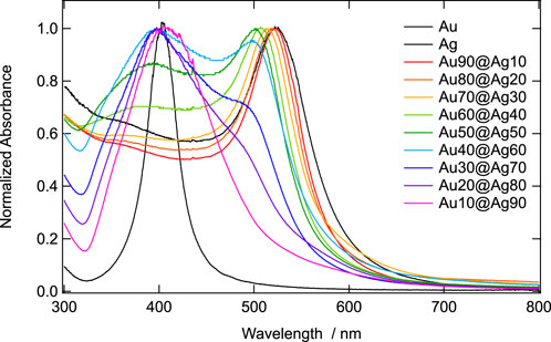
Figure 2. Normalized UV-visible absorbance spectra for the Aux@Ag100-x core-shell nanoparticles with x varying from 0 to 100 with a 12 nm diameter gold core.
In order to perform the necessary corrections to the first hyperpolarizability recorded during the HRS experiments and due to the SPR shifts, it is mandatory to determine the molar extinction coefficient at both the fundamental and the harmonic wavelengths. The HRS experiments were conducted at the fundamental wavelength of 784 nm and the harmonic wavelength of 392 nm so the molar extinction coefficients were determined at these wavelengths. However, as seen from Figure 2 where absorbance for all samples was negligible at 784 nm, this molar extinction coefficient was disregarded. On the opposite, the molar extinction coefficient at 392 nm was determined performing UV-visible absorption spectroscopy as a function of the nanoparticle concentration. Supplementary Figure S1 was obtained and the molar extinction coefficient ε392 determined from the slopes, using a light path length of 1 cm, see Figure 3. Interestingly, this molar extinction coefficient ε392 increases as the gold molar content decreases but remains largely superior to the molar extinction coefficient of the pure silver nanoparticles. This latter feature results from the remaining existence of the 12 nm diameter gold core that possesses interband transitions in the spectral domain around 400 nm, see Figure 2.

Figure 3. Molar extinction coefficient at 392 nm as a function of the gold molar fraction x of the Aux@Ag100-x core-shell nanoparticles with a 12 nm diameter gold core.
A log-log plot of this exponential-like decrease of the molar extinction coefficient ε392 as a function of the gold molar content yields a value of −1.44±0.04, see Supplementary Figure S2. Note that the gold core being 12 nm in diameter, the shell thickness ranges from zero for no silver up to 13 nm for the Au10@Ag90 composite core-shell nanoparticles. The value reported here for the pure silver nanoparticles corresponds to a size of 13 nm in diameter.
Nonlinear optical properties
In these experiments where the HRS intensity is collected, the monochromaticity of the second harmonic light generated was always assessed to prevent any spurious contributions from luminescence, see Supplementary Figure S3. This experimental procedure is necessary to ensure that the observed process is indeed the conversion of two photons at the fundamental frequency into one photon at the harmonic frequency and to determine the level of photo-excited luminescence. Luminescence from gold nanoparticles has already been reported in the past and is usually attributed to surface trap states owing to the coating of the particles. These results clearly call for a careful analysis of the data in order to truly determine the first hyperpolarizabilities of the Aux@Ag100-x core-shell nanoparticles. Hereafter, the HRS intensity is always taken as the height of the Gaussian line extracted through an adjustment procedure, see Supplementary Material and Supplementary Figure S4. Note that the area of the HRS band remains constant and therefore would lead to similar results.
The HRS experiments performed as a function of the nanoparticles concentration allowed for the determination of the absolute value of the first hyperpolarizability of the Aux@Ag100-x core-shell nanoparticles. Indeed, the HRS intensity is given by (Clays and Persoons, 1992):
where I is the fundamental intensity,

Figure 4. Normalized HRS intensity at 392 nm as a function of the Aux@Ag100-x core-shell nanoparticles concentration with a 12 nm diameter gold core.
The
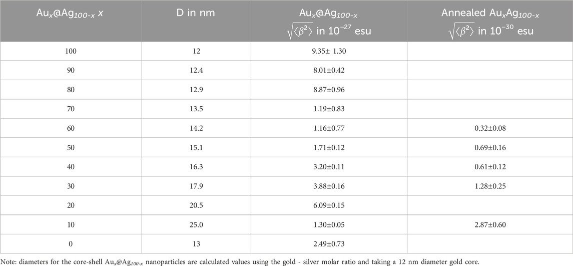
Table 1. Parameters extracted from the UV-visible absorbance spectroscopy and HRS measurements for the Aux@Ag100-x core-shell and annealed alloyed AuxAg100-x nanoparticles.
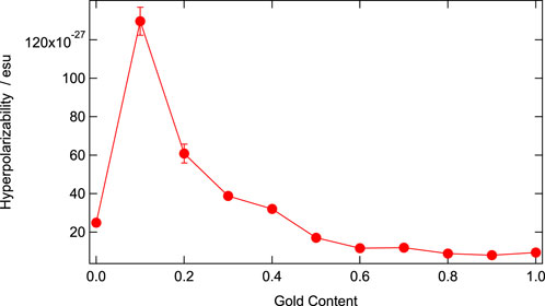
Figure 5. Absolute value of the first hyperpolarizability at the fundamental wavelength of 784 nm and the harmonic wavelength of 392 nm for the Aux@Ag100-x core-shell nanoparticles with x going from 0 to 100 and a 12 nm diameter gold core as a function of gold content.
In order to rationalize this dependence, one may take a closely look at the dependences of the first hyperpolarizability as a function of the nanoparticle diameter and resonances (Russier-Antoine et al., 2007; Nappa et al., 2006). As far as retardation effects of the electromagnetic fields can be neglected, the first hyperpolarizability
Laser annealing
The importance of plasmonic nanoparticles in sensor development, imaging, photo-diagnostics and optoelectronics has resulted in a strong interest toward the development of straightforward synthetic methods for their preparation. Hence, it has been found that the physical laser ablation method can be used to prepare metal nanoparticles in solution from a bulk metal piece (Major et al., 2009). In this line furthermore, nanosecond laser-induced heating of composite nanoparticles can transform Au@Ag core-shell nanoparticles into homogeneous alloyed AuAg nanoparticles. The transformation comes from the selective laser heating caused by resonant electronic excitation. In the present case, by looking at Figure 2, it appears that a 532 nm wavelength light can indeed achieve this operation. The spectra presented in Supplementary Figure S5 show the optical changes for the Au40@Ag60 core-shell nanoparticles induced by an unfocused 10 ns laser irradiation at 532 nm and an average power of 90 mW to avoid a too strong energy deposition in the sample. The irradiation was stopped after 23 min when no further changes were appearing in the spectra. As can be seen, the absorption spectra are considerably modified with the characteristics of the core-shell nanoparticles disappearing and a single SPR band appearing. The position of this new band depends on the composition of the nanoparticles, indicating the formation of single-phase alloyed nanoparticles. This single SPR band position located at 450 nm is in very good agreement with the value of 445 nm found for synthesized alloyed Au40Ag60 nanoparticles. Five Aux@Ag100-x solutions were thus irradiated (x = 10, 30, 40, 50 and 60) to obtain the annealed solutions noted AuxAg100-x. The other solutions were found too instable to proceed further with the annealing step. This feature may stem from the silver content increase as the size of the nanoparticle increases as well since the synthesis is based on the growth of a shell on a fixed size gold nanoparticle. Furthermore, it is known that light may affect the nanoparticles in their morphology (Stamplecoskie and Scaiano, 2010) as well and finally citrate may also loose its stabilizing role in this situation although it can preserve the nanoparticles from silver oxidation. Determination of the absolute value of the first hyperpolarizability magnitude for these five AuxAg100-x nanoparticles thus followed similarly to the above measurements performed for the core-shell nanoparticles.
Figure 6 presents the final normalized UV-visible absorbance spectra for the four AuxAg100-x samples irradiated whereas Supplementary Figures S5, S7 present the absorbance as a function of the concentration and the molar extinction coefficient as a function of the gold content.
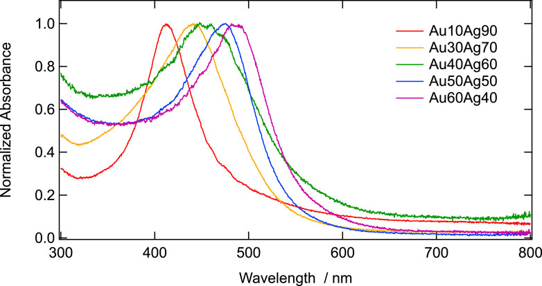
Figure 6. Normalized UV-visible absorbance spectra for the annealed alloyed AuxAg100-x nanoparticles with x varying from 0 to 100.
As expected, and because the single SPR band gets closer to the harmonic wavelength at 392 nm, absorbance is larger for the nanoparticles with the lowest amount of gold at fixed concentration, see Supplementary Figure S6.
Hence, the molar extinction coefficient is larger for nanoparticles with the lowest amount of gold, see Figure 7. In this respect, the molar extinction coefficient for the annealed AuxAg100-x nanoparticles follows a trend similar to that of the core-shell corresponding nanoparticles with a slope of −1.49±0.17, similar to the core-shell nanoparticles, see Supplementary Figure S7.
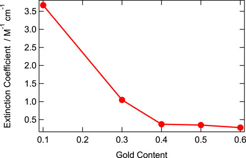
Figure 7. Molar extinction coefficient at 392 nm as a function of the gold molar fraction x of the annealed alloyed AuxAg100-x nanoparticles with a 12 nm diameter gold core.
Finally, HRS measurements were then performed as a function of the nanoparticle concentration, yielding the standard linear plots obeying Equation 1 in Figure 8. From the slope normalized at vanishing concentration to the neat aqueous solvent, the first hyperpolarizability magnitude of the annealed nanoparticles were then determined, see Figure 9.

Figure 8. Normalized HRS intensity at 392 nm as a function of the annealed alloyed AuxAg100-x nanoparticles concentration.
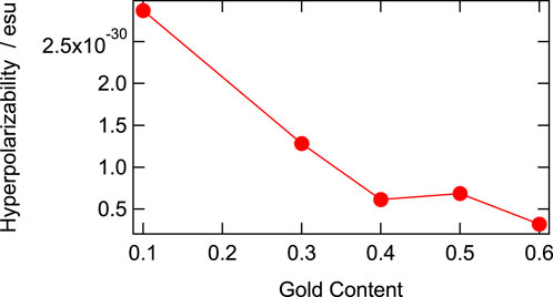
Figure 9. Absolute value of the first hyperpolarizability at the fundamental wavelength of 784 nm and the harmonic wavelength of 392 nm for the annealed alloyed AuxAg100-x nanoparticles with x going from 0 to 100 as a function of gold content.
As expected, the first hyperpolarizabilities of the annealed nanoparticles follow the same trend as the molar extinction coefficient reported on Figure 7. The slope of the log-log plot of the first hyperpolarizability with respect to the gold content is found to be −1.11±0.21, see Supplementary Figure S8. This slope is however somewhat weaker than the corresponding one reported for the molar extinction coefficient. This result nevertheless suggest that the general trend observed for the first hyperpolarizability of the annealed nanoparticles is driven by the SPR resonance enhancement and its shifts due to the gold molar content.
Comparison of the first hyperpolarizability measured for the core-shell nanoparticles appears therefore to be three orders of magnitude larger with values ranging from 1×10⁻²⁷ to 10 × 10−27 esu as compared to that of the corresponding annealed nanoparticles, the first hyperpolarizability of which ranges from 0.5 × 10⁻³⁰ up to 3 × 10⁻³⁰ esu. The core-shell morphology appears to be largely superior in efficiency towards the SHG process, as observed in scattering measurement. This feature may stem from the existence of the two interfaces, namely, the surrounding medium–silver metal nanoparticles interface and the silver–gold metal interface. These results indicate that besides the efficient SPR enhancement that drives the dependence with the gold–silver molar ratio, it is likely that the two interface nonlinearities, localized in a relatively close position with respect to each other since the silver layer never exceeds 13 nm, provide a constructive in-phase SHG response.
Conclusion
In this present work, the first hyperpolarizability of Aux@Ag100-x core-shell nanoparticles, the gold core of which is 12 nm in diameter, is determined and compared to that of the annealed nanoparticles. Laser annealing transforms the initially composite Au@Ag core-shell nanoparticles into alloyed AuAg homogeneous nanoparticles. It appears that in both cases, the first hyperpolarizability magnitude is driven by the SPR resonance enhancement occurring at the harmonic wavelength. As the gold molar content increases, the SPR band redshifts away from the harmonic wavelength at 392 nm and consequently the first hyperpolarizability decreases. However, in a striking manner, the first hyperpolarizability magnitude of the Au@Ag core-shell nanoparticles remains three orders of magnitude larger than that of the annealed alloyed AuAg nanoparticles. This feature is attributed to the existence of two nonlinearities constructively contributing due to their close localization, the surface nonlinearity due to the surrounding medium–silver layer interface and the silver metal–gold metal interface. These results call for the use of composite core-shell nanoparticles as imaging probes or for sensing purposes for instance.
Data availability statement
The original contributions presented in the study are included in the article/Supplementary Material, further inquiries can be directed to the corresponding author.
Author contributions
IR-A: Data curation, Investigation, Methodology, Writing–review and editing, Formal Analysis. CJ: Data curation, Formal Analysis, Investigation, Methodology, Writing–review and editing, Validation. EB: Formal Analysis, Investigation, Validation, Writing–review and editing. FB: Investigation, Writing–review and editing, Methodology, Resources. P-FB: Investigation, Methodology, Resources, Writing–review and editing, Conceptualization, Data curation, Project administration, Supervision, Validation, Writing–original draft.
Funding
The author(s) declare that financial support was received for the research, authorship, and/or publication of this article. The authors acknowledge financial support from the ANR project MetaTrap (ANR-22-CE09-0011).
Conflict of interest
The authors declare that the research was conducted in the absence of any commercial or financial relationships that could be construed as a potential conflict of interest.
Generative AI statement
The author(s) declare that no Generative AI was used in the creation of this manuscript.
Publisher’s note
All claims expressed in this article are solely those of the authors and do not necessarily represent those of their affiliated organizations, or those of the publisher, the editors and the reviewers. Any product that may be evaluated in this article, or claim that may be made by its manufacturer, is not guaranteed or endorsed by the publisher.
Supplementary material
The Supplementary Material for this article can be found online at: https://www.frontiersin.org/articles/10.3389/fphot.2025.1548555/full#supplementary-material
References
Abid, J.-P., Nappa, J., Girault, H. H., and Brevet, P. F. (2004). Pure surface plasmon resonance enhancement of the first hyperpolarizability of gold core–silver shell nanoparticles. J. Chem. Phys. 121, 12577–12582. doi:10.1063/1.1826053
Amendola, V., Pilot, R., Frasconi, M., Maragò, O. M., and Iati, M. A. (2017). Surface plasmon resonance in gold nanoparticles: a review. J. Phys. Condens. Matter 29, 203002. doi:10.1088/1361-648x/aa60f3
Brevet, P. F., and Girault, H. H. (1996). in Second harmonic generation at liquid/liquid interfaces in liquid-liquid interfaces, Theory and methods. Editors A. G. Volkov, and D. W. Deamer (Boca Raton: CRC Press).
Cao, Y. W., Jin, R., and Mirkin, C. A. (2001). DNA-modified Core−Shell Ag/Au nanoparticles. J. Am. Chem. Soc. 123, 7961–7962. doi:10.1021/ja011342n
Clays, K., and Persoons, A. (1992). Hyper-Rayleigh scattering in solution. Rev. Sci. Instrum. 63, 3285–3289. doi:10.1063/1.1142538
Creighton, J. A., Blatchford, C. G., and Albrecht, M. G. (1979). Plasma resonance enhancement of Raman scattering by pyridine adsorbed on silver or gold sol particles of size comparable to the excitation wavelength. J. Chem. Soc. Faraday Trans. 2 (75), 790. doi:10.1039/f29797500790
Duboisset, J., and Brevet, P. F. (2019). Second harmonic scattering defined topological classes for nano-objects. J. Phys. Chem. C 123, 25303–25308. doi:10.1021/acs.jpcc.9b04810
Duboisset, J., Matar, G., Russier-Antoine, I., Benichou, E., Bachelier, G., Jonin, Ch., et al. (2010). First hyperpolarizability of the natural aromatic amino acids tryptophan, tyrosine and phenylalanine and the tripeptide lysine-tryptophan-lysine determined by hyper Rayleigh scattering. J. Phys. Chem. B 114, 13861–13865. doi:10.1021/jp105554s
Galletto, P., Brevet, P. F., Girault, H. H., Antoine, R., and Broyer, M. (1999). Size dependence of the surface plasmon enhanced second harmonic response of gold colloids: towards a new calibration method. Chem. Commun., 581–582. doi:10.1039/a900230h
Hao, E. C., Schatz, G. C., Johnson, R. C., and Hupp, J. T. (2002). Hyper-Rayleigh scattering from silver nanoparticles. J. Chem. Phys. 117, 5963–5966. doi:10.1063/1.1510439
Hicks, J. M., Kemnitz, K., Eisenthal, K. B., and Heinz, T. F. (1986). Studies of liquid surfaces by second harmonic generation. J. Chem. Phys. 90, 560–562. doi:10.1021/j100276a015
Johnson, R. C., Li, J., Hupp, J. T., and Schatz, G. C. (2002). Hyper-Rayleigh scattering studies of silver, copper, and platinum nanoparticle suspensions. Chem. Phys. Lett. 356, 534–540. doi:10.1016/s0009-2614(02)00407-4
Kim, Y., C Johnson, R., Li, J., Hupp, J. T., and Schatz, G. C. (2002). Synthesis, linear extinction, and preliminary resonant hyper-Rayleigh scattering studies of gold-core/silver-shell nanoparticles: comparisons of theory and experiment. Chem. Phys. Lett. 352, 421–428. doi:10.1016/s0009-2614(01)01506-8
Link, S., Wang, Z. L., and El-Sayed, M. A. (1999). Alloy Formation of Gold−Silver nanoparticles and the dependence of the plasmon absorption on their composition. J. Phys. Chem. B 103, 3529–3533. doi:10.1021/jp990387w
Macnulty, B. J., and Woollard, L. D. (1955). The use of Rhodamine B in analytical chemistry: the determination of small quantities of gold. Anal. Chim. Acta 13, 154. doi:10.1016/S0003-2670(00)87917-9
Major, K. J., De, C., and Obare, S. O. (2009). The study of surface plasmon in Au/Ag core/shell compound nanoparticles. Plasmonics 4, 61. doi:10.1007/s11468-012-9336-6
Mulvaney, P., Giersig, M., and Henglein, A. (1993). Electrochemistry of multilayer colloids: preparation and absorption spectrum of gold-coated silver particles. J. Phys. Chem. 97, 7061–7064. doi:10.1021/j100129a022
Myroshnychenko, V., Rodríguez-Fernández, J., Pastoriza-Santos, I., Funston, A. M., Novo, C., Mulvaney, P., et al. (2008). Modelling the optical response of gold nanoparticles. Chem. Soc. Rev. 37, 1792. doi:10.1039/b711486a
Nappa, J., Revillod, G., Russier-Antoine, I., Benichou, E., Jonin, Ch., and Brevet, P. F. (2005). Electric dipole origin of the second harmonic generation of small metallic particles. Phys. Rev. B 71, 165407. doi:10.1103/physrevb.71.165407
Nappa, J., Russier-Antoine, I., Benichou, E., Jonin, C., and Brevet, P. F. (2006). Second harmonic generation from small gold metallic particles: from the dipolar to the quadrupolar response. J. Chem. Phys. 125, 184712. doi:10.1063/1.2375095
Pérez-Juste, J., Pastoriza-Santos, I., Liz-Marzán, L. M., and Mulvaney, P. (2005)Gold nanorods: synthesis, characterization and applications. Coord. Chem. Rev. 249, 1870. doi:10.1016/j.ccr.2005.01.030
Rodriguez-Gonzalez, B., Sanchez-Iglesias, A., Giersig, M., and Liz-Marzan, L. M. (2004). AuAg bimetallic nanoparticles: formation, silica-coating and selective etching. Faraday Disc 125, 133–144. doi:10.1039/b303205a
Russier-Antoine, I., Bachelier, G., Sablonière, V., Duboisset, J., Benichou, E., Jonin, Ch., et al. (2008). Surface heterogeneity in AuAg nanoparticles probed by hyper Rayleigh scattering. Phys. Rev. B 78, 035436. doi:10.1103/physrevb.78.035436
Russier-Antoine, I., Benichou, E., Bachelier, G., Jonin, Ch., and Brevet, P. F. (2007). Multipolar contributions of the second harmonic generation from silver and gold nanoparticles. J. Phys. Chem. C 111, 9044–9048. doi:10.1021/jp0675025
Shen, Y. R. (1986). Surface second harmonic generation: a new technique for surface studies. Ann. Rev. Mater. Sci. 16, 69–86. doi:10.1146/annurev.matsci.16.1.69
Stamplecoskie, K. G., and Scaiano, J. C. (2010). Light emitting diode irradiation can control the morphology and optical properties of silver nanoparticles. J. Am. Chem. Soc. 132, 1825–1827. doi:10.1021/ja910010b
Keywords: hyper Rayleigh scattering, gold - silver core-shell nanoparticles, first hyperpolarizability, laser annealed nanoparticles, resonance enhancement
Citation: Russier-Antoine I, Jonin C, Benichou E, Bertorelle F and Brevet P-F (2025) Large first hyperpolarizability of the Au@Ag core-shell nanoparticles as compared to the corresponding alloyed nanoparticles resulting from laser annealing. Front. Photonics 6:1548555. doi: 10.3389/fphot.2025.1548555
Received: 19 December 2024; Accepted: 03 February 2025;
Published: 03 March 2025.
Edited by:
Armando Genco, Polytechnic University of Milan, ItalyReviewed by:
Ilia L. Rasskazov, SunDensity Inc., United StatesDai Zhang, University of Tübingen, Germany
Copyright © 2025 Russier-Antoine, Jonin, Benichou, Bertorelle and Brevet. This is an open-access article distributed under the terms of the Creative Commons Attribution License (CC BY). The use, distribution or reproduction in other forums is permitted, provided the original author(s) and the copyright owner(s) are credited and that the original publication in this journal is cited, in accordance with accepted academic practice. No use, distribution or reproduction is permitted which does not comply with these terms.
*Correspondence: Pierre-François Brevet, cGZicmV2ZXRAdW5pdi1seW9uMS5mcg==