- 1Laboratory of Functional and Structural Biology, Institute of Biological Sciences, Federal University of Pará, Belém, Brazil
- 2Department of Morphology and Physiological Sciences, Center of Sciences Biological and Health, State University of Pará, Belém, Brazil
Alcohol consumption is common in many societies and has increased considerably, resulting in many socioeconomic and public health problems. In this sense, studies have been carried out in order to understand the mechanisms involved in alcohol consumption and related harmful effects. This study aimed to identify and map the knowledge and to perform bibliometric analysis of the neurotoxicology of alcohol based on the 100 most cited articles. A search was carried out in the Web of Science Core Collection database and information was extracted regarding the journal, authors, keywords, year of publication, number of citations, country and continent of the corresponding author. For each selected manuscript, the study design, alcohol exposure model, dose, period of exposure, and effect on the central nervous system and research hotspots were mapped. The journal with the highest number of publications was Alcoholism: Clinical and Experimental Research (n = 11 papers), the author who contributed the most was Crews FT (n = 8 papers), the studies had a total of 288 keywords and 75% of the publications were from the United States of America. The experimental studies evaluated the effects of prenatal and postnatal exposure and were conducted in rats and mice using doses ranging from 2.5 to 14 g/kg/day, with administration by subcutaneous, intraperitoneal, intragastric, or inhalation route or with free access through drinking bottles. Among the studies mapped, the oldest one (1989) aimed to understand the systemic damage and mechanisms of action involved, while the most recent focused on understanding the receptors and mechanisms involved in addiction, as well as genetic factors. Our results show the panorama of the most widespread scientific production in the scientific community on the neurotoxicology of ethanol, a high prevalence was observed in studies that addressed fetal alcohol syndrome and/or the effects of ethanol on neurodevelopment.
1 Introduction
Worldwide, the use of substances such as alcohol, tobacco, and illicit drugs has increased over time. Alcohol consumption is a cultural habit of many societies, and it is estimated that more than 2 billion people use this substance worldwide (WHO, 2019). In addition, its use is one of the main causes of long-term disability and causes 3 million deaths annually in the world (Tolomeo et al., 2021) and as a result, it has become a growing public health and socioeconomic concern (Bitew et al., 2020).
In the United States, excessive ethanol consumption is responsible for 88,000 deaths each year, and one in every 10 deaths of adults of working age (Kanny et al., 2018). In 2004, the National Institute on Alcohol Abuse and Alcoholism (NIAAA) defined excessive alcohol consumption (binge drinking), as consuming four or more drinks for women and five or more for men in a 2-h interval or consumption that leads to a blood alcohol concentration above 0.08 g/dL (Dejong et al., 2019; Currie et al., 2020). In addition to this pattern of consumption, the term “high-intensity consumption” was recently adopted in reference to consumption two or three times greater than excessive alcohol consumption; however there is not yet consensus on use of this term (Chung et al., 2018; Patrick and Azar, 2018).
The effects of ethanol are influenced by the pattern of consumption and the concentration of alcohol in the blood is dose-dependent, being proportional to the type and duration of exposure and determined by the speed at which alcohol is absorbed, distributed, metabolized, and excreted (Zakhari, 2006).
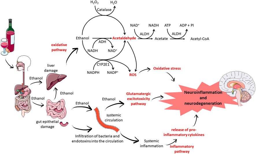
Regardless of the consumption pattern and the amount ingested, alcohol is essentially metabolized in the liver by enzymes such as alcohol dehydrogenase (ADH) and aldehyde dehydrogenase (ALDH). However, alcohol metabolism can also occur in other tissues, such as the brain, mediated by cytochrome p450 enzymes–especially CYP2E1—and catalase. The action of ADH and ALDH produces highly reactive and toxic intermediate metabolites in addition to reactive oxygen species (ROS), which can trigger changes in the redox state, triggering damage to various cells, tissues and organs (Zakhari, 2006; Fernandes et al., 2018a; Fernandes et al., 2018; Frazão et al., 2020; da Silva et al., 2018; Fernandes et al., 2015; Fernandes et al., 2018c). In Figure 1 we present the main mechanisms of damage triggered by ethanol to the nervous system (Figure 1).
The nervous system has been shown to be very sensitive to these oxidative changes, which can trigger neuroinflammatory conditions and thus trigger apoptotic processes (Salim, 2017). However, the mechanisms involved are not fully elucidated. Therefore, several groups have focused on understanding the mechanisms involved, including receptors, neurochemical changes, genetic factors and tissue response (Teixeira et al., 2014; Oliveira et al., 2015; Hamada and Lasek, 2020; Panula, 2020; García-Gutiérrez et al., 2022).
Given the socioeconomic impact of alcohol consumption, it is of great importance to know, understand and map these effects to search for alternatives for the prevention or treatment of damage to the nervous system induced by alcohol. Therefore, our objective was to identify, evaluate, and map the knowledge of the 100 most cited papers evaluating the neurotoxic effects of ethanol.
2 Materials and methods
2.1 Search strategy
To map the information produced about the neurotoxicology of alcohol, bibliometric analysis tools were used, as described in previous studies by our group (de Lima et al., 2022).
Data from this study were collected in December 2022 from the Web of Science Core Collection (WoS-CC) database, with no restrictions on language or year of publication, using a search strategy that used terms related to alcohol and neurotoxicity (Table 1).
2.2 Selection of studies
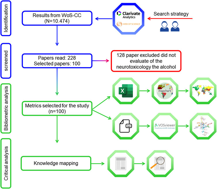
In WoS-CC, the results were organized in descending order of the number of citations. Two researchers (P.F.S.M. and D.C.B.-d.-S) independently carried out the selection of articles and data extraction after reading the title and abstract and then the full text, and differences of opinion were resolved by a third examiner (Nascimento et al., 2022; Corôa et al., 2023; de Sousa Né et al., 2023). The inclusion criteria were that the publications selected had to be articles that addressed the neurotoxicology of alcohol exposure. Editorials, comments, letters, and conference papers were excluded (Figure 2) (de Sousa Né et al., 2023).
2.3 Data extraction of bibliometric parameters
After selecting the 100 most cited articles, the following bibliometric parameters were collected: journal, authors, article title, keywords, number of citations, citation density, year of publication, DOI/URL, and country and continent of the corresponding author.
The rank was established based on decreasing order of the number of citations in the WoS-CC, and in the case of articles with identical numbers of citations, the citation density was used. The Scopus and Google Scholar databases were used for the purposes of comparison with the number of WoS-CC citations, since each database has unique criteria for quantification and data recording (Kulkarni et al., 2009).
2.4 Data analysis
The extracted data were exported to the software Visualization of Similarities Viewer (VOSviewer) version 1.6.16 (Centre for Science and Technology Studies, University of Leiden, the Netherlands) for analysis and construction of the collaboration network regarding the author and co-authorship and occurrences of the authors’ keywords (van Eck and Waltman, 2010). To create the networks, the authors’ names and keywords of authors with at least one article were introduced into the software as a unit of analysis. This software organizes terms into closely related nodes (clusters), represented by different colors, where the number of clusters is determined proportionally to the resolution parameter. The descriptive analysis of the data was performed using spreadsheet editing software (Microsoft Excel 365) and the graphical representation of the countries was generated using the MapChart tool (mapchart.net/index.html) (Figure 2).
2.5 Content analysis
For knowledge mapping, the selected articles were read in full and information such as the study design, model of exposure to ethanol, damage mechanisms associated with ethanol, pharmacological receptors, dose, exposure pathway, period of exposure, and cells and regions affected by use were extracted (Figure 2).
For the study design classification, the Cochrane Collaboration glossary was adopted as a reference: studies were classified as literature review, systematic review, laboratory studies (in vitro, in vivo, and ex vivo), practice guideline, case control, cohort study, case report, case series, cross-sectional study, randomized clinical study, or non-randomized clinical study.
3 Results
3.1 Selected studies and bibliometric analysis
The WoS-CC search yielded a total of 10,474 results, of which 228 were read, and the 100 most cited were selected (Figure 2). The selected articles received a total of 37,743 citations; the most cited article was “Critical Periods of Vulnerability for the Developing Nervous System: Evidence from Humans and Animal Models” (1,998 citations) (Rice and Barone, 2000) and the least cited was “Selective impairment of hippocampal neurogenesis by chronic alcoholism: Protective effects of an antioxidant” (Herrera et al., 2003; 209 citations). The average number of citations per article was 377.63. For most articles, the number of citations in WoS-CC was lower than the number of citations in Google Scholar and Scopus. The mean citation density in WoS-CC was 37.55. The article with the lowest citation density (7.38) was “Ethanol and the Nervous-System” (Charness et al., 1989) and the one with the highest citation density (86.67) was “Critical Periods of Vulnerability for the Developing Nervous System: Evidence from Humans and Animal Models” (Rice and Barone, 2000) (Table 2).

TABLE 2. General data of the 100 selected articles about the effects of alcohol on the central nervous system.
3.1.1 Period of publication
The two oldest articles were published in 1989 and the most recent in 2018 (Figure 3). The most recent article was “Prevalence of Fetal Alcohol Spectrum Disorders in 4 US Communities” (May et al., 2018) which had the objective of estimating the prevalence of fetal alcohol spectrum disorders and received 353 citations, with a citation density of 70.60. The oldest articles were “Ethanol and the nervous system” (Charness et al., 1989) and “Mechanism of Action of Ethanol: initial Central Nervous System Actions” (Deitrich et al., 1989) both of which proposed to evaluate the effects of alcohol and/or its metabolites on the nervous system, with the first receiving 251 citations and 7.38 citations per year while the second received 381 citations and an average of 11.21 citations per year.
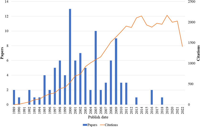
FIGURE 3. Publications and number of citations each year of the top 100 articles related to use of alcohol and central nervous system effects.
The selected articles were published between the years 1989 and 2018; 2% of the articles were published in 1989, 25% between 1990 and 1999, 63% between 2000 and 2009, and 10% between 2010 and 2018. The year 2000 was the period with the highest number of publications selected in this study (n = 13) and the year 2005 had the second highest number of publications (n = 10). Regarding the number of citations, there was annual growth until the year 2011, when it reached 1,902. In 2012, there was a slight reduction in the number of citations, but in 2013 and 2014 the growth in the number of citations recovered, reaching 2,149 in 2014. In 2015, there was a reduction that remained stable until 2018. In 2019, the number of citations grew again, reaching 2,165, the maximum reached in the evaluated period (Figure 3).
Regarding the number of citations, there was annual growth until the year 2011, when it reached 1,902. In 2012, there was a slight reduction in the number of citations, but in 2013 and 2014 the growth in the number of citations recovered, reaching 2,149 in 2014. In 2015, there was a reduction that remained stable until 2018. In 2019, the number of citations grew again, reaching 2,165, the maximum reached in the evaluated period (Figure 3).
3.1.2 Journal of publication
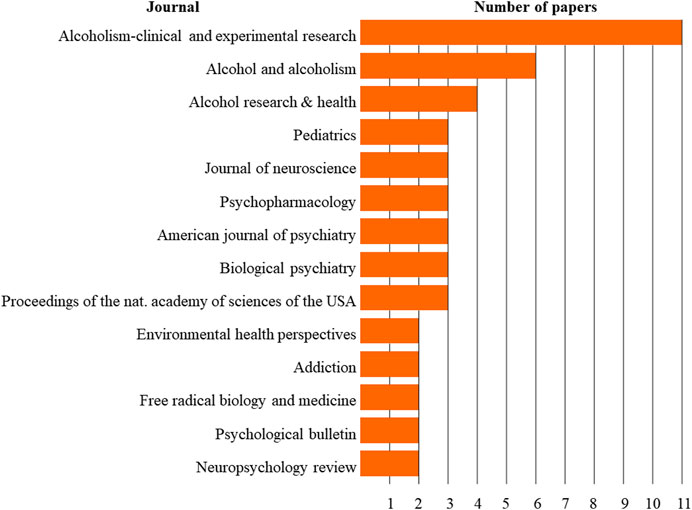
The articles were published in 65 journals, of which Alcoholism: Clinical and Experimental Research was the one with the most papers published (n = 11), followed by Alcohol and Alcoholism (n = 6) and Alcohol Research & Health (n = 4). Most of the journals contributed only one article (78.46%), 7.69% contributed two articles each, and 9.23% contributed three articles each (Figure 4).
3.1.3 Contributing authors
A total of 371 authors contributed to the selected papers. Crews FT, Guerri C, Koob GF, and Riley EP were the authors with the highest number of publications (n = 8, n = 6, n = 5, and n = 5, respectively). The most cited authors were Koob GF and Crews FT, who received 3,513 and 2,813 citations, respectively. Crews FT was first author on five articles and last author on three articles, while Koob GF was sole author on two articles, co-author on two articles and last author on one article. (Figure 5).
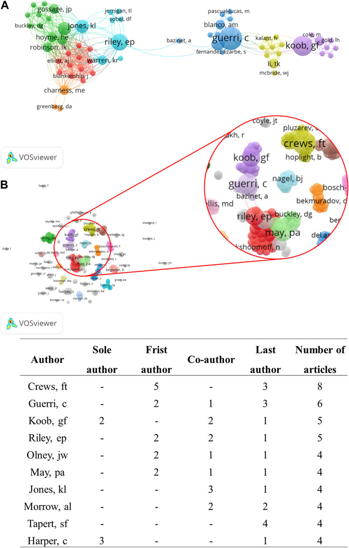
FIGURE 5. Network visualization of co-authorship: (A) The largest set of connected authors (n = 72 authors); (B) All authors with at least one papers (n = 371authors), showing the authors with the largest number of articles published about the neurotoxicology of alcohol. The node size represents the number of documents: the larger the node, the greater the number of articles. Clusters are represented by different colors and the lines indicate co-citation links between authors.
3.1.4 Keywords
A total of 288 author keywords were part of the articles, with an occurrence variation of between 1 and 14 occurrences. The words most used by the authors were “alcoholism”, “alcohol”, and “ethanol”, used 14, 13, and 11 times respectively. These words also had the highest binding strengths among the keywords (Figure 6).
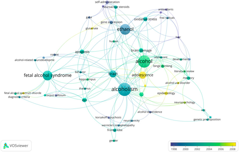
FIGURE 6. Overlay visualization of co-occurrence of author-keywords with a no minimum of two occurrences. Clusters are represented by different colors and the lines indicate co-citation links between keywords.
3.1.5 Geographical distribution of corresponding authors of the top 100 articles
The most relevant scientific production regarding the effects of alcohol on the nervous system is distributed across several countries; however, it is concentrated in the North American continent (n = 78 articles), more specifically in the United States, which contributed with 75 articles and 77.41% of the citations. The European continent contributed with 13 articles, a result mainly due to articles with corresponding authors from Spain (n = 7), England (n = 2), and France (n = 2). Africa, Central America, and South America did not present selected articles in this study (Figure 7).
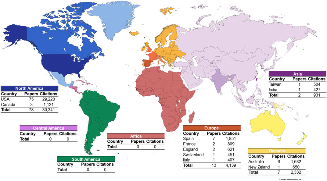
FIGURE 7. Geographical distribution of the corresponding authors of the 100 most cited articles on the neurotoxicology of alcohol.
3.2 Content analysis
3.2.1 Study design
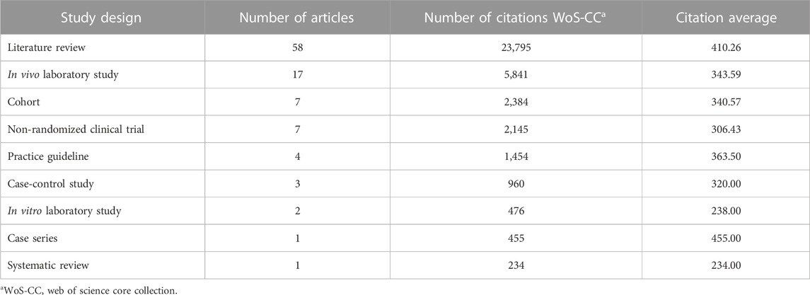
Among the 100 most cited articles, the most frequent study models were literature review (58%), in vivo laboratory study (17%), cohort (7%), and non-randomized clinical trial (7%). These study types received a total of 90.52% (34,165) of the total number of citations (Table 3).
3.2.2 Knowledge maps
In primary studies, several aspects were covered, such as mechanisms associated with alcohol-induced damage (He and Crews, 2008) and changes in the morphology (Caine et al., 1997) and function (Townshend and Duka, 2005) of different regions of the central nervous system. In addition, there were studies that addressed the prevalence of fetal alcohol syndrome (Clarren et al., 1997; May et al., 2018), updating of fetal alcohol syndrome diagnostic criteria (Astley and Clarren, 2000; Chudley et al., 2005; Hoyme et al., 2005; Hoyme et al., 2016), and improving the knowledge on Werneck encephalopathy criteria in people with alcohol use disorders (Caine et al., 1997).
Some studies evaluated these alterations in specific regions of the nervous system. Reductions in the cortes cortex (Kril et al., 1997) and corpus callosum volume (Riley et al., 1995; Bookstein et al., 2002) as well as the hippocampus region (De Bellis et al., 2000; Nagel et al., 2005) were explored. Furthermore, it was observed that women are more sensitive to these ethanol-induced damages (Hommer et al., 2001; Medina et al., 2008).
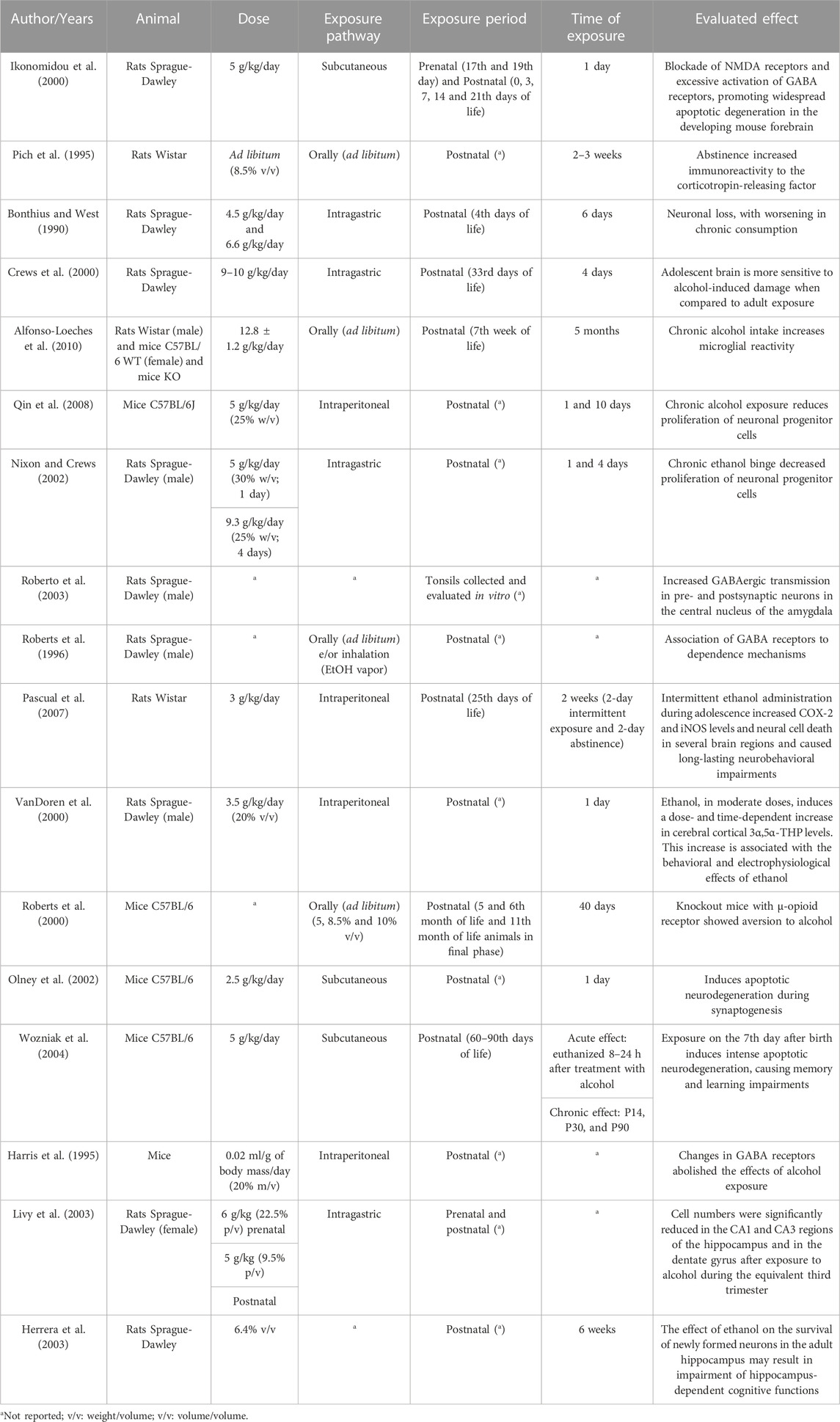
Among the experimental studies, doses ranging from 2.5 to 14 g/kg, with a dose of 5 g/kg being used more frequently (29.41% of the articles). These studies were carried out in different experimental models: in rats (Wistar and Sprague-Dawley) and mice (C56BL/7 and C57BL/6J); Sprague-Dawley rats were the most frequently used species (n = 9 articles, 52.94%). The majority of the selected articles addressed prenatal and postnatal exposure to alcohol, with postnatal exposure being the most frequently studied (n = 15 articles, 88.23%). Regarding the assessment of damage, most studies started the investigation in the first days of the animal’s life (Table 4).
Among the observed results, the authors reported that alcohol consumption induces intense neuroinflammation with consequent apoptotic neurodegeneration, inducing alterations in several regions of the central nervous system. The hippocampus was the region of greatest interest, where it was shown that alcohol abuse induces a reduction in the population of neurons in this brain region which is associated with a decrease in learning and memory capacity.
4 Discussion
This study was based on the 100 most cited articles and focused on mapping knowledge about the neurotoxicology of alcohol. In the present study, it was observed that most of the articles were literature reviews and that they received a high average number of citations. The selected studies were carried out in vitro, in humans, or animals, with different modes of exposure to ethanol. In animals, exposure was evaluated in acute and chronic protocols, both with potentially dangerous doses, equivalent to heavy alcohol consumption (National Institute on Alcohol Abuse and Alcoholism, 2004). The main effects of ethanol neurotoxicity were evaluated especially during pregnancy and adolescence, when abusive consumption can lead to impaired neurodevelopment.
Several factors contribute differently to the outcome of ethanol exposure, among them the amount, frequency, and pattern of consumption (Molina and Nelson, 2018). The damages induced by ethanol consumption are proportional to the type and duration of exposure (Zakhari, 2006), so intense and episodic consumption has received special attention from several researchers due to this pattern of consumption providing high concentration of alcohol in the blood and this remain a longer period, given the limited ability to metabolize alcohol in determining time (Molina and Nelson, 2018; Simon et al., 2022). In this perspective, the studies selected for this mapping made use of an alcohol exposure model that mimics exposure in binge drinking in humans, making it clear that scientists are concerned with this exposure model, given the risk of this form of exposure to induce damage. Biochemical, molecular, and tissue effects in part and/or throughout the nervous system.
Excessive alcohol consumption induces neurodegeneration and cortical dysfunction, this dysfunction associated with impulsivity and contribute to the consumption of dangerous amounts of alcohol. In addition, frequent consumption of high doses of alcohol induces neuroadaptive processes that lead to tolerance, dependence, and manifestations of withdrawal syndrome (Fadda and Rossetti, 1998; Crews and Nixon, 2009).
Considering the historical perspective of ethanol neurotoxicology, this analysis identified and selected studies considered “classic”, which influenced the knowledge of this topic and thus contributed to the development of new researches (Ahmad et al., 2020). The number of citations was considered as an indicator of the quality and impact of a scientific work (Ahmad et al., 2020). Among these articles, the most cited article reviewed the critical period of neurodevelopment, investigating the impact of alcohol exposure during different periods in humans and rats (Rice and Barone, 2000).
The WoS-CC was used as a reference database for using citation metrics from 1945, while a comparison was performed with the Scopus database, whose metrics date from 1966, and Google Scholar. Google citation values were higher than citations in WoS-CC and Scopus, since the Google database counts citations in articles, books, congress annals, academic works (dissertations and theses), reports, and pre-prints differently from the WoS-CC and Scopus platforms. Thus, the difference in number of citations between the databases for the same article is due to the exclusive method of recording and counting citations in each database (Bakkalbasi et al., 2006; Kulkarni et al., 2009).
The three journals that published the most were the journals “Alcoholim-clinical and experimental research”, “alcohol and alcoholis” and “Alcohol research and health”, specific journals on ethanol, not appearing among the journals that most published any public health journal and/or collective health, showing that much of the most cited production is centred on biological and behavioral aspects of the effects of ethanol.
The most cited author was Koob GF, with 3513 citations and five articles. This researcher is recognized worldwide for dedicating his career to the study of alcohol and the neurobiology of alcohol and other drugs. He is currently Director of the NIAAA, which investigates several aspects of alcohol research, ranging from basic science to diagnosis, prevention, treatment, and epidemiology. Koob’s articles are among the 100 most cited, addressing important aspects of ethanol neurotoxicology, such as mechanisms involved in alcohol dependence, including reward pathways involved in this abuse process, in addition to the evaluation of therapeutic alternatives for the treatment of problems related to alcohol.
When evaluating the geographic issue and the production of knowledge by country, it is important to consider that alcohol consumption has become a social problem with an impact on public health in several countries (Harper, 2009; Petrella et al., 2020), among them the United States, where alcohol is the most consumed intoxicant among adolescents (Medina et al., 2008). It is estimated that 19% of adolescents and 7% of adults have some degree of dependence or excessive consumption, and such abusive use has been found to be one of the leading causes of death in the United States (Esser et al., 2020), responsible for 10% of premature deaths in working-age adults (Stahre et al., 2014). These data justify the North American community’s great interest in studying alcohol abuse and its effects, especially on the central nervous system. Our results showed that 75% of the selected articles are by authors affiliated with North American institutions and 60% of the articles state that they received funding from the National Institutes of Health (NIH).
Among the 37 selected primary studies, 20 assessed the effects of alcohol through prenatal exposure. Prenatal exposure studies are of great relevance, especially in the initial period of neurodevelopment, allowing a better understanding of the mechanisms involved in changes in this process, since the effects of alcohol are associated with its ability to cross the blood-brain barrier. In this sense, alcohol can induce damage to the developing nervous system by several mechanisms.
Prenatal exposure occurs at a critical period in neurodevelopment, leading to changes in proliferation, migration, differentiation, synaptogenesis, myelination, and apoptosis (Rice and Barone, 2000). Prenatal alcohol exposure results in learning, memory, and attention deficits due to the teratogenic effect of alcohol, which causes abnormal morphofunctional development of the hippocampus (Berman and Hannigan, 2000); in vitro and in vivo studies have demonstrated abnormal projections of axons and reduction of pyramidal neurons in the hippocampus as a result of prenatal alcohol exposure (Berman and Hannigan, 2000). Furthermore, an association with depressive and psychotic disorders in adulthood has been reported (Olney et al., 2006).
During embryogenesis, glial cells play a critical role; radial glia provide guidance for the migration of neurons, in addition to acting as a neural precursor cell with potential for self-renewal and generation of neurons and oligodendrocytes (Guerri et al., 2009). In addition, synaptogenesis is a critical period in synaptic function, where glial cells promote synapse formation and regulate neurotransmitters and energy in the developing brain (Guerri et al., 2009). Alterations in these mechanisms, as occur in ethanol exposure, can induce irreversible damage to the nervous system (Guerri et al., 2009).
Postnatal exposure was evaluated by 17 articles, in which it was observed that the effects are directly proportional to the type and period of exposure: the earlier the exposure to ethanol, the greater the intensity of damage (Eckardt et al., 1998; Nixon and Crews, 2002; Goodlett et al., 2005; Guerri et al., 2009; Thompson et al., 2009). Among human studies, exposure to alcohol in adolescence has received particular attention (n = 8 papers). Adolescence is considered a period sensitive to the neurotoxic effects of alcohol use (Medina et al., 2008). This period is marked by the development of the prefrontal cortex, structures of the limbic system, and white matter association fibers (Bava and Tapert, 2010). These processes are associated with the development of more sophisticated cognitive functions and emotional processing (Bava and Tapert, 2010).
Excessive alcohol consumption (binge drinking or heavy alcohol use) is prevalent and severe in this period and can lead to changes in the development of these structures (De Bellis et al., 2000; Bava and Tapert, 2010), and the frequent consumption of high doses of alcohol induces neuro-adaptive processes that lead to tolerance, dependence, and manifestations of the withdrawal syndrome (Fadda and Rossetti, 1998; Paus et al., 2008; Crews and Nixon, 2009).
The selected studies showed differences between genders, with women being more sensitive and more vulnerable to the hazardous effects induced by chronic exposure to alcohol (Mumenthaler et al., 1999; Hommer et al., 2001; Medina et al., 2008; Courtney and Polich, 2009). Hommer et al. (2001) and Medina et al. (2008) demonstrated that women addicted to alcohol had smaller volumes of white and gray matter than non-dependent women, and these differences were smaller when the same groups were compared among men. These results may be related to the difference in blood glucose levels and levels of aldehyde dehydrogenase between genders, in addition to the fact that women reach higher concentrations of alcohol with alcohol consumption in a similar dose to men (Mumenthaler et al., 1999).
Several mechanisms, acting synergistically and in different regions, have been proposed to explain brain damage induced by alcohol exposure (Harper and Matsumoto, 2005). Selected studies assessed injury to the hippocampus (n = 6 papers), cortex (n = 5 papers), corpus callosum (n = 2 papers), amygdala (n = 1 papers), and thalamic nucleus (n = 1 papers).
The nervous system is characterized by high metabolic activity with high oxygen consumption and has a high content of polyunsaturated fatty acids and catecholamines, both of which are easily oxidizable. Neuronal death induced by alcohol consumption is associated with increased oxidative stress and expression of pro-inflammatory proteins (Crews and Nixon, 2009). The oxidation of alcohol by the activity of catalase and alcohol dehydrogenase leads to the formation of free radicals (Nordmann et al., 1992). In this context, Pascual et al. (2007) demonstrated increases in cyclooxygenage-2 (COX-2) and nitric oxide synthase (iNOS) and consequent neuronal death in an animal model.
Chronic alcohol consumption leads to regulation of N-methyl-D-aspartate (NMDA) receptors, as well as excitatory amino acid receptors (De Bellis et al., 2000). During abstinence, there is an increase in excitatory transmission, which can lead to excitotoxicity due to the increased stimulation of these receptors (De Bellis et al., 2000). Alcohol modulates several neurochemical systems or their relationship, among them the GABA, glutamatergic, serotonergic, dopaminergic, and opioid neuronal systems (Eckardt et al., 1998).
During the mapping of the most cited articles, a search to understand the mechanisms of addiction and dependence involved in alcohol drug addiction was observed. In this sense, Roberts el al. (1996) (Roberts et al., 1996) and Roberto et al. (2003) evaluated neuropharmacological mechanisms (GABA and glutamate) related to the reinforcement and pathways of neurotransmitters involved in reward in alcohol addiction and thus opening the possibility of investigations into new pharmacological therapies and advances in research. These studies reinforce ethanol–GABA interaction in the central nucleus of the amygdala (Roberts et al., 1996; Roberto et al., 2003). In addition, the cannabinoid system is distributed throughout the nervous system and is associated with functions of motor control, cognition, emotion, and motivation. In this context, de de Fonseca et al. (2005) associated the endogenous cannabinoid system with drug addiction to alcohol, suggesting the use of a cannabinoid receptor antagonist as a therapeutic alternative in dependence, and relapse related to alcohol abuse.
Among the selected articles, the effects of alcohol on the prefrontal cortex (Moselhy et al., 2001; Medina et al., 2008; Crews and Boettiger, 2009), hippocampus (Berman and Hannigan, 2000; De Bellis et al., 2000; Nixon and Crews, 2002; Livy et al., 2003), cerebellum (da Silva et al., 2018; Lamarão-Vieira et al., 2019), thalamic nucleus (Harding et al., 2000), amygdala (Pich et al., 1995; Roberto et al., 2003), and corpus callosum (Riley et al., 1995; Bookstein et al., 2002). However, the mechanisms of alcohol neurotoxicity are still not completely understood, requiring studies that evaluate other regions of the nervous system, such as the spinal cord, where initial studies show sensitivity to alcohol (da Silva et al., 2018) as well as to other toxic substances (da Silva et al., 2022; Eiró-Quirino et al., 2023).
The bibliometric study is based on the number of citations and, therefore, allowing to identify the “hot topics”, dynamic trend and current trends on a topic (Jani et al., 2020; Yanbing et al., 2020), is one of the limitations of this type of study, since it is a purely quantitative evaluation. The limitation of the present assessment relies on the absence of analysis of the quality of the selected study or scientific method nor the certainty of evidence of these articles. In addition, more recent and robust articles may be outside the classification of the selected list, since there has not been enough time to accumulate the number of citations and appear among the 100 most cited articles. The results presented here do not allow for decision-making in public and/or collective health, but which still bring important elucidations showing the need for well-designed epidemiological studies with greater dissemination in the scientific community. There are gaps regarding studies with more evidence and it is relevant for researchers to pay attention to the need for clinical studies, the growing consumption of alcohol by women and young people, new patterns of consumption, such as heavy alcohol use, and the consequences of short- and long-term abstinence.
5 Conclusion
This study identified and analyzed the 100 most cited articles on the neurotoxicology of alcohol using the citation analysis method; most articles were selected from literature reviews and had corresponding authors affiliated with North American institutions. The articles assess the effects of pre- and postnatal alcohol exposure. Prenatal exposure presents data of great interest regarding the risk of experiencing fetal alcohol syndrome, while postnatal exposure, especially in adolescence, is associated with cognitive and memory impairment.
Data availability statement
The original contributions presented in the study are included in the article/Supplementary material, further inquiries can be directed to the corresponding author.
Author contributions
Conceptualization: PM and RL; methodology: PM, DB-S, and RL; formal analysis: PM, DB-S, and RL; investigation: PM, DB-S, and RL; resources: RL; data curation: PM, DB-S, and RL; writing original draft preparation: PM, DB-S, WM, and LB; writing, review, and editing: PM, DB-S, LB, RS-R, CM, and RL; visualization: PM, LB, RL; supervision: RL. All authors contributed to the article and approved the submitted version.
Funding
PM and LB received a scholarship from FAPESPA—Fundação Amazônia de Amparo a Estudos e Pesquisas. RL is a researcher from Conselho Nacional de Desenvolvimento Científico e Tecnológico (CNPq) and received a grant under number 312275/2021-8. Also, this research was funded by PROCAD Amazônia—CAPES (23038.005350/2018-78). The APC was funded by Pró-Reitoria de Pesquisa e Pós-graduação from Federal University of Pará (PROPESP-UFPA).
Acknowledgments
We are grateful to CNPq, CAPES, FADESP, and PROPESP for all the fellowship in developing this research.
Conflict of interest
The authors declare that the research was conducted in the absence of any commercial or financial relationships that could be construed as a potential conflict of interest.
Publisher’s note
All claims expressed in this article are solely those of the authors and do not necessarily represent those of their affiliated organizations, or those of the publisher, the editors and the reviewers. Any product that may be evaluated in this article, or claim that may be made by its manufacturer, is not guaranteed or endorsed by the publisher.
References
Ahmad, S. J. S., Ahmed, A. R., Kowalewski, K. F., Nickel, F., Rostami, K., Stocker, C. J., et al. (2020). Citation classics in general medical journals: Assessing the quality of evidence; A systematic review. Gastroenterol Hepatol Bed Bench 13 (2), 101–114.
Alfonso-Loeches, S., Pascual-Lucas, M., Blanco, A. M., Sanchez-Vera, I., and Guerri, C. (2010). Pivotal role of TLR4 receptors in alcohol-induced neuroinflammation and brain damage. J. Neurosci. 30 (24), 8285–8295. doi:10.1523/JNEUROSCI.0976-10.2010
Astley, S. J., and Clarren, S. K. (2000). Diagnosing the full spectrum of fetal alcohol-exposed individuals: Introducing the 4-digit diagnostic code. Alcohol Alcohol 35 (4), 400–410. doi:10.1093/alcalc/35.4.400
Bakkalbasi, N., Bauer, K., Glover, J., and Wang, L. (2006). Three options for citation tracking: Google scholar, Scopus and Web of science. Biomed. Digit. Libr. 3, 7–8. doi:10.1186/1742-5581-3-7
Bava, S., and Tapert, S. F. (2010). Adolescent brain development and the risk for alcohol and other drug problems. Neuropsychol. Rev. 20 (4), 398–413. doi:10.1007/s11065-010-9146-6
Begleiter, H., and Porjesz, B. (1999). What is inherited in the predisposition toward alcoholism? A proposed model. Alcohol Clin. Exp. Res. 23 (7), 1125–1135. doi:10.1111/j.1530-0277.1999.tb04269.x
Berman, R. F., and Hannigan, J. H. (2000). Effects of prenatal alcohol exposure on the hippocampus: Spatial behavior, electrophysiology, and neuroanatomy. Hippocampus 10 (1), 94–110. doi:10.1002/(SICI)1098-1063(2000)10:1<94::AID-HIPO11>3.0.CO;2-T
Bitew, M. S., Zewde, M. F., Wubetu, M., and Alehegn Alemu, A. (2020). Consumption of alcohol and binge drinking among pregnant women in Addis Ababa, Ethiopia: Prevalence and determinant factors. PLoS One 15 (12), e0243784. doi:10.1371/journal.pone.0243784
Boden, J. M., and Fergusson, D. M. (2011). Alcohol and depression. Addiction 106 (5), 906–914. doi:10.1111/j.1360-0443.2010.03351.x
Bonthius, D. J., and West, J. R. (1990). Alcohol-induced neuronal loss in developing rats: Increased brain damage with binge exposure. Alcohol Clin. Exp. Res. 14 (1), 107–118. doi:10.1111/j.1530-0277.1990.tb00455.x
Bookstein, F. L., Streissguth, A. P., Sampson, P. D., Connor, P. D., and Barr, H. M. (2002). Corpus callosum shape and neuropsychological deficits in adult males with heavy fetal alcohol exposure. Neuroimage 15 (1), 233–251. doi:10.1006/nimg.2001.0977
Brower, K. J. (2003). Insomnia, alcoholism and relapse. Sleep. Med. Rev. 7 (6), 523–539. doi:10.1016/s1087-0792(03)90005-0
Caine, D., Halliday, G. M., Kril, J. J., and Harper, C. G. (1997). Operational criteria for the classification of chronic alcoholics: Identification of wernicke’s encephalopathy. J. Neurol. Neurosurg. Psychiatry 62 (1), 51–60. doi:10.1136/jnnp.62.1.51
Casey, B. J., Jones, R. M., and Somerville, L. H. (2011). Braking and accelerating of the adolescent brain. J. Res. Adolesc. 21 (1), 21–33. doi:10.1111/j.1532-7795.2010.00712.x
Chanraud, S., Martelli, C., Delain, F., Kostogianni, N., Douaud, G., Aubin, H. J., et al. (2007). Brain morphometry and cognitive performance in detoxified alcohol-dependents with preserved psychosocial functioning. Neuropsychopharmacology 32 (2), 429–438. doi:10.1038/sj.npp.1301219
Charness, M. E., Simon, R. P., and Greenberg, D. A. (1989). Ethanol and the nervous system. N. Engl. J. Med. 321 (7), 442–454. doi:10.1056/nejm198908173210706
Chudley, A. E., Conry, J., Cook, J. L., Loock, C., Rosales, T., LeBlanc, N., et al. (2005). Fetal alcohol spectrum disorder: Canadian guidelines for diagnosis. Can. Med. Assoc. J. 172, S1–S21. doi:10.1503/cmaj.1040302
Chung, T., Creswell, K. G., Bachrach, R., Clark, D. B., and Martin, C. S. (2018). Adolescent binge drinking. Alcohol Res. 39 (1), 5–15.
Clarren, S. K., Dehaene, P., and Bookstein, F. L. (1997). Incidence of fetal alcohol syndrome and prevalence of alcohol-related neurodevelopmental disorder. Teratology 56 (5), 317–326. doi:10.1002/(SICI)1096-9926(199711)56:5<317::AID-TERA5>3.0.CO;2-U
Corôa, M. C. P., Mendes, P. F. S., Baia-da-Silva, D. C., Souza-Monteiro, D., Ferreira, M. K. M., Braga, G. L. C., et al. (2023). What is known about midazolam? A bibliometric approach of the literature. Healthc 11 (1), 96. doi:10.3390/healthcare11010096
Costa, L. G., Aschner, M., Vitalone, A., Syversen, T., and Soldin, O. P. (2004). Developmental neuropathology of environmental agents. Annu. Rev. Pharmacol. Toxicol. 44 (1), 87–110. doi:10.1146/annurev.pharmtox.44.101802.121424
Courtney, K. E., and Polich, J. (2009). Binge drinking in young adults: Data, definitions, and determinants. Psychol. Bull. 135 (1), 142–156. doi:10.1037/a0014414
Crews, F. T., Bechara, R., Brown, L. A., Guidot, D. M., Mandrekar, P., Oak, S., et al. (2006). Cytokines and alcohol. Alcohol Clin. Exp. Res. 30 (4), 720–730. doi:10.1111/j.1530-0277.2006.00084.x
Crews, F. T., and Boettiger, C. A. (2009). Impulsivity, frontal lobes and risk for addiction. Pharmacol. Biochem. Behav. 93 (3), 237–247. doi:10.1016/j.pbb.2009.04.018
Crews, F. T., Braun, C. J., Hoplight, B., Switzer, R. C., and Knapp, D. J. (2000). Binge ethanol consumption causes differential brain damage in young adolescent rats compared with adult rats. Alcohol Clin. Exp. Res. 24 (11), 1712–1723. doi:10.1111/j.1530-0277.2000.tb01973.x
Crews, F. T., Morrow, A. L., Criswell, H., and Breese, G. (1996). Effects of ethanol on ion channels. Int. Rev. Neurobiol. 39 (39), 283–367. doi:10.1016/s0074-7742(08)60670-4
Crews, F. T., and Nixon, K. (2009). Mechanisms of neurodegeneration and regeneration in alcoholism. Alcohol Alcohol 44 (2), 115–127. doi:10.1093/alcalc/agn079
Currie, C. L., Sanders, J. L., Swanepoel, L. M., and Davies, C. M. (2020). Maternal adverse childhood experiences are associated with binge drinking during pregnancy in a dose-dependent pattern: Findings from the All Our Families cohort. Child. Abus Negl. 101 (1), 104348. doi:10.1016/j.chiabu.2019.104348
da Silva, D. C. B., Bittencourt, L. O., Baia-Da-silva, D. C., Chemelo, V. S., Eiró-Quirino, L., Nascimento, P. C., et al. (2022). Methylmercury causes neurodegeneration and downregulation of myelin basic protein in the spinal cord of offspring rats after maternal exposure. Int. J. Mol. Sci. 23 (7), 3777. doi:10.3390/ijms23073777
da Silva, F. B. R., Cunha, P. A., Ribera, P. C., Barros, M. A., Cartágenes, S. C., Fernandes, L. M. P., et al. (2018). Heavy chronic ethanol exposure from adolescence to adulthood induces cerebellar neuronal loss and motor function damage in female rats. Front. Behav. Neurosci. 12, 88–11. doi:10.3389/fnbeh.2018.00088
De Bellis, M. D., Clark, D. B., Beers, S. R., Soloff, P. H., Boring, A. M., Hall, J., et al. (2000). Hippocampal volume in adolescent-onset alcohol use disorders. Am. J. Psychiatry 157 (5), 737–744. doi:10.1176/appi.ajp.157.5.737
De Bellis, M. D. (2002). Developmental traumatology: A contributory mechanism for alcohol and substance use disorders. Psychoneuroendocrinology 27 (1–2), 155–170. doi:10.1016/s0306-4530(01)00042-7)
de Fonseca, F. R., del Arco, I., Bermudez-Silva, F. J., Bilbao, A., Cippitelli, A., and Navarro, M. (2005). The endocannabinoid system: Physiology and pharmacology. Alcohol Alcohol 40 (1), 2–14. doi:10.1093/alcalc/agh110
de Lima, W. F., Né, Y. G. S., Aragão, W. A. B., Eiró-Quirino, L., Baia-da-Silva, D. C., Cirovic, A., et al. (2022). Global scientific research landscape on aluminum Toxicology. Biol. Trace Elem. Res. 201, 3210–3224. doi:10.1007/s12011-022-03427-9
de Sousa Né, Y. G., Lima, W. F., Mendes, P. F. S., Baia-da-Silva, D. C., Bittencourt, L. O., Nascimento, P. C., et al. (2023). Dental caries and salivary oxidative stress: Global scientific research landscape. Antioxidants 12 (2), 330. doi:10.3390/antiox12020330
Deitrich, R. A., Dunwiddie, T. V., Harris, R. A., and Erwin, V. G. (1989). Mechanism of action of ethanol: Initial central nervous system actions. Pharmacol. Rev. 41 (4), 489–537.
Dejong, K., Olyaei, A., and Lo, J. O. (2019). Alcohol use in pregnancy. Clin. Obstet. Gynecol. 62 (1), 142–155. doi:10.1097/GRF.0000000000000414
Diamond, I., and Gordon, A. S. (1997). Cellular and molecular neuroscience of alcoholism. Physiol. Rev. 77 (1), 1–20. doi:10.1152/physrev.1997.77.1.1
Dunwiddie, T. V., and Masino, S. a. (2001). The role and regulation of adenosine in the central nervous system. Annu. Rev. Neurosci. 24, 31–55. doi:10.1146/annurev.neuro.24.1.31
Eckardt, M. J., File, S. E., Gessa, G. L., Grant, K. A., Guerri, C., Hoffman, P. L., et al. (1998). Effects of moderate alcohol consumption on the central nervous system. Alcohol Clin. Exp. Res. 22 (5), 998–1040. doi:10.1111/j.1530-0277.1998.tb03695.x
Eiró-Quirino, L., Lima, W. F., Aragão, W. A. B., Bittencourt, L. O., Mendes, P. F. S., Fernandes, R. M., et al. (2023). Exposure to tolerable concentrations of aluminum triggers systemic and local oxidative stress and global proteomic modulation in the spinal cord of rats. Chemosphere 313, 137296–137298. doi:10.1016/j.chemosphere.2022.137296
Esser, M. B., Sherk, A., Liu, Y., Naimi, T. S., Stockwell, T., and Stahre, M. (2020). Correction and republication: Deaths and years of potential life lost from excessive alcohol use — United States, 2011–2015. MMWR Surveill. Summ. 69 (39), 1427. doi:10.15585/mmwr.mm6930a1
Fadda, F., and Rossetti, Z. L. (1998). Chronic ethanol consumption:from neuroadaptation to neurodegeneration. Prog. Neurobiol. [Internet] 56 (4), 385–431. doi:10.1016/s0301-0082(98)00032-x
Fernandes, L. M. P., Cartágenes, S. C., Barros, M. A., Carvalheiro, T. C. V. S., Castro, N. C. F., Schamne, M. G., et al. (2018c). Repeated cycles of binge-like ethanol exposure induce immediate and delayed neurobehavioral changes and hippocampal dysfunction in adolescent female rats. Behav. Brain Res. 350, 99–108. doi:10.1016/j.bbr.2018.05.007
Fernandes, L. M. P., Lopes, K. S., Santana, L. N. S., Fontes-Júnior, E. A., Ribeiro, C. H. M. A., Silva, M. C. F., et al. (2018a). Repeated cycles of binge-like ethanol intake in adolescent female rats induce motor function impairment and oxidative damage in motor cortex and liver, but not in blood. Oxid. Med. Cell. Longev. 2018, 3467531. doi:10.1155/2018/3467531
Fernandes, L. M. P., Teixeira, F. B., Junior, S. M. A., Pinheiro, J. de J. V., Maia C do, S. F., and Lima, R. R. (2015). Immunohistochemical changes and atrophy after chronic ethanol intoxication in rat salivary glands. Histol. Histopathol. 30 (9), 1069–1078. doi:10.14670/HH-11-604
Fernandes, R. M., Nascimento, P. C., Bittencourt, L. O., de Oliveira, A. C. A., Puty, B., Leão, L. K. R., et al. (2018b). Chronic ethanol forced administration from adolescence to adulthood reduces cell density in the rat spinal cord. Tissue Cell. 55, 77–82. doi:10.1016/j.tice.2018.10.001
Fernandez-Lizarbe, S., Pascual, M., and Guerri, C. (2009). Critical role of TLR4 response in the activation of microglia induced by ethanol. J. Immunol. 183 (7), 4733–4744. doi:10.4049/jimmunol.0803590
Fiala, J. C., Spacek, J., and Harris, K. M. (2002). Dendritic spine pathology: Cause or consequence of neurological disorders? Brain Res. Rev. 39 (1), 29–54. doi:10.1016/s0165-0173(02)00158-3
Frazão, D. R., do Socorro Ferraz Maia, C., dos Santos Chemelo, V., Monteiro, D., de Oliveira Ferreira, R., Bittencourt, L. O., et al. (2020). Ethanol binge drinking exposure affects alveolar bone quality and aggravates bone loss in experimentally-induced periodontitis. PLoS One 15 (7), 1–12. doi:10.1371/journal.pone.0236161
García-Gutiérrez, M. S., Navarrete, F., Gasparyan, A., Navarro, D., Morcuende, Á., Femenía, T., et al. (2022). Role of cannabinoid CB2 receptor in alcohol use disorders: From animal to human studies. Int. J. Mol. Sci. 23 (11), 5908. doi:10.3390/ijms23115908
Goodlett, C. R., Horn, K. H., and Zhou, F. C. (2005). Alcohol teratogenesis: Mechanisms of damage and strategies for intervention. Exp. Biol. Med. 230 (6), 394–406. doi:10.1177/15353702-0323006-07
Grace, A. A. (2000). The tonic/phasic model of dopamine system regulation and its implications for understanding alcohol and psychostimulant craving. Addiction 95 (8), S119–S128. doi:10.1080/09652140050111690
Grobin, A. C., Matthews, D. B., Devaud, L. L., and Morrow, A. L. (1998). The role of GABA A receptors in the acute and chronic effects of ethanol. Psychopharmacol. Berl. 139 (1–2), 2–19. doi:10.1007/s002130050685
Guerri, C., Bazinet, A., and Riley, E. P. (2009). Foetal alcohol spectrum disorders and alterations in brain and behaviour. Alcohol Alcohol 44 (2), 108–114. doi:10.1093/alcalc/agn105
Guerri, C., and Pascual, M. (2010). Mechanisms involved in the neurotoxic, cognitive, and neurobehavioral effects of alcohol consumption during adolescence. Alcohol 44 (1), 15–26. doi:10.1016/j.alcohol.2009.10.003
Hamada, K., and Lasek, A. W. (2020). Receptor tyrosine kinases as therapeutic targets for alcohol use disorder. Neurotherapeutics 17 (1), 4–16. doi:10.1007/s13311-019-00795-4
Haorah, J., Ramirez, S. H., Floreani, N., Gorantla, S., Morsey, B., and Persidsky, Y. (2008). Mechanism of alcohol-induced oxidative stress and neuronal injury. Free Radic. Biol. Med. 45 (11), 1542–1550. doi:10.1016/j.freeradbiomed.2008.08.030
Harding, A., Halliday, G., Caine, D., and Kril, J. (2000). Degeneration of anterior thalamic nuclei differentiates alcoholics with amnesia. Brain 123 (1), 141–154. doi:10.1093/brain/123.1.141
Harper, C., and Matsumoto, I. (2005). Ethanol and brain damage. Curr. Opin. Pharmacol. 5 (1), 73–78. doi:10.1016/j.coph.2004.06.011
Harper, C. (2009). The neuropathology of alcohol-related brain damage. Alcohol Alcohol 44 (2), 136–140. doi:10.1093/alcalc/agn102
Harper, C. (1998). The neuropathology of alcohol-specific brain damage, or does alcohol damage the brain? J. Neuropathol. Exp. Neurol. [Internet] 57 (2), 101–110. doi:10.1097/00005072-199802000-00001
Harris, R. A., McQuilkin, S. J., Paylor, R., Abeliovich, A., Tonegawa, S., and Wehner, J. M. (1995). Mutant mice lacking the gamma isoform of protein kinase C show decreased behavioral actions of ethanol and altered function of gamma-aminobutyrate type A receptors. Proc. Natl. Acad. Sci. 92 (9), 3658–3662. doi:10.1073/pnas.92.9.3658
He, J., and Crews, F. T. (2008). Increased MCP-1 and microglia in various regions of the human alcoholic brain. Exp. Neurol. 210 (2), 349–358. doi:10.1016/j.expneurol.2007.11.017
Heinz, A., Jones, D. W., Mazzanti, C., Goldman, D., Ragan, P., Hommer, D., et al. (2000). A relationship between serotonin transporter genotype and in vivo protein expression and alcohol neurotoxicity. Biol. Psychiatry 47 (7), 643–649. doi:10.1016/s0006-3223(99)00171-7
Herrera, D. G., Yagüe, A. G., Johnsen-Soriano, S., Bosch-Morell, F., Collado-Morente, L., Muriach, M., et al. (2003). Selective impairment of hippocampal neurogenesis by chronic alcoholism: Protective effects of an antioxidant. Proc. Natl. Acad. Sci. U. S. A. 100 (13), 7919–7924. doi:10.1073/pnas.1230907100
Hommer, D. W., Momenan, R., Kaiser, E., and Rawlings, R. R. (2001). Evidence for a gender-related effect of alcoholism on brain volumes. Am. J. Psychiatry 158 (2), 198–204. doi:10.1176/appi.ajp.158.2.198
Horrocks, L. A., and Yeo, Y. K. (1999). Health benefits of docosahexaenoic acid (DHA). Pharmacol. Res. 40 (3), 211–225. doi:10.1006/phrs.1999.0495
Hoyme, H. E., Kalberg, W. O., Elliott, A. J., Blankenship, J., Buckley, D., Marais, A-S., et al. (2016). Updated clinical guidelines for diagnosing fetal alcohol spectrum disorders. Pediatrics 138 (2), e20154256. doi:10.1542/peds.2015-4256
Hoyme, H. E., May, P. A., Kalberg, W. O., Kodituwakku, P., Gossage, J. P., Trujillo, P. M., et al. (2005). A practical clinical approach to diagnosis of fetal alcohol spectrum disorders: Clarification of the 1996 institute of medicine criteria. Pediatrics 115 (1), 39–47. doi:10.1542/peds.2004-0259
Ikonomidou, C., Bittigau, P., Koch, C., Genz, K., Stefovska, V., Hörster, F., et al. (2000). Ethanol-induced apoptotic neurodegeneration and fetal alcohol syndrome. Science 287 (5455), 1056–1060. doi:10.1126/science.287.5455.1056
Jani, R. H., Prabhu, A. V., Zhou, J. J., Alan, N., and Agarwal, N. (2020). Citation analysis of the most influential articles on traumatic spinal cord injury. J. Spinal Cord. Med. 43 (1), 31–38. doi:10.1080/10790268.2019.1576426
Kanny, D., Naimi, T. S., Liu, Y., Lu, H., and Brewer, R. D. (2018). Annual total binge drinks consumed by U.S. Adults, 2015. Am. J. Prev. Med. 54 (4), 486–496. doi:10.1016/j.amepre.2017.12.021
Koob, G. F. (2008). A role for brain stress systems in addiction. Neuron 59 (1), 11–34. doi:10.1016/j.neuron.2008.06.012
Koob, G. F. (1992). Drugs of abuse: Anatomy, pharmacology and function of reward pathways. Trends Pharmacol. Sci. 13 (C), 177–184. doi:10.1016/0165-6147(92)90060-j
Kril, J. J., Halliday, G. M., Svoboda, M. D., and Cartwright, H. (1997). The cerebral cortex is damaged in chronic alcoholics. Neuroscience 79 (4), 983–998. doi:10.1016/s0306-4522(97)00083-3
Kulkarni, A. V., Aziz, B., Shams, I., and Busse, J. W. (2009). Comparisons of citations in Web of Science, Scopus, and Google Scholar for articles published in general medical journals. Jama 302 (10), 1092–1096. doi:10.1001/jama.2009.1307
Kumar, S., Porcu, P., Werner, D. F., Matthews, D. B., Diaz-Granados, J. L., Helfand, R. S., et al. (2009). The role of GABAA receptors in the acute and chronic effects of ethanol: A decade of progress. Psychopharmacol. Berl. 205 (4), 529–564. doi:10.1007/s00213-009-1562-z
Lamarão-Vieira, K., Pamplona-Santos, D., Nascimento, P. C., Corrêa, M. G., Bittencourt, L. O., Dos Santos, S. M., et al. (2019). Physical exercise attenuates oxidative stress and morphofunctional cerebellar damages induced by the ethanol binge drinking paradigm from adolescence to adulthood in rats. Oxid. Med. Cell. Longev. 2019, 6802424. doi:10.1155/2019/6802424
LeMarquand, D., Pihl, R. O., and Benkelfat, C. (1994). Serotonin and alcohol intake, abuse, and dependence: Clinical evidence. Biol. Psychiatry 36 (5), 326–337. doi:10.1016/0006-3223(94)90630-0
Lewohl, J. M., Wang, L., Miles, M. F., Zhang, L., Dodd, P. R., and Adron Harris, R. (2000). Gene expression in human alcoholism: Microarray analysis of frontal cortex. Alcohol Clin. Exp. Res. 24 (12), 1873–1882. doi:10.1111/j.1530-0277.2000.tb01993.x
Liou, H. H., Tsai, M. C., Chen, C. J., Jeng, J. S., Chang, Y. C., Chen, S. Y., et al. (1997). Environmental risk factors and Parkinson's disease: A case-control study in taiwan. Neurology 48 (6), 1583–1588. doi:10.1212/wnl.48.6.1583
Livy, D. J., Miller, E. K., Maier, S. E., and West, J. R. (2003). Fetal alcohol exposure and temporal vulnerability: Effects of binge-like alcohol exposure on the developing rat hippocampus. Neurotoxicol Teratol. 25 (4), 447–458. doi:10.1016/s0892-0362(03)00030-8
Maier, S. E., and West, J. R. (2001). Drinking patterns and alcohol-related birth defects. Alcohol Res. Health 25, 168–174.
Martin, P. R., Singleton, C. K., and Hiller-Sturmhöfel, S. (2003). The role of thiamine deficiency in alcoholic brain disease. Alcohol Res. Heal 27 (2), 134–142.
May, P. A., Chambers, C. D., Kalberg, W. O., Zellner, J., Feldman, H., Buckley, D., et al. (2018). Prevalence of fetal alcohol spectrum disorders in 4 US Communities. JAMA 319 (5), 474–482. doi:10.1001/jama.2017.21896
May, P. A., and Gossage, J. P. (2001). Estimating the prevalence of fetal alcohol syndrome. A summary. Alcohol Res. Health 25 (3), 159–167.
McBride, W. J., and Li, T-K. (1998). Animal models of alcoholism: Neurobiology of high alcohol-drinking behavior in rodents. Crit. Rev. Neurobiol. 12 (4), 339–369. doi:10.1615/critrevneurobiol.v12.i4.40
McKinney, A. M., Short, J., Truwit, C. L., McKinney, Z. J., Kozak, O. S., SantaCruz, K. S., et al. (2007). Posterior reversible encephalopathy syndrome: Incidence of atypical regions of involvement and imaging findings. Am. J. Roentgenol. 189 (4), 904–912. doi:10.2214/AJR.07.2024
Medina, K. L., McQueeny, T., Nagel, B. J., Hanson, K. L., Schweinsburg, A. D., and Tapert, S. F. (2008). Prefrontal cortex volumes in adolescents with alcohol use disorders: Unique gender effects. Alcohol Clin. Exp. Res. [Internet] 32 (3), 386–394. doi:10.1111/j.1530-0277.2007.00602.x
Mehta, A., Prabhakar, M., Kumar, P., Deshmukh, R., and Sharma, P. L. (2013). Excitotoxicity: Bridge to various triggers in neurodegenerative disorders. Eur. J. Pharmacol. 698 (1–3), 6–18. doi:10.1016/j.ejphar.2012.10.032
Molina, P. E., and Nelson, S. (2018). Binge drinking’s effects on the body. Alcohol Res. 39 (1), 99–109.
Moselhy, H. F., Georgiou, G., and Kahn, A. (2001). Frontal lobe changes in alcoholism: A review of the literature. Alcohol Alcohol 36 (5), 357–368. doi:10.1093/alcalc/36.5.357
Mumenthaler, M. S., Taylor, J. L., O’Hara, R., and Yesavage, J. A. (1999). Gender differences in moderate drinking effects. Alcohol Res. Health 23 (1), 55–64.
Nagel, B. J., Schweinsburg, A. D., Phan, V., and Tapert, S. F. (2005). Reduced hippocampal volume among adolescents with alcohol use disorders without psychiatric comorbidity. Psychiatry Res. - Neuroimaging 139 (3), 181–190. doi:10.1016/j.pscychresns.2005.05.008
Nascimento, P. C., Ferreira, M. K. M., Bittencourt, L. O., Martins-Júnior, P. A., and Lima, R. R. (2022). Global research trends on maternal exposure to methylmercury and offspring health outcomes. Front. Pharmacol. 13, 973118–973119. doi:10.3389/fphar.2022.973118
National Institute on Alcohol Abuse and Alcoholism (2004). NIAAA council approves definition of binge drinking. NIAAA News 3, 3.
Nixon, K., and Crews, F. T. (2002). Binge ethanol exposure decreases neurogenesis in adult rat hippocampus. J. Neurochem. 83 (5), 1087–1093. doi:10.1046/j.1471-4159.2002.01214.x
Nordmann, R., Ribière, C., and Rouach, H. (1992). Implication of free radical mechanisms in ethanol-induced cellular injury. Free Radic. Biol. Med. 12 (3), 219–240. doi:10.1016/0891-5849(92)90030-k
Oliveira, A. C., Pereira, M. C. S., Santana Ln da, S., Fernandes, R. M. P., Teixeira, F. B., Oliveira, G. B., et al. (2015). Chronic ethanol exposure during adolescence through early adulthood in female rats induces emotional and memory deficits associated with morphological and molecular alterations in hippocampus. J. Psychopharmacol. 29 (6), 712–724. doi:10.1177/0269881115581960
Olney, J. W., Tenkova, T., Dikranian, K., Qin, Y-Q., Labruyere, J., and Ikonomidou, C. (2002). Ethanol-induced apoptotic neurodegeneration in the developing C57BL/6 mouse brain. Dev. Brain Res. 133 (2), 115–126. doi:10.1016/s0165-3806(02)00279-1
Olney, J. W., Wozniak, D. F., Jevtovic-Todorovic, V., Farber, N. B., Bittigau, P., and Ikonomidou, C. (2006). Drug-induced apoptotic neurodegeneration in the developing brain. Brain Pathol. 12 (4), 488–498. doi:10.1111/j.1750-3639.2002.tb00467.x
Panula, P. (2020). Histamine, histamine H 3 receptor, and alcohol use disorder. Br. J. Pharmacol. [Internet 177 (3), 634–641. doi:10.1111/bph.14634
Pascual, M., Blanco, A. M., Cauli, O., Miñarro, J., and Guerri, C. (2007). Intermittent ethanol exposure induces inflammatory brain damage and causes long-term behavioural alterations in adolescent rats. Eur. J. Neurosci. [Internet] 25 (2), 541–550. doi:10.1111/j.1460-9568.2006.05298.x
Paus, T., Keshavan, M., and Giedd, J. N. (2008). Why do many psychiatric disorders emerge during adolescence? Nat. Rev. Neurosci. 9 (12), 947–957. doi:10.1038/nrn2513
Petrella, C., Carito, V., Carere, C., Ferraguti, G., Ciafrè, S., Natella, F., et al. (2020). Oxidative stress inhibition by resveratrol in alcohol-dependent mice. Nutrition 79 (80), 110783–110786. doi:10.1016/j.nut.2020.110783
Pich, E., Lorang, M., Yeganeh, M., Rodriguez de Fonseca, F., Raber, J., Koob, G., et al. (1995). Increase of extracellular corticotropin-releasing factor-like immunoreactivity levels in the amygdala of awake rats during restraint stress and ethanol withdrawal as measured by microdialysis. J. Neurosci. 15 (8), 5439–5447. doi:10.1523/JNEUROSCI.15-08-05439.1995
Polich, J., and Criado, J. R. (2006). Neuropsychology and neuropharmacology of P3a and P3b. Int. J. Psychophysiol. 60 (2), 172–185. doi:10.1016/j.ijpsycho.2005.12.012
Popova, S., Lange, S., Shield, K., Mihic, A., Chudley, A. E., Mukherjee, R. A. S., et al. (2016). Comorbidity of fetal alcohol spectrum disorder: A systematic review and meta-analysis. Lancet 387 (10022), 978–987. doi:10.1016/S0140-6736(15)01345-8
Porjesz, B., Rangaswamy, M., Kamarajan, C., Jones, K. A., Padmanabhapillai, A., and Begleiter, H. (2005). The utility of neurophysiological markers in the study of alcoholism. Clin. Neurophysiol. 116 (5), 993–1018. doi:10.1016/j.clinph.2004.12.016
Qin, L., He, J., Hanes, R. N., Pluzarev, O., Hong, J., and Crews, F. T. (2008). Increased systemic and brain cytokine production and neuroinflammation by endotoxin following ethanol treatment. J. Neuroinflammation 17, 10–17. doi:10.1186/1742-2094-5-10
Rangaswamy, M., Porjesz, B., Chorlian, D. B., Wang, K., Jones, K. A., Bauer, L. O., et al. (2002). Beta power in the EEG of alcoholics. Biol. Psychiatry 52 (8), 831–842. doi:10.1016/s0006-3223(02)01362-8
Rice, D., and Barone, S. (2000). Critical periods of vulnerability for the developing nervous system: Evidence from humans and animal models. Environ. Health Perspect. 108, 511–533. doi:10.1289/ehp.00108s3511
Riley, E. P., Infante, M. A., and Warren, K. R. (2011). Fetal alcohol spectrum disorders: An overview. Neuropsychol. Rev. 21 (2), 73–80. doi:10.1007/s11065-011-9166-x
Riley, E. P., Mattson, S. N., Sowell, E. R., Jernigan, T. L., Sobel, D. F., and Jones, K. L. (1995). Abnormalities of the corpus callosum in children prenatally exposed to alcohol. Alcohol Clin. Exp. Res. [Internet] 19 (5), 1198–1202. doi:10.1111/j.1530-0277.1995.tb01600.x
Roberto, M., Madamba, S. G., Moore, S. D., Tallent, M. K., and Siggins, G. R. (2003). Ethanol increases GABAergic transmission at both pre- and postsynaptic sites in rat central amygdala neurons. Proc. Natl. Acad. Sci. 100 (4), 2053–2058. doi:10.1073/pnas.0437926100
Roberts, A. J., Cole, M., and Koob, G. F. (1996). Intra-amygdala muscimol decreases operant ethanol self-administration in dependent rats. Alcohol Clin. Exp. Res. 20 (7), 1289–1298. doi:10.1111/j.1530-0277.1996.tb01125.x
Roberts, A. J., McDonald, J. S., Heyser, C. J., Kieffer, B. L., Matthes, H. W. D., Koob, G. F., et al. (2000). mu-Opioid receptor knockout mice do not self-administer alcohol. J. Pharmacol. Exp. Ther. 293 (3), 1002–1008.
Romero, F. J., Bosch-Morell, F., Romero, M. J., Jareño, E. J., Romero, B., Marín, N., et al. (1998). Lipid peroxidation products and antioxidants in human disease. Environ. Health Perspect. 106 (5), 1229–1234. doi:10.1289/ehp.98106s51229
Salim, S. (2017). Oxidative stress and the central nervous system. J. Pharmacol. Exp. Ther. 360 (1), 201–205. doi:10.1124/jpet.116.237503
Sayette, M. A. (1993). An appraisal-disruption model of alcohol’s effects on stress responses in social drinkers. Psychol. Bull. [Internet] 114 (3), 459–476. doi:10.1037/0033-2909.114.3.459
Simon, L., Souza-Smith, F. M., and Molina, P. E. (2022). Alcohol-associated tissue injury: Current views on pathophysiological mechanisms. Annu. Rev. Physiol. 84, 87–112. doi:10.1146/annurev-physiol-060821-014008
Squeglia, L. M., Jacobus, J., and Tapert, S. F. (2009). The influence of substance use on adolescent brain development. Clin. EEG Neurosci. 40 (1), 31–38. doi:10.1177/155005940904000110
Stahre, M., Roeber, J., Kanny, D., Brewer, R. D., and Zhang, X. (2014). Contribution of excessive alcohol consumption to deaths and years of potential life lost in the United States. Prev. Chronic Dis. 11, E109–E112. doi:10.5888/pcd11.130293
Sullivan, E. V., and Pfefferbaum, A. (2005). Neurocircuitry in alcoholism: A substrate of disruption and repair. Psychopharmacol. Berl. 180 (4), 583–594. doi:10.1007/s00213-005-2267-6
Sullivan, E. V., Rosenbloom, M. J., and Pfefferbaum, A. (2000). Pattern of motor and cognitive deficits in detoxified alcoholic men. Alcohol Clin. Exp. Res. 24 (5), 611–621. doi:10.1111/j.1530-0277.2000.tb02032.x
Teixeira, F. B., Santana, L. N. D. S., Bezerra, F. R., De Carvalho, S., Fontes-Júnior, E. A., Prediger, R. D., et al. (2014). Chronic ethanol exposure during adolescence in rats induces motor impairments and cerebral cortex damage associated with oxidative stress. PLoS One 9 (6), e101074. doi:10.1371/journal.pone.0101074
Thompson, B. L., Levitt, P., and Stanwood, G. D. (2009). Prenatal exposure to drugs: Effects on brain development and implications for policy and education. Nat. Rev. Neurosci. 10 (4), 303–312. doi:10.1038/nrn2598
Tolomeo, S., Macfarlane, J. A., Baldacchino, A., Koob, G. F., and Steele, J. D. (2021). Alcohol binge drinking: Negative and positive valence system abnormalities. Biol. Psychiatry Cogn. Neurosci. Neuroimaging 6 (1), 126–134. doi:10.1016/j.bpsc.2020.09.010
Townshend, J. M., and Duka, T. (2005). Binge drinking, cognitive performance and mood in a population of young social drinkers. Alcohol Clin. Exp. Res. 29 (3), 317–325. doi:10.1097/01.alc.0000156453.05028.f5
Tsai, G., Gastfriend, D. R., and Coyle, J. T. (1995). The glutamatergic basis of human alcoholism. Am. J. Psychiatry 152 (3), 332–340. doi:10.1176/ajp.152.3.332
van Eck, N. J., and Waltman, L. (2010). Software survey: VOSviewer, a computer program for bibliometric mapping. Scientometrics 84 (2), 523–538. doi:10.1007/s11192-009-0146-3
VanDoren, M. J., Matthews, D. B., Janis, G. C., Grobin, A. C., Devaud, L. L., and Morrow, A. L. (2000). Neuroactive steroid 3alpha-hydroxy-5alpha-pregnan-20-one modulates electrophysiological and behavioral actions of ethanol. J. Neurosci. 20, 1982–1989. doi:10.1523/JNEUROSCI.20-05-01982.2000
Weiss, F., Ciccocioppo, R., Parsons, L. H., Katner, S., Liu, X., Zorrilla, E. P., et al. (2006). Compulsive drug-seeking behavior and relapse. Ann. N. Y. Acad. Sci. 937 (1), 1–26. doi:10.1111/j.1749-6632.2001.tb03556.x
WHO (2019). Global status report on alcohol and health 2018. Geneva, Switzerland: World Health Organization.
Windle, M., Spear, L. P., Fuligni, A. J., Angold, A., Brown, J. D., Pine, D., et al. (2008). Transitions into underage and problem drinking: Developmental processes and mechanisms between 10 and 15 years of age. Pediatrics 121 (4), 273–289. doi:10.1542/peds.2007-2243C
Wozniak, D. F., Hartman, R. E., Boyle, M. P., Vogt, S. K., Brooks, A. R., Tenkova, T., et al. (2004). Apoptotic neurodegeneration induced by ethanol in neonatal mice is associated with profound learning/memory deficits in juveniles followed by progressive functional recovery in adults. Neurobiol. Dis. 17 (3), 403–414. doi:10.1016/j.nbd.2004.08.006
Yanbing, S., Ruifang, Z., Chen, W., Shifan, H., Hua, L., and Zhiguang, D. (2020). Bibliometric analysis of journal of nursing management from 1993 to 2018. J. Nurs. Manag. 28 (2), 317–331. doi:10.1111/jonm.12925
Zakhari, S. (2006). Overview: How is alcohol metabolized by the body? Alcohol Res. Health 29, 245–254.
Keywords: alcohol abuse, central nervous system, alcohol, alcoholism, bibliometric
Citation: Mendes PFS, Baia-da-Silva DC, Melo WWP, Bittencourt LO, Souza-Rodrigues RD, Fernandes LMP, Maia CdSF and Lima RR (2023) Neurotoxicology of alcohol: a bibliometric and science mapping analysis. Front. Pharmacol. 14:1209616. doi: 10.3389/fphar.2023.1209616
Received: 21 April 2023; Accepted: 11 July 2023;
Published: 01 August 2023.
Edited by:
Christopher Joseph Hammond, Johns Hopkins University, United StatesReviewed by:
Katie Davis-Anderson, Los Alamos National Laboratory (DOE), United StatesSrinivas Muvvala, Yale University, United States
Copyright © 2023 Mendes, Baia-da-Silva, Melo, Bittencourt, Souza-Rodrigues, Fernandes, Maia and Lima. This is an open-access article distributed under the terms of the Creative Commons Attribution License (CC BY). The use, distribution or reproduction in other forums is permitted, provided the original author(s) and the copyright owner(s) are credited and that the original publication in this journal is cited, in accordance with accepted academic practice. No use, distribution or reproduction is permitted which does not comply with these terms.
*Correspondence: Rafael Rodrigues Lima, cmFmYWxpbWFAdWZwYS5icg==
 Paulo Fernando Santos Mendes
 Daiane Claydes Baia-da-Silva
 Wallacy Watson Pereira Melo1
 Leonardo Oliveira Bittencourt
 Renata Duarte Souza-Rodrigues
 Luanna Melo Pereira Fernandes
 Cristiane do Socorro Ferraz Maia
 Rafael Rodrigues Lima