 Paolo Zuccolini
 Paola Gavazzo
 Michael Pusch
94% of researchers rate our articles as excellent or good
Learn more about the work of our research integrity team to safeguard the quality of each article we publish.
Find out more
MINI REVIEW article
Front. Pharmacol., 24 May 2022
Sec. Pharmacology of Ion Channels and Channelopathies
Volume 13 - 2022 | https://doi.org/10.3389/fphar.2022.906608
BK (KCa 1.1, Slo-1) is a K+ channel characterized by an allosteric regulation of the gating mechanism by Ca2+ binding and voltage, and a high unitary conductance. The channel is expressed in many different tissues, where it is involved in the regulation or the fine-tuning of many physiological processes. Among other organs, BK is expressed in the pancreatic duct, a part of the gland important for the correct ionic composition of the pancreatic juice. Unfortunately, the pancreatic duct is also the site where one of the deadliest cancer types, the pancreatic duct adenocarcinoma (PDAC), develops. In the past years, it has been reported that continuous exposure of cancer cells to BK openers can have a significant impact on cell viability as well as on the ability to proliferate and migrate. Here, we first summarize the main BK channel properties and its roles in pancreatic duct physiology. Then we focus on the potential role of BK as a pharmacological target in PDAC. Moreover, we discuss how results obtained when employing BK activators on cancer cells can, in some cases, be misleading.
The large-conductance Ca2+- and voltage-activated K+ (BK) channel has been intensively studied for its important physiological roles and its peculiar features. The first evidence of a Ca2+-induced K+ conductance was found by Meech in Aplysia californica and Helix aspersa in the early 1970s (Meech, 1972; Meech and Standen, 1974; Bentzen et al., 2007). Almost a decade later, two distinct groups characterized BK single channel currents in excised membrane patches from bovine chromaffin and mouse muscle cells, respectively (Marty, 1981; Pallotta et al., 1981). The gene encoding the BK channel α subunit (KCNMA1) was cloned in the early 1990s from Drosophila melanogaster (Atkinson et al., 1991; Adelman et al., 1992) and Mus musculus (Butler et al., 1993). The BK pore-forming α subunit associates with β and γ subunits, which modulate channel properties and pharmacology; such functional diversity is further expanded by different splice variants and posttranslational modifications (Li and Yan, 2016; Latorre et al., 2017; Gonzalez-Perez and Lingle, 2019). BK channels are ubiquitously expressed in a large variety of excitable and non-excitable cells, where they play an important role in many physiological processes (Latorre et al., 2017; Kshatri et al., 2018; Sancho and Kyle, 2021).
PDAC is one of the deadliest types of cancer, given the lack of specific symptoms and resistance to chemotherapy (Fan et al., 2020; Zhang et al., 2020). BK has been found to be expressed both in the pancreatic duct epithelia cells (PDECs) (Gray et al., 1990; Hede et al., 2005; Venglovecz et al., 2011) and in PDAC cell lines (Zuccolini et al., 2022). Many studies in the literature report that sustained opening of the BK channel with activators reduces the ability to proliferate and migrate of different cancer cell lines. Unfortunately, according to new available data, this does not seem to be the case for Panc-1, a cell line derived from a primary PDAC.
BK displays a dual gating mechanism that regulates channel opening via depolarization and Ca2+ binding and is characterized by a high single channel conductance. Indeed, for both human recombinant and native channel, conductance values of 170 up to 230 pS are reported, which depend on the experimental conditions, for example symmetrical or physiological K+ concentrations (Ahring et al., 1997; Ransom and Sontheimer, 2001; Sandle et al., 2007). The complex interplay between the two gating processes became clearer thanks to recently published cryo-EM structures (Hite et al., 2017; Tao et al., 2017; Tao and MacKinnon, 2019). The channel architecture is similar to that of tetrameric voltage- and ligand-gated K+ channels, with a central pore surrounded by four voltage-sensing domains. Each BK subunit includes a voltage sensor domain formed by five transmembrane segments (VSD, S0-S4), a pore domain (PD, S5 and S6) and a cytosolic tail domain (CTD). BK channels are less sensitive to voltage compared to Shaker-like Kv channels, likely because the S4 segment has only three positively charged residues and a proper gating charge transfer center is lacking. The CTD comprises two regulators of K+ conductance (RCK) domains termed RCK1 and RCK2, and an additional regulatory site that can bind Mg2+ ions is located at the interface between the so-called gating ring and the transmembrane region (Shi et al., 2002; Xia et al., 2002; Yang et al., 2008).
Ca2+ binding and voltage sensor activation enhance channel opening by a dual-allosteric mechanism described in a Markov model (Horrigan and Aldrich, 2002) and confirmed by structures (Hite et al., 2017; Tao et al., 2017). Ca2+ binding to the CTD induces conformational changes that enhance pore transition to the open state. However, at (negative) resting membrane potentials, the S4 segment and the S4-S5 linker are in a conformation that negatively interferes with this mechanism. Moreover, as previously mentioned, the VSD is poorly voltage-sensitive. Therefore, significant changes in open probability -at low Ca2+ levels-are appreciable only at very positive voltages. Local increase in Ca2+ concentration favors the CTD conformational change to the bound form, which in turn exerts a displacement of the S4-S5 linker and pulls the S6, the segment that lines the pore. Therefore, an increase of intracellular calcium concentration ([Ca2+]i) would left-shift the channel voltage-dependence toward much less positive potentials.
As the channel is activated by calcium binding, it can exert a negative feedback mechanism on oscillations of [Ca2+]i. Such a mechanism is, for example, important for the regulation of the degree of tension in vascular and urinary smooth muscles, where local increases of [Ca2+]i induce BK opening, which hyperpolarize the membrane potential deactivating Ca2+ channels (Brenner et al., 2000; Herrera et al., 2000). Another negative feedback mechanism has recently been found in neurons, where Ca2+ entry via NMDARs activates BK, inducing a membrane repolarization which, in turn, halts NMDAR activity (Zhang et al., 2018; Gomez et al., 2021). BK is also important for flow-induced K+ secretion and K+ homeostasis in the distal nephron as well as for the regulation of calcium oscillation in α- and β-cells of the pancreas (Rieg et al., 2007; Jacobson et al., 2010; Dickerson et al., 2019).
BK channel are sensitive to different activators and inhibitors (Bentzen et al., 2014). In the following paragraphs, we report examples for the employment of such molecules; a list of well-known BK openers and blockers mentioned in this mini-review is reported in Table 1.
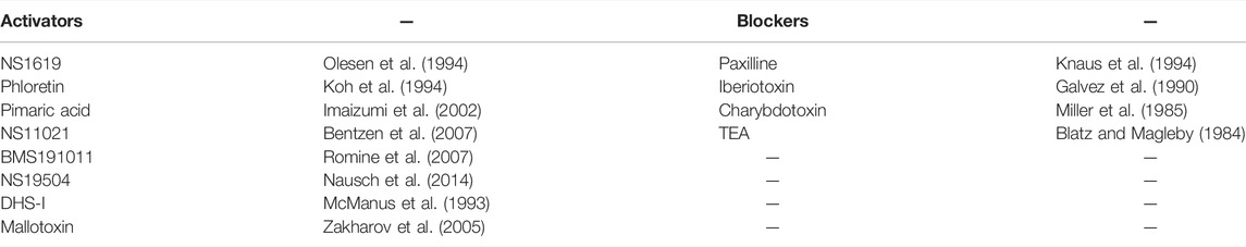
TABLE 1. commonly used BK activators and inhibitors.
In exocrine pancreas electrolyte secretion is carried out mainly by PDECs which are particularly specialized in bicarbonate (HCO3−) secretion (Lee et al., 2012). The main physiological role of PDECs is indeed to secrete a HCO3−-rich fluid, which transports the enzymes to the duodenum and raises the pH of the small intestine in order to neutralize the acidic stomach content (Venglovecz et al., 2015). In the luminal membrane of PDECs, HCO3− secretion is mediated by Cl−/HCO3− exchangers, which work in concert with the CFTR Cl− channel (Wang et al., 2006; Stewart et al., 2009). The efflux of HCO3− is crucial for proper functioning of the pancreas, indeed insufficient ductal HCO3− and fluid secretion can seriously damage the gland (Durie, 1989; Scheele et al., 1996). The BK channel is expressed in PDECs and has been found both on the basolateral as well as the luminal membrane (Gray et al., 1990; Hede et al., 2005; Venglovecz et al., 2011). The localization of the channel within ductal cells seems to vary between the two most studied animal models, namely rat and guinea pigs (Hayashi and Novak, 2013). In PDECs, BK is believed to contribute to creating the proper driving force for HCO3− secretion from the luminal membrane (Gray et al., 1990; Venglovecz et al., 2011; Lee et al., 2012) given its large conductance for K+ ions: cell-attached measurements with K+-rich pipette solution in isolated rat PDECs revealed a conductance of ∼170 pS (Hayashi et al., 2012). Expression levels of the four BK β subunits in the rat pancreatic tissue were estimated by RT-PCR in 2005 by Hede and others, who found that only β1 was expressed (Hede et al., 2005).
In the early 1990s it was proposed that the channel is involved in the secretion increase in response to secretagogues like secretin via a cAMP/PKA signaling pathway (Gray et al., 1990). The secretin receptor is coupled to adenylyl cyclase; therefore, its activation increases cAMP concentration (Folsch et al., 1980; de Ondarza and Hootman, 1995). Gray and others found that basolateral stimulation of rat PDECs with secretin induced a Ca2+-independent increase of the channel open probability, which remained in excised patches (Gray et al., 1990). Further experiments of the same study showed that channel activity is increased by phosphorylation of the channel or regulatory subunits (possibly β1, the only β subunit expressed in the tissue) by PKA, suggesting that, under physiological conditions, BK channels play an essential role in cAMP-stimulated HCO3− secretion. However, years later, it was observed that in guinea pig ducts the BK specific blocker iberiotoxin did not alter bicarbonate secretion induced by secretin, suggesting that BK is not involved in the stimulatory effect of the hormone in this animal model (Venglovecz et al., 2011). Moreover, it has been reported that channel response to PKA phosphorylation depends on the splice-variant of the α-subunit: the ZERO variant is activated by PKA, which conversely inhibits another variant named STREX-1 (Tian et al., 2001). Nevertheless, BK is probably important for bicarbonate secretion, as the luminal application of the channel opener NS11021 induces an increase of HCO3− release (Venglovecz et al., 2011).
BK-assisted HCO3− efflux was found to be important for PDECs first response to low concentrations of bile acid in the duct, in the initial phases of a gallstone-induced pancreatitis. It is indeed reported that low luminal doses of one of the primary bile acids, chenodeoxycholate (CDC), induce an increase of HCO3− secretion associated with an elevation of [Ca2+]i (Venglovecz et al., 2008). In 2010, Venglovecz and others identified in BK the apical Ca2+-dependent channel responsible for the observed phenomenon (Venglovecz et al., 2011). The authors suggest that opening of BK hyperpolarizes the cell membrane, increasing the driving force for anion secretion.
In addition to the above-mentioned studies, it is also worth to mention that BK is thought to be involved in the modulation of the secretion induced by ATP and UTP binding to purinergic receptors P2Y, although the mechanism in not completely clear (Hede et al., 1999; Hede et al., 2005; Ha and Cheong, 2017). PDECs express G-protein coupled P2Y and ligand-gated ion channel P2X receptors; it has been observed that the perfusion of ATP/UTP on the basolateral side reduced fluid and HCO3− secretion (Ishiguro et al., 1999). In this context, stimulation of P2Y receptors was shown to decrease K+ conductance in PDECs, which would in turn slow down HCO3− secretion (Hede et al., 1999). Further experiments with heterologously expressed P2Y2 and BK demonstrated that activation of the purinergic receptor is able to inhibit BK, providing an explanation for the basolateral ATP/UTP-induced decrease in HCO3− secretion (Hede et al., 2005). However, the picture turned out to be more complex as the activation of P2Y4 (also present in PDECs) resulted in an increase of BK current, and both P2Y4 and P2Y2 were able to activate another Ca2+-gated K+ channels, i.e. the intermediate conductance (IK) channel, also expressed in PDECs (Hede et al., 2005). Therefore, it is not clear what is the specific contribution of BK in the response to ATP and UTP.
In the past years ion channels have emerged as important actors for tumor biology (Lastraioli et al., 2015). As mentioned above, BK opening can induce a strong hyperpolarization in the membrane potential. Alterations in the membrane voltage have been proposed to be extremely important in controlling the cell cycle (Abdul Kadir et al., 2018); this suggests that BK can play a key role in cancer and indeed; many studies in the literature show that the activation of BK can have a significant impact on cancer cell proliferation and migration.
For example, it has been observed that BK opening can reduce the migration of glioma cells. Bordey and others reported that prolonged exposure to acetylcholine (Ach) and muscarine led to a rise of [Ca2+]i and stopped the migration of astrocytoma cells U373MG (Bordey et al., 2000). Further investigation showed that the increase of [Ca2+]i was followed by BK activation. It remained nevertheless unclear if the inhibition of cell migration was directly due to BK channel gating per se or if the cause was the increase of Ca2+ or other second messenger signals. Few years later, Kraft et al. suggested a significant impact of BK activation on the migration of glioma cells (Kraft et al., 2003). They found that BK activators phloretin and NS1619, as well as ACh, were able to reduce cell migration velocity by approximately 50% and that the latter effect could be reversed by BK blockers.
Moreover, it has been reported that BK openers induce U251 glioma and small cell lung cancer (SCLC) cells to swell (Hoa et al., 2007; Hoa et al., 2014). Also, the opening of BK has been found to induce a decrease in the viability and the migration of breast cancer cells. In a recent work, Sizemore and collaborators treated MDA-MB-231 cells with two distinct BK activators: BMS-191011 and NS-11021. The latter were able to induce a strong membrane hyperpolarization and, after prolonged treatment, also a reduction of live cells and migration (Sizemore et al., 2020). Moreover, low doses of the BK agonist BMS-191011 triggered apoptosis in MDA-MB-231 cells, where cleavage of procaspase three into caspase three was observed. In another work, Han et al., demonstrated that the BK activator NS1619 is able to inhibit proliferation and to induce apoptosis in ovarian cancer cells (Han et al., 2008).
Our group has recently investigated the effect of different BK openers on PDAC cells, which had not yet been exhaustively evaluated for this type of cancer (Remigante et al., 2021). We tested the impact on Panc-1 cell viability, migration and proliferation of two of the newest and most effective known BK activators, namely NS11021 and NS19504.
It has been reported for other types of cancers that the neoplastic tissue can differ from the normal one in terms of BK splice variants and regulatory subunits expression. For example, Egland et al. observed in pancreatic cancer tissues samples the expression of the γ1 protein LRRC26 (also known as CAPC), which left-shifts the voltage-dependence of the channel activation to more hyperpolarized voltages (Egland et al., 2006; Gonzalez-Perez et al., 2022). For the parotid BK channel V1/2 values of 50 mV have been measured when the channel was co-expressed in CHO cells with the LRRC26 protein and of 189 mV for the α subunit expressed alone (Almassy and Begenisich, 2012). Moreover, Liu et al. reported the presence of the splice variant gBK in a pancreatic adenocarcinoma sample (Liu et al., 2002). This splice variant is characterized by a normal BK unitary conductance of ∼250 pS but displays slower activation and higher sensitivity to [Ca2+]i than its closest homolog hbr5 (Liu et al., 2002). The endogenous currents displayed by Panc-1 cells at low [Ca2+]i (∼20 nM) are characterized by a deviation from linearity and a sudden increase of current density at voltages ≥ +80 mV, which are likely due to the activation of BK channels (Zuccolini et al., 2022). No clear evidence of LRRC26-mediated strong left shift of BK voltage-dependence can be observed in this cell line. This is in agreement with what has been reported by gene expression analysis (see (Klijn et al., 2015), EGA: EGAS00001000610).
Both NS11021 and NS19504 are able to induce a Paxilline-sensitive outward current in Panc-1 cells, which maintains a certain degree of voltage-dependency. We then tested the long-term effects of the two molecules on Panc-1 cell viability (72 h exposure). NS11021 decreased cell viability within 72 h. However, several lines of evidence raised serious doubts that this effect is dependent on BK activation. First, another cancer cell line (melanoma IGR37) displayed a similar viability drop after a 72 hours-long treatment with NS11021 even though the expression of the channel is ∼104 times lower in IGR37 cells than in Panc-1 cells (Ferrera et al., 2021; Zuccolini et al., 2022), and no BK like currents were activated by NS11021 in IGR37 cells (Remigante et al., 2021). Moreover, the other tested BK activator, NS19504, did not have any impact on Panc-1 viability in the same experimental conditions. Subsequently, we investigated the effect of the activators on Panc-1 migration ability with wound healing assays: neither NS11021 nor NS19504 induced a reduction in cell migration with respect to untreated Panc-1 cells. Data from real-time cell proliferation experiments were similar to those from cell viability assays: NS19504 did not have any effect, while NS11021 slowed down the proliferation of Panc-1 cells but also of the negative control IGR37. Most importantly, imaging experiments revealed that both molecules induced an increase of [Ca2+]i. In the case of NS19504, the calcium increase is partially correlated with BK activation as Paxilline strongly reduced the raise in [Ca2+]i. Conversely, NS11021 is able to activate a calcium conductance within the cell membrane in a BK-independent manner. This could explain why the molecule affected also the negative control IGR37 cells, where the channel is poorly present. Surprisingly, when we tested the ability of BMS191011, another reportedly specific BK opener (Table 1, Romine et al., 2007), to elicit BK current in Panc-1 cells we found that the compound was much less efficient than NS11021 and NS19504. Induced currents at +120 mV compared to those measured in standard bath solution were 14 times higher for NS11021, 7 times higher for NS19504 while only 2 times higher for BMS19504.
According to these recent data, it seems that the activation of the BK channel is not a good strategy for slowing down the survival and carcinogenicity of PDAC cells, although it has been suggested to be efficient for other cancer cell types. Indeed, even the more specific BK activator NS19504 did not affect viability nor proliferation or migration of Panc-1 cells. Moreover, the activation of BK can induce, in this cell type, an increase in [Ca2+]i which is a signal related to many aspects of cellular physiology.
BK is a ubiquitously expressed channel which has attracted scientists’ attention for decades. Because of its peculiar calcium- and voltage-dependent dual gating mechanism and its large conductance, this channel plays a role in many physiological pathways involving membrane hyperpolarization. Among the different tissues, BK is expressed in the epithelial cells of the pancreatic duct. Here, it has been proposed to contribute in different ways to bicarbonate efflux especially in the presence of bile acid in the duct. A deep understanding of the BK involvement in the molecular pathways underlying the HCO3- secretion regulation appears problematic. Different observations made in different animal models or focused on different cell compartments, might appear contradictory. Given the large number of channels and transporters expressed in PDECs, their specific localization in the luminal vs. apical membrane, their complex interactions and the different external stimuli that can modulate them it can actually become complicated to put together the various pieces of information in a general scheme in which the BK function is completely defined. In this regard, to get an idea of the complexity of the system, we recommend specific reviews: (Lee et al., 2012; Hayashi and Novak, 2013; Venglovecz et al., 2015; Schnipper et al., 2020; Venglovecz et al., 2021). The channel is expressed also in the neoplastic formation that originates in this fundamental part of the digestive system, the PDAC.
Studies in literature report that the prolonged application of various BK channel openers decreases viability and migration in different cancer cell lines. Unfortunately, recent experiments from our laboratory revealed that prolonged treatment with BK activators does not have such effects on PDAC cells, namely Panc-1. Indeed, Panc-1 cells exposed to different types of openers did not show any alteration neither in viability, nor in proliferation or migration. Moreover, similar results were obtained also with the melanoma line IGR39. It is important to highlight that the impact of BK activation on membrane voltage (and in turn cells duplication) may differ between different cell lines. Indeed, the gating range of the BK channel can considerably vary between different cell types, which might express different BK splice variants and/or auxiliary subunits and might have different basal levels of [Ca2+]i. For example, BMS191011 induced membrane hyperpolarization in breast cancer cells (Sizemore et al., 2020). However, Sizemore and collaborators did not test if BMS191011 actually activated BK in the breast cancer cells. This is relevant in that our group has recently found that the compound only poorly activated the BK channel in PDAC and melanoma lines (Remigante et al., 2021). A possible explanation for the divergent effects of BK agonists between Panc-1 and other cancer cells, could be that the basal Ca2+ concentration in Panc-1 cells [around 200 nM (Remigante et al., 2019; Zuccolini et al., 2022)] is not high enough to reach significant channel activation at negative voltages even with the activator present because the gating range is too right-shifted. Nevertheless, it can be hypothesized that, during the constitutive presence of the activators, local spatiotemporal fluctuations can increase the concentration of calcium in certain cell compartments rather than in the entire cytoplasm. In addition, since membrane potential and calcium concentration vary in critical points of the cell cycle, the action of the activators can occur only in those key moments, but is still enough to overall slow down cancer cell growth. Another issue to be considered is that regulatory subunits can affect BK response to activators. McManus and others observed that the channel α subunit was insensitive to the agonist DHS-I (McManus et al., 1993; Giangiacomo et al., 1998) when expressed alone in Xenopus oocytes, while the activator worked when α and β subunits were co-expressed in the heterologous system (McManus et al., 1995). Conversely, we recently discovered that the VRAC blocker DCPIB is able to activate the BK channel even when the α subunit is expressed alone in HEK293 cells (Zuccolini et al., 2022). The action of another BK agonist, Mallotoxin (MTX), is not affected by β subunits (Zakharov et al., 2005). Nevertheless, in the presence of the above-mentioned LRRC26 protein, which moves the activation voltage window to less negative potentials, MTX does not induce any further left-shift of the voltage dependence (Almassy and Begenisich, 2012). It seems therefore that a deep understanding of the channel composition in the neoplastic tissue of interest is crucial for designing and planning experiments with positive outcome. For example, it would be important to determine if and how much LRRC26 is expressed in the analyzed cancer cell line, based on available publications (Egland et al., 2006) and databases like the Cancer Cell Lines Encyclopedia (CCLE, https://ctd2-data.nci.nih.gov/Public/TGen/CCLE_RNA-seq_Analysis/). According to CCLE, the LRRC26 γ1 regulatory subunit seems to be poorly expressed in the cancer lines on which BK activators had been shown to have anti-cancer effects, although it could be detected by RT-qPCR in MDA-MB-231 cells (Egland et al., 2006). However, unfortunately, not all the studies in which BK activators are employed on cancer cells provide detailed information about the cells resting state and the expression of regulatory proteins. This might be a major problem when one approaches the study of the potentiality of BK as a target for cancer treatment.
Another point we would like to stress is that BK activators should be used with caution, as some of them may have side effects on other membrane proteins. For example, NS1619 had been thought for years to be a specific and potent activator of the BK channel, enhancing the open probability by interacting with the channel from the intracellular side (Gessner et al., 2012; Malysz et al., 2013). Unfortunately, it has been reported that NS1619 can induce Ca2+ release in pig smooth muscle cells (Yamamura et al., 2001). Other experiments by Wrzosek showed that NS1619 directly affects SERCA activity in sarcoplasmic reticulum vesicles, increasing Ca2+ leakage from isolated vesicles (Wrzosek, 2014). Another known side effect of this molecule is the blockade of the intermediate conductance calcium-gated K+ channel KCa3.1 (Cai et al., 1998). NS1619 has also been reported to affect the gating of L-type Ca2+ channels and Ca2+-gated Cl− channels (Park et al., 2007; Saleh et al., 2007). NS11021 is a potent “newer generation” BK activator, able to increase the open probability of the channel by altering gating kinetics without affecting the single-channel conductance (Bentzen et al., 2007). The compound seemed to have a direct and Ca2+-independent action on BK, as Rockman et al. reported that it could increase the open probability of a truncated channel lacking the CTD (Rockman et al., 2020). Nevertheless, in Panc-1 and IGR39 cancer cell lines, in addition to directly activating BK, the compound activated a Ca2+ conductance leading to intracellular calcium increase (Remigante et al., 2021). Among the BK activators, NS19504 appears to be the most specific. It has been tested by radioligand binding assays against 68 different channels and receptors (Nausch et al., 2014). Effects were observed only against norepinephrine transporter (SLC6A2), dopamine transporter (SLC6A3), and sigma nonopioid intracellular receptor 1 (σ1R), while the other 65 channels and receptors were not affected (Nausch et al., 2014). It is worth to specify that the molecular mechanisms by which activators modulate channel opening can be very diverse. While some of them bind the channel from the intracellular side, like NS11021 or NS1619, (Olesen et al., 1994; Bentzen et al., 2007), others like MTX act from the outside (Zakharov et al., 2005; Wu et al., 2007). It was indeed reported that in whole-cell experiments MTX affected channel gating when perfused from the outside but not when applied to the cytosol via patch pipette (Zakharov et al., 2005). Similarly, also DCPIB binds to the extracellular side of the channel, increasing the open probability (Zuccolini et al., 2022).
Another critical point in using BK activators in Panc-1 is the fact that, in this cell type, BK opening itself might generate an increase of [Ca2+]i, possibly by creating the electrical driving force for calcium entrance. For this reason it would be hard to understand if effects observed after treatments with BK activators are solely dependent on their action on BK channels or rather due to the increase of intracellular [Ca2+]i. Finally, the principle according to which membrane hyperpolarization slows down cancer growth has been proposed to work for some cancer cell types but, in contrast, other studies report anti-cancer action for BK blockers (Goda et al., 2018; Li et al., 2018; Noda et al., 2020).
We therefore conclude that the employment of BK activators presents quite some issues to which scientists should pay attention. In this regard, it appears important that studies in which BK activators are used on cancer cells, investigate several parameters that are relevant for a proper interpretation of effects on viability, migration and proliferation. In particular, basal cellular conditions [resting potential, resting (Ca2+)i, BK splice variants, BK subunit expression] need to be assessed and the biophysical properties of the native BK current under these conditions need to be determined. Furthermore, the employed molecules need to be tested for side-effects on other proteins and, in particular, on unspecific increases of intracellular calcium. In the absence of this information, the outcome of administration of BK openers is hard to interpret, as one cannot distinguish whether a given effect is due to the specific activation of BK or is the result of off-target effects. Such a variety in the impact of BK agonists and blockers does not necessarily question the results reported in the literature nor preclude the importance of these molecules in specific cases, but it can raise an alarm flag regarding the use of presumably specific BK channel modulators as anti-cancer agents.
PZ wrote the text, PG and MP supervised the writing process.
This work was supported by the grant from the Fondazione AIRC per la Ricerca sul Cancro (grant # IG 21558) and the Italian Research Ministry (PRIN 20174TB8KW) to MP.
The authors declare that the research was conducted in the absence of any commercial or financial relationships that could be construed as a potential conflict of interest.
All claims expressed in this article are solely those of the authors and do not necessarily represent those of their affiliated organizations, or those of the publisher, the editors and the reviewers. Any product that may be evaluated in this article, or claim that may be made by its manufacturer, is not guaranteed or endorsed by the publisher.
Abdul kadir, L., Stacey, M., and Barrett-Jolley, R. (2018). Emerging Roles of the Membrane Potential: Action beyond the Action Potential. Front. Physiol. 9, 1661. doi:10.3389/fphys.2018.01661
Adelman, J. P., Shen, K. Z., Kavanaugh, M. P., Warren, R. A., Wu, Y. N., Lagrutta, A., et al. (1992). Calcium-activated Potassium Channels Expressed from Cloned Complementary DNAs. Neuron 9, 209–216. doi:10.1016/0896-6273(92)90160-f
Ahring, P. K., Strøbaek, D., Christophersen, P., Olesen, S. P., and Johansen, T. E. (1997). Stable Expression of the Human Large-Conductance Ca2+-Activated K+ Channel Alpha- and Beta-Subunits in HEK293 Cells. FEBS Lett. 415, 67–70. doi:10.1016/s0014-5793(97)01096-x
Almassy, J., and Begenisich, T. (2012). The LRRC26 Protein Selectively Alters the Efficacy of BK Channel Activators. Mol. Pharmacol. 81, 21–30. doi:10.1124/mol.111.075234
Atkinson, N. S., Robertson, G. A., and Ganetzky, B. (1991). A Component of Calcium-Activated Potassium Channels Encoded by the Drosophila Slo Locus. Science 253, 551–555. doi:10.1126/science.1857984
Bentzen, B. H., Nardi, A., Calloe, K., Madsen, L. S., Olesen, S. P., and Grunnet, M. (2007). The Small Molecule NS11021 Is a Potent and Specific Activator of Ca2+-Activated Big-Conductance K+ Channels. Mol. Pharmacol. 72, 1033–1044. doi:10.1124/mol.107.038331
Bentzen, B. H., Olesen, S. P., Rønn, L. C., and Grunnet, M. (2014). BK Channel Activators and Their Therapeutic Perspectives. Front. Physiol. 5, 389. doi:10.3389/fphys.2014.00389
Blatz, A. L., and Magleby, K. L. (1984). Ion Conductance and Selectivity of Single Calcium-Activated Potassium Channels in Cultured Rat Muscle. J. Gen. Physiol. 84, 1–23. doi:10.1085/jgp.84.1.1
Bordey, A., Sontheimer, H., and Trouslard, J. (2000). Muscarinic Activation of BK Channels Induces Membrane Oscillations in Glioma Cells and Leads to Inhibition of Cell Migration. J. Membr. Biol. 176, 31–40. doi:10.1007/s00232001073
Brenner, R., Peréz, G. J., Bonev, A. D., Eckman, D. M., Kosek, J. C., Wiler, S. W., et al. (2000). Vasoregulation by the Beta1 Subunit of the Calcium-Activated Potassium Channel. Nature 407, 870–876. doi:10.1038/35038011
Butler, A., Tsunoda, S., Mccobb, D. P., Wei, A., and Salkoff, L. (1993). mSlo, a Complex Mouse Gene Encoding "maxi" Calcium-Activated Potassium Channels. Science 261, 221–224. doi:10.1126/science.7687074
Cai, S., Garneau, L., and Sauvé, R. (1998). Single-channel Characterization of the Pharmacological Properties of the K(Ca2+) Channel of Intermediate Conductance in Bovine Aortic Endothelial Cells. J. Membr. Biol. 163, 147–158. doi:10.1007/s002329900379
De ondarza, J., and Hootman, S. R. (1995). Regulation of Cyclic AMP Levels in guinea Pig Pancreatic Ducts and Cultured Duct Epithelial Monolayers. Pancreas 11, 261–270. doi:10.1097/00006676-199510000-00008
Dickerson, M. T., Dadi, P. K., Altman, M. K., Verlage, K. R., Thorson, A. S., Jordan, K. L., et al. (2019). Glucose-mediated Inhibition of Calcium-Activated Potassium Channels Limits α-cell Calcium Influx and Glucagon Secretion. Am. J. Physiol. Endocrinol. Metab. 316, E646–E659. doi:10.1152/ajpendo.00342.2018
Durie, P. R. (1989). The Pathophysiology of the Pancreatic Defect in Cystic Fibrosis. Acta Paediatr. Scand. Suppl. 363, 41–44. doi:10.1111/apa.1989.78.s363.41
Egland, K. A., Liu, X. F., Squires, S., Nagata, S., Man, Y. G., Bera, T. K., et al. (2006). High Expression of a Cytokeratin-Associated Protein in Many Cancers. Proc. Natl. Acad. Sci. U. S. A. 103, 5929–5934. doi:10.1073/pnas.0601296103
Fan, J. Q., Wang, M. F., Chen, H. L., Shang, D., Das, J. K., and Song, J. (2020). Current Advances and Outlooks in Immunotherapy for Pancreatic Ductal Adenocarcinoma. Mol. Cancer 19, 32. doi:10.1186/s12943-020-01151-3
Ferrera, L., Barbieri, R., Picco, C., Zuccolini, P., Remigante, A., Bertelli, S., et al. (2021). TRPM2 Oxidation Activates Two Distinct Potassium Channels in Melanoma Cells through Intracellular Calcium Increase. Int. J. Mol. Sci. 22 (16), 8359. doi:10.3390/ijms22168359
Fölsch, U. R., Fischer, H., Söling, H. D., and Creutzfeldt, W. (1980). Effects of Gastrointestinal Hormones and Carbamylcholine on cAMP Accumulation in Isolated Pancreatic Duct Fragments from the Rat. Digestion 20, 277–292. doi:10.1159/000198449
Galvez, A., Gimenez-Gallego, G., Reuben, J. P., Roy-Contancin, L., Feigenbaum, P., Kaczorowski, G. J., et al. (1990). Purification and Characterization of a Unique, Potent, Peptidyl Probe for the High Conductance Calcium-Activated Potassium Channel from Venom of the Scorpion Buthus Tamulus. J. Biol. Chem. 265, 11083–11090. doi:10.1016/s0021-9258(19)38560-6
Gessner, G., Cui, Y. M., Otani, Y., Ohwada, T., Soom, M., Hoshi, T., et al. (2012). Molecular Mechanism of Pharmacological Activation of BK Channels. Proc. Natl. Acad. Sci. U. S. A. 109, 3552–3557. doi:10.1073/pnas.1114321109
Giangiacomo, K. M., Kamassah, A., Harris, G., and Mcmanus, O. B. (1998). Mechanism of Maxi-K Channel Activation by Dehydrosoyasaponin-I. J. Gen. Physiol. 112, 485–501. doi:10.1085/jgp.112.4.485
Goda, A. A., Siddique, A. B., Mohyeldin, M., Ayoub, N. M., and El Sayed, K. A. (2018). The Maxi-K (BK) Channel Antagonist Penitrem A as a Novel Breast Cancer-Targeted Therapeutic. Mar. Drugs 16 (5), 157. doi:10.3390/md16050157
Gómez, R., Maglio, L. E., Gonzalez-Hernandez, A. J., Rivero-Pérez, B., Bartolomé-Martín, D., and Giraldez, T. (2021). NMDA Receptor-BK Channel Coupling Regulates Synaptic Plasticity in the Barrel Cortex. Proc. Natl. Acad. Sci. U. S. A. 118 (35), e2107026118. doi:10.1073/pnas.2107026118
Gonzalez-perez, V., and Lingle, C. J. (2019). Regulation of BK Channels by Beta and Gamma Subunits. Annu. Rev. Physiol. 81, 113–137. doi:10.1146/annurev-physiol-022516-034038
Gonzalez-perez, V., Zhou, Y., Ciorba, M. A., and Lingle, C. J. (2022). The LRRC Family of BK Channel Regulatory Subunits: Potential Roles in Health and Disease. J. Physiol. 600, 1357–1371. doi:10.1113/JP281952
Gray, M. A., Greenwell, J. R., Garton, A. J., and Argent, B. E. (1990). Regulation of Maxi-K+ Channels on Pancreatic Duct Cells by Cyclic AMP-dependent Phosphorylation. J. Membr. Biol. 115, 203–215. doi:10.1007/BF01868636
Ha, G. E., and Cheong, E. (2017). Calcium-activated Chloride Channels: a New Target to Control the Spiking Pattern of Neurons. BMB Rep. 50, 109–110. doi:10.5483/bmbrep.2017.50.3.033
Han, X., XI, L., Wang, H., Huang, X., Ma, X., Han, Z., et al. (2008). The Potassium Ion Channel Opener NS1619 Inhibits Proliferation and Induces Apoptosis in A2780 Ovarian Cancer Cells. Biochem. Biophys. Res. Commun. 375, 205–209. doi:10.1016/j.bbrc.2008.07.161
Hayashi, M., and Novak, I. (2013). Molecular Basis of Potassium Channels in Pancreatic Duct Epithelial Cells. Channels (Austin) 7, 432–441. doi:10.4161/chan.26100
Hayashi, M., Wang, J., Hede, S. E., and Novak, I. (2012). An Intermediate-Conductance Ca2+-Activated K+ Channel Is Important for Secretion in Pancreatic Duct Cells. Am. J. Physiol. Cell Physiol. 303, C151–C159. doi:10.1152/ajpcell.00089.2012
Hede, S. E., Amstrup, J., Christoffersen, B. C., and Novak, I. (1999). Purinoceptors Evoke Different Electrophysiological Responses in Pancreatic Ducts. P2Y Inhibits K(+) Conductance, and P2X Stimulates Cation Conductance. J. Biol. Chem. 274, 31784–31791. doi:10.1074/jbc.274.45.31784
Hede, S. E., Amstrup, J., Klaerke, D. A., and Novak, I. (2005). P2Y2 and P2Y4 Receptors Regulate Pancreatic Ca(2+)-Activated K+ Channels Differently. Pflugers Arch. 450, 429–436. doi:10.1007/s00424-005-1433-3
Herrera, G. M., Heppner, T. J., and Nelson, M. T. (2000). Regulation of Urinary Bladder Smooth Muscle Contractions by Ryanodine Receptors and BK and SK Channels. Am. J. Physiol. Regul. Integr. Comp. Physiol. 279, R60–R68. doi:10.1152/ajpregu.2000.279.1.R60
Hite, R. K., Tao, X., and Mackinnon, R. (2017). Structural Basis for Gating the High-Conductance Ca2+-Activated K+ Channel. Nature 541, 52–57. doi:10.1038/nature20775
Hoa, N. T., Ge, L., Tajhya, R. B., Beeton, C., Cornforth, A. N., Abolhoda, A., et al. (2014). Small Cell Lung Cancer Cells Express the Late Stage gBK Tumor Antigen: a Possible Immunotarget for the Terminal Disease. Am. J. Transl. Res. 6 (3), 188–205.
Hoa, N. T., Zhang, J. G., Delgado, C. L., Myers, M. P., Callahan, L. L., Vandeusen, G., et al. (2007). Human Monocytes Kill M-CSF-Expressing Glioma Cells by BK Channel Activation. Lab. Invest. 87, 115–129. doi:10.1038/labinvest.3700506
Horrigan, F. T., and Aldrich, R. W. (2002). Coupling between Voltage Sensor Activation, Ca2+ Binding and Channel Opening in Large Conductance (BK) Potassium Channels. J. Gen. Physiol. 120, 267–305. doi:10.1085/jgp.20028605
Imaizumi, Y., Sakamoto, K., Yamada, A., Hotta, A., Ohya, S., Muraki, K., et al. (2002). Molecular Basis of Pimarane Compounds as Novel Activators of Large-Conductance Ca(2+)-Activated K(+) Channel Alpha-Subunit. Mol. Pharmacol. 62, 836–846. doi:10.1124/mol.62.4.836
Ishiguro, H., Naruse, S., Kitagawa, M., Hayakawa, T., Case, R. M., and Steward, M. C. (1999). Luminal ATP Stimulates Fluid and HCO3- Secretion in guinea-pig Pancreatic Duct. J. Physiol. 519 Pt 2, 551–558. doi:10.1111/j.1469-7793.1999.0551m.x
Jacobson, D. A., Mendez, F., Thompson, M., Torres, J., Cochet, O., and Philipson, L. H. (2010). Calcium-activated and Voltage-Gated Potassium Channels of the Pancreatic Islet Impart Distinct and Complementary Roles during Secretagogue Induced Electrical Responses. J. Physiol. 588, 3525–3537. doi:10.1113/jphysiol.2010.190207
Klijn, C., Durinck, S., Stawiski, E. W., Haverty, P. M., Jiang, Z., Liu, H., et al. (2015). A Comprehensive Transcriptional Portrait of Human Cancer Cell Lines. Nat. Biotechnol. 33, 306–312. doi:10.1038/nbt.3080
Knaus, H. G., Mcmanus, O. B., Lee, S. H., Schmalhofer, W. A., Garcia-Calvo, M., Helms, L. M., et al. (1994). Tremorgenic Indole Alkaloids Potently Inhibit Smooth Muscle High-Conductance Calcium-Activated Potassium Channels. Biochemistry 33, 5819–5828. doi:10.1021/bi00185a021
Koh, D. S., Reid, G., and Vogel, W. (1994). Activating Effect of the Flavonoid Phloretin on Ca(2+)-Activated K+ Channels in Myelinated Nerve Fibers of Xenopus laevis [corrected]. Neurosci. Lett. 165, 167–170. doi:10.1016/0304-3940(94)90736-6
Kraft, R., Krause, P., Jung, S., Basrai, D., Liebmann, L., Bolz, J., et al. (2003). BK Channel Openers Inhibit Migration of Human Glioma Cells. Pflugers Arch. 446, 248–255. doi:10.1007/s00424-003-1012-4
Kshatri, A. S., Gonzalez-Hernandez, A., and Giraldez, T. (2018). Physiological Roles and Therapeutic Potential of Ca2+ Activated Potassium Channels in the Nervous System. Front. Mol. Neurosci. 11, 258. doi:10.3389/fnmol.2018.00258
Lastraioli, E., Iorio, J., and Arcangeli, A. (2015). Ion Channel Expression as Promising Cancer Biomarker. Biochim. Biophys. Acta 1848, 2685–2702. doi:10.1016/j.bbamem.2014.12.016
Latorre, R., Castillo, K., Carrasquel-Ursulaez, W., Sepulveda, R. V., Gonzalez-Nilo, F., Gonzalez, C., et al. (2017). Molecular Determinants of BK Channel Functional Diversity and Functioning. Physiol. Rev. 97, 39–87. doi:10.1152/physrev.00001.2016
Lee, M. G., Ohana, E., Park, H. W., Yang, D., and Muallem, S. (2012). Molecular Mechanism of Pancreatic and Salivary Gland Fluid and HCO3 Secretion. Physiol. Rev. 92, 39–74. doi:10.1152/physrev.00011.2011
Li, N., Liu, L., Li, G., Xia, M., Du, C., and Zheng, Z. (2018). The Role of BKCa in Endometrial Cancer HEC-1-B Cell Proliferation and Migration. Gene 655, 42–47. doi:10.1016/j.gene.2018.02.055
Li, Q., and Yan, J. (2016). Modulation of BK Channel Function by Auxiliary Beta and Gamma Subunits. Int. Rev. Neurobiol. 128, 51–90. doi:10.1016/bs.irn.2016.03.015
Liu, X., Chang, Y., Reinhart, P. H., Sontheimer, H., and Chang, Y. (2002). Cloning and Characterization of Glioma BK, a Novel BK Channel Isoform Highly Expressed in Human Glioma Cells. J. Neurosci. 22, 1840–1849. doi:10.1523/jneurosci.22-05-01840.2002
Malysz, J., Rovner, E. S., and Petkov, G. V. (2013). Single-channel Biophysical and Pharmacological Characterizations of Native Human Large-Conductance Calcium-Activated Potassium Channels in Freshly Isolated Detrusor Smooth Muscle Cells. Pflugers Arch. 465, 965–975. doi:10.1007/s00424-012-1214-8
Marty, A. (1981). Ca-dependent K Channels with Large Unitary Conductance in Chromaffin Cell Membranes. Nature 291, 497–500. doi:10.1038/291497a0
Mcmanus, O. B., Harris, G. H., Giangiacomo, K. M., Feigenbaum, P., Reuben, J. P., Addy, M. E., et al. (1993). An Activator of Calcium-dependent Potassium Channels Isolated from a Medicinal Herb. Biochemistry 32, 6128–6133. doi:10.1021/bi00075a002
Mcmanus, O. B., Helms, L. M., Pallanck, L., Ganetzky, B., Swanson, R., and Leonard, R. J. (1995). Functional Role of the Beta Subunit of High Conductance Calcium-Activated Potassium Channels. Neuron 14, 645–650. doi:10.1016/0896-6273(95)90321-6
Meech, R. W. (1972). Intracellular Calcium Injection Causes Increased Potassium Conductance in Aplysia Nerve Cells. Comp. Biochem. Physiol. A Comp. Physiol. 42, 493–499. doi:10.1016/0300-9629(72)90128-4
Meech, R. W., and Standen, N. B. (1974). Calcium-mediated Potassium Activation in Helix Neurones. J. Physiol. 237, 43P–44P. doi:10.1113/jphysiol.1974.sp010481
Miller, C., Moczydlowski, E., Latorre, R., and Phillips, M. (1985). Charybdotoxin, a Protein Inhibitor of Single Ca2+-Activated K+ Channels from Mammalian Skeletal Muscle. Nature 313, 316–318. doi:10.1038/313316a0
Nausch, B., Rode, F., Jørgensen, S., Nardi, A., Korsgaard, M. P., Hougaard, C., et al. (2014). NS19504: a Novel BK Channel Activator with Relaxing Effect on Bladder Smooth Muscle Spontaneous Phasic Contractions. J. Pharmacol. Exp. Ther. 350, 520–530. doi:10.1124/jpet.113.212662
Noda, S., Chikazawa, K., Suzuki, Y., Imaizumi, Y., and Yamamura, H. (2020). Involvement of the γ1 Subunit of the Large-Conductance Ca2+-Activated K+ Channel in the Proliferation of Human Somatostatinoma Cells. Biochem. Biophys. Res. Commun. 525, 1032–1037. doi:10.1016/j.bbrc.2020.02.176
Olesen, S. P., Munch, E., Moldt, P., and Drejer, J. (1994). Selective Activation of Ca(2+)-dependent K+ Channels by Novel Benzimidazolone. Eur. J. Pharmacol. 251, 53–59. doi:10.1016/0014-2999(94)90442-1
Pallotta, B. S., Magleby, K. L., and Barrett, J. N. (1981). Single Channel Recordings of Ca2+-Activated K+ Currents in Rat Muscle Cell Culture. Nature 293, 471–474. doi:10.1038/293471a0
Park, W. S., Kang, S. H., Son, Y. K., Kim, N., Ko, J. H., Kim, H. K., et al. (2007). The Mitochondrial Ca2+-Activated K+ Channel Activator, NS 1619 Inhibits L-type Ca2+ Channels in Rat Ventricular Myocytes. Biochem. Biophys. Res. Commun. 362, 31–36. doi:10.1016/j.bbrc.2007.07.057
Ransom, C. B., and Sontheimer, H. (2001). BK Channels in Human Glioma Cells. J. Neurophysiol. 85, 790–803. doi:10.1152/jn.2001.85.2.790
Remigante, A., Morabito, R., and Marino, A. (2019). Natural Antioxidants Beneficial Effects on Anion Exchange through Band 3 Protein in Human Erythrocytes. Antioxidants (Basel) 9 (1), 25. doi:10.3390/antiox9010025
Remigante, A., Zuccolini, P., Barbieri, R., Ferrera, L., Morabito, R., Gavazzo, P., et al. (2021). NS-11021 Modulates Cancer-Associated Processes Independently of BK Channels in Melanoma and Pancreatic Duct Adenocarcinoma Cell Lines. Cancers (Basel) 13 (23), 6144. doi:10.3390/cancers13236144
Rieg, T., Vallon, V., Sausbier, M., Sausbier, U., Kaissling, B., Ruth, P., et al. (2007). The Role of the BK Channel in Potassium Homeostasis and Flow-Induced Renal Potassium Excretion. Kidney Int. 72, 566–573. doi:10.1038/sj.ki.5002369
Rockman, M. E., Vouga, A. G., and Rothberg, B. S. (2020). Molecular Mechanism of BK Channel Activation by the Smooth Muscle Relaxant NS11021. J. Gen. Physiol. 152 (6), e201912506. doi:10.1085/jgp.201912506
Romine, J. L., Martin, S. W., Meanwell, N. A., Gribkoff, V. K., Boissard, C. G., Dworetzky, S. I., et al. (2007). 3-[(5-Chloro-2-hydroxyphenyl)methyl]-5-[4-(trifluoromethyl)phenyl ]-1,3,4-Oxadiazol-2(3h)-One, BMS-191011: Opener of Large-Conductance Ca(2+)-Activated Potassium (Maxi-K) Channels, Identification, Solubility, and SAR. J. Med. Chem. 50, 528–542. doi:10.1021/jm061006n
Saleh, S. N., Angermann, J. E., Sones, W. R., Leblanc, N., and Greenwood, I. A. (2007). Stimulation of Ca2+-Gated Cl- Currents by the Calcium-dependent K+ Channel Modulators NS1619 [1,3-Dihydro-1-[2-Hydroxy-5-(trifluoromethyl)phenyl]-5-(trifluoromethyl)-2h-Benzimidazol-2-One] and Isopimaric Acid. J. Pharmacol. Exp. Ther. 321, 1075–1084. doi:10.1124/jpet.106.118786
Sancho, M., and Kyle, B. D. (2021). The Large-Conductance, Calcium-Activated Potassium Channel: A Big Key Regulator of Cell Physiology. Front. Physiol. 12, 750615. doi:10.3389/fphys.2021.750615
Sandle, G. I., Perry, M. D., Mathialahan, T., Linley, J. E., Robinson, P., Hunter, M., et al. (2007). Altered Cryptal Expression of Luminal Potassium (BK) Channels in Ulcerative Colitis. J. Pathol. 212, 66–73. doi:10.1002/path.2159
Scheele, G. A., Fukuoka, S. I., Kern, H. F., and Freedman, S. D. (1996). Pancreatic Dysfunction in Cystic Fibrosis Occurs as a Result of Impairments in Luminal pH, Apical Trafficking of Zymogen Granule Membranes, and Solubilization of Secretory Enzymes. Pancreas 12, 1–9. doi:10.1097/00006676-199601000-00001
Schnipper, J., Dhennin-Duthille, I., Ahidouch, A., and Ouadid-Ahidouch, H. (2020). Ion Channel Signature in Healthy Pancreas and Pancreatic Ductal Adenocarcinoma. Front. Pharmacol. 11, 568993. doi:10.3389/fphar.2020.568993
Shi, J., Krishnamoorthy, G., Yang, Y., Hu, L., Chaturvedi, N., Harilal, D., et al. (2002). Mechanism of Magnesium Activation of Calcium-Activated Potassium Channels. Nature 418, 876–880. doi:10.1038/nature00941
Sizemore, G., Mclaughlin, S., Newman, M., Brundage, K., Ammer, A., Martin, K., et al. (2020). Opening Large-Conductance Potassium Channels Selectively Induced Cell Death of Triple-Negative Breast Cancer. BMC Cancer 20, 595. doi:10.1186/s12885-020-07071-1
Stewart, A. K., Yamamoto, A., Nakakuki, M., Kondo, T., Alper, S. L., and Ishiguro, H. (2009). Functional Coupling of Apical Cl-/HCO3- Exchange with CFTR in Stimulated HCO3- Secretion by guinea Pig Interlobular Pancreatic Duct. Am. J. Physiol. Gastrointest. Liver Physiol. 296, G1307–G1317. doi:10.1152/ajpgi.90697.2008
Tao, X., Hite, R. K., and Mackinnon, R. (2017). Cryo-EM Structure of the Open High-Conductance Ca2+-Activated K+ Channel. Nature 541, 46–51. doi:10.1038/nature20608
Tao, X., and Mackinnon, R. (2019). Molecular Structures of the Human Slo1 K+ Channel in Complex with β4. Elife 8, e51409. doi:10.7554/eLife.51409
Tian, L., Duncan, R. R., Hammond, M. S., Coghill, L. S., Wen, H., Rusinova, R., et al. (2001). Alternative Splicing Switches Potassium Channel Sensitivity to Protein Phosphorylation. J. Biol. Chem. 276, 7717–7720. doi:10.1074/jbc.C000741200
Venglovecz, V., Hegyi, P., Rakonczay, Z., Tiszlavicz, L., Nardi, A., Grunnet, M., et al. (2011). Pathophysiological Relevance of Apical Large-Conductance Ca²+-Activated Potassium Channels in Pancreatic Duct Epithelial Cells. Gut 60, 361–369. doi:10.1136/gut.2010.214213
Venglovecz, V., Rakonczay, Z., Gray, M. A., and Hegyi, P. (2015). Potassium Channels in Pancreatic Duct Epithelial Cells: Their Role, Function and Pathophysiological Relevance. Pflugers Arch. 467, 625–640. doi:10.1007/s00424-014-1585-0
Venglovecz, V., Rakonczay, Z., Ozsvári, B., Takács, T., Lonovics, J., Varró, A., et al. (2008). Effects of Bile Acids on Pancreatic Ductal Bicarbonate Secretion in guinea Pig. Gut 57, 1102–1112. doi:10.1136/gut.2007.134361
Venglovecz, V. H. P., and Gray, M. A. (2021). Ion Channels in Pancreatic Duct Epithelial Cells in Health and Disease. Ann Arbor, MI: Pancreapedia.
Wang, Y., Soyombo, A. A., Shcheynikov, N., Zeng, W., Dorwart, M., Marino, C. R., et al. (2006). Slc26a6 Regulates CFTR Activity In Vivo to Determine Pancreatic Duct HCO3- Secretion: Relevance to Cystic Fibrosis. EMBO J. 25, 5049–5057. doi:10.1038/sj.emboj.7601387
Wrzosek, A. (2014). The Potassium Channel Opener NS1619 Modulates Calcium Homeostasis in Muscle Cells by Inhibiting SERCA. Cell Calcium 56, 14–24. doi:10.1016/j.ceca.2014.03.005
Wu, S. N., Wang, Y. J., and Lin, M. W. (2007). Potent Stimulation of Large-Conductance Ca2+-Activated K+ Channels by Rottlerin, an Inhibitor of Protein Kinase C-Delta, in Pituitary Tumor (GH3) Cells and in Cortical Neuronal (HCN-1A) Cells. J. Cell Physiol. 210, 655–666. doi:10.1002/jcp.20866
Xia, X. M., Zeng, X., and Lingle, C. J. (2002). Multiple Regulatory Sites in Large-Conductance Calcium-Activated Potassium Channels. Nature 418, 880–884. doi:10.1038/nature00956
Yamamura, H., Ohi, Y., Muraki, K., Watanabe, M., and Imaizumi, Y. (2001). BK Channel Activation by NS-1619 Is Partially Mediated by Intracellular Ca2+ Release in Smooth Muscle Cells of Porcine Coronary Artery. Br. J. Pharmacol. 132, 828–834. doi:10.1038/sj.bjp.0703885
Yang, H., Shi, J., Zhang, G., Yang, J., Delaloye, K., and Cui, J. (2008). Activation of Slo1 BK Channels by Mg2+ Coordinated between the Voltage Sensor and RCK1 Domains. Nat. Struct. Mol. Biol. 15, 1152–1159. doi:10.1038/nsmb.1507
Zakharov, S. I., Morrow, J. P., Liu, G., Yang, L., and Marx, S. O. (2005). Activation of the BK (SLO1) Potassium Channel by Mallotoxin. J. Biol. Chem. 280, 30882–30887. doi:10.1074/jbc.M505302200
Zhang, J., Guan, X., Li, Q., Meredith, A. L., Pan, H. L., and Yan, J. (2018). Glutamate-activated BK Channel Complexes Formed with NMDA Receptors. Proc. Natl. Acad. Sci. U. S. A. 115, E9006–E9014. doi:10.1073/pnas.1802567115
Zhang, W., Zhang, K., Zhang, P., Zheng, J., Min, C., and Li, X. (2020). Research Progress of Pancreas-Related Microorganisms and Pancreatic Cancer. Front. Oncol. 10, 604531. doi:10.3389/fonc.2020.604531
Keywords: BK channel, pancreatic duct, pancreatic duct adenocarcinoma (PDAC), BK activators, cancer, NS11021, NS19504, BMS191011
Citation: Zuccolini P, Gavazzo P and Pusch M (2022) BK Channel in the Physiology and in the Cancer of Pancreatic Duct: Impact and Reliability of BK Openers. Front. Pharmacol. 13:906608. doi: 10.3389/fphar.2022.906608
Received: 28 March 2022; Accepted: 10 May 2022;
Published: 24 May 2022.
Edited by:
Ricardo Gómez, Cold Spring Harbor Laboratory, United StatesReviewed by:
Christopher Lingle, Washington University in St. Louis, United StatesCopyright © 2022 Zuccolini, Gavazzo and Pusch. This is an open-access article distributed under the terms of the Creative Commons Attribution License (CC BY). The use, distribution or reproduction in other forums is permitted, provided the original author(s) and the copyright owner(s) are credited and that the original publication in this journal is cited, in accordance with accepted academic practice. No use, distribution or reproduction is permitted which does not comply with these terms.
*Correspondence: Paolo Zuccolini, cGFvbG8uenVjY29saW5pQGliZi5jbnIuaXQ=
Disclaimer: All claims expressed in this article are solely those of the authors and do not necessarily represent those of their affiliated organizations, or those of the publisher, the editors and the reviewers. Any product that may be evaluated in this article or claim that may be made by its manufacturer is not guaranteed or endorsed by the publisher.
Research integrity at Frontiers
Learn more about the work of our research integrity team to safeguard the quality of each article we publish.