- 1Research Institute of Nephrology, Zhengzhou University, The First Affiliated Hospital of Zhengzhou University, Zhengzhou, China
- 2Department of Integrated Traditional and Western Nephrology, The First Affiliated Hospital of Zhengzhou University, Zhengzhou, China
- 3Henan Province Research Center for Kidney Disease, The First Affiliated Hospital of Zhengzhou University, Zhengzhou, China
- 4Blood Purification Center, The First Affiliated Hospital of Zhengzhou University, Zhengzhou, China
- 5Department of Pharmacy, The First Affiliated Hospital of Zhengzhou University, Zhengzhou, China
- 6School of Laboratory Medicine, Xinxiang Medical University, Xinxiang, China
- 7Clinical Systems Biology Laboratories, The First Affiliated Hospital of Zhengzhou University, Zhengzhou, China
Acute kidney injury (AKI), a common and serious clinical kidney syndrome with high incidence and mortality, is caused by multiple pathogenic factors, such as ischemia, nephrotoxic drugs, oxidative stress, inflammation, and urinary tract obstruction. Cell death, which is divided into several types, is critical for normal growth and development and maintaining dynamic balance. Ferroptosis, an iron-dependent nonapoptotic type of cell death, is characterized by iron overload, reactive oxygen species accumulation, and lipid peroxidation. Recently, growing evidence demonstrated the important role of ferroptosis in the development of various kidney diseases, including renal clear cell carcinoma, diabetic nephropathy, and AKI. However, the exact mechanism of ferroptosis participating in the initiation and progression of AKI has not been fully revealed. Herein, we aim to systematically discuss the definition of ferroptosis, the associated mechanisms and key regulators, and pharmacological progress and summarize the most recent discoveries about the role and mechanism of ferroptosis in AKI development. We further conclude its potential therapeutic strategies in AKI.
Introduction
Acute kidney injury (AKI), which is characterized by a rapid decline in renal function, is caused by various physiological and pathological factors (Figure 1) (Kellum et al., 2021). In recent years, the incidence and mortality rates of AKI have remained high and exhibit an annual increasing trend (Liu et al., 2020). According to epidemiological investigation, the incidence rate of AKI is increasing with the increase of the aging population, the incidence rate of AKI in general inpatients is 5%, and the mortality rate of severe patients is over 50% (Ronco et al., 2019). The main clinical manifestation of AKI is the sudden or continuous decline of renal function in a short time (several hours to weeks), resulting in the accumulation of harmful and toxic metabolites in the body, and severe patients may also progress to renal failure or even death (Deep et al., 2021).
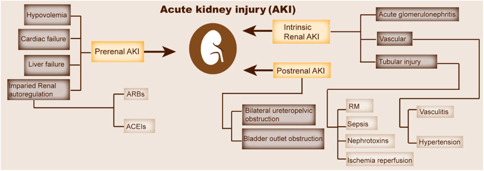
FIGURE 1. Major pathogenesis of AKI. This figure indicates the pathophysiology and etiology of AKI. CHF, congestive heart failure; ARBs, angiotensin receptor blockers; ACEIs, angiotensin-converting enzyme inhibitors; RM, rhabdomyolysis.
Given the pathogenesis of AKI, oxidative stress, inflammation, hypoxia, and apoptosis caused by surgery, rhabdomyolysis (RM), infection, ischemia–reperfusion injury (IRI), sepsis, and nephrotoxic drugs are the main causes (Ow et al., 2021). In clinical treatment, although blood purification can improve renal function after AKI, it cannot completely cure the disease. Relevant studies have also shown that the occurrence of AKI increases the potential risk of patients developing chronic kidney disease (CKD) and end-stage renal disease.
Cell death is critical for maintaining dynamic balance in the body. In recent years, other forms of cell death besides apoptosis, including ferroptosis, necroptosis, and pyroptosis, have been gradually recognized with the deepening of research (D'Arcy, 2019). Direct evidence shows that ferroptosis inhibitors possess renal protective effects in various animal models of AKI, suggesting that ferroptosis plays an important role in the initiation and progression of AKI (Hu et al., 2019). Therefore, it is of great significance to explore the mechanism of ferroptosis in AKI and clarify the effects of ferroptosis on the prognosis of AKI to CKD.
Overview of Ferroptosis
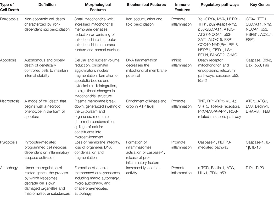
Iron is an indispensable trace element in the human body, which plays multiple important biological functions, including the induction of ATP production, synthesis of DNA and heme, and many other physiological activities (Milto et al., 2016). Intracellular iron content, especially ferrous iron overload, could induce lipid peroxidation. In 2012, Dixon et al. officially named this iron-dependent cell death mediated by excessive lipid peroxidation as ferroptosis for the first time. Intracellular iron retention, reduced glutathione (GSH) content, and lipid reactive oxygen species (ROS) accumulation are the main characteristics of ferroptosis (Dixon et al., 2012). The excessive accumulation of ROS can activate intracellular oxidative stress response and damage proteins, nucleic acids, and lipids, finally resulting in the occurrence of ferroptosis (Tang et al., 2021). Ferroptosis is different from the previously found regulatory cell death patterns such as apoptosis, necrosis, pyroptosis, and autophagy in morphology, genetics, and mechanisms (Hirschhorn and Stockwell, 2019) (Table 1).
ACSL4, acyl-CoA synthetase long-chain family member 4; ALOX-15, arachidonate lipoxygenase 15; AP-1, activator protein-1; ATG5, autophagy-related 5; ATG7, autophagy-related 7; COQ10, coenzyme Q10; DRAM3, damage regulated autophagy modulator 3; FSP1, ferroptosis suppressor protein 1; GPX4, glutathione peroxidase 4; HSPβ1, heat shock protein beta-1; Keap1, Keleh-like ECH-associated protein 1, MAPK, mitogen-activated protein kinase; MLKL, mixed lineage kinase domain like protein; mTOR, mammalian target of rapamycin; MVA, mevalonate; LC3, microtubule-associated protein 1 light chain3; NCOA4, nuclear receptor coactivator 4; Nrf2, nuclear factor erythroid 2-related factor 2; PKC, protein kinase C; RIP, receptor-interacting serine/threonine kinase; ROS, reactive oxygen species; SAT1, spermidine/spermine N1-acetyltransferase 1; SLC7A11, solute carrier family 7 member 11; system Xc−, cysteine/glutamate transporter receptor; TFEB, transcription factor EB; TFR1, transferrin receptor 1; TNF-α, tumor necrosis factor α.
A number of studies have investigated ferroptosis, however, the specific mechanisms of ferroptosis are still unknown. With the deepening of research on ferroptosis, various regulators and mechanismas have been developed successively, which provides new insights into the treatment of ferroptosis-associated diseases.
Mechanisms and Key Regulators of Ferroptosis
During the past few years, the process and functions of ferroptosis has been well studied. In addition, several regulators of ferroptosis have been extensively investigated, including system Xc−, GPX4, p53, FSP1, and nuclear factor erythroid 2-related factor (Nrf2). In this part, we will briefly describe the major regulators and related molecular mechanisms of ferroptosis (Figure 2).
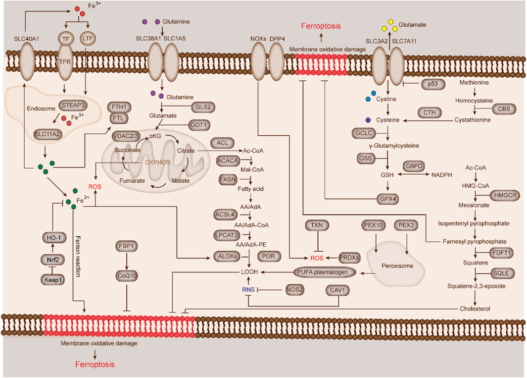
FIGURE 2. Mechanisms and key regulators of ferroptosis. The major mechanisms and key regulators of ferroptosis can be roughly divided into two important mechanisms (iron metabolism and lipid peroxidation) and seven key regulators (system Xc−, GPX4, p53, acyl-CoA synthetase long-chain family member 4 [ACSL4], FSP1, and Nrf2). In addition, other mechanisms and regulators, such as p53/SLC7A11 and VDAC2/3 are also involved in lipid regulation and ferroptosis.
Iron Metabolism
As an important inducer of ROS production through enzymatic or non-enzymatic reactions, iron plays an important role in ferroptosis. In general, under normal conditions, extracellular Fe3+ ions first bind to transferrin (TFR), and then are transported into cells via membrane transferrin receptor 1 (TFR1) and stored in the form of iron protein complex (mainly ferritin). Fe3+ ions are reduced to Fe2+ and subsequently transported and stored in the cell iron pool, while excess Fe2+ ions are stored in ferritin (van Swelm et al., 2020). Ferritin is an iron storage protein complex, which is composed of ferritin light chain and ferritin heavy chain 1 (FTH1). In the case of iron metabolic disorder, the low expression of FTH1 and the overexpression of TFR1 often lead to the excessive accumulation of Fe2+ ions, which induce the production and accumulation of a large amount of ROS through Fenton reaction and eventually promote cell ferroptosis (Daher and Karim, 2017). TFR1 has been considered as the only known protein that controls iron input in mammalian cells and indispensable in iron metabolism (Kawabata, 2019).
Lipid Peroxidation
Lipid peroxidation plays a driving role in the occurrence of ferroptosis, which can be completed by non-enzymatic or enzymatic reaction. Compared with unsaturated fatty acids and monounsaturated fatty acids, polyunsaturated fatty acids (PUFAs) are more prone to lipid peroxidation and ferroptosis (Latunde-Dada, 2017). The abundance and location of PUFAs determine the degree of lipid peroxidation and ferroptosis. Free PUFAs are substrates for the synthesis of lipid signal transduction mediators, but they must be esterified into membrane phospholipids and oxidized to initiate ferroptosis. The formation of PUFA coenzyme A derivatives is a necessary condition for the generation of ferroptosis, and the involvement of regulatory enzymes in the biosynthesis of PUFAs in membrane phospholipids can trigger or prevent ferroptosis (Yang et al., 2016).
ACSL4 and lysophosphatidylcholine acyltransferase 3 are involved in the biosynthesis of phosphatidylethanolamine in the cell membrane. The deletion of these genes will increase the cell resistance to ferroptosis. By contrast, cells supplemented with arachidonic acid or other PUFAs increases their sensitivity to ferroptosis (Doll et al., 2017; Shimbara-Matsubayashi et al., 2019). Lipoxygenases (LOXs) are important enzyme systems that mediate the formation of ferroptosis peroxides. Free PUFAs are the preferred substrate of LOX; knocking out LOXs can alleviate the injury caused by ferroptosis. In addition, phosphatidylethanolamine can be further oxidized under the catalysis of LOXs, thereby inducing cell ferroptosis (Stockwell et al., 2017).
System Xc−
System Xc−, a cystine glutamate transporter that widely expressed on the cell membrance, is composed of light chain subunit (SLC7A11) and heavy chain subunit (SLC3A2) (Dixon et al., 2012). Cystine and glutamate are exchanged at a ratio of 1:1 inside and outside the cell via system Xc−. System Xc− transfers cystine or cystine sulfide into cells and reduces it to cysteine (Chen et al., 2021a). Under extracellular oxidation, the exchange of cystine/cystine sulfide and glutamate is the upstream of ferroptosis (Hirschhorn and Stockwell, 2019). The exchange of extracellular cystine and intracellular glutamate affects the synthesis of GSH and maintains the stability of intracellular GSH, thereby protecting cells from oxidative stress (Bersuker et al., 2019).
GSH is mainly composed of glutamate, cysteine, and glycine, and the sulfhydryl structure of GSH can be oxidized and dehydrogenated, making GSH an important antioxidant and free radical scavenger in the body (Bannai and Tateishi, 1986). GSH is an essential cofactor for GPXs to exert antioxidant function (Lash, 2005). Reduced GSH is an important intracellular antioxidant in mammals. Glutamic acid, cysteine, and glycine generate GSH in two steps under the catalysis of glutamyl cysteine ligase and glutathione synthase (Bjørklund et al., 2021). Therefore, the availability of glutamate and cysteine affects GSH biosynthesis. Recently, studies have found that ferroptosis induced by erastin and sulfasalazine can inhibit system Xc−, reduce intracellular cystine uptake, decrease GSH synthesis, induce lipid peroxide accumulation, and finally lead to cell ferroptosis (Floros et al., 2021). Jiang et al. reported that pachymic acid could alleviate IRI-induced AKI (IRI-AKI) by upregulating Nrf2, SLC7A11, and HO-1 expression, which indicated the importance of system Xc− in kidney injury (Jiang et al., 2021). Tumor suppressor protein p53 plays a critical role in cellular response to various life activities, such as DNA damage, hypoxia, oncogene activation, and ferroptosis (Kang et al., 2019). p53 downregulates SLC7A11 and inhibits the uptake of cysteine via system Xc−, thus affecting GPX4 activity, resulting in renal cell ferroptosis (Jiang et al., 2015a). Li et al. showed that α-lipoic acid ameliorates folic acid (FA)-induced AKI by inhibiting p53 and up-regulating SLC7A11 expression (Li et al., 2021a). Additionally, Beclin1, a key regulator of autophagy, has been proved to be a new driver of ferroptosis and can promote ferroptosis by regulating the action of system Xc− in tumor cells (Kang et al., 2018).
In summary, system Xc− can maintain GSH levels by maintaining the balance between intracellular and extracellular cystine and glutamate. Once the equilibrium state is broken, the depletion of intracellular GSH level will decrease the synthesis and activity of GPX4, finally resulting in the occurrence of cell ferroptosis.
GPX4
GPXs, one family of antioxidant enzymes that are widely expressed in different tissues and can reduce peroxides (Jiao et al., 2017). The GPXs contains eight members, however, onlyGPX1, GPX3, and GPX4 are found to be expressed in the kidney; notably, GPX4 plays a more important role in ferroptosis (Brigelius-Flohé and Maiorino, 2013). Ursini et al. firstly isolated GPX4 from pig liver in 1982 and found that it had the ability to inhibit iron-triggered lipid peroxidation in microsomes (Ursini et al., 1982). At present, GPX4 is the only known enzyme that can directly remove lipid peroxidation, which can reduce harmful lipid peroxidation into harmless substances, so as to interrupt the occurrence of lipid peroxidation and finally inhibit cell ferroptosis (Forcina and Dixon, 2019). The inhibition of GPX4 activity can lead to lipid peroxide accumulation, which is a marker of ferroptosis (Seibt et al., 2019).
Chemical compounds such as RSL3, DPI7, and DPI10 can directly act on GPX4 and block its activity, thus leading to the inhibition of cellular antioxidant capacity and ROS accumulation and finally resulting in the development of ferroptosis (Liang et al., 2019). Yang et al. found that GPX4 was a key regulator of ferroptosis and inhibiting GPX4 could induce tumor cell ferroptosis (Yang et al., 2014). Gaschler et al. found that five-membered cyclic peroxides 1 and 2 could increase the sensitivity of cells to ferroptosis by indirectly inactivating GPX4 (Gaschler et al., 2018). Kinowaki et al. found that GPX4 was significantly correlated with the prognosis of diffuse B-cell lymphoma and that GPX4-depleted cells were sensitive to ROS-induced cell ferroptosis (Kinowaki et al., 2018). The mechanism of ferroptosis induced by erastin and RSL3 is not exactly the same. Erastin mainly inhibits system Xc−, thus suppressing GSH synthesis, but indirectly inhibiting GPX4, which results in cell ferroptosis. By contrast, RSL3 can directly bind to GPX4, resulting in cell lipid peroxidation and ROS accumulation and finally leading to cell ferroptosis (Friedmann Angeli et al., 2014).
p53
p53 is an important tumor suppressor. Aside from its effects on cell death, autophagy, apoptosis, and pyroptosis, p53 is also involved in regulating cell ferroptosis. p53 can promote ferroptosis by decreasing the expression of SLC7A11. It also suppresses ferroptosis via the direct downregulation of dipeptidyl peptidase 4 (DPP4) or upregulation of cyclin dependent kinase inhibitor 1A/p21 expression. For example, Jiang et al. firstly found that p53 inhibited the uptake of cystine by blocking SLC7A11, which caused a significant reduction of GSH, and induction of cell ferroptosis (Jiang et al., 2015a). Chu et al. revealed that knockout of arachidonate 12-lipoxygenase (ALOX12) specifically blocked the occurrence of ferroptosis, which proved that p53 indirectly activated the function of ALOX12 by inhibiting SLC7A11, resulting in ALOX12-dependent ferroptosis after ROS stress (Chu et al., 2019). Xie et al. demonstrated that p53 could inhibit the ferroptosis sensitivity of tumor cells induced by erastin by blocking DPP4 activity (Xie et al., 2017). In conclusion, p53 can not only regulate apoptosis and cell cycle arrest, but also inhibit the occurrence and development of tumors by regulating ferroptosis.
FSP1
FSP1, previously known as apoptosis-inducing factor mitochondrion-associated 2, has been recently proved as an endogenous ferroptosis inhibitor (Doll et al., 2019). Studies demonstrated that FSP1 was a key component of the non-mitochondrial coenzyme Q (CoQ) antioxidant system and could promote the generation of lipophilic free radicals and the recruitment of antioxidants (RTA) by decreasing the expression of coenzyme Q10. RTA can effectively relieve ferroptosis by neutralizing the accumulation of lipid peroxide (Santoro, 2020). Therefore, FSP1 is considered as a ferroptosis suppressor in various cancer cells. Bersuker et al. recently found that when GPX4 was inactivated, FSP1 could maintain the growth of lung cancer cells, which implied that FSP1 contributed to the resistance against cell ferroptosis and might be one important drug target for cancer treatment (Bersuker et al., 2019). Additionally, through high-throughput screening among 10,000 small molecule drugs, researchers found that the inhibitor iFSP1 could significantly upregulate the sensitivity of tumor cells to ferroptosis by inhibiting FSP1. Meanwhile, Dai et al. reported that FSP1 could block the occurrence of cell ferroptosis by affecting ubiquinol metabolism (Dai et al., 2020).
Nrf2
Nrf2 is an important regulator of cellular antioxidant responses (Baird and Yamamoto, 2020).
Studies have reported that Nrf2 can transcriptionally regulate the expression of a series of downstream genes related to ferroptosis, including NAD(P)H:quinone oxidoreductase 1, HO-1, SLC7A11, and GPX4 (Abdalkader et al., 2018). Thus, Nrf2 is considered as an important negative regulator of ferroptosis. Fan et al. reported that overexpression of Nrf2 could up-regulate SLC7A11, thereby inhibiting the cell ferroptosis (Fan et al., 2017). Tian et al. found Nrf2 attenuated myocardial ischemia-reperfusion injury in diabetic rats via blocking ferroptosis (Tian et al., 2022). In addition, Sun et al. reported that the p62/Keap1/Nrf2 axis was activated under the stimulation of ferroptosis inducers, causing the activation and transcription of HO-1 and FTH-1 and finally decreasing the sensitivity to ferroptosis in cells (Sun et al., 2016). Additionally, Nrf2 directly or indirectly regulates the expression and function of GPX4 (Hayes and Dinkova-Kostova, 2014). Liao et al. showed that DJ-1 could inhibit trophoblast ferroptosis of preeclampsia by upregulating the expression of Nrf2 and its downstream gene GPX4 (Liao et al., 2022). Ge et al. demonstrated that Zinc attenuated contusion spinal cord injury by inhibiting ferroptosis via activating Nrf2/GPX4 pathway (Ge et al., 2021). On the contrary, during the past few years, it was found that Nrf2 activation could induce ferroptosis. For example, Wei et al. revealed that Tagitinin C promoted cell ferroptosis in colorectal cancer by activating PERK/Nrf2/HO-1 pathway (Wei et al., 2021). Chen et al. found Nrf2 was the downstream gene of tumor suppressor ARF, and ARF could induce ferroptosis of tumor cells by inhibiting Nrf2 activation, which uncovered that Nrf2 activation might be a positive regulator in ferroptosis (Chen et al., 2017). Nrf2 activation could upregulate the expression of HO-1, however, several studied supported that HO-1 overexpression would trigger ferroptosis by promoting iron overloading and lipid peroxidation (Xu et al., 2019). Therefore, these studies suggested a dual role of Nrf2 in ferroptosis.
Recently, Nrf2 was also proved to participate in regulating ferroptosis in kidney injury. Li et al. reported that upregulating Nrf2 could rescue kidney injury and renal failure by inhibiting ferroptosis in diabetic mice (Li et al., 2021b). Meanwhile, it found that roxadustat treatment could attenuate FA-induced AKI by blocking ferroptosis through regulating Nrf2 (Li et al., 2020a). Meng et al. discovered that the activation of Nrf2 by ADAMTS-13 could inhibit ferroptosis to ameliorate cisplatin-induced AKI (Meng et al., 2021). Jiang et al. found that pachymic acid exerted a protective effect on IRI-induced AKI mice through the inhibition of ferroptosis by activating Nrf2 (Jiang et al., 2021). Yang et al. demonstrated that dimethyl fumarate could prevent ferroptosis to attenuate AKI by upregulating Nrf2 (Yang et al., 2021). Deng et al. revealed that the inhibition of mitochondrial iron overload could alleviate aristolactam I-induced AKI by regulating the Nrf2/HO-1/GPX4 axis (Deng et al., 2020). The above studies showed that Nrf2 played a key role in renal injury caused by ferroptosis. Since the specific mechanism of Nrf2-mediated ferroptosis in AKI renal injury has not been fully clarified, future work should focus on identifying the target gene of Nrf2 and further clarify the mechanism of this gene in specifically regulating ferroptosis in AKI, which is of great significance for the development of therapeutic drugs for AKI in the future.
ACSL4
ACSL4 is a member of the long-chain acyl coenzyme A synthetase family, which can catalyze the activation of fatty acids to synthesize acyl coenzyme A. Therefore, it is considered as a key enzyme in fatty acid catabolism (Qu et al., 2021). Different from other ACSL family members, ACSL4 can activate long-chain PUFAs and participate in the synthesis of membrane phospholipids. Meanwhile, knockout of ACSL4 will cause ferroptosis (Doll et al., 2017).
The long-chain PUFAs on these membranes are easily oxidized, and ferroptosis inducers such as RSL3 can induce cell ferroptosis. ] ACSl4−/− knockout cells displayed resistance against ferroptosis, and the inhibitory effect of lipid peroxide on GPX4 required the participation of ACSL4 (Shimbara-Matsubayashi et al., 2019). Doll et al. found that thiazolidinediones could protect ACSL4 knockout embryonic fibroblasts, reduce the degree of membrane lipid oxidation and cell death, and significantly prolong the survival time of ACSL4 knockout mice (Doll et al., 2017). Wang et al. demonstrated thaterastin and RSL3 induced renal tubular cell death accompanied by high ACSL4 levels in vitro, while the ACSL4 inhibitor rosiglitazone improved the survival rate and alleviated kidney injury in diabetic nephropathy mice, and decreased lipid peroxidation products and iron content (Wang et al., 2020). Li et al. reported that ACSL4 played a critical role in ferroptosis-mediated tissue injury in IR mouse model, and special protein 1 could increase ACSL4 transcription and further promote the progression of ferroptosis (Li et al., 2019a). Feng et al. revealed that miRNA-17-92 exerted protective effects on endothelial cells by inhibiting erastin-induced ferroptosis via targeting the A20/ACSL4 pathway (Xiao et al., 2019a). Cui et al. showed that ACSL4 induced neuronal death by increasing lipid peroxidation and promoted ischemic stroke by enhancing ferroptosis-induced brain injury and neuroinflammation (Cui et al., 2021). These studies suggested that ACSL4 might be a crucial determinant of ferroptosis (Yuan et al., 2016).
Other Mechanisms
Several other pathways might also be closely related to the occurrence of ferroptosis. Voltage-dependent anion channels (VDACs), a type of channel proteins located in the mitochondrial outer membrane, play an essential role in the communication between mitochondria and other organelles (Skonieczna et al., 2017). Yagoda et al. revealed that erastin could directly combine with VDAC2/3 to reduce the oxidation rate of NADH, and induce cell ferroptosis (Yagoda et al., 2007). In addition, the PI3K/Akt/mTOR (Yi et al., 2020), p53/SLC7A11 (Jiang et al., 2015b), and glutamine metabolic pathways (Gao et al., 2015) play negative regulatory roles in the occurrence of ferroptosis. To sum up, the research on ferroptosis is still in the preliminary stage, and the specific mechanisms have not been completely identified.
Pharmacological Progress of Ferroptosis
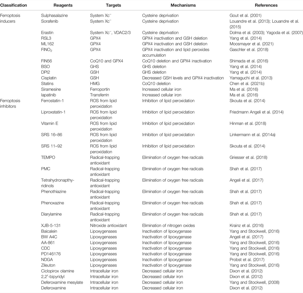
In recent years, numerous natural and synthetic drugs related to ferroptosis, including inducers and inhibitors, have been identified. Generally, for ferroptosis in non-neoplastic diseases (e.g., AKI, CKD), ferroptosis inhibitors can be used to reduce iron levels, restrain lipid peroxidation, and relieve renal function insufficiency. For carcinoma, cell death can be promoted by ferroptosis inducers to block tumor cell proliferation by increasing ROS levels and inducing the progression of iron-dependent lipid peroxidation (Xie et al., 2016) (Table 2).
Ferroptosis is Involved in the Development of AKI
AKI is a disease featured by acute renal insufficiency, which is caused by many factors. Although significant progress has been made, no specific drugs have been developed for the prevention and treatment of AKI (Kellum et al., 2021). Recent findings revealed that ferroptosis is a promising therapeutic target, especially in diseases dominated by kidney tubular injury (Carney, 2021). Therefore, studies on RM-, ischemia-reperfusion-, sepsis-, and nephrotoxic drug-induced AKI animal models have provided direct evidence to confirm the participation of ferroptosis in AKI.
Ferroptosis and AKI Caused by RM
RM refers to a clinical syndrome caused by the entry of a large number of intracellular components such as myoglobin (Mb), phosphocreatine kinase, and lactate dehydrogenase into the peripheral blood, which accounts for 13%–50% of cases with AKI (Cabral et al., 2020). The accumulation of Mb in kidney is the core factor leading to kidney injury, and Mb-mediated lipid peroxidation in renal tubular epithelial cells is closely related to glutamate metabolism and can mediate proximal tubular ferroptosis (Vanholder et al., 2021). Guerrero-Hue et al. confirmed that ferroptosis was involved in the development of RM-associated AKI, and curcumin could alleviate RM-induced kidney injury via inhibiting ferroptosis of renal tubular epithelial cells (Guerrero-Hue et al., 2019). Zarjou et al. demonstrated that FTH−/- mice displayed a higher mortality and more serious kidney injury in RM-induced AKI model compared with wild-type mice, indicating that FTH exerted a protective effect on renal tubular injury in AKI (Zarjou et al., 2013).
Ferroptosis and IRI-AKI
IRI refers to injury caused by the restoration of blood flow after the interruption or decline of blood flow in the body or organs for a period of time (Sharfuddin and Molitoris, 2011). Renal IRI is one of the main causes of AKI, and the pathophysiology of IRI-AKI includes mitochondrial dysfunction, inflammation, ROS, and lipid peroxidation (Malek and Nematbakhsh, 2015). Recent study demonstrated ferroptosis might be a new driver in initiating IRI-AKI (Yan et al., 2020). Huang et al. found that in an IRI-AKI mouse model, augmenter of liver regeneration (ALR) could regulate the development of ferroptosis through GSH/GPX4. Ding et al. reported that microRNAs also participated in the regulation of ferroptosis in an IRI-AKI mouse model (Ding et al., 2020). Additionally, Wang et al. showed ACSL4 knockout significanlty inhibited the ferroptosis of renal tubular epithelial cells in IRI-AKI mice, which indicated ACSL4 might be a key target for AKI treatment (Wang et al., 2022). XJB-5-131 is a new generation of antioxidant, which exerts dual effects of mitochondrial targeting and free radical scavenging. Zhao et al. confirmed that XJB-5-131 could specifically inhibit ferroptosis by inhibiting lipid peroxidation, so as to alleviate IRI-AKI (Zhao et al., 2020). Recently, the application of ferroptosis inhibitors, including Fer-1 and liproxstatin-1, had been found to exhibit protective effects against functional acute renal failure and structural organ damage in IRI-AKI mice (Yan et al., 2020). The latest study from Tonnus et al. showed that the deletion of FSP1 or GPX4 improved the sensitivity of tubular ferroptosis in IRI-AKI mice, which supported that the dysfunction of ferroptosis-surveilling systems enhanced the sensitivity of mice to tubular ferroptosis during AKI (Tonnus et al., 2021).
Ferroptosis and Sepsis-Associated AKI
Sepsis refers to the systemic inflammatory responses caused by infection and can develop into severe sepsis, septic shock, and multiple organ dysfunction. Sepsis has been considered as one of the main causes of AKI, which is called SA-AKI (Bagshaw et al., 2009). The mechanism of SA-AKI is complicated, and the ideal therapeutic effects have not been achieved. A large number of studies revealed that renal tubular cell necrosis, apoptosis, and autophagy occur in SA-AKI, but these typical modes of cell death cannot fully explain the renal pathological changes in SA-AKI. Recently, Xiao et al. reported that ferroptosis of renal tubular epithelial cells was occurred in SA-AKI mice, and the results demonstrated that Maresin conjugates in tissue regeneration 1 (MCTR1) could significantly suppress cell ferroptosis in SA-AKI by activating Nrf2 pathway (Xiao et al., 2021). Zhang et al. demonstrated miR-124-3p.1 displayed inhibitory effect on the ferroptosis in SA-AKI by inhibiting the up-regulation of lysophosphatidylcholine acyltransferase 3, which indicated miR-124-3p.1 might be a biomarker and potential therapeutical target in AKI (Wu et al., 2022).
Ferroptosis in Nephrotoxic Drug-Induced AKI
Nephrotoxic drugs (e.g., FA, cisplatin) is another key factor triggering AKI. FA at a high dosage might rapidly form crystals in renal tubules, resulting in AKI. Guo et al. found that lipid peroxidation occured in the kidney tissue of FA-induced AKI mouse model, and blocking Rev-erb-α/β could ameliorate FA-induced AKI by restraining ferroptosis (Guo et al., 2021). Martin-Sanchez et al. confirmed that treatment with the ferroptosis inhibitor Fer-1 could significantly alleviate renal function and reduce kidney damage in FA-induced AKI mice (Martin-Sanchez et al., 2017).
Cisplatin is a commonly used anticancer drug; however, it often causes nephrotoxicity, limiting its use. Previous studies had shown that the incidence of cisplatin-induced AKI was 20%–30%. Recently, studies showed that cisplatin administration in FTH knockout mice significantly induced proximal tubule injury compared with that in wild-type mice (Zarjou et al., 2013). Lu et al. reported that Ras homolog enriched in brain could relieve cisplatin-induced AKI by maintaining mitochondrial homeostasis (Lu et al., 2020). Zhou et al. recently reported that polydatin enabled to attenuate the cell ferroptosis in cisplatin-induced AKI via regulating system Xc−/GSH/GPX4 pathway and inhibiting iron metabolism disorders (Zhou et al., 2022). Deng et al. found Se/Albumin nanoparticles could alleviate cisplatin-induced AKI by inhibiting ferroptosis with a decrease of superoxide dismutase, and up-regulation of GSH and GPX4 (Deng et al., 2022).
Biological Reactions Associated With Ferroptosis During AKI
Previous studies had shown that biological reactions such as necroptosis, inflammation, autophagy, and ferroptosis displayed a close relationship with the occurrence and development of various diseases in the human body. Therefore, in this part, we aim to summarize the recent studies about the crosstalk between various biological reactions and ferroptosis during AKI (Figure 3).
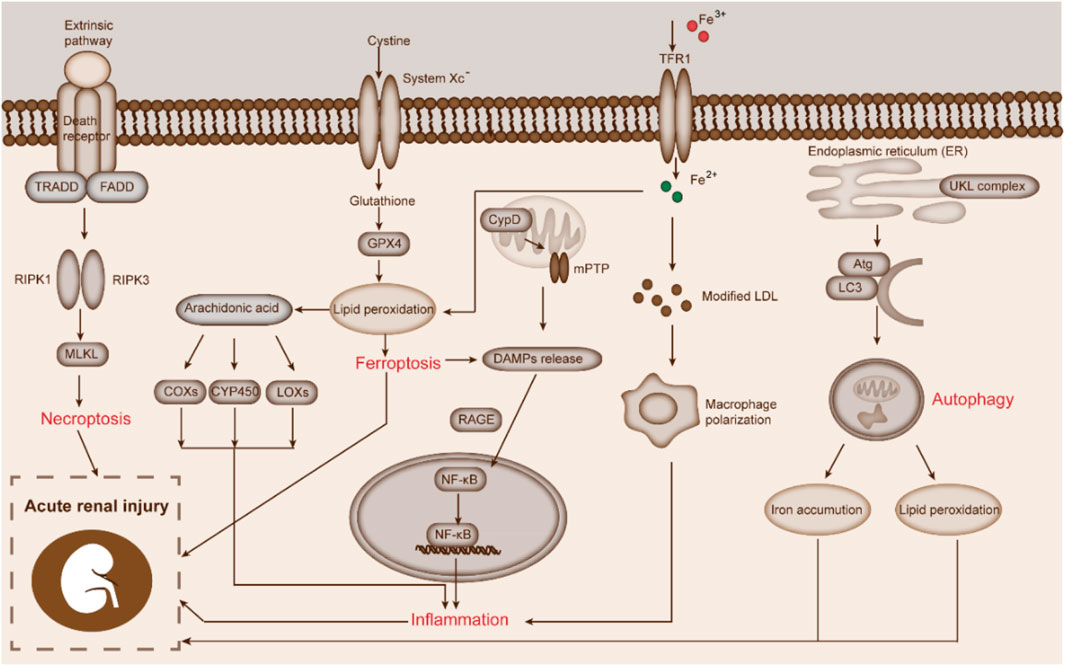
FIGURE 3. Biological responses correlated to ferroptosis during AKI. This figure displays the crosstalk between necroptosis, inflammation, autophagy, and ferroptosis in AKI.
Necroptosis
Among various types of cell death, necroptosis is a recently discovered and controllable programmed cell death mode. Generally, necroptosis is mediated by receptor-interacting serine/threonine protein kinase 3 and its substrate mixed lineage kinase domain-like protein (MLKL) (Chen et al., 2020). Several studies had shown that the imbalance of necroptosis was closely related to the development of various human diseases, such as inflammatory diseases, autoimmune diseases, tumors, kidney diseases, and degenerative diseases (Westman et al., 2019).
Since the discovery of ferroptosis and necroptosis, the role of necroptosis in the development of AKI and AKI to CKD progression had been deeply investigated. A growing body of evidence had described the essential roles of ferroptosis and necroptosis in AKI. Angeli et al. reported that necroptosis inhibitor protected the kidney injury from inhibiting necroptosis and ferroptosis in IRI- and cisplatin-induced AKI mice (Friedmann Angeli et al., 2014). Using MLKI−/− mice to establish IRI model, Müller et al. found that inhibiting necroptosis only exerted a slight protective effect on renal tubule injury and ferroptosis development. However, when ferroptosis inhibitors were used, they detected the high sensitivity of renal tubular epithelial cells to necroptosis, suggesting a “selective” interaction between necroptosis and ferroptosis (Müller et al., 2017). Martin-Sanchez et al. revealed that necroptosis-related proteins contributed to FA-induced AKI, but ferroptosis was still the main cause of FA-induced AKI. In addition, the secondary inflammatory responses triggered by ferroptosis further promoted renal injury in AKI, which suggested the association of ferroptosis and necroptosis to FA-induced AKI in mice (Martin-Sanchez et al., 2017).
Autophagy
Autophagy is a biological process, in which cells wrap their cytoplasmic proteins or organelles to form autophagic vesicles, fuse with lysosomes, and degrade their inclusions, so as to realize metabolic requirements and organelle renewal in cells (Mizushima and Komatsu, 2011). Studies showed that autophagy was closely related to the initiation of cancers, neurodegenerative diseases, and kidney diseases (Luan et al., 2021). IRI, ROS, and inflammatory responses will affect autophagy, and the impairment of autophagy function will accelerate cell death (Mizushima and Levine, 2020). In mice models intervened by ferroptosis inducers, researchers observed the accumulation of autophagy lysosomes and the abnormal expression of autophagy-related genes such as ATG3, ATG5, and Beclin1. Interestingly, autophagy can trigger the occurrence of ferroptosis. Hou et al. found that knockout of autophagy-related genes limited the development of erastin-induced ferroptosis by down-regulating intracellular iron concentrations and lipid peroxidation (Hou et al., 2016). Masaldan et al. demonstrated that senescent cells were significantly resistant against ferroptosis, and autophagy activators could induce ferritin degradation, resulting in the down-regulation of TFR1 and ferritin (Masaldan et al., 2018). Additionally, Chen et al. revealed that legumain deficiency alleviated the development of renal tubular injury and ferroptosis in AKI mice, which proved legumain enhanced renal tubular epithelial cell ferroptosis by facilitating autophagy in AKI(Chen et al., 2021c).
Inflammation
During ferroptosis, the shrinkage of cell and organelle membranes and the increase of permeability cause cell content release, including damage-associated molecular pattern (DAMPs) (Feng et al., 2020), which leads to local inflammatory responses (Mu et al., 2021). Li et al. reported that ferroptosis inhibition alleviated angiotensin II-induced inflammation and protected the brain from external injury by activating Nrf2/HO-1 pathway (Li et al., 2021c). In oxalate crystallization-induced AKI mouse mode, Proneth et al. observed necrotizing inflammatory response in tissues with ferroptosis, such as DAMPs release and production of proinflammatory factors. They also found that the inflammatory response could be effectively blocked by ferroptosis inhibitor (Proneth and Conrad, 2019). Linkermann et al. found the proinflammatory factor TNF-α released by macrophages exhibited side effects on the stability of ferroptosis-related protein GPX4 (Linkermann et al., 2014b). In addition, Li et al. observed that Fer-1 inhibited TLR4/Trif/type I IFN pathway and alleviated the inflammatory response in cardiac IRI mouse model (Li et al., 2019b). Guerrero-Hue et al. described that curcumin ameliorated kidney injury by decreasing inflammation-mediated cell ferroptosis (Guerrero-Hue et al., 2019).
Other Metabolic Pathways
Recently, several other metabolic pathways closely related to ferroptosis were explored. Pannexin 1 is a channel protein that can release ATP and Su et al. found Pannexin 1 deficiency could reduce the ferroptosis of renal tubular cells in IRI-AKI mice by regulating the MAPK/ERK pathway and NOCA4-mediated ferritin autophagy (Su et al., 2019). CoQ, also known as ubiquinone, is a lipid-soluble quinone compound. CoQ produced by the mevalonate pathway is not only an antioxidant in cells, but also an effective inhibitor of ferroptosis. FIN56 could exhaust CoQ by regulating the activity of squalene synthase, leading to the accumulation of lethal lipid peroxide and occurrence of ferroptosis (Baschiera et al., 2021). Dixon et al. found that statins could deplete CoQ and enhance the cell sensitivity to ferroptosis by blocking 3-hydroxy-3-methylglutaryl coenzyme A reductase of the mevalonate pathway (Dixon et al., 2015). Besides, tremendous studies revealed that the p62/Keap1/Nrf2, Atg5/NCOA4, as well as glutamine metabolic pathway were also played key roles in ferroptosis by effectively regulating the intracellular iron content and ROS level (Li et al., 2020b).
Treatment of AKI by Targeting Ferroptosis
Inhibiting ferroptosis of renal tubular epithelial cells could effectively alleviate the progression of renal injury in AKI. With the gradual recognition of the role of ferroptosis in AKI, the treatment of AKI by inhibiting ferroptosis has become a hot topic in the field of nephrology (Figure 4).

FIGURE 4. Treatment of AKI by targeting ferroptosis of renal tubular epithelial cells. This figure shows that abnormal increase of Fe2+ and H2O2 promotes the Fenton chemical reaction and lipid peroxidation, and mediates ferroptosis, thus leading to AKI. RM initiates ferroptosis by increasing the levels of Fe2+; IRI induces ferroptosis by suppressing the transformation of Fe2+ to Fe3+. Other pathogenic factors such as FA, cisplatin, sepsis, and dexamethasone can also induce ferroptosis and lead to AKI. Ferrostatin-1, liproxstatin-1, deferoxamine, XJB-5-131, and vitamin E can alleviate or delay the development of AKI by inhibiting ferroptosis.
At present, the main therapeutic advances include ferroptosis inhibitors, lipid peroxidation pathway inhibitors, iron homeostasis regulators, and ROS generation inhibitors. Skouta et al. demonstrated that Fer-1 decreased ROS levels, reduced lipid peroxides, scavenged oxygen free radicals, and inhibited cell death in diverse disease models including AKI (Skouta et al., 2014). Meng et al. reported that similar to Fer-1, ADAMTS13 could also relieve cisplatin-induced inflammatory response and oxidative stress in AKI mice by regulating the Nrf2-mediated ferroptosis (Meng et al., 2021). Liproxstatin-1 has been considered as another ferroptosis inhibitor via delaying lipid hydroperoxide accumulation (Zilka et al., 2017). Friedmann Angeli et al. found liproxstatin-1 could suppress ferroptosis of human renal tubule epithelial cells in IRI-AKI mouse model (Friedmann Angeli et al., 2014). Ma et al. observed that ferroptosis inhibition by liproxstatin-1 significantly reduced kidney lipid peroxidation and the degree of renal histopathological injury in AKI rats (Ma et al., 2021). Yang et al. found that lysyl oxidase inhibitors inhibited ferroptosis in AKI by blocking lipid peroxidation (Yang et al., 2016). Zhang et al. displayed that liproxstatin-1 alleviated the unilateral ureteral obstruction-induced renal injury in AKI mice by inhibiting the ferroptosis of renal tubular epithelial cells (Zhang et al., 2021). In addition to common ferroptosis inhibitors, several other antioxidants and iron chelators (e.g., vitamin E and deferoxamine) were also observed to inhibit ferroptosis by regulating ROS, iron metabolism, and lipid peroxidation. Xiao et al. indicated that miR-212-5p attenuated neuronal ferroptosis during traumatic brain injury by targeting PTGS2 (Xiao et al., 2019b).
Previously, dexamethasone was used as immunosuppressant for the treatment of inflammatory diseases. A latest in ferroptosis research revealed that dexamethasone significantly promoted the ferroptosis in AKI by increasing GR-mediated dipeptidase-1 expression and decreasing the GSH level, which indicated dexamethasone might be an potential ferroptosis agonist (von Massenhausen et al., 2022).
In addition, with the development of traditional Chinese medicines, several herb components were also confirmed to exert anti-ferroptosis activity and further alleviated renal injury in AKI. Wang et al. demonstrated that quercetin could alleviate kidney injury in IRI- or FA-induced AKI mice by inhibiting ferroptosis alleviating (Wang et al., 2021a). . Cheng et al. showed that vitamin D inhibited ferroptosis by activating the Nrf2/GPX4 pathway in zebrafish liver cells (Cheng et al., 2021). Hu et al. also demonstrated that vitamin D receptor activation attenuated cisplatin-induced AKI by inhibiting renal tubular epithelial cells ferroptosis via targeting GPX4 (Hu et al., 2020).
Currently, several studies on the development of ferroptosis inhibitors has been performed in various AKI animal models, but they still lack clinical application potential. Therefore, clinical research on the prevention and targeted treatment of ferroptosis in AKI should be conducted in the future.
Conclusion
AKI is a type of kidney disease caused by various pathogenic conditions, such as acute tissue ischemia, toxic substance injury, and immune system disorders (Kellum et al., 2021). In view of the complex pathogenesis of AKI, effective strategies for the prevention and treatment of AKI are still lacking (Mercado et al., 2019). Therefore, in-depth analysis of the pathogenesis of AKI is of great significance to alleviate the renal injury and improve the survival of patients with AKI.
Ferroptosis affects the development of various cancers and nervous system diseases (Zheng and Conrad, 2020). With the deepening of research, ferroptosis has been reported to play an important role in the progression of kidney injury, including various AKI models and CKDs. The pathogenesis of AKI is complicated by apoptosis, necrosis, and other forms of cell death in the pathophysiological process, however, the degree of ferroptosis in AKI is different (Han et al., 2020). In addition, the crosstalk between ferroptosis and autophagy, inflammation, oxidative stress, and other regulatory mechanisms in the progression of kidney injury should be further explored (Hu et al., 2019).
Through summarizing the relationship between AKI and ferroptosis, researchers found that ferroptosis inhibition has renal protective effect in different AKI models, but some questions remain to be answered. For example, the mechanism of ferroptosis triggered by Fe2+ and lipid peroxide accumulation remains unknown, and the regulatory mechanism of AKI leading to ferroptosis of renal tubular epithelial cells has not been fully clarified. Moreover, further studies are needed to elucidate the specific involvement of ferroptosis especially when multiple cell death modes coexist in AKI and whether different factors affect it. At present, there are no specific biomarkers of ferroptosis compared with apoptosis (caspase activation) and autophagy (autophagy lysosome formation). Therefore, exploring specific biomarkers of ferroptosis is an urgent direction for the study of ferroptosis (Li et al., 2020b). Although the clinical treatment of AKI by targeting ferroptosis has not been reported, there are several reports on the progress of ferroptosis in other diseases, which supports the potential of ferroptosis as a therapeutic target. For example, Kim et al. found cell ferroptosis occurred in diabetic patient kidney tubules with significant decrease of SLC7A11 and GPX4, and ferroptosis inhibition effectively reduced the renal tubules injury (Kim et al., 2021). Clinically, IRI is the main cause of AKI after cardiac surgery. Choi et al. found the level of iron binding protein was decreased during operation, which indicated the Fe2+ released by the body was not treated in time, resulting in the increase of unstable iron pool and induction of kidney damage (Choi et al., 2019). Aside from the classical and reported signaling pathways, are there any other ferroptosis regulatory pathways? How should we apply the research findings of ferroptosis to the clinical treatment of AKI? The above problems need to be solved urgently. Therefore, we need to consider how to carry out research to regulate the progress of ferroptosis to prevent the progress of kidney injury. More comprehensive and in-depth research on ferroptosis in AKI and other renal diseases is needed to expand our knowledge and treatment of renal damage, so as to benefit from future clinical results (Wang et al., 2021b).
Overall, ferroptosis plays a key role in the progression of AKI-induced renal injury. Given that there is no effective treatment for AKI, ferroptosis is expected to become a new target for the treatment of this disease.
Author Contributions
QF, HL, and YY designed and wrote the manuscript. QF, HL, YY, XY, YQ, SP, RW, BZ, HW, and K-DR revised the manuscript, and all authors reviewed the manuscript. All authors have seen and approved the final version of the manuscript being submitted.
Funding
This work is financially supported by grants from: Key R&D and promotion Special Projects of Henan Province (No. 212102310194), the Medical Science and Technology Research Project of Henan Province (LHGJ20190265, LHGJ20190227, SBGJ202102145, and SBGJ202103079), and the National Natural Science Foundation of China (No. 31900502).
Conflict of Interest
The authors declare that the research was conducted in the absence of any commercial or financial relationships that could be construed as a potential conflict of interest.
Publisher’s Note
All claims expressed in this article are solely those of the authors and do not necessarily represent those of their affiliated organizations, or those of the publisher, the editors and the reviewers. Any product that may be evaluated in this article, or claim that may be made by its manufacturer, is not guaranteed or endorsed by the publisher.
References
Abdalkader, M., Lampinen, R., Kanninen, K. M., Malm, T. M., and Liddell, J. R. (2018). Targeting Nrf2 to Suppress Ferroptosis and Mitochondrial Dysfunction in Neurodegeneration. Front. Neurosci. 12, 466. doi:10.3389/fnins.2018.00466
Angeli, J. P. F., Shah, R., Pratt, D. A., and Conrad, M. (2017). Ferroptosis Inhibition: Mechanisms and Opportunities. Trends Pharmacol. Sci. 38, 489–498. doi:10.1016/j.tips.2017.02.005
Bagshaw, S. M., Lapinsky, S., Dial, S., Arabi, Y., Dodek, P., Wood, G., et al. (2009). Acute Kidney Injury in Septic Shock: Clinical Outcomes and Impact of Duration of Hypotension Prior to Initiation of Antimicrobial Therapy. Intensive Care Med. 35, 871–881. doi:10.1007/s00134-008-1367-2
Baird, L., and Yamamoto, M. (2020). The Molecular Mechanisms Regulating the KEAP1-NRF2 Pathway. Mol. Cell Biol 40 (13), e00099–20. doi:10.1128/MCB.00099-20
Bannai, S., and Tateishi, N. (1986). Role of Membrane Transport in Metabolism and Function of Glutathione in Mammals. J. Membr. Biol. 89, 1–8. doi:10.1007/BF01870891
Baschiera, E., Sorrentino, U., Calderan, C., Desbats, M. A., and Salviati, L. (2021). The Multiple Roles of Coenzyme Q in Cellular Homeostasis and Their Relevance for the Pathogenesis of Coenzyme Q Deficiency. Free Radic. Biol. Med. 166, 277–286. doi:10.1016/j.freeradbiomed.2021.02.039
Bersuker, K., Hendricks, J. M., Li, Z., Magtanong, L., Ford, B., Tang, P. H., et al. (2019). The CoQ Oxidoreductase FSP1 Acts Parallel to GPX4 to Inhibit Ferroptosis. Nature 575, 688–692. doi:10.1038/s41586-019-1705-2
Bjørklund, G., Peana, M., Maes, M., Dadar, M., and Severin, B. (2021). The Glutathione System in Parkinson's Disease and its Progression. Neurosci. Biobehav Rev. 120, 470–478. doi:10.1016/j.neubiorev.2020.10.004
Brigelius-Flohé, R., and Maiorino, M. (2013). Glutathione Peroxidases. Biochim. Biophys. Acta 1830, 3289–3303. doi:10.1016/j.bbagen.2012.11.020
Cabral, B. M. I., Edding, S. N., Portocarrero, J. P., and Lerma, E. V. (2020). Rhabdomyolysis. Disease-a-Month 66, 101015. doi:10.1016/j.disamonth.2020.101015
Carney, E. F. (2021). Ferroptotic Stress Promotes the AKI to CKD Transition. Nat. Rev. Nephrol. 17, 633. doi:10.1038/s41581-021-00482-8
Chen, C. a., Wang, D., Yu, Y., Zhao, T., Min, N., Wu, Y., et al. (2021). Legumain Promotes Tubular Ferroptosis by Facilitating Chaperone-Mediated Autophagy of GPX4 in AKI. Cell Death Dis 12, 65. doi:10.1038/s41419-020-03362-4
Chen, D., Tavana, O., Chu, B., Erber, L., Chen, Y., Baer, R., et al. (2017). NRF2 Is a Major Target of ARF in P53-independent Tumor Suppression. Mol. Cell 68, 224–e4. doi:10.1016/j.molcel.2017.09.009
Chen, K. W., Demarco, B., and Broz, P. (2020). Beyond Inflammasomes: Emerging Function of Gasdermins during Apoptosis and NETosis. EMBO J. 39, e103397. doi:10.15252/embj.2019103397
Chen, X., Kang, R., Kroemer, G., and Tang, D. (2021). Broadening Horizons: the Role of Ferroptosis in Cancer. Nat. Rev. Clin. Oncol. 18, 280–296. doi:10.1038/s41571-020-00462-0
Chen, X., Yu, C., Kang, R., Kroemer, G., and Tang, D. (2021). Cellular Degradation Systems in Ferroptosis. Cell Death Differ 28, 1135–1148. doi:10.1038/s41418-020-00728-1
Cheng, K., Huang, Y., and Wang, C. (2021). 1,25(OH)2D3 Inhibited Ferroptosis in Zebrafish Liver Cells (ZFL) by Regulating Keap1-Nrf2-GPx4 and NF-kappaB-Hepcidin Axis. Int. J. Mol. Sci. 22 (21), 11334. doi:10.3390/ijms222111334
Choi, N., Whitlock, R., Klassen, J., Zappitelli, M., Arora, R. C., Rigatto, C., et al. (2019). Early Intraoperative Iron-Binding Proteins Are Associated with Acute Kidney Injury after Cardiac Surgery. J. Thorac. Cardiovasc. Surg. 157, 287–e2. doi:10.1016/j.jtcvs.2018.06.091
Chu, B., Kon, N., Chen, D., Li, T., Liu, T., Jiang, L., et al. (2019). ALOX12 Is Required for P53-Mediated Tumour Suppression through a Distinct Ferroptosis Pathway. Nat. Cell Biol 21, 579–591. doi:10.1038/s41556-019-0305-6
Cui, Y., Zhang, Y., Zhao, X., Shao, L., Liu, G., Sun, C., et al. (2021). ACSL4 Exacerbates Ischemic Stroke by Promoting Ferroptosis-Induced Brain Injury and Neuroinflammation. Brain Behav. Immun. 93, 312–321. doi:10.1016/j.bbi.2021.01.003
D'Arcy, M. S. (2019). Cell Death: a Review of the Major Forms of Apoptosis, Necrosis and Autophagy. Cell Biol Int 43, 582–592. doi:10.1002/cbin.11137
Daher, R., and Karim, Z. (2017). Iron Metabolism: State of the Art. Transfus. Clin. Biol. 24, 115–119. doi:10.1016/j.tracli.2017.06.015
Dai, E., Zhang, W., Cong, D., Kang, R., Wang, J., and Tang, D. (2020). AIFM2 Blocks Ferroptosis Independent of Ubiquinol Metabolism. Biochem. Biophys. Res. Commun. 523, 966–971. doi:10.1016/j.bbrc.2020.01.066
Deep, A., Bansal, M., and Ricci, Z. (2021). Acute Kidney Injury and Special Considerations during Renal Replacement Therapy in Children with Coronavirus Disease-19: Perspective from the Critical Care Nephrology Section of the European Society of Paediatric and Neonatal Intensive Care. Blood Purif. 50, 150–160. doi:10.1159/000509677
Deng, H. F., Yue, L. X., Wang, N. N., Zhou, Y. Q., Zhou, W., Liu, X., et al. (2020). Mitochondrial Iron Overload-Mediated Inhibition of Nrf2-HO-1/gpx4 Assisted ALI-Induced Nephrotoxicity. Front. Pharmacol. 11, 624529. doi:10.3389/fphar.2020.624529
Deng, L., Xiao, M., Wu, A., He, D., Huang, S., Deng, T., et al. (2022). Se/Albumin Nanoparticles for Inhibition of Ferroptosis in Tubular Epithelial Cells during Acute Kidney Injury. ACS Appl. Nano Mater. 5 (1), 227–236. doi:10.1021/acsanm.1c02706
Ding, C., Ding, X., Zheng, J., Wang, B., Li, Y., Xiang, H., et al. (2020). miR-182-5p and miR-378a-3p Regulate Ferroptosis in I/R-induced Renal Injury. Cell Death Dis 11, 929. doi:10.1038/s41419-020-03135-z
Dixon, S. J., Lemberg, K. M., Lamprecht, M. R., Skouta, R., Zaitsev, E. M., Gleason, C. E., et al. (2012). Ferroptosis: an Iron-dependent Form of Nonapoptotic Cell Death. Cell 149, 1060–1072. doi:10.1016/j.cell.2012.03.042
Dixon, S. J., Winter, G. E., Musavi, L. S., Lee, E. D., Snijder, B., Rebsamen, M., et al. (2015). Human Haploid Cell Genetics Reveals Roles for Lipid Metabolism Genes in Nonapoptotic Cell Death. ACS Chem. Biol. 10, 1604–1609. doi:10.1021/acschembio.5b00245
Doll, S., Freitas, F. P., Shah, R., Aldrovandi, M., da Silva, M. C., Ingold, I., et al. (2019). FSP1 Is a Glutathione-independent Ferroptosis Suppressor. Nature 575, 693–698. doi:10.1038/s41586-019-1707-0
Doll, S., Proneth, B., Tyurina, Y. Y., Panzilius, E., Kobayashi, S., Ingold, I., et al. (2017). ACSL4 Dictates Ferroptosis Sensitivity by Shaping Cellular Lipid Composition. Nat. Chem. Biol. 13, 91–98. doi:10.1038/nchembio.2239
Dolma, S., Lessnick, S. L., Hahn, W. C., and Stockwell, B. R. (2003). Identification of Genotype-Selective Antitumor Agents Using Synthetic Lethal Chemical Screening in Engineered Human Tumor Cells. Cancer Cell 3, 285–296. doi:10.1016/s1535-6108(03)00050-3
Fan, Z., Wirth, A. K., Chen, D., Wruck, C. J., Rauh, M., Buchfelder, M., et al. (2017). Nrf2-Keap1 Pathway Promotes Cell Proliferation and Diminishes Ferroptosis. Oncogenesis 6, e371. doi:10.1038/oncsis.2017.65
Feng, Q., Liu, D., Lu, Y., and Liu, Z. (2020). The Interplay of Renin-Angiotensin System and Toll-like Receptor 4 in the Inflammation of Diabetic Nephropathy. J. Immunol. Res. 2020, 6193407. doi:10.1155/2020/6193407
Floros, K. V., Cai, J., Jacob, S., Kurupi, R., Fairchild, C. K., Shende, M., et al. (2021). MYCN-amplified Neuroblastoma Is Addicted to Iron and Vulnerable to Inhibition of the System Xc-/Glutathione Axis. Cancer Res. 81, 1896–1908. doi:10.1158/0008-5472.CAN-20-1641
Forcina, G. C., and Dixon, S. J. (2019). GPX4 at the Crossroads of Lipid Homeostasis and Ferroptosis. Proteomics 19, e1800311. doi:10.1002/pmic.201800311
Friedmann Angeli, J. P., Schneider, M., Proneth, B., Tyurina, Y. Y., Tyurin, V. A., Hammond, V. J., et al. (2014). Inactivation of the Ferroptosis Regulator Gpx4 Triggers Acute Renal Failure in Mice. Nat. Cell Biol 16, 1180–1191. doi:10.1038/ncb3064
Gao, M., Monian, P., Quadri, N., Ramasamy, R., and Jiang, X. (2015). Glutaminolysis and Transferrin Regulate Ferroptosis. Mol. Cell 59, 298–308. doi:10.1016/j.molcel.2015.06.011
Gaschler, M. M., Andia, A. A., Liu, H., Csuka, J. M., Hurlocker, B., Vaiana, C. A., et al. (2018). FINO2 Initiates Ferroptosis through GPX4 Inactivation and Iron Oxidation. Nat. Chem. Biol. 14, 507–515. doi:10.1038/s41589-018-0031-6
Ge, M. H., Tian, H., Mao, L., Li, D. Y., Lin, J. Q., Hu, H. S., et al. (2021). Zinc Attenuates Ferroptosis and Promotes Functional Recovery in Contusion Spinal Cord Injury by Activating Nrf2/GPX4 Defense Pathway. CNS Neurosci. Ther. 27 (9), 1023–1040. doi:10.1111/cns.13657
Gout, P. W., Buckley, A. R., Simms, C. R., and Bruchovsky, N. (2001). Sulfasalazine, a Potent Suppressor of Lymphoma Growth by Inhibition of the X(c)- Cystine Transporter: a New Action for an Old Drug. Leukemia 15, 1633–1640. doi:10.1038/sj.leu.2402238
Griesser, M., Shah, R., Van Kessel, A. T., Zilka, O., Haidasz, E. A., and Pratt, D. A. (2018). The Catalytic Reaction of Nitroxides with Peroxyl Radicals and its Relevance to Their Cytoprotective Properties. J. Am. Chem. Soc. 140, 3798–3808. doi:10.1021/jacs.8b00998
Guerrero-Hue, M., García-Caballero, C., Palomino-Antolín, A., Rubio-Navarro, A., Vázquez-Carballo, C., Herencia, C., et al. (2019). Curcumin Reduces Renal Damage Associated with Rhabdomyolysis by Decreasing Ferroptosis-Mediated Cell Death. FASEB J. 33, 8961–8975. doi:10.1096/fj.201900077R
Guo, L., Zhang, T., Wang, F., Chen, X., Xu, H., Zhou, C., et al. (2021). Targeted Inhibition of Rev-Erb-α/β Limits Ferroptosis to Ameliorate Folic Acid-Induced Acute Kidney Injury. Br. J. Pharmacol. 178, 328–345. doi:10.1111/bph.15283
Han, C., Liu, Y., Dai, R., Ismail, N., Su, W., and Li, B. (2020). Ferroptosis and its Potential Role in Human Diseases. Front. Pharmacol. 11, 239. doi:10.3389/fphar.2020.00239
Hayes, J. D., and Dinkova-Kostova, A. T. (2014). The Nrf2 Regulatory Network Provides an Interface between Redox and Intermediary Metabolism. Trends Biochem. Sci. 39, 199–218. doi:10.1016/j.tibs.2014.02.002
Hinman, A., Holst, C. R., Latham, J. C., Bruegger, J. J., Ulas, G., McCusker, K. P., et al. (2018). Vitamin E Hydroquinone Is an Endogenous Regulator of Ferroptosis via Redox Control of 15-lipoxygenase. PLoS One 13, e0201369. doi:10.1371/journal.pone.0201369
Hirschhorn, T., and Stockwell, B. R. (2019). The Development of the Concept of Ferroptosis. Free Radic. Biol. Med. 133, 130–143. doi:10.1016/j.freeradbiomed.2018.09.043
Hou, W., Xie, Y., Song, X., Sun, X., Lotze, M. T., Zeh, H. J., et al. (2016). Autophagy Promotes Ferroptosis by Degradation of Ferritin. Autophagy 12, 1425–1428. doi:10.1080/15548627.2016.1187366
Hu, Z., Zhang, H., Yang, S. K., Wu, X., He, D., Cao, K., et al. (2019). Emerging Role of Ferroptosis in Acute Kidney Injury. Oxid Med. Cell Longev 2019, 8010614. doi:10.1155/2019/8010614
Hu, Z., Zhang, H., Yi, B., Yang, S., Liu, J., Hu, J., et al. (2020). VDR Activation Attenuate Cisplatin Induced AKI by Inhibiting Ferroptosis. Cell Death Dis 11, 73. doi:10.1038/s41419-020-2256-z
Jiang, G. P., Liao, Y. J., Huang, L. L., Zeng, X. J., and Liao, X. H. (2021). Effects and Molecular Mechanism of Pachymic Acid on Ferroptosis in Renal Ischemia Reperfusion Injury. Mol. Med. Rep. 23. doi:10.3892/mmr.2020.11704
Jiang, L., Hickman, J. H., Wang, S. J., and Gu, W. (2015). Dynamic Roles of P53-Mediated Metabolic Activities in ROS-Induced Stress Responses. Cell Cycle 14, 2881–2885. doi:10.1080/15384101.2015.1068479
Jiang, L., Kon, N., Li, T., Wang, S. J., Su, T., Hibshoosh, H., et al. (2015). Ferroptosis as a P53-Mediated Activity during Tumour Suppression. Nature 520, 57–62. doi:10.1038/nature14344
Jiao, Y., Wang, Y., Guo, S., and Wang, G. (2017). Glutathione Peroxidases as Oncotargets. Oncotarget 8, 80093–80102. doi:10.18632/oncotarget.20278
Kang, R., Kroemer, G., and Tang, D. (2019). The Tumor Suppressor Protein P53 and the Ferroptosis Network. Free Radic. Biol. Med. 133, 162–168. doi:10.1016/j.freeradbiomed.2018.05.074
Kang, R., Zhu, S., Zeh, H. J., Klionsky, D. J., and Tang, D. (2018). BECN1 Is a New Driver of Ferroptosis. Autophagy 14, 2173–2175. doi:10.1080/15548627.2018.1513758
Kawabata, H. (2019). Transferrin and Transferrin Receptors Update. Free Radic. Biol. Med. 133, 46–54. doi:10.1016/j.freeradbiomed.2018.06.037
Kellum, J. A., Romagnani, P., Ashuntantang, G., Ronco, C., Zarbock, A., and Anders, H. J. (2021). Acute Kidney Injury. Nat. Rev. Dis. Primers 7, 52. doi:10.1038/s41572-021-00284-z
Kim, S., Kang, S.-W., Joo, J., Han, S. H., Shin, H., Nam, B. Y., et al. (2021). Characterization of Ferroptosis in Kidney Tubular Cell Death under Diabetic Conditions. Cell Death Dis 12, 160. doi:10.1038/s41419-021-03452-x
Kinowaki, Y., Kurata, M., Ishibashi, S., Ikeda, M., Tatsuzawa, A., Yamamoto, M., et al. (2018). Glutathione Peroxidase 4 Overexpression Inhibits ROS-Induced Cell Death in Diffuse Large B-Cell Lymphoma. Lab. Invest. 98, 609–619. doi:10.1038/s41374-017-0008-1
Krainz, T., Gaschler, M. M., Lim, C., Sacher, J. R., Stockwell, B. R., and Wipf, P. (2016). A Mitochondrial-Targeted Nitroxide Is a Potent Inhibitor of Ferroptosis. ACS Cent. Sci. 2, 653–659. doi:10.1021/acscentsci.6b00199
Lash, L. H. (2005). Role of Glutathione Transport Processes in Kidney Function. Toxicol. Appl. Pharmacol. 204, 329–342. doi:10.1016/j.taap.2004.10.004
Latunde-Dada, G. O. (2017). Ferroptosis: Role of Lipid Peroxidation, Iron and Ferritinophagy. Biochim. Biophys. Acta Gen. Subj 1861, 1893–1900. doi:10.1016/j.bbagen.2017.05.019
Li, J., Cao, F., Yin, H. L., Huang, Z. J., Lin, Z. T., Mao, N., et al. (2020). Ferroptosis: Past, Present and Future. Cell Death Dis 11, 88. doi:10.1038/s41419-020-2298-2
Li, S., Zheng, L., Zhang, J., Liu, X., and Wu, Z. (2021). Inhibition of Ferroptosis by Up-Regulating Nrf2 Delayed the Progression of Diabetic Nephropathy. Free Radic. Biol. Med. 162, 435–449. doi:10.1016/j.freeradbiomed.2020.10.323
Li, S., Zhou, C., Zhu, Y., Chao, Z., Sheng, Z., Zhang, Y., et al. (2021). Ferrostatin-1 Alleviates Angiotensin II (Ang II)- Induced Inflammation and Ferroptosis in Astrocytes. Int. Immunopharmacol 90, 107179. doi:10.1016/j.intimp.2020.107179
Li, W., Feng, G., Gauthier, J. M., Lokshina, I., Higashikubo, R., Evans, S., et al. (2019). Ferroptotic Cell Death and TLR4/Trif Signaling Initiate Neutrophil Recruitment after Heart Transplantation. J. Clin. Invest. 129, 2293–2304. doi:10.1172/JCI126428
Li, X., Zou, Y., Fu, Y. Y., Xing, J., Wang, K. Y., Wan, P. Z., et al. (2021). A-lipoic Acid Alleviates Folic Acid-Induced Renal Damage through Inhibition of Ferroptosis. Front. Physiol. 12, 680544. doi:10.3389/fphys.2021.680544
Li, X., Zou, Y., Xing, J., Fu, Y. Y., Wang, K. Y., Wan, P. Z., et al. (2020). Pretreatment with Roxadustat (FG-4592) Attenuates Folic Acid-Induced Kidney Injury through Antiferroptosis via Akt/GSK-3β/Nrf2 Pathway. Oxid Med. Cell Longev 2020, 6286984. doi:10.1155/2020/6286984
Li, Y., Feng, D., Wang, Z., Zhao, Y., Sun, R., Tian, D., et al. (2019). Ischemia-induced ACSL4 Activation Contributes to Ferroptosis-Mediated Tissue Injury in Intestinal Ischemia/reperfusion. Cell Death Differ 26, 2284–2299. doi:10.1038/s41418-019-0299-4
Liang, C., Zhang, X., Yang, M., and Dong, X. (2019). Recent Progress in Ferroptosis Inducers for Cancer Therapy. Adv. Mater. 31, e1904197. doi:10.1002/adma.201904197
Liao, T., Xu, X., Ye, X., and Yan, J. (2022). DJ-1 Upregulates the Nrf2/GPX4 Signal Pathway to Inhibit Trophoblast Ferroptosis in the Pathogenesis of Preeclampsia. Sci. Rep. 12, 2934. doi:10.1038/s41598-022-07065-y
Linkermann, A., Skouta, R., Himmerkus, N., Mulay, S. R., Dewitz, C., De Zen, F., et al. (2014). Synchronized Renal Tubular Cell Death Involves Ferroptosis. Proc. Natl. Acad. Sci. U S A. 111, 16836–16841. doi:10.1073/pnas.1415518111
Linkermann, A., Stockwell, B. R., Krautwald, S., and Anders, H. J. (2014). Regulated Cell Death and Inflammation: an Auto-Amplification Loop Causes Organ Failure. Nat. Rev. Immunol. 14, 759–767. doi:10.1038/nri3743
Liu, K. D., Goldstein, S. L., Vijayan, A., Parikh, C. R., Kashani, K., Okusa, M. D., et al. (2020). AKI!Now Initiative: Recommendations for Awareness, Recognition, and Management of AKI. Clin. J. Am. Soc. Nephrol. 15, 1838–1847. doi:10.2215/CJN.15611219
Louandre, C., Ezzoukhry, Z., Godin, C., Barbare, J. C., Mazière, J. C., Chauffert, B., et al. (2013). Iron-dependent Cell Death of Hepatocellular Carcinoma Cells Exposed to Sorafenib. Int. J. Cancer 133, 1732–1742. doi:10.1002/ijc.28159
Louandre, C., Marcq, I., Bouhlal, H., Lachaier, E., Godin, C., Saidak, Z., et al. (2015). The Retinoblastoma (Rb) Protein Regulates Ferroptosis Induced by Sorafenib in Human Hepatocellular Carcinoma Cells. Cancer Lett. 356, 971–977. doi:10.1016/j.canlet.2014.11.014
Lu, Q., Wang, M., Gui, Y., Hou, Q., Gu, M., Liang, Y., et al. (2020). Rheb1 Protects against Cisplatin-Induced Tubular Cell Death and Acute Kidney Injury via Maintaining Mitochondrial Homeostasis. Cell Death Dis 11, 364. doi:10.1038/s41419-020-2539-4
Luan, Y., Luan, Y., Feng, Q., Chen, X., Ren, K. D., and Yang, Y. (2021). Emerging Role of Mitophagy in the Heart: Therapeutic Potentials to Modulate Mitophagy in Cardiac Diseases. Oxid Med. Cell Longev 2021, 3259963. doi:10.1155/2021/3259963
Ma, D., Li, C., Jiang, P., Jiang, Y., Wang, J., and Zhang, D. (2021). Inhibition of Ferroptosis Attenuates Acute Kidney Injury in Rats with Severe Acute Pancreatitis. Dig. Dis. Sci. 66, 483–492. doi:10.1007/s10620-020-06225-2
Ma, S., Henson, E. S., Chen, Y., and Gibson, S. B. (2016). Ferroptosis Is Induced Following Siramesine and Lapatinib Treatment of Breast Cancer Cells. Cell Death Dis 7, e2307. doi:10.1038/cddis.2016.208
Malek, M., and Nematbakhsh, M. (2015). Renal Ischemia/reperfusion Injury; from Pathophysiology to Treatment. J. Ren. Inj Prev 4, 20–27. doi:10.12861/jrip.2015.06
Martin-Sanchez, D., Ruiz-Andres, O., Poveda, J., Carrasco, S., Cannata-Ortiz, P., Sanchez-Niño, M. D., et al. (2017). Ferroptosis, but Not Necroptosis, Is Important in Nephrotoxic Folic Acid-Induced AKI. J. Am. Soc. Nephrol. 28, 218–229. doi:10.1681/ASN.2015121376
Masaldan, S., Clatworthy, S. A. S., Gamell, C., Meggyesy, P. M., Rigopoulos, A. T., Haupt, S., et al. (2018). Iron Accumulation in Senescent Cells Is Coupled with Impaired Ferritinophagy and Inhibition of Ferroptosis. Redox Biol. 14, 100–115. doi:10.1016/j.redox.2017.08.015
Meng, X., Huang, W., Mo, W., Shu, T., Yang, H., and Ning, H. (2021). ADAMTS-13-regulated Nuclear Factor E2-Related Factor 2 Signaling Inhibits Ferroptosis to Ameliorate Cisplatin-Induced Acute Kidney Injuy. Bioengineered 12, 11610–11621. doi:10.1080/21655979.2021.1994707
Mercado, M. G., Smith, D. K., and Guard, E. L. (2019). Acute Kidney Injury: Diagnosis and Management. Am. Fam. Physician 100, 687–694.
Milto, I. V., Suhodolo, I. V., Prokopieva, V. D., and Klimenteva, T. K. (2016). Molecular and Cellular Bases of Iron Metabolism in Humans. Biochemistry (Mosc) 81, 549–564. doi:10.1134/S0006297916060018
Mizushima, N., and Komatsu, M. (2011). Autophagy: Renovation of Cells and Tissues. Cell 147, 728–741. doi:10.1016/j.cell.2011.10.026
Mizushima, N., and Levine, B. (2020). Autophagy in Human Diseases. N. Engl. J. Med. 383, 1564–1576. doi:10.1056/NEJMra2022774
Moosmayer, D., Hilpmann, A., Hoffmann, J., Schnirch, L., Zimmermann, K., Badock, V., et al. (2021). Crystal Structures of the Selenoprotein Glutathione Peroxidase 4 in its Apo Form and in Complex with the Covalently Bound Inhibitor ML162. Acta Crystallogr. D Struct. Biol. 77, 237–248. doi:10.1107/S2059798320016125
Mu, Q., Chen, L., Gao, X., Shen, S., Sheng, W., Min, J., et al. (2021). The Role of Iron Homeostasis in Remodeling Immune Function and Regulating Inflammatory Disease. Sci. Bull. 66, 1806–1816. doi:10.1016/j.scib.2021.02.010
Müller, T., Dewitz, C., Schmitz, J., Schröder, A. S., Bräsen, J. H., Stockwell, B. R., et al. (2017). Necroptosis and Ferroptosis Are Alternative Cell Death Pathways that Operate in Acute Kidney Failure. Cell Mol Life Sci 74, 3631–3645. doi:10.1007/s00018-017-2547-4
Ow, C. P. C., Trask-Marino, A., Betrie, A. H., Evans, R. G., May, C. N., and Lankadeva, Y. R. (2021). Targeting Oxidative Stress in Septic Acute Kidney Injury: From Theory to Practice. J. Clin. Med. 10 (17), 3798. doi:10.3390/jcm10173798
Probst, L., Dächert, J., Schenk, B., and Fulda, S. (2017). Lipoxygenase Inhibitors Protect Acute Lymphoblastic Leukemia Cells from Ferroptotic Cell Death. Biochem. Pharmacol. 140, 41–52. doi:10.1016/j.bcp.2017.06.112
Proneth, B., and Conrad, M. (2019). Ferroptosis and Necroinflammation, a yet Poorly Explored Link. Cell Death Differ 26, 14–24. doi:10.1038/s41418-018-0173-9
Qu, X. F., Liang, T. Y., Wu, D. G., Lai, N. S., Deng, R. M., Ma, C., et al. (2021). Acyl-CoA Synthetase Long Chain Family Member 4 Plays Detrimental Role in Early Brain Injury after Subarachnoid Hemorrhage in Rats by Inducing Ferroptosis. CNS Neurosci. Ther. 27, 449–463. doi:10.1111/cns.13548
Ronco, C., Bellomo, R., and Kellum, J. A. (2019). Acute Kidney Injury. Lancet 394, 1949–1964. doi:10.1016/S0140-6736(19)32563-2
Santoro, M. M. (2020). The Antioxidant Role of Non-mitochondrial CoQ10: Mystery Solved!. Cell Metab 31, 13–15. doi:10.1016/j.cmet.2019.12.007
Seibt, T. M., Proneth, B., and Conrad, M. (2019). Role of GPX4 in Ferroptosis and its Pharmacological Implication. Free Radic. Biol. Med. 133, 144–152. doi:10.1016/j.freeradbiomed.2018.09.014
Shah, R., Margison, K., and Pratt, D. A. (2017). The Potency of Diarylamine Radical-Trapping Antioxidants as Inhibitors of Ferroptosis Underscores the Role of Autoxidation in the Mechanism of Cell Death. ACS Chem. Biol. 12, 2538–2545. doi:10.1021/acschembio.7b00730
Sharfuddin, A. A., and Molitoris, B. A. (2011). Pathophysiology of Ischemic Acute Kidney Injury. Nat. Rev. Nephrol. 7, 189–200. doi:10.1038/nrneph.2011.16
Shimada, K., Skouta, R., Kaplan, A., Yang, W. S., Hayano, M., Dixon, S. J., et al. (2016). Global Survey of Cell Death Mechanisms Reveals Metabolic Regulation of Ferroptosis. Nat. Chem. Biol. 12, 497–503. doi:10.1038/nchembio.2079
Shimbara-Matsubayashi, S., Kuwata, H., Tanaka, N., Kato, M., and Hara, S. (2019). Analysis on the Substrate Specificity of Recombinant Human Acyl-CoA Synthetase ACSL4 Variants. Biol. Pharm. Bull. 42, 850–855. doi:10.1248/bpb.b19-00085
Skonieczna, M., Cieslar-Pobuda, A., Saenko, Y., Foksinski, M., Olinski, R., Rzeszowska-Wolny, J., et al. (2017). The Impact of DIDS-Induced Inhibition of Voltage-dependent Anion Channels (VDAC) on Cellular Response of Lymphoblastoid Cells to Ionizing Radiation. Med. Chem. 13, 477–483. doi:10.2174/1573406413666170421102353
Skouta, R., Dixon, S. J., Wang, J., Dunn, D. E., Orman, M., Shimada, K., et al. (2014). Ferrostatins Inhibit Oxidative Lipid Damage and Cell Death in Diverse Disease Models. J. Am. Chem. Soc. 136, 4551–4556. doi:10.1021/ja411006a
Stockwell, B. R., Friedmann Angeli, J. P., Bayir, H., Bush, A. I., Conrad, M., Dixon, S. J., et al. (2017). Ferroptosis: A Regulated Cell Death Nexus Linking Metabolism, Redox Biology, and Disease. Cell 171, 273–285. doi:10.1016/j.cell.2017.09.021
Su, L., Jiang, X., Yang, C., Zhang, J., Chen, B., Li, Y., et al. (2019). Pannexin 1 Mediates Ferroptosis that Contributes to Renal Ischemia/reperfusion Injury. J. Biol. Chem. 294, 19395–19404. doi:10.1074/jbc.RA119.010949
Sun, X., Ou, Z., Chen, R., Niu, X., Chen, D., Kang, R., et al. (2016). Activation of the P62-Keap1-NRF2 Pathway Protects against Ferroptosis in Hepatocellular Carcinoma Cells. Hepatology 63, 173–184. doi:10.1002/hep.28251
Tang, D., Chen, X., Kang, R., and Kroemer, G. (2021). Ferroptosis: Molecular Mechanisms and Health Implications. Cell Res 31, 107–125. doi:10.1038/s41422-020-00441-1
Tian, H., Xiong, Y., Zhang, Y., Leng, Y., Tao, J., Li, L., et al. (2022). Activation of NRF2/FPN1 Pathway Attenuates Myocardial Ischemia-Reperfusion Injury in Diabetic Rats by Regulating Iron Homeostasis and Ferroptosis. Cell Stress Chaperones 27, 149–164.
Tonnus, W., Meyer, C., Steinebach, C., Belavgeni, A., von Mässenhausen, A., Gonzalez, N. Z., et al. (2021). Dysfunction of the Key Ferroptosis-Surveilling Systems Hypersensitizes Mice to Tubular Necrosis during Acute Kidney Injury. Nat. Commun. 12, 4402. doi:10.1038/s41467-021-24712-6
Ursini, F., Maiorino, M., Valente, M., Ferri, L., and Gregolin, C. (1982). Purification from Pig Liver of a Protein Which Protects Liposomes and Biomembranes from Peroxidative Degradation and Exhibits Glutathione Peroxidase Activity on Phosphatidylcholine Hydroperoxides. Biochim. Biophys. Acta 710, 197–211. doi:10.1016/0005-2760(82)90150-3
van Swelm, R. P. L., Wetzels, J. F. M., and Swinkels, D. W. (2020). The Multifaceted Role of Iron in Renal Health and Disease. Nat. Rev. Nephrol. 16, 77–98. doi:10.1038/s41581-019-0197-5
Vanholder, R., Sükrü Sever, M., and Lameire, N. (2021). Kidney Problems in Disaster Situations. Nephrol. Ther. 17S, S27–S36. doi:10.1016/j.nephro.2020.02.009
von Massenhausen, A., Zamora Gonzalez, N., Maremonti, F., Belavgeni, A., Tonnus, W., Meyer, C., et al. (2022). Dexamethasone Sensitizes to Ferroptosis by Glucocorticoid Receptor-Induced Dipeptidase-1 Expression and Glutathione Depletion. Sci. Adv. 8, eabl8920. doi:10.1126/sciadv.abl8920
Wang, J., Liu, Y., Wang, Y., and Sun, L. (2021). The Cross-Link between Ferroptosis and Kidney Diseases. Oxid Med. Cell Longev 2021, 6654887. doi:10.1155/2021/6654887
Wang, Y., Bi, R., Quan, F., Cao, Q., Lin, Y., Yue, C., et al. (2020). Ferroptosis Involves in Renal Tubular Cell Death in Diabetic Nephropathy. Eur. J. Pharmacol. 888, 173574. doi:10.1016/j.ejphar.2020.173574
Wang, Y., Quan, F., Cao, Q., Lin, Y., Yue, C., Bi, R., et al. (2021). Quercetin Alleviates Acute Kidney Injury by Inhibiting Ferroptosis. J. Adv. Res. 28, 231–243. doi:10.1016/j.jare.2020.07.007
Wang, Y., Zhang, M., Bi, R., Su, Y., Quan, F., Lin, Y., et al. (2022). ACSL4 Deficiency Confers protection against Ferroptosis-Mediated Acute Kidney Injury. Redox Biol. 51, 102262. doi:10.1016/j.redox.2022.102262
Wei, R., Zhao, Y., Wang, J., Yang, X., Li, S., Wang, Y., et al. (2021). Tagitinin C Induces Ferroptosis through PERK-Nrf2-HO-1 Signaling Pathway in Colorectal Cancer Cells. Int. J. Biol. Sci. 17, 2703–2717. doi:10.7150/ijbs.59404
Westman, J., Grinstein, S., and Marques, P. E. (2019). Phagocytosis of Necrotic Debris at Sites of Injury and Inflammation. Front. Immunol. 10, 3030. doi:10.3389/fimmu.2019.03030
Wu, H., Zhang, H., Qian, J., Li, S., Sang, L., Wang, P., et al. (2022). The Regulation of LPCAT3 by miR-124-3p.1 in Acute Kidney Injury Suppresses Cell Proliferation by Disrupting Phospholipid Metabolism. Biochem. Biophysical Res. Commun. 604, 37–42. doi:10.1016/j.bbrc.2022.03.009
Xiao, F. J., Zhang, D., Wu, Y., Jia, Q. H., Zhang, L., Li, Y. X., et al. (2019). miRNA-17-92 Protects Endothelial Cells from Erastin-Induced Ferroptosis through Targeting the A20-ACSL4 axis. Biochem. Biophys. Res. Commun. 515, 448–454. doi:10.1016/j.bbrc.2019.05.147
Xiao, J., Yang, Q., Zhang, Y., Xu, H., Ye, Y., Li, L., et al. (2021). Maresin Conjugates in Tissue Regeneration-1 Suppresses Ferroptosis in Septic Acute Kidney Injury. Cell Biosci 11, 221. doi:10.1186/s13578-021-00734-x
Xiao, X., Jiang, Y., Liang, W., Wang, Y., Cao, S., Yan, H., et al. (2019). miR-212-5p Attenuates Ferroptotic Neuronal Death after Traumatic Brain Injury by Targeting Ptgs2. Mol. Brain 12, 78. doi:10.1186/s13041-019-0501-0
Xie, Y., Hou, W., Song, X., Yu, Y., Huang, J., Sun, X., et al. (2016). Ferroptosis: Process and Function. Cell Death Differ 23, 369–379. doi:10.1038/cdd.2015.158
Xie, Y., Zhu, S., Song, X., Sun, X., Fan, Y., Liu, J., et al. (2017). The Tumor Suppressor P53 Limits Ferroptosis by Blocking DPP4 Activity. Cell Rep 20, 1692–1704. doi:10.1016/j.celrep.2017.07.055
Xu, T., Ding, W., Ji, X., Ao, X., Liu, Y., Yu, W., et al. (2019). Molecular Mechanisms of Ferroptosis and its Role in Cancer Therapy. J. Cell Mol Med 23, 4900–4912. doi:10.1111/jcmm.14511
Yagoda, N., von Rechenberg, M., Zaganjor, E., Bauer, A. J., Yang, W. S., Fridman, D. J., et al. (2007). RAS-RAF-MEK-dependent Oxidative Cell Death Involving Voltage-dependent Anion Channels. Nature 447, 864–868. doi:10.1038/nature05859
Yamaguchi, H., Hsu, J. L., Chen, C. T., Wang, Y. N., Hsu, M. C., Chang, S. S., et al. (2013). Caspase-independent Cell Death Is Involved in the Negative Effect of EGF Receptor Inhibitors on Cisplatin in Non-small Cell Lung Cancer Cells. Clin. Cancer Res. 19, 845–854. doi:10.1158/1078-0432.CCR-12-2621
Yan, H. F., Tuo, Q. Z., Yin, Q. Z., and Lei, P. (2020). The Pathological Role of Ferroptosis in Ischemia/reperfusion-Related Injury. Zool Res. 41, 220–230. doi:10.24272/j.issn.2095-8137.2020.042
Yang, W. S., Kim, K. J., Gaschler, M. M., Patel, M., Shchepinov, M. S., and Stockwell, B. R. (2016). Peroxidation of Polyunsaturated Fatty Acids by Lipoxygenases Drives Ferroptosis. Proc. Natl. Acad. Sci. U S A. 113, E4966–E4975. doi:10.1073/pnas.1603244113
Yang, W. S., SriRamaratnam, R., Welsch, M. E., Shimada, K., Skouta, R., Viswanathan, V. S., et al. (2014). Regulation of Ferroptotic Cancer Cell Death by GPX4. Cell 156, 317–331. doi:10.1016/j.cell.2013.12.010
Yang, W. S., and Stockwell, B. R. (2016). Ferroptosis: Death by Lipid Peroxidation. Trends Cell Biol 26, 165–176. doi:10.1016/j.tcb.2015.10.014
Yang, W. S., and Stockwell, B. R. (2008). Synthetic Lethal Screening Identifies Compounds Activating Iron-dependent, Nonapoptotic Cell Death in Oncogenic-RAS-Harboring Cancer Cells. Chem. Biol. 15, 234–245. doi:10.1016/j.chembiol.2008.02.010
Yang, Y., Cai, F., Zhou, N., Liu, S., Wang, P., Zhang, S., et al. (2021). Dimethyl Fumarate Prevents Ferroptosis to Attenuate Acute Kidney Injury by Acting on NRF2. Clin. Transl Med. 11, e382. doi:10.1002/ctm2.382
Yi, J., Zhu, J., Wu, J., Thompson, C. B., and Jiang, X. (2020). Oncogenic Activation of PI3K-AKT-mTOR Signaling Suppresses Ferroptosis via SREBP-Mediated Lipogenesis. Proc. Natl. Acad. Sci. U S A. 117, 31189–31197. doi:10.1073/pnas.2017152117
Yuan, H., Li, X., Zhang, X., Kang, R., and Tang, D. (2016). Identification of ACSL4 as a Biomarker and Contributor of Ferroptosis. Biochem. Biophys. Res. Commun. 478, 1338–1343. doi:10.1016/j.bbrc.2016.08.124
Zarjou, A., Bolisetty, S., Joseph, R., Traylor, A., Apostolov, E. O., Arosio, P., et al. (2013). Proximal Tubule H-Ferritin Mediates Iron Trafficking in Acute Kidney Injury. J. Clin. Invest. 123, 4423–4434. doi:10.1172/JCI67867
Zhang, B., Chen, X., Ru, F., Gan, Y., Li, B., Xia, W., et al. (2021). Liproxstatin-1 Attenuates Unilateral Ureteral Obstruction-Induced Renal Fibrosis by Inhibiting Renal Tubular Epithelial Cells Ferroptosis. Cell Death Dis 12, 843. doi:10.1038/s41419-021-04137-1
Zhao, Z., Wu, J., Xu, H., Zhou, C., Han, B., Zhu, H., et al. (2020). XJB-5-131 Inhibited Ferroptosis in Tubular Epithelial Cells after Ischemia-Reperfusion Injury. Cell Death Dis 11, 629. doi:10.1038/s41419-020-02871-6
Zheng, J., and Conrad, M. (2020). The Metabolic Underpinnings of Ferroptosis. Cell Metab 32, 920–937. doi:10.1016/j.cmet.2020.10.011
Zhou, L., Yu, P., Wang, T. T., Du, Y. W., Chen, Y., Li, Z., et al. (2022). Polydatin Attenuates Cisplatin-Induced Acute Kidney Injury by Inhibiting Ferroptosis. Oxid Med. Cell Longev 2022, 9947191. doi:10.1155/2022/9947191
Keywords: ferroptosis, acute kidney injury (AKI), mechanisms, regulators, treatment progress
Citation: Feng Q, Yu X, Qiao Y, Pan S, Wang R, Zheng B, Wang H, Ren K-D, Liu H and Yang Y (2022) Ferroptosis and Acute Kidney Injury (AKI): Molecular Mechanisms and Therapeutic Potentials. Front. Pharmacol. 13:858676. doi: 10.3389/fphar.2022.858676
Received: 20 January 2022; Accepted: 04 April 2022;
Published: 19 April 2022.
Edited by:
Marta Ruiz-Ortega, Autonomous University of Madrid, SpainReviewed by:
Juan Antonio Moreno, University of Cordoba, SpainWulf Tonnus, University Hospital Carl Gustav Carus, Germany
Copyright © 2022 Feng, Yu, Qiao, Pan, Wang, Zheng, Wang, Ren, Liu and Yang. This is an open-access article distributed under the terms of the Creative Commons Attribution License (CC BY). The use, distribution or reproduction in other forums is permitted, provided the original author(s) and the copyright owner(s) are credited and that the original publication in this journal is cited, in accordance with accepted academic practice. No use, distribution or reproduction is permitted which does not comply with these terms.
*Correspondence: Qi Feng, ZmVuZ3FpMjAxOUB6enUuZWR1LmNu; Hui Liu, aHNkbGl1aHVpQDE2My5jb20=; Yang Yang, eWFuZ3lhbmdiaW9AMTYzLmNvbQ==
 Qi Feng
 Xiaoyue Yu1,2,3
 Kai-Di Ren
 Hui Liu
 Yang Yang