
94% of researchers rate our articles as excellent or good
Learn more about the work of our research integrity team to safeguard the quality of each article we publish.
Find out more
REVIEW article
Front. Pharmacol. , 06 April 2022
Sec. Ethnopharmacology
Volume 13 - 2022 | https://doi.org/10.3389/fphar.2022.754191
This article is part of the Research Topic Medicinal Plants and Their Active Constituents in the Treatment of Metabolic Syndrome View all 18 articles
 Xiao-Yan Gao1Guan-Cheng Liu1Jian-Xiu Zhang1Ling-He Wang2Chang Xu1Zi-An Yan2Ao Wang1Yi-Fei Su1Jung-Joon Lee3
 Guang-Chun Piao1,2,3*
 Hai-Dan Yuan1,2,3*
Ginsenoside Re is a protopanaxatriol-type saponin extracted from the berry, leaf, stem, flower bud, and root of Panax ginseng. In recent years, ginsenoside Re (Re) has been attracting attention as a dietary phytochemical. In this review, studies on Re were compiled by searching a combination of keywords, namely “pharmacology,” “pharmacokinetics,” and “toxicology,” in the Google Scholar, NCBI, PubMed, and Web of Science databases. The aim of this review was to provide an exhaustive overview of the pharmacological activities, pharmacokinetics, and toxicity of Re, focusing on clinical evidence that has shown effectiveness in specific diseases, such as diabetes mellitus, nervous system diseases, inflammation, cardiovascular disease, and cancer. Re is also known to eliminate virus, enhance the immune response, improve osteoporosis, improve skin barrier function, enhance intracellular anti-oxidant actions, regulate cholesterol metabolism, alleviate allergic responses, increase sperm motility, reduce erectile dysfunction, promote cyclic growth of hair follicles, and reduce gastrointestinal motility dysfunction. Furthermore, this review provides data on pharmacokinetic parameters and toxicological factors to examine the safety profile of Re. Such data will provide a theoretical basis and reference for Re-related studies and future applications.
Ginseng is a perennial herb belonging to the family Araliaceae and genus Panax (P.). The plant has been used as a tonic in Chinese traditional medicine for more than 2000 years. It is also extensively used as a medicinal supplement across Asia and America (Jiang et al., 2020; Xu et al., 2020). P. ginseng Meyer (Asian ginseng), P. quinquefolium L. (American ginseng), and Eleutherococcus senticosus (Siberian ginseng) are the most common types of ginseng (Kiefer and Pantuso, 2003). All of these species are in the Araliaceae plant family. Extensive preclinical and clinical evidence in scientific literature support the significant beneficial effects of P. ginseng and P. quinquefolius L. in significant central nervous system, metabolic, infectious, and neoplastic diseases (Mancuso and Santangelo, 2017). Active components of most P. ginseng species include ginsenoside, polysaccharide, peptide, polyacetylenic alcohol and fatty acids (Dong et al., 2017). Of the active components, ginsenoside (i.e., ginseng saponin or triterpene saponin) is an important component responsible for many biochemical and pharmacological properties of the herb (Gillis, 1997). Currently, more than 30 natural ginsenosides have been extracted and their chemical structures have been identified. The main active ginsenosides are categorized into two groups based on the types of aglycone. The 20(S)-protopanaxadiol group includes ginsenosides Rb1, Rb2, Rb3, Rc, Rd, Rh2, compound K, and Rg3, and the 20(S)-protopanaxatriol group (PPT) comprises ginsenosides Re, Rf, Rg1, Rg2, and Rh1 (Ma et al., 2005). Of these, Re (C48H82O18, PubChem CID: 441921) is a major component (0.15%) of P. ginseng. We chose Re in the present study because of its high concentration in a number of commercially available P. ginseng extracts (Harkey et al., 2001). This water-soluble compound (Xie et al., 2005b) accounts for 23% of total saponins and is abundant in the leaves, stems, flower buds, berries, and roots of the plant (Joo et al., 2010; Bae et al., 2012; Kim et al., 2009). Previous research has shown that Re is more abundant in leaves, berries, and flower buds than in roots, and that it is the major saponin in P. ginseng fruits (Attele et al., 2002; Xie et al., 2004; Su et al., 2014). The percentage weight of Re extracts from American P. ginseng were 4.79, 3.5, and 0.4% in leaves, berries, and roots, respectively (Xie et al., 2005a; Han et al., 2012). This work showed that P. ginseng leaves and berries had the highest Re concentration, and that Re is the major ginsenoside in P. ginseng leaves. These findings also revealed that the Re content is different in various parts of the P. ginseng plant. In recent years, Re has been attracting attention as a dietary phytochemical, likely attributed to advantages such as ease of availability, low cost, high efficacy, straightforward isolation and purification techniques, and low side effects and toxicity risks (Quan et al., 2012). Re is a white crystalline powder that is readily soluble in methanol and ethanol. Its chemical properties include; melting point: 201–203°C; boiling point: 1011.8 ± 65.0°C; density: 1.38 ± 0.1 g/cm3; and acidity coefficient: 12.85 ± 0.70 (https://www.chemicalbook.com/ProductChemicalPropertiesCB5210824.htm). Previous research revealed in vivo and in vitro mechanisms that mediated diverse pharmacological activities of Re. Re has anti-diabetic (Table 1), neuroregulatory (Table 2), anti-inflammatory (Table 3), pro-cardiac (Table 4), anti-cancer (Table 5), anti-viral, anti-fungal and anti-oxidant effects. It is also known to improve skin barrier function, regulate cholesterol metabolism, alleviate allergic responses, enhance the immune response, improve osteoporosis, increase sperm motility, reduce erectile dysfunction, promote cyclic growth of hair follicles, and reduce gastrointestinal motility dysfunction (Table 6). In this review, the pharmacological actions and associated molecular mechanisms, pharmacokinetic characteristics, and toxicology of Re were summarized after researching major online databases. This review also describes the limitations of Re.
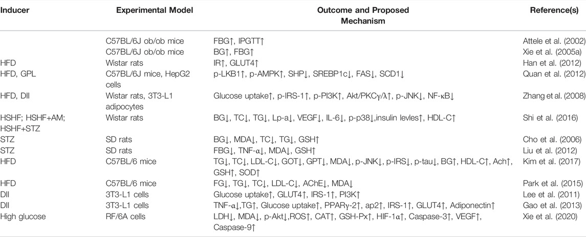
TABLE 1. Summary of anti-diabetes effects of Re.
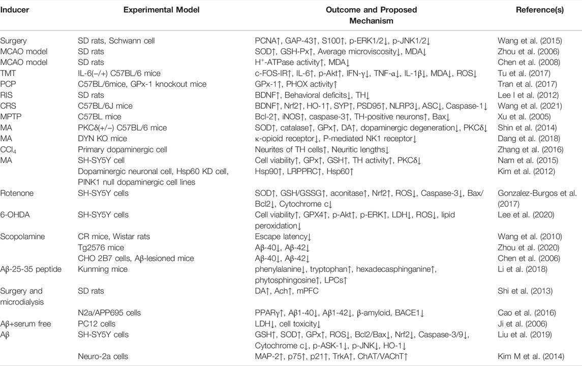
TABLE 2. Summary of nervous system disease effects of Re.
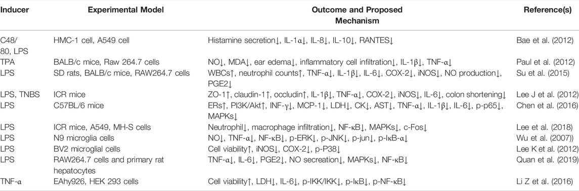
TABLE 3. Summary of anti-inflammation effects of Re.
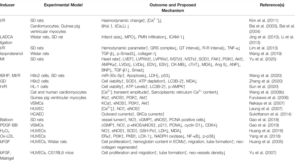
TABLE 4. Summary of cardiovascular disease effects of Re.
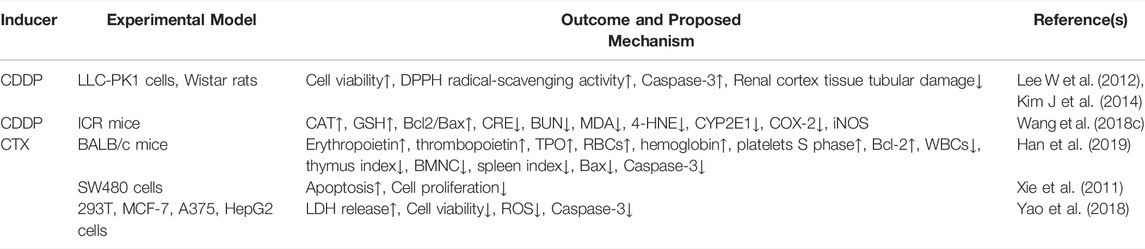
TABLE 5. Summary anti-cancer effects of Re.
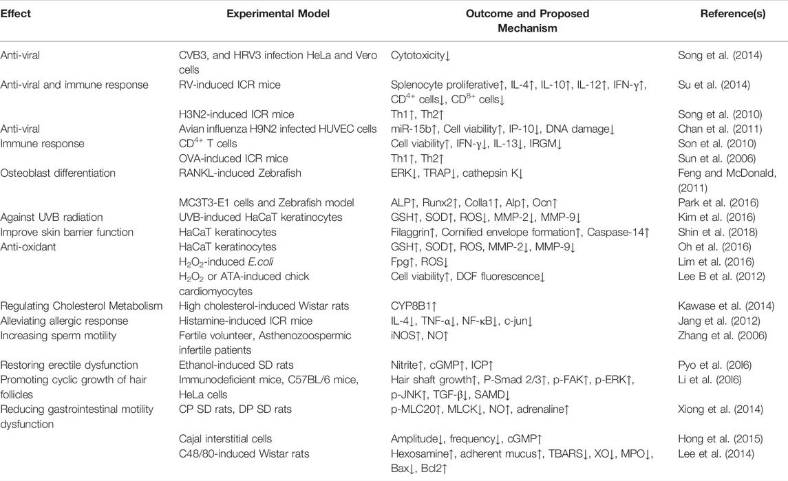
TABLE 6. Summary of other disease effects of Re.
Pharmacokinetic studies are necessary for observing and predicting the actions and interactions of drugs and for determining their efficacy and toxicity. The pharmacokinetics of Re have been studied in both animals and humans (Table 7), with major parameters, such as maximum concentration (Tmax), T1/2, and bioavailability examined. However, there is still little known about its metabolic and pharmacokinetic profiles.
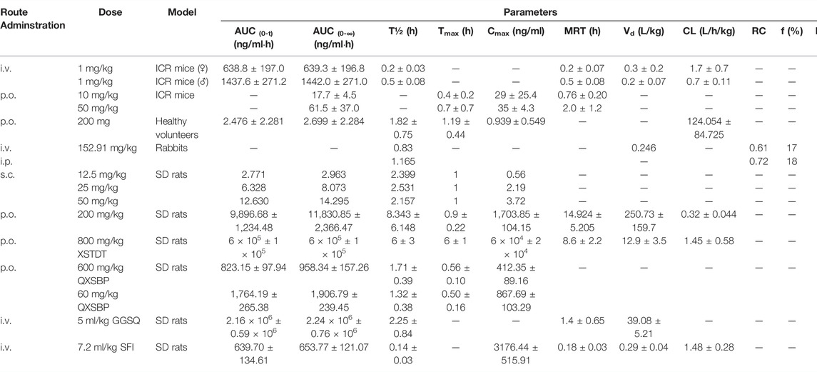
TABLE 7. The main pharmacokinetic parameters of Re.
The time for saponins to reach Tmax in rat plasma was less than 2 h, indicating that saponins are rapidly absorbed and readily distributed in tissues (Li et al., 2006; Gui et al., 2007). In humans, Liu et al. (2011) reported that the Tmax of Re was 1.19 ± 0.44 h after oral ingestion. Another study showed that the Tmax of Re was 0.75 h after oral administration of total P. notoginsenoside powder in rats, suggesting rapid absorption of Re in the gastrointestinal tract. The absolute bioavailability of Re was 7.06% (Li et al., 2006). Joo et al. (2010) revealed that the Tmax of Re was 0.4 ± 0.2 h in ICR mice. The same study also showed that the oral bioavailability was 0.19–0.28%, suggesting that the absorption rate of Re was lower after oral administration. Shi et al. (2013) demonstrated that Re (12.5, 25 and 50 mg/kg, s.c. injection) was rapidly distributed to the cerebrospinal fluid and exhibited linear pharmacokinetics in rats, and that the Tmax of Re was 1 h for all doses. However, for the lowest dose of 12.5 mg/kg, Re was not detectable in dialysates after 4 h. Extensive gastrointestinal metabolism, poor membrane permeability, and low solubility of deglycosylated products may limit the absorption of ginsenosides in the intestines. Therefore, the dose of test compounds must be high to detect ginsenoside content in plasma (Qi et al., 2011).
According to preclinical trials, several types of saponins, including ginsenosides Rg2, Rh1, F1, Rg1, and protopanaxatriol, may be metabolites of Re in human plasma and urine samples (Liu et al., 2011). After administration of Re (200 mg/kg, p.o. for 24 h), the major excreted ginsenoside metabolites in rat urine included Rg1 and Re. In feces, the main metabolite was Rg1, but other deglycosylated metabolites, including F1 and protopanaxatriol, were also detected (Kim et al., 2013). Yang et al. (2009) identified 11 and nine metabolites together with Re in rat urine collected after intravenous (50 mg/kg, i.v.) and oral (100 mg/kg, p.o.) administration of Re, respectively. The metabolites included Rg1, Rg2, Rh1, and F1. Oral and intravenous doses of Re showed distinct metabolism patterns in the rat, but there were also certain characteristics in common. Deglycosylation was found to be the major metabolic pathway of Re in rats, indicating that a large part of Re was metabolized and transformed in the gastrointestinal tract to ginsenosides with more biological effects (Christensen, 2009). The Re may be metabolized into ginsenosides Rh1 and F1 by human intestinal microflora, and subsequently absorbed into the blood (Bae et al., 2005). After oral administration of 100 mg/kg Re to rats, Chen et al. (2009) detected six metabolites of Re in feces, including ginsenosides Rg2, Rh1, Rh1, F1, Rh1, and PPT. In general, Re may be hydrolyzed by gastric fluids to ginsenoside Rg2 that is then converted in the intestine into ginsenoside Rh1 by the elimination of rhamnose through intestinal bacteria. Intact Re also reaches the large intestine where it can be metabolized by bacteria into ginsenoside F1 and 20(S)-PPT via ginsenoside Rg1. Like intestinal bacteria, several food microorganisms produce specific forms of ginsenosides. (Chi and Ji, 2005) tested the biotransformation of Re by cell extracts from various food-grade edible microorganisms, and found Re was transformed into Rh1 via Rg2 by Bifidobacterium sp. Int57 and SJ32, Re was transformed into Rh1 via Rg1 by Aspergillus niger KCTC 6906, and Re was transformed into Rg2 by A. usamii var. shirousamii KCTC 6956.
Joo et al. (2010) found that Re was rapidly cleared from the bodies of male or female mice within 0.2 ± 0.03 and 0.5 ± 0.08 h, respectively, after intravenous administration. Chen et al. (1980) estimated that the half-life of Ren in rabbits, after intravenous administration, was about 0.83 h, and the elimination half-life of Re after i.p. injection could be measured from urine (1.165 h) but not plasma samples. In healthy volunteers, the half-life of Re after oral ingestion of Re tablets (200 mg/tablets, p.o.) was reported to be 1.82 ± 0.75 h (Liu et al., 2011). A randomized, double-blind, placebo-controlled trial reported that researchers were unable to detect Re in plasma of obese adults, even though the subjects were prescribed large daily oral doses of P. ginseng and Re for 30 days and ingested the last dose 30 min before collection of blood samples to assess Re concentrations. The absence of Re may be explained by the quick elimination of ginsenoside (Reeds et al., 2011). Pharmacokinetic studies of Re in rats and human volunteers were consistent with this statement. After intragastric (i.g.) administration of Banxia Xiexin Decoction in rats, plasma concentrations of Re at most time points were lower than the lower limit of quantification (Wang et al., 2008a). Pharmacokinetic studies of Re in rats and volunteers following i.v. administration of Shen Mai indicated that Re was quickly eliminated in the body, and that pharmacokinetic characteristics fitted the two-compartment model (Liu et al., 2005; Xia et al., 2008). Altogether, evidence from pharmacokinetic and metabolic studies of Re demonstrated that 1) the absorption of Re was fast in the gastrointestinal tract; 2) Re may be metabolized mainly into Rh1 and F1 by intestinal microflora before absorption into blood; and 3) Re was quickly cleared from the body (Peng et al., 2012).
We included articles that were published from January 2000 to March 2021. Because more than 344 articles were found, we opted to focus on those specifically pertaining to new reports of the pharmacology, pharmacokinetics, and toxicology of Re. We searched four electronic databases, Google Scholar, NCBI, PubMed, and Web of Science, and compiled data according to the grade of evidence that was found. Systematic searches were performed in four electronic databases and the reference lists of most papers in the past 20 years were checked for further relevant publications. All articles containing original data on pharmacological activity, pharmacokinetics, and toxicology of Re were included. In addition, we only included studies written in English. Approximately 140 articles were used in the review process, across a variety of in vitro and in vivo studies, case reports, and randomized controlled trials.
Attele et al. (2002) found that Re (20 mg/kg, i.p. for 12 days) had marked anti-hyperglycemic activities, with no effect on the body weight of C57BL/6J ob/ob mice. This finding suggests that Re has potential as an anti-diabetic agent. Re (10 mg/kg, i.p. for 12 days) significantly reduced fasting blood glucose levels and promoted glucose tolerance (GT) and systemic insulin sensitivity (IS) in ob/ob mice without affecting body weight (Xie et al., 2005a). These findings suggest Re may provide a therapeutic role in ameliorating GT and insulin resistance (IR) in patients with type 2 diabetes mellitus (T2DM). Administration of Re (0.2 mg/ml for 90 min) rapidly normalized IR and muscle glucose transport induced by high-fat diet (HFD) in the epitrochlearis and soleus muscles of rats (Han et al., 2012). Re may have specifically acted to ameliorate IR in muscles of rats because it failed to modify HFD-induced muscle glucose transport resistance following stimulation by contraction or hypoxia. Muscle contraction and hypoxia exert an insulin-like-stimulating effect on glucose transport. However, Re did not affect basal or insulin-stimulated muscle glucose transport in chow-fed rats. According to these animal studies, P. ginseng or ginsenoside appeared to improve oral GT and accelerate insulin-stimulated glucose disposal (Xie et al., 2004). The Re-induced improvement in IS may or may not be associated with weight loss. Therefore, it remains unclear whether the amelioration was due to weight loss or insulin-sensitizing traits. These studies demonstrated the association between the anti-hyperglycemic activity of Re and improved IS, whereas body weight was unaffected. The improvement may be attributed to the insulin-sensitizing properties of Re. Quan et al. (2012) studied the potential anti-glycemic role of Re in HFD-induced diabetes in mice. Administration of Re (20 mg/kg, i.g. for 3 weeks) markedly lowered BG and triglyceride levels and prevented hepatic steatosis in C57BL/6J mice on a HFD. The hypoglycemic effect was associated with suppression of hepatic gluconeogenesis, possibly associated with AMP-activated protein kinase (AMPK) activation. In rats on a HFD, Re (40 mg/kg, i.p. for 2 weeks, twice a day) improved IR by inhibiting c-Jun N-terminal kinase (JNK) and nuclear factor (NF)-kB activation (Zhang et al., 2008). Several studies have concluded that the anti-hyperglycemic effect of Re was primarily responsible for improved microvasculopathy or reduced cognitive impairment in HFD-induced diabetic mouse models. In such models, Re (20 mg/kg, i.g. for 8 weeks) exerted a protective and anti-angiopathy effect in DM, such as the initial stages of high-sucrose-HFD (HSHF)-induced diabetes, HSHF+alloxan monohydrate-induced Type 1 diabetes mellitus (T1DM), and HSHF+streptozotocin (STZ)-induced T2DM. Administration of Re reduced BG levels, regulated increasing insulin levels, improved lipid metabolism, and reduced endothelial cell dysfunction. The underlying mechanism was possibly associated with p38 mitogen-activated protein kinase (MAPK) activation, and extracellular signal-regulated kinase (ERK) 1/2 and JNK signaling (Shi et al., 2016). In addition, Re (20 mg/kg, i.g. for 2 weeks) had an anti-diabetic microvasculopathy effect, including protective actions against oxidative stress in the kidneys and eyes, and increased BG and lipid levels in rats with STZ-induced diabetes (Cho et al., 2006). In rats with STZ-induced T1DM, Re (40 mg/kg, i.g. for 8 weeks) improved diabetes-related cognitive decline while decreasing fasting BG levels, although it did not affect BG, which was associated with oxidative stress and inflammation (Liu et al., 2012). In mice, Re improved HFD-induced IR through amelioration of hyperglycemia by protecting the brain cholinergic and antioxidant systems (Kim et al., 2017). Specifically, Re (5, 10 and 20 mg/kg/d, i.g. for 4 weeks) improved diabetes-associated cognitive impairment, and was possibly associated with improvement of the anti-oxidant and cholinergic systems in brain tissue. In HFD-induced hyperglycemic C57BL/6 mice, Re played a positive role through amelioration of insulin tolerance and BG levels. Re possibly improved learning and memory disorders related to HFD-induced diabetes. As the major ginsenoside in the P. ginseng berry ethyl acetate fraction (blended with drinking water 20 and 50 mg/kg, p.o. for 4 weeks), Re ameliorated cognitive decline in a dose-dependent manner because of its cholinergic activity, and it decreased oxidative stress in mice with HFD-induced T2DM and behavioral deficiency (Park et al., 2015).
In 3T3-L1 adipocytes, Re (10 μM for 24 h) improved IR by inhibiting the inflammatory signaling cascade and activating the insulin signaling pathway (Zhang et al., 2008). Further results demonstrated that Re (1–10 μΜ for 0.5 h) increased glucose uptake in mature 3T3-L1 cells by significantly enhancing glucose transporter 4 (GLUT4) mRNA expression through the phosphoinositide 3-kinase (PI3K)-dependent pathway involving insulin receptor substrate-1 (IRS-1) in the glucose transport system cascade (Lee et al., 2011). Gao et al. (2013) demonstrated that Re (30, 60 μM for 5 days) reduced IR in adipocytes by directly enhancing the expression of peroxisome proliferator-activated receptor-γ (PPARγ)-2 and the corresponding AP2 genes, increasing adiponectin and IRS-1 expression, inhibiting inflammatory cytokine tumor nuclear factor-α (TNF-α) expression and production, and promoting GLUT4 translocation. The regulation of these factors facilitated adipocyte glucose uptake and disposal, although it failed to enhance GLUT4 expression. Another study found that Re (20 μM for 3 h) suppressed glucose generation in HepG2 cells, possibly by triggering the expression of the orphan nuclear receptor small heterodimer partner gene via AMPK activation (Quan et al., 2012). These results indicate that Re improved IR through reduction of lipotoxicity in the muscles and liver by enhancing adipocyte lipid storage capacity and promoting GLUT4 translocation to plasma membranes. Thus, Re compound regulation of insulin-stimulated glucose ingestion led to improved IR. Furthermore, Re (3 μM for 24 h) was proposed to exert anti-angiogenetic effects in diabetic retinopathy through the PI3K/Akt-mediated hypoxia-inducible factor-1-alpha (HIF-1α)/vascular endothelial growth factor (VEGF) signaling pathway in high-glucose-induced retinal endothelial RF/6A cells (Xie et al., 2020).
Overall, in vivo and in vitro data suggest four possible mechanisms underlying Re-induced improvement of diabetes and diabetes-related complications: 1) regulation of insulin resistance and insulin secretion, 2) modulation of glucose or lipid metabolism, 3) modulation of inflammatory cytokines, and 4) activation of oxidative stress.
In rats with sciatic nerve crush injury, Re (2.0 mg/kg, i.p. for 4 weeks) promoted functional recovery, nerve regeneration, and proliferation of injured sciatic nerves. The Re compound promoted Schwann cell proliferation, differentiation, and migration during the course of peripheral neural repair after crush injury. This effect was possibly mediated by the regulation of ERK1/2 and JNK1/2 signaling pathways (Wang et al., 2015).
One study reported the anti-oxidant effects of Re (5, 10 and 20 mg/kg, i.g. for 1 week) in rats with cerebral ischemia-reperfusion (I/R) injury. The Re compound considerably increased membrane fluidity of brain mitochondria, activated anti-oxidative enzymes, and decreased lipid peroxidation products, including malondialdehyde (Zhou et al., 2006). Neuroprotective effects of Re (5, 10 and 20 mg/kg, i.g. for 1 week) against cerebral I/R injury in rats were associated with a reduction in malondialdehyde levels and mitochondrial swelling, leading to an increase in H+-ATPase activity (Chen et al., 2008).
Tu et al. (2017) reported that Re (20 mg/kg, i.p. for 3 days) attenuated convulsive behaviors, oxidative damage, pro-apoptotic potential and neuronal degeneration through the interleukin-6 (IL-6)-dependent PI3K/Akt signaling pathway in mice with trimethyltin-induced neurotoxicity. Treatment with Re (20 mg/kg, i.p. for 1 day) markedly decreased phencyclidine-induced neurotoxic alterations, including behavioral changes and mitochondrial dysfunction. These Re-mediated alterations were due to interactive modulation between glutathione peroxidase-1 (GPx-1) and NADPH oxidase in mice (Tran et al., 2017).
Administration of Re (50 mg/kg, i.p. for 10 days) before immobilization stress markedly improved body weight, serum corticosterone levels, behavioral alterations, and cognitive deficits in rats. These effects were mediated through modulation of the central noradrenergic system and hypothalamic corticotrophin-releasing factor in the brain (Lee B et al., 2012). Another study showed Re (20, 40 mg/kg, i.p. for 3 weeks) inhibited memory deficits induced by chronic restraint stress (Wang et al., 2021). The protective effects were related to anti-inflammatory and anti-oxidant activities of the Re compound, as well as positive regulation of brain-derived neurotrophic factor and plasticity-associated proteins in the hippocampus.
Administration of Re can effectively prevent onset of Alzheimer’s disease (AD) by improving the activity of dopamine (DA) neurons. One study found that Re (6.5, 13 and 26 mg/kg, i.g. for 13 days) prevented apoptosis of substantia nigra dopaminergic neurons induced by 1-methyl-4-phenyl-1,2,3,6-tetrahydropyridine in C57BL mice (Xu et al., 2005). The effect was mediated by reversing the abnormal expression of apoptosis regulatory proteins and inhibiting caspase-3 activation. Administration of Re (10, 20 mg/kg, i.p. for 2 weeks, twice a day) rescued methamphetamine-induced dopaminergic neurotoxicity. The effect was associated with potentiating oxidative burdens, compensative induction of GPx activity, mitochondrial dysfunction, pro-inflammatory changes, apoptotic cellular degeneration, and dopaminergic degeneration through inactivation of the protein kinase Cẟ (PKCδ) gene (Shin et al., 2014). Another study reported that Re (20 mg/kg, i.p. for 5 days, twice a day) protected methamphetamine-treated prodynorphin knockout mice against dopaminergic neurotoxicity through anti-oxidant, anti-inflammatory, and anti-apoptotic actions. The effects were facilitated by dynorphin-induced upregulation of the κ-opioid receptor, followed by substance P-mediated downregulation of the NK1 receptor (Dang et al., 2018).
Administration of Re (10 µM) and ginsenoside Rd (5 µM for 48 h) provided considerable neuroprotective effects on primary dopaminergic midbrain neurons treated with CCl4. The neuroprotective effects were in part due to the lowering of oxidative stress and alleviation of inflammatory responses (Zhang et al., 2016). In addition, Re treatment (50, 100 μM for 24 h) of SH-SY5Y cells rescued methamphetamine-induced mitochondrial burden (compensative induction of cytosolic and mitochondrial GPx activity, mitochondrial oxidative stress, mitochondrial dysfunction, and mitochondrial translocation of cleaved PKCδ, and pro-apoptosis through genetic inhibition of PKCδ) (Nam et al., 2015). Kim et al. (2012) investigated the actions of Re on mitochondrial dysfunction in a PD model. They found that Re (3 µM) targeted mitochondrial dysfunction and rescued the defective PINK1-Hsp90/LRPPRC-Hsp60-complex IV signaling axis of PINK1-null neurons by restoring nitric oxide (NO) levels. Co-treatment using Rd and Re (0.5, 1 μM for 24 h) protected SH-SY5Y cells against rotenone-induced toxicity by regulating molecular mechanisms that enhanced cell viability, including prevention of morphological changes, lowered oxidative stress, improved mitochondrial integrity and function, and inhibited apoptosis owing to oxidative stress (Gonzalez-Burgos et al., 2017). The anti-oxidant mechanism of Re in PD remains unclear. In SH-SY5Y cells treated with 6-hydroxydopamine to induce oxidative stress, the Re compound (25 µM for 9 h) mediated its anti-oxidant effect by upregulating a key antioxidant gene GPX4 via PI3K/Akt and ERK cascades (Lee et al., 2020).
Kai-Xin-San, a Chinese herbal formula, has been clinically administered at 3 g/kg (i.g. for 4 weeks) to treat animals with AD and neurosis. P. ginseng, a component of Kai-Xin-San, is known to enhance learning ability and memory. In addition, positive effects of Re and Rb1, the most abundant saponins, on learning ability and memory were reported (Wang et al., 2010). Amyloid β (Aβ) peptide plays an important role in AD. Zhou et al. reported that Re may interfere with AD progression by affecting the Aβ peptide (Zhou et al., 2020). Oral administration of Re (25 mg/kg, i.g. for 18 h) considerably reduced Aβ1-40 and Aβ1-42 levels in brains of Tg2576 mice (Chen et al., 2006). Furthermore, Li et al. (2018) demonstrated that Re (4 mg/kg, i.g. for 40 days) improved cognitive impairment, reduced Aβ accumulation, and restored biomarker levels, including amino acids, lecithin, and sphingolipids in the plasma of AD mice. Because of its effect on Aβ peptides, Re is increasingly considered a potential alternative drug for AD treatment. In addition, Re exhibits anti-dementia activity. The Re compound improved extracellular levels of DA and acetylcholine (Ach), particularly in the hippocampus. Also, Re (12.5, 25 and 50 mg/kg, s.c.) increased extracellular levels of DA and Ach in the medial prefrontal cortex (Shi et al., 2013).
Treatment with Re has been reported to improve AD by affecting Aβ peptide levels in several cell models. Liang et al. reported that Re markedly reduced the generation of Aβ proteins in N2a/APP695 cells. The effect of Re (50–100 μM for 24 h) on Aβ generation was mediated by PPARγ activation in combination with Aβ-site precursor protein-cleaving enzyme 1 inhibition (Cao et al., 2016). Treatment with Re (0.1–100 μM for 2 h) considerably reduced cell toxicity and increased the release of lactate dehydrogenase, thereby attenuating PC12 cell damage induced by Aβ peptides (Ji et al., 2006). In addition, Re (25 µM for 48 h) exhibited neuroprotective activity against neurotoxicity arising from Aβ25-35 in SH-SY5Y cells by reducing oxidative damage and neuronal cell apoptosis. The neuroprotective activity was associated with the activation of nuclear factor erythroid-2 associated factor 2/heme oxygenase-1 anti-oxidant response pathways and inhibition of reactive oxygen species (ROS)-dependent apoptosis signal-regulated kinase 1/JNK/Bax apoptosis pathways (Liu et al., 2019). Furthermore, Kim et al. demonstrated that Re (5 μg/ml for 48 h) effectively upregulated the expression of choline acetyltransferase and vesicular acetylcholine transporter, and Ach production in Neuro-2a cells, thus countering symptoms during AD progression (Kim J et al., 2014).
In vivo and in vitro data suggest six possible mechanisms of Re-mediated improvement of complications associated with nervous system diseases: 1) regulation of central cholinergic pathways, 2) modulation of the apoptotic signaling pathway, 3) modulation of inflammatory responses, 4) modulation of mitochondrial burden, 5) regulation of anti-oxidant signaling pathways, and 6) reduction of Aβ peptide accumulation and loss of midbrain DA neurons.
Treatment with Re considerably inhibited neutrophil infiltration in a model of skin inflammation arising from 12-O-tetradecanoylphorbol-13-acetate. It also improved paw and ear oedema, increased malondialdehyde levels in paw fluid during c-carrageenan-induced edema, and suppressed interleukin-1β (IL-1β) and TNF-α expression in lipopolysaccharide (LPS)-stimulated murine Raw 264.7 macrophages (Paul et al., 2012). Moreover, Re (1 mg/kg, i.v. for 15 min) suppressed the LPS-induced increase in body temperature, white blood cell count, and pro-inflammatory mediators (Su et al., 2015). In LPS-induced systemic inflammation, Re (10, 20 mg/kg, i.g. for 4 h) suppressed serum levels of IL-1β and TNF-α in mice. Similarly, in 2,4,6-trinitrobenzene sulfonic acid-induced colitic mice, Re (10, 20 mg/kg, i.g. for 3 days) suppressed the expression of IL-1β, TNF-α, cyclooxygenase-2, and inducible nitric oxide synthase, and the activation of transcription factor NF-κB. However, it enhanced the expression of anti-inflammatory cytokine IL-10, indicating that Re can suppress Th1 rather than Th2 cell activation (Lee I et al., 2012). Administration of Re (15 mg/kg, i.g. for 1 week) also prevented NF-κB activation and LPS-induced myocardial inflammation in mice. The action of Re in cardiac dysfunction involves both MAPK inhibition and preserved activation of estrogen receptors and the PI3K/Akt signaling pathway (Chen et al., 2016). Treatment with Re (6–50 mg/kg, p.o. for 2 h) produced strong and significant inhibitory actions against LPS-induced lung inflammation in mice, and decreased inflammatory cell infiltration into lung tissue. The effect was mediated by inhibiting the activation of MAPK and transcription factors NF-κB and c-Fos (Lee et al., 2018).
An in vitro investigation of the anti-inflammatory effects of Re (5, 10 μΜ for 30 min) in macrophages showed that it suppressed the expression of pro-inflammatory cytokines (TNF-α and IL-1β) and activation of transcription factor NF-κB by preventing the binding between LPS and toll-like receptor 4 (TLR4). However, Re did not suppress pro-inflammatory cytokines in peptidoglycan- or TNF-α-stimulated peritoneal macrophages (Lee J et al., 2012), highlighting its action in reducing inflammation by suppressing the LPS and TLR4 interaction in macrophages. Su et al. (2015) demonstrated that Re (50 μg/ml for 1 h) competed with LPS binding to the TLR4, and blocked the LPS-triggered signaling pathway in LPS-stimulated RAW264.7 cells. Extracellular Re was shown to compete with LPS binding to the TLR4, consistent with its role in the activation of extracellular TLR4 (Su et al., 2012). In addition, Wu et al. reported an anti-inflammatory role of Re (10–100 μΜ for 48 h) in LPS-induced activated N9 microglial cells. Re mediated its effects by inhibiting the generation of NO and TNF-α through downregulation of NF-κB activation (Wu et al., 2007). Treatment with Re (2 μg/ml for 24 h) reduced neuroinflammation by reducing the levels of inducible nitric oxide synthase and cyclooxygenase-2, and activating p38 MAPK in LPS-treated BV2 microglial cells (Lee K et al., 2012). Moreover, Quan H et al. (2019) reported that Re (10–40 μΜ for 24 h) inhibited LPS-induced TNF-α and IL-6 production in RAW264.7 cells, and reduced IL-6, NO, prostaglandin E2, and TNF-α secretion in primary rat hepatocytes via MAPK and NF-κB signaling pathways. Re is an effective component of Shen Fu, and was reported to exert anti-inflammatory effects by suppressing the NF-κB signaling pathway in TNF-α-stimulated EAhy926 cells (Li P et al., 2016). Incubation with Re (1.7 μg/ml for 24 h) decreased histamine secretion in human mast cells, and reduced IL-1α, IL-8, and IL-10 levels, and regulated T-cell-expressed and secreted protein secretion in A549 cells (Bae et al., 2012).
Altogether, in vivo and in vitro study data indicate that the possible mechanism of anti-inflammatory activities of Re involves NF-κB inactivation and reduced inflammatory cytokine release.
Kim et al. (2011) showed that Re improved ischemia/reperfusion (I/R) dysfunction by reversing the hemodynamic change (aortic flow, coronary flow, perfusion pressure, and cardiac output) and inhibiting the level of intracellular Ca2+ ([Ca2+]i). This study indicated that the anti-ischemic effect of Re was mediated by inhibiting an increase of [Ca2+]i. Additionally, Re prevented heart mitochondrial Ca2+ accumulation in I/R injury. In isolated single cardiomyocytes, Re suppressed the L-type Ca2+ current and strengthened the slowly activating delayed rectifier K+ current (IKs). This may be the underlying mechanism that prevented mitochondrial Ca2+ overload (Bai et al., 2003; Bai et al., 2004).
A rat model showed that Re (20 mg/kg, i.g. for 15 days) provided an effective treatment for myocardial infraction arising from left anterior descending coronary artery ligation. Treatment with Re improved the parameters of myocardial injury by downregulating the expression of intercellular adhesion molecule-1 and inhibiting polymorphonuclear leukocyte infiltration (Jing et al., 2010; Li et al., 2013). In this research, Re was reported to exhibit a protective role in ischemia-induced myocardial injury by regulating calcium transport, preserving mitochondrial structure and function, enhancing anti-oxidant capacity, and recovering myocardial blood flow.
In addition, Re lowered myocardial injury and suppressed cardiac hypertrophy in experimental models with cardiac dysfunction. Lim et al. (2013) proposed that Re (100 μM, injected into the aortic line for 3 min) exerted beneficial effects on cardiac function in rats with I/R injury, considerably improved hemodynamic functions and left ventricular developed pressure, ameliorated electrocardiographic abnormalities, and decreased the production of TNF-α. Treatment with Re (5, 20 mg/kg, i.g. for 4 weeks) also reduced isoproterenol-induced myocardial fibrosis, increased heart weight and hydroxyproline content, and reduced heart failure. The molecular mechanisms underlying the protective role of Re were possibly related to regulation of the transforming growth factor-beta 1 (TGF-β1)/Smad3 pathway (Wang et al., 2019). In a rat model of myocardial injury, Re (135 mg/kg, i.g. for 4 weeks) preserved cardiac function and structure, reduced myocardial injury and stress, and decreased left ventricular fibrosis by regulating the AMPK/TGF-β1/Smad2/3 and FAK/PI3K/Akt signaling pathways (Yu et al., 2020). These findings suggest a possible therapeutic role for Re in suppressing ventricular remodeling and promoting postinfarction healing. Overall, Re restored blood supply quickly and also delayed detrimental ventricular remodeling during chronic myocardial infraction rehabilitation.
Wang et al. found that Re (200 μg/ml for 24 h) increased H9c2 cell viability after tertbutyl hydroperoxide treatment and reduced lactate dehydrogenase release and cell apoptosis (Wang et al., 2020). Treatment with Re (100 μM for 3 h) inhibited glucose deprivation-induced autophagy of H9c2 cardiac muscle cells, an effect which may be associated with the inhibition of autophagy, increase in cellular ATP content and viability, and alleviation of oxidative stress (Zhang et al., 2020). In addition, in the hypoxia/reoxygenation injury model, Re (100 µM for 21 h) increased HL-1 cell viability and ATP levels. The possible mechanism was that Re acted on the binding interface between HIF-1α and von Hippel-Lindau protein to prevent the binding of these proteins, thereby suppressing HIF-1α ubiquitination (Sun et al., 2020).
Administration of Re (≥10 nM) effectively suppressed the electromechanical alternans of cardiomyocytes in cats and humans by increasing sarcoplasmic reticulum Ca2+-release channels, and thereby improving arrhythmia (Wang et al., 2008b). Furukawa et al. (2006) showed that Re (3 μM) increased IKs, [Ca2+]i, activation of eNOS, and NO production through a c-Src/PI3K/Akt-dependent mechanism related to the non-genomic pathway of sex steroid receptors. Similarly, in vascular smooth muscle cells (VSMCs), Re non-genomically and dose dependently activated KCa currents and eNOS (EC50 = 4.1 ± 0.3 μM) through the c-Src/PI3-kinase/Akt pathway of the estrogen receptor (Nakaya et al., 2007). A study on human umbilical vein endothelial cells (HUVECs) revealed that Re augmented [Ca2+]i and NO production in a dose-dependent manner (EC50 of 316 and 615 nM, respectively) (Leung et al., 2007). In human coronary artery endothelial cells, Re (1 μM) induced vasorelaxation by increasing small-conductance Ca2+-activated K+ (SKCa) channel activity, stimulating NO production, and promoting vasodilation (Sukrittanon et al., 2014).
Abnormal structure and function of VSMCs may result in the development and progression of arteriosclerosis. Enhanced proliferation and migration of VSMCs represent critical events during the course of atherosclerotic lesion development (Bennett et al., 2016). Gao et al. (2018) demonstrated that Re (25 or 50 mg/kg, i.g. for 2 weeks) inhibited VSMC proliferation by suppressing phenotypic modulation and inhibiting vascular neointimal hyperplasia in balloon-injured rats through the eNOS/NO/cyclic guanosine monophosphate (cGMP) pathway. Re improved platelet-derived growth factor-BB-induced VSMC proliferation through G0/G1 cell cycle arrest, which was associated with eNOS/NO/cGMP pathway activation (Gao et al., 2019).
In contrast, endothelial cells provide an interface between circulating blood in the lumen and other vessel walls. Endothelial cells exhibit great sensitivity and vulnerability to toxic substances circulating in blood vessels. Endothelial dysfunction is an important contributor to the pathobiology of atherosclerosis (Gimbrone and García-Cardeña, 2016). Huang et al. (2016) found that Re (4, 16, and 64 μmol/L for 24 h) attenuated oxidative damage in H2O2-induced HUVECs and increased the production of NO and eNOS, superoxide dismutase (SOD), and GPx activities. The protective effects were associated with an oxidative stress response, protein synthesis and mitochondrial function. In addition, Yang et al. demonstrated that Re (120 μg/ml for 12 h) improved oxidized low-density lipoprotein-induced endothelial cell apoptosis. The effect was possibly elicited through regulation of oxidative stress, inhibition of inflammatory mediators, and recovery of balanced pro- and anti-apoptotic protein expression via p38/MAPK/NF-κB and PI3K/Akt/NF-κB pathways. These pathways may be regulated by the lectin-like oxidized low-density lipoprotein receptor-1, NADPH oxidase, and estrogen receptor α (Yang et al., 2018). Therefore, Re is a potential anti-oxidant that may be used to protect HUVECs from damage by oxidative stress through the anti-oxidant defense system. The Re compound also inhibited VSMC proliferation, attenuated endothelial dysfunction, and possibly promoted NO production, thereby reducing atherosclerosis.
Re is a pro-angiogenic compound with high stability that upregulates in vitro proliferation, migration, chemo-invasion, and tube formation of HUVECs. It also affects ex vivo aortic sprouting and in vivo neovascularization. In vitro results revealed that Re (10–30 μg/ml for 48 h) dose dependently enhanced the proliferation, migration, and tube formation of HUVECs (Huang et al., 2005). Additionally, extracellular matrix incorporating Re (70 μg for 1 week and 1 month) induced angiogenesis and enhanced tissue regeneration by increasing neocapillary density and tissue hemoglobin in a rat model (Yu et al., 2007). These findings indicate that Re can serve as an angiogenic agent to accelerate tissue regeneration.
In summary, in vivo and in vitro reports suggest five possible mechanisms by which Re may improve the cardiovascular system: 1) attenuation of myocardial ischemia, 2) inhibition of [Ca2+]i and activation of IKs, 3) increased NO production, 4) reduced cardiomyocyte apoptosis autophagy, and 5) the regulation of oxidative stress.
A combination of Re and cisplatin increased the survival rate of LLC-PK1 cells by 21.4%. However, the renoprotective effects of Re were weaker than that of Maillard reaction products in Re-leucine/serine and glucose-leucine mixtures. Moreover, Maillard reaction products reduced cisplatin-induced oxidative kidney damage by increasing 1,1-diphenyl-2 picrylhydrazyl radical-scavenging activity and decreasing the expression of cleaved caspase-3 protein in rats (Lee W et al., 2012; Kim M et al., 2014). (Wang et al., 2008c) found that Re (25 mg/kg, i.g. for 10 days) considerably suppressed acute kidney injury induced by cisplatin in mice, by inhibiting the oxidative stress damage, inflammatory response, and apoptosis. Re (5, 10 mg/kg, i.p. for 1 week) also improved cyclophosphamide-induced myelosuppression, alleviated clinical symptoms of myelosuppression, and promoted recovery of bone marrow hematopoietic functions. The possible mechanisms involved the regulation of hematopoiesis-related cytokine levels, promotion of cellular entry to the normal cell cycle, and improvement of bone marrow nucleated cell apoptosis-related protein expression (Han et al., 2019).
One mg/mL of American P. ginseng berry extract (containing 15.1 mg/g of Re for 72 h) exhibited strong anti-proliferative effects and triggered morphological alterations in SW480 human colorectal cancer cells (Xie et al., 2011). Re-carbon dots (0.5 mg/ml for 4 h) inhibited cancer cell proliferation (A375, HepG2, and MCF-7 cells) through the ROS-mediated pathway. However, the inhibitory effect on A375 cells was higher than that on other cells. Re induced apoptosis via the ROS- and caspase-mediated pathways (Yao et al., 2018). These findings demonstrate that Re can be used as a potential anti-cancer adjuvant for preventing and treating various cancers.
Altogether, in vivo and in vitro data show three possible mechanisms underlying the anti-cancer activities of Re: 1) inhibition of cell proliferation, 2) induction of cell apoptosis and 3) modulation of oxidative damage.
Song et al. (2014) demonstrated that Re (100 μg/ml for 48 h) had potential therapeutic efficacy in CVB3 and HRV3 infections in HeLa and Vero cells, respectively. Su et al. (2014) showed that co-administration of Re (5.0 mg/kg. s.c. for 3 weeks) with the rabies virus vaccine remarkably increased the serum antibody response in mice. Other studies have shown that co-administration of Re (50 μg, s.c. for 3 weeks) with inactivated influenza virus A/Fujian/411/2002 (H3N2) markedly amplified serum-specific antibody responses (IgG, IgG1, IgG2a, and IgG2b), hemagglutination inhibition titers, lymphocyte proliferation responses, and IL-5 and IFN-γ production (Song et al., 2010). Chan et al. (2011) reported that Re (50 μΜ for 16 h) protected HUVECs from H9N2/G1 influenza virus-induced apoptosis. CD4+ T cells are important immune cells in the human immune system. Son et al. found that Re (10, 20 and 40 μg/ml for 24 h) enhanced the viability of activated CD4+ T cells by downregulating IFN-γ production, which interfered with autophagy by reducing immunity-associated GTPase family proteins (Son et al., 2010). Re also enhanced the expression of Th1-type-related and Th2-type-related cytokines (Su et al., 2014). Administration of Re (10, 25 and 50 μg, s.c. for 2 weeks) had considerable adjuvant effects on specific antibody and cellular responses in ovalbumin-immunized mice, affecting the immune system favoring Th1- or Th2-type responses, as shown by enhanced titers of IgG1 and IgG2b isotypes (Sun et al., 2006). These results indicated Re-mediated activation of Th1 and Th2 immune responses in mouse models. Therefore, these studies indicate that Re can enhance the host immune system as a vaccine adjuvant.
An optimal balance of osteoblasts and osteoclasts is crucial for bone remodeling. Impaired bone homeostasis potentially causes bone disease, such as bone fracture and osteoporosis (Feng and McDonald, 2011). It was demonstrated that Re had dual effects promoting osteoblast differentiation and inhibiting osteoclast differentiation. This research showed that Re (2.5, 5 and 10 μM for 48 h) dose dependently inhibited osteoclast differentiation and decreased nuclear factor of activated T cell cytoplasmic 1 and tartrate-resistant acid phosphatase mRNA levels, which are osteoclast differentiation markers. These effects were elicited by blocking the ERK signaling pathway in bone marrow-derived macrophages stimulated with the receptor activator of NF-κB ligand. Osteoclast generation in zebrafish scales was inhibited by Re (10 μM for 5 weeks), shown by reduced expression of osteoclast marker genes tartrate-resistant acid phosphatase and cathepsin K (Park et al., 2016). Kim et al. (2016) found that Re affected the differentiation and mineralization of osteoblasts both in vitro and in vivo models. Treatment with Re (50 µM for 5 weeks) promoted the expression of osteoblastic markers, including alkaline phosphatase activity, and mRNA levels of alkaline phosphatase, type 1 collagen, and osteocalcin in mouse osteoblast precursor MC3T3-E1 cells. Moreover, Re amplified the mineralization of osteoblasts in mouse MC3T3-E1 cells and zebrafish scales.
Treatment with Re (5, 12, and 30 μM for 0.5 h) provided potential anti-photo-ageing activity in HaCaT keratinocytes under UVB radiation. This activity was possibly elicited through downregulation of UVB-induced intracellular ROS formation, production and secretion of pro-matrix metalloproteinase-2 and -9, and upregulation of total GPx levels and SOD activity (Shin et al., 2018). In addition, Oh et al. (2016) found that Re (5, 12 and 30 μM for 1 h) improved skin barrier functions, shown by enhanced cornified cell envelope formation, filaggrin levels and caspase-14 activity in HaCaT keratinocytes. Furthermore, Re (5, 12 and 30 μM for 24 h) demonstrated anti-oxidative activity through the upregulation of anti-oxidant components including total GPx and SOD under normal conditions. Re also prevented oxidative stress in HaCaT keratinocytes (Lim et al., 2016).
Re (0.05, 0.1 and 0.5 mg/ml for 2 h) protected chick cardiomyocytes from exogenous H2O2- and endogenous antimycin A-induced oxidative stress. The underlying mechanism for this protective effect involved scavenging of H2O2 and hydroxyl radicals. However, in an electron spin resonance spectroscopy study, Re did not reduce the 1,1-diphenyl-2 picrylhydrazyl-induced electron spin resonance signals for xanthine oxidase or H2O2 (Xie et al., 2006). Therefore, direct scavenging of free radicals was impossible through a single anti-oxidation pathway in vivo. The anti-oxidative effects of Re were achieved through activation or enhancement of the intracellular anti-oxidant system.
Kawase et al. (2014) reported that Re (0.1–1 μM for 24 h) exerted a positive effect on cholesterol metabolism, increasing the expression level of sterol 12a-hydroxylase mRNA in rat primary hepatocytes, thereby facilitating cholic acid generation within bile acids.
Jang et al. (2012) reported that Re (25 mg/kg, p.o. for 6 h) potently alleviated scratching behavior in mice with histamine-induced itch, by inhibiting the activation of transcription factors (NF-κB and c-jun), as well as the expression of IL-4 and TNF-α.
Zhang et al. (2006) demonstrated that Re (100 μM for 2 h) improved sperm motility from fertile and asthenozoospermic infertile human subjects by enhancing NOS activity to promote endogenous NO generation.
The Re-enriched fraction (containing 109.0 mg/g of Re, 54.5 mg/kg, i.g. for 5 weeks) of P. ginseng berries effectively restored ethanol-induced erectile dysfunction in male rats through the NO-cGMP pathway (Pyo et al., 2016).
(Li Z et al., 2016) reported that topical treatment (5 mg/day, topical application on the back for 9 weeks) with Re markedly triggered hair shaft growth through selective suppression of hair growth phase transition-associated signaling pathways and TGF-β signaling cascades in nude mice.
Re-mediated bidirectional regulation is dependent on the jejunal contractile status and requires the co-existence of the enteric nervous system, Ca2+, and Cajal interstitial cells. The stimulatory role of Re (10 μM) on jejunal contractility of rat isolated jejunal segments was associated with cholinergic stimulation, whereas its inhibitory role was associated with adrenergic activation and the NO-relaxing mechanism (Xiong et al., 2014). In addition, Re (40 μΜ) inhibited pacemaker potentials through ATP-sensitive K+ channels and the cGMP/NO-dependent pathway in cultured Cajal interstitial cells obtained from the small intestine of mice (Hong et al., 2015). Re (20, 100 mg/kg, i.g. for 30 min) ameliorated acute gastric mucosal lesions induced by compound 48/80, possibly by triggering mucus secretion and decreasing neutrophil infiltration, inflammation, and oxidative stress in gastric mucosa (Lee et al., 2014).
An acute toxicity study in mice treated with P. ginseng extract found LD50 values of 10–30 g/kg (Brekhman and Dardymov, 1969). Chronic treatment of mice and rats with P. ginseng extract (5 g/kg, p.o. for 2 years) produced almost no toxic effects, and the appearance, behavior, weight, and various physiological/histological indexes were within reasonable ranges (National Toxicology Program, 2011). Likewise, (Lu et al., 2012) found that the LD50 of Re was 5.0 g/kg in mice. In addition, in a chronic toxicity study, male and female SD rats treated with 375 mg/kg/day (orally) Re for 26 weeks, well below the typically non-toxic range (5–15 g/kg) of chemical substances (Hayes and Loomis, 1996), did not exhibit death, adverse reactions, and organ abnormalities (Lu et al., 2012).
In vitro rat embryo cultures found that 50 μg/ml Re induced severe developmental delay and significantly reduced the morphological scores of all organ systems, but was not teratogenic to specific organ systems (Chan et al., 2004). However, in vitro embryotoxicity may not reflect the human situation, and limited information about the blood concentration of Re in humans was available from the medical literature. Further investigations are necessary to evaluate the pharmacokinetics and placental transfer of ginsenosides in humans.
No chronic carcinogenicity studies of Re in experimental animals have been found in the literature.
Several studies reported that some patients had vaginal bleeding and breast pain owing to the estrogen-like effects of P. ginseng (Palmer et al., 1978; Greenspan, 1983; Kabalak et al., 2004). The Re compound has an estrogen-like effect (Bae et al., 2005), and may have similar side effects, but these have not been reported in the literature.
Previous studies have shown that Re is abundant in the leaves, berries, flower buds, and roots of P. ginseng plants (Gao et al., 2018), in which the Re compound accounts for more than 30% of the total ginsenoside content (Wang et al., 2008a). Its pharmaco-economical merits support its use in natural supplements or drug formulations. Although Re is a relatively abundant ginsenoside with well-known pharmacological effects, to date, little is known about its pharmacokinetic profiles. Several studies have shown that because of its low bioavailability after oral absorption, its therapeutic effect is poor. Therefore, in-depth pharmacokinetic studies of Re should be performed to examine the presence of active metabolites. The identification of these metabolites may provide pivotal information regarding the bioactive forms of the ginsenoside Re and its pharmacological mechanisms. The potential therapeutic effect of Re may be improved by modifying the mode of administration or chemical structure. Structural changes in ginsenoside after heat processing may be strongly related to improvement in biological activity. After heat processing, Re demonstrated improved therapeutic efficacy, including anti-oxidant and anti-cancer activities (Lee B et al., 2012). Therefore, this area could be a new focus for future research.
Studies have shown that the Re compound has therapeutic efficacy on DM, neurological disorders, inflammatory responses, CVD and cancer. Moreover, multiple studies had shown a role for Re in treating hyperglycemia and hyperlipidemia in models of diabetes. Literature searches indicated that Re-induced improvement in the above-mentioned conditions were associated with anti-oxidant and anti-inflammatory properties, part of which were elicited through suppression of the p38-MAPK-mediated signaling pathway or activation of the PI3K/Akt and NF-κB signaling pathways. The anti-oxidant effect of Re was achieved by activating or enhancing the intracellular anti-oxidant system.
In conclusion, the beneficial properties of Re for DM, nervous system diseases, inflammatory responses, CVD, cancers, viral infections, oxidative stress, cholesterol metabolism, allergic and immune responses (Figure 1) indicate its potential as a novel treatment agent, but these properties need to be verified by future clinical experiments.
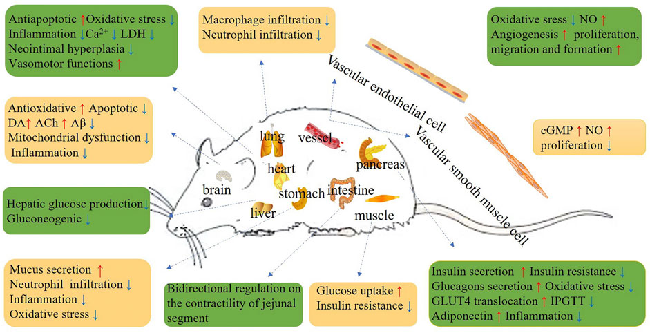
FIGURE 1. Schematic diagram depicting the beneficial effects of Re.
X-YG completed the document collection and manuscript writing with the help of G-CL, J-XZ, L-HW, CX, Z-AY, AW, and Y-FS. The revision of the manuscript was collaboratively finished by G-CP and finally approved by H-DY.
This study was supported by the National Natural Science Foundation of China (No. 82060674), Science and Technology Planning Project of the Jilin Provincial Education Department (JJKH20210587KJ), Higher Education Discipline Innovation Project (111 Project, D18012), and Jilin Scientific and Technological development program (20190304055YY). We thank Liwen Bianji (Edanz) (www.liwenbianji.cn), for editing the English text of a draft of this manuscript.
The authors declare that the research was conducted in the absence of any commercial or financial relationships that could be construed as a potential conflict of interest.
All claims expressed in this article are solely those of the authors and do not necessarily represent those of their affiliated organizations, or those of the publisher, the editors and the reviewers. Any product that may be evaluated in this article, or claim that may be made by its manufacturer, is not guaranteed or endorsed by the publisher.
Attele, A. S., Zhou, Y. P., Xie, J. T., Wu, J. A., Zhang, L., Dey, L., et al. (2002). Antidiabetic Effects of Panax Ginseng berry Extract and the Identification of an Effective Component. Diabetes 51 (6), 1851–1858. doi:10.2337/diabetes.51.6.1851
Bae, E. A., Shin, J. E., and Kim, D. H. (2005). Metabolism of Ginsenoside Re by Human Intestinal Microflora and its Estrogenic Effect. Biol. Pharm. Bull. 28 (10), 1903–1908. doi:10.1248/bpb.28.1903
Bae, H. M., Cho, O. S., Kim, S. J., Im, B. O., Cho, S. H., Lee, S., et al. (2012). Inhibitory Effects of Ginsenoside Re Isolated from Ginseng berry on Histamine and Cytokine Release in Human Mast Cells and Human Alveolar Epithelial Cells. J. Ginseng Res. 36 (4), 369–374. doi:10.5142/jgr.2012.36.4.369
Bai, C. X., Sunami, A., Namiki, T., Sawanobori, T., and Furukawa, T. (2003). Electrophysiological Effects of Ginseng and Ginsenoside Re in guinea Pig Ventricular Myocytes. Eur. J. Pharmacol. 476 (1-2), 35–44. doi:10.1016/s0014-2999(03)02174-5
Bai, C. X., Takahashi, K., Masumiya, H., Sawanobori, T., and Furukawa, T. (2004). Nitric Oxide-dependent Modulation of the Delayed Rectifier K+ Current and the L-type Ca2+ Current by Ginsenoside Re, an Ingredient of Panax Ginseng, in guinea-pig Cardiomyocytes. Br. J. Pharmacol. 142 (3), 567–575. doi:10.1038/sj.bjp.0705814
Bennett, M. R., Sinha, S., and Owens, G. K. (2016). Vascular Smooth Muscle Cells in Atherosclerosis. Circ. Res. 118 (4), 692–702. doi:10.1161/CIRCRESAHA.115.306361
Brekhman, I. I., and Dardymov, I. V. (1969). New Substances of Plant Origin Which Increase Nonspecific Resistance. Annu. Rev. Pharmacol. 9, 419–430. doi:10.1146/annurev.pa.09.040169.002223
Cao, G., Su, P., Zhang, S., Guo, L., Zhang, H., LiangQin, Y. C., et al. (2016). Ginsenoside Re Reduces Aβ Production by Activating PPARγ to Inhibit BACE1 in N2a/APP695 Cells. Eur. J. Pharmacol. 793, 101–108. doi:10.1016/j.ejphar.2016.11.006
Chan, L. Y., Chiu, P. Y., and Lau, T. K. (2004). Embryotoxicity Study of Ginsenoside Rc and Re in In Vitro Rat Whole Embryo Culture. Reprod. Toxicol. 19 (1), 131–134. doi:10.1016/j.reprotox.2004.06.001
Chan, L. Y., Kwok, H. H., Chan, R. W., Peiris, M. J., Mak, N. K., Wong, R. N., et al. (2011). Dual Functions of Ginsenosides in Protecting Human Endothelial Cells against Influenza H9N2-Induced Inflammation and Apoptosis. J. Ethnopharmacol. 137 (3), 1542–1546. doi:10.1016/j.jep.2011.08.022
Chen, F., Eckman, E. A., and Eckman, C. B. (2006). Reductions in Levels of the Alzheimer's Amyloid Beta Peptide after Oral Administration of Ginsenosides. FASEB. J. 20 (8), 1269–1271. doi:10.1096/fj.05-5530fje
Chen, G., Yang, M., and Guo, D. (2009). [Metabolic Study of Ginsenoside Re in Rats]. Zhongguo Zhong Yao Za Zhi 34 (12), 1540–1543.
Chen, L., Liu, L., Wang, Q., JiangTian, Y. H., and Tian, H. (2021). Comparative Pharmacokinetics Study of Six Effective Components between Two Dosage Forms of Qixue-Shuangbu Prescription in Rats by UPLC-MS/MS. Biomed. Chromatogr. 35 (10), e5179. doi:10.1002/bmc.5179
Chen, L. M., Zhou, X. M., Cao, Y. L., and Hu, W. X. (2008). Neuroprotection of Ginsenoside Re in Cerebral Ischemia-Reperfusion Injury in Rats. J. Asian Nat. Prod. Res. 10 (5-6), 439–445. doi:10.1080/10286020801892292
Chen, R. C., Wang, J., Yang, L., Sun, G. B., and Sun, X. B. (2016). Protective Effects of Ginsenoside Re on Lipopolysaccharide-Induced Cardiac Dysfunction in Mice. Food Funct. 7 (5), 2278–2287. doi:10.1039/c5fo01357g
Chen, S. E., Sawchuk, R. J., and Staba, E. J. (1980). American Ginseng. III. Pharmacokinetics of Ginsenosides in the Rabbit. Eur. J. Drug Metab. Pharmacokinet. 5 (3), 161–168. doi:10.1007/bf03189460
Chen, Y. B., Wang, Y. F., Hou, W., Wang, Y. P., Xiao, S. Y., Fu, Y. Y., et al. (2017). Effect of B-Complex Vitamins on the Antifatigue Activity and Bioavailability of Ginsenoside Re after Oral Administration. J. Ginseng. Res. 41 (2), 209–214. doi:10.1016/j.jgr.2016.03.006
Chi, H., and Ji, G. E. (2005). Transformation of Ginsenosides Rb1 and Re from Panax Ginseng by Food Microorganisms. Biotechnol. Lett. 27 (11), 765–771. doi:10.1007/s10529-005-5632-y
Cho, W. C., Chung, W. S., Lee, S. K., Leung, A. W., Cheng, C. H., and Yue, K. K. (2006). Ginsenoside Re of Panax Ginseng Possesses Significant Antioxidant and Antihyperlipidemic Efficacies in Streptozotocin-Induced Diabetic Rats. Eur. J. Pharmacol. 550 (1-3), 173–179. doi:10.1016/j.ejphar.2006.08.056
Christensen, L. P. (2009). Ginsenosides Chemistry, Biosynthesis, Analysis, and Potential Health Effects. Adv. Food Nutr. Res. 55, 1–99. doi:10.1016/s1043-4526(08)00401-4
Dai, G., Jiang, Z., Zhu, L., Zhang, Q., Zong, Y., Liu, S., et al. (2016). Simultaneous Determination of Notoginsenoside R1 and Ginsenoside Re in Rat Plasma by Ultra High Performance Liquid Chromatography with Tandem Mass Spectrometry and its Application to a Pharmacokinetic Study. J. Sep. Sci. 39 (17), 3368–3374. doi:10.1002/jssc.201600522
Dang, D. K., Shin, E. J., Kim, D. J., Tran, H. Q., Jeong, J. H., Jang, C. G., et al. (2018). Ginsenoside Re Protects Methamphetamine-Induced Dopaminergic Neurotoxicity in Mice via Upregulation of Dynorphin-Mediated κ-opioid Receptor and Downregulation of Substance P-Mediated Neurokinin 1 Receptor. J. Neuroinflammation 15 (1), 52. doi:10.1186/s12974-018-1087-7
Dong, W.-W., Zhao, J., Zhong, F.-L., Zhu, W.-J., Jiang, J., Wu, S., et al. (2017). Biotransformation of Panax Ginseng Extract by Rat Intestinal Microflora: Identification and Quantification of Metabolites Using Liquid Chromatography-Tandem Mass Spectrometry. J. Ginseng Res. 41 (4), 540–547. doi:10.1016/j.jgr.2016.11.002
Feng, X., and McDonald, J. M. (2011). Disorders of Bone Remodeling. Annu. Rev. Pathol. 6, 121–145. doi:10.1146/annurev-pathol-011110-130203
Furukawa, T., Bai, C. X., Kaihara, A., Ozaki, E., Kawano, T., Nakaya, Y., et al. (2006). Ginsenoside Re, a Main Phytosterol of Panax Ginseng, Activates Cardiac Potassium Channels via a Nongenomic Pathway of Sex Hormones. Mol. Pharmacol. 70 (6), 1916–1924. doi:10.1124/mol.106.028134
Gao, Y., Gao, C. Y., Zhu, P., Xu, S. F., Luo, Y. M., DengYang, J. D. L., et al. (2018). Ginsenoside Re Inhibits Vascular Neointimal Hyperplasia in Balloon-Injured Carotid Arteries through Activating the eNOS/NO/cGMP Pathway in Rats. Biomed. Pharmacother. 106, 1091–1097. doi:10.1016/j.biopha.2018.07.044
Gao, Y., Yang, M. F., Su, Y. P., Jiang, H. M., You, X. J., Yang, Y. J., et al. (2013). Ginsenoside Re Reduces Insulin Resistance through Activation of PPAR-γ Pathway and Inhibition of TNF-α Production. J. Ethnopharmacol. 147 (2), 509–516. doi:10.1016/j.jep.2013.03.057
Gao, Y., Zhu, P., Xu, S. F., Li, Y. Q., Deng, J., and Yang, D. L. (2019). Ginsenoside Re Inhibits PDGF-BB-Induced VSMC Proliferation via the eNOS/NO/cGMP Pathway. Biomed. Pharmacother. 115, 108934. doi:10.1016/j.biopha.2019.108934
Gillis, C. N. (1997). Panax Ginseng Pharmacology: a Nitric Oxide Link? Biochem. Pharmacol. 54 (1), 1–8. doi:10.1016/s0006-2952(97)00193-7
Gimbrone, M. A., and García-Cardeña, G. (2016). Endothelial Cell Dysfunction and the Pathobiology of Atherosclerosis. Circ. Res. 118 (4), 620–636. doi:10.1161/CIRCRESAHA.115.306301
González-Burgos, E., Fernández-Moriano, C., Lozano, R., Iglesias, I., and Gómez-Serranillos, M. P. (2017). Ginsenosides Rd and Re Co-treatments Improve Rotenone-Induced Oxidative Stress and Mitochondrial Impairment in SH-Sy5y Neuroblastoma Cells. Food Chem. Toxicol. 109 (Pt 1), 38–47. doi:10.1016/j.fct.2017.08.013
Greenspan, E. M. (1983). Ginseng and Vaginal Bleeding. Jama 249 (15), 2018. doi:10.1001/jama.1983.03330390026012
Gui, F. J., Yang, X. W., Li, L. Y., and Tian, J. M. (2007). Simultaneous Enantiomer Determination of 20 (R)- and 20 (S)-ginsenoside-Rg2 in Rat Plasma after Intravenous Administration Using HPLC Method. J. Chromatogr. B. Analyt. Technol. Biomed. Life Sci. 850 (1-2), 1–6. doi:10.1016/j.jchromb.2006.11.008
Han, D. H., Kim, S. H., Higashida, K., Jung, S. R., Polonsky, K. S., Klein, S., et al. (2012). Ginsenoside Re Rapidly Reverses Insulin Resistance in Muscles of High-Fat Diet Fed Rats. Metabolism 61 (11), 1615–1621. doi:10.1016/j.metabol.2012.04.008
Han, J., Xia, J., Zhang, L., Cai, E., Zhao, Y., Fei, X., et al. (2019). Studies of the Effects and Mechanisms of Ginsenoside Re and Rk3 on Myelosuppression Induced by Cyclophosphamide. J. Ginseng Res. 43 (4), 618–624. doi:10.1016/j.jgr.2018.07.009
Harkey, M. R., Henderson, G. L., Gershwin, M. E., Stern, J. S., and Hackman, R. M. (2001). Variability in Commercial Ginseng Products: an Analysis of 25 Preparations. Am. J. Clin. Nutr. 73 (6), 1101–1106. doi:10.1093/ajcn/73.6.1101
Hong, N. R., Park, H. S., Ahn, T. S., Kim, H. J., Ha, K. T., and Kim, B. J. (2015). Ginsenoside Re Inhibits Pacemaker Potentials via Adenosine Triphosphate-Sensitive Potassium Channels and the Cyclic Guanosine Monophosphate/nitric Oxide-dependent Pathway in Cultured Interstitial Cells of Cajal from Mouse Small Intestine. J. Ginseng Res. 39 (4), 314–321. doi:10.1016/j.jgr.2015.02.004
Huang, G. D., Zhong, X. F., Deng, Z. Y., and Zeng, R. (2016). Proteomic Analysis of Ginsenoside Re Attenuates Hydrogen Peroxide-Induced Oxidative Stress in Human Umbilical Vein Endothelial Cells. Food Funct. 7 (5), 2451–2461. doi:10.1039/c6fo00123h
Huang, Y. C., Chen, C. T., Chen, S. C., Lai, P. H., Liang, H. C., Chang, Y., et al. (2005). A Natural Compound (Ginsenoside Re) Isolated from Panax Ginseng as a Novel Angiogenic Agent for Tissue Regeneration. Pharm. Res. 22 (4), 636–646. doi:10.1007/s11095-005-2500-3
Jang, S. E., Jung, I. H., Joh, E. H., Han, M. J., and Kim, D. H. (2012). Antibiotics Attenuate Anti-scratching Behavioral Effect of Ginsenoside Re in Mice. J. Ethnopharmacol. 142 (1), 105–112. doi:10.1016/j.jep.2012.04.022
Ji, B., Zhao, X., Yu, P., Meng, L., Zhao, Y., and Yu, Z. (2017). Simultaneous Determination and Pharmacokinetics of Fourteen Bioactive Compounds in Rat Plasma by LC-ESI-MS/MS Following Intravenous Injection of Gegen-Sanqi Compatibility Solution. J. Chromatogr. B. Analyt. Technol. Biomed. Life Sci. 1068-1069-1069, 164–172. doi:10.1016/j.jchromb.2017.10.023
Ji, Z. N., Dong, T. T., Ye, W. C., Choi, R. C., Lo, C. K., and Tsim, K. W. (2006). Ginsenoside Re Attenuate Beta-Amyloid and Serum-free Induced Neurotoxicity in PC12 Cells. J. Ethnopharmacol. 107 (1), 48–52. doi:10.1016/j.jep.2006.02.004
Jiang, J., Sun, X., Akther, M., Lian, M. L., Quan, L. H., Koppula, S., et al. (2020). Ginsenoside Metabolite 20(S)-protopanaxatriol from Panax Ginseng Attenuates Inflammation-Mediated NLRP3 Inflammasome Activation. J. Ethnopharmacol. 251, 112564. doi:10.1016/j.jep.2020.112564
Jing, L., Wang, M., Zheng, Z., Peng, J., Wei, Y., Zheng, Z., et al. (2010). Ginsenoside Re Downregulates ICAM-1 Expression, Inhibits Polymorphonuclear Infiltration, and Ameliorates Ischemia-Reperfusion Injury. Med. Chem. Res. 19 (8), 962–969. doi:10.1007/s.00044-009-9242-410.1007/s00044-009-9242-4
Joo, K. M., Lee, J. H., Jeon, H. Y., Park, C. W., Hong, D. K., Jeong, H. J., et al. (2010). Pharmacokinetic Study of Ginsenoside Re with Pure Ginsenoside Re and Ginseng berry Extracts in Mouse Using Ultra Performance Liquid Chromatography/mass Spectrometric Method. J. Pharm. Biomed. Anal. 51 (1), 278–283. doi:10.1016/j.jpba.2009.08.013
Kabalak, A. A., Soyal, O. B., Urfalioglu, A., Saracoglu, F., and Gogus, N. (2004). Menometrorrhagia and Tachyarrhythmia after Using Oral and Topical Ginseng. J. Womens Health (Larchmt) 13 (7), 830–833. doi:10.1089/jwh.2004.13.830
Kawase, A., Yamada, A., Gamou, Y., Tahara, C., Takeshita, F., Murata, K., et al. (2014). Effects of Ginsenosides on the Expression of Cytochrome P450s and Transporters Involved in Cholesterol Metabolism. J. Nat. Med. 68 (2), 395–401. doi:10.1007/s11418-013-0791-y
Kim, H. B., Lim, K. H., Kang, C.-W., Kim, B. S., Roh, Y. S., Kwon, J., et al. (2011). Influence of Ginsenoside-Re against Myocardial Infarction in Isolated Heart. Mol. Cel. Toxicol. 7 (1), 15–24. doi:10.1007/s13273-011-0003-3
Kim, H. M., Kim, D. H., Han, H. J., Park, C. M., Ganipisetti, S. R., Valan Arasu, M., et al. (2016). Ginsenoside Re Promotes Osteoblast Differentiation in Mouse Osteoblast Precursor MC3T3-E1 Cells and a Zebrafish Model. Molecules 22 (1), 42. doi:10.3390/molecules22010042
Kim, J. H., Han, I. H., Yamabe, N., Kim, Y. J., Lee, W., Eom, D. W., et al. (2014). Renoprotective Effects of Maillard Reaction Products Generated during Heat Treatment of Ginsenoside Re with Leucine. Food Chem. 143, 114–121. doi:10.1016/j.foodchem.2013.07.075
Kim, J. M., Park, C. H., Park, S. K., Seung, T. W., Kang, J. Y., Ha, J. S., et al. (2017). Ginsenoside Re Ameliorates Brain Insulin Resistance and Cognitive Dysfunction in High Fat Diet-Induced C57BL/6 Mice. J. Agric. Food Chem. 65 (13), 2719–2729. doi:10.1021/acs.jafc.7b00297
Kim, K. H., Song, K., Yoon, S. H., Shehzad, O., Kim, Y. S., and Son, J. H. (2012). Rescue of PINK1 Protein Null-specific Mitochondrial Complex IV Deficits by Ginsenoside Re Activation of Nitric Oxide Signaling. J. Biol. Chem. 287 (53), 44109–44120. doi:10.1074/jbc.M112.408146
Kim, M. S., Yu, J. M., Kim, H. J., Kim, H. B., Kim, S. T., Jang, S. K., et al. (2014). Ginsenoside Re and Rd Enhance the Expression of Cholinergic Markers and Neuronal Differentiation in Neuro-2a Cells. Biol. Pharm. Bull. 37 (5), 826–833. doi:10.1248/bpb.b14-00011
Kim, U., Park, M. H., Kim, D. H., and Yoo, H. H. (2013). Metabolite Profiling of Ginsenoside Re in Rat Urine and Faeces after Oral Administration. Food Chem. 136 (3-4), 1364–1369. doi:10.1016/j.foodchem.2012.09.050
Kim, Y. K., Yoo, D. S., Xu, H., Park, N. I., Kim, H. H., Choi, J. E., et al. (2009). Ginsenoside Content of Berries and Roots of Three Typical Korean Ginseng (Panax Ginseng) Cultivars. Nat. Prod. Commun. 4 (7), 903–906. doi:10.1177/1934578X0900400704
Lee, B., Shim, I., Lee, H., and Hahm, D. H. (2012). Effect of Ginsenoside Re on Depression- and Anxiety-like Behaviors and Cognition Memory Deficit Induced by Repeated Immobilization in Rats. J. Microbiol. Biotechnol. 22 (5), 708–720. doi:10.4014/jmb.1112.12046
Lee, G. H., Lee, W. J., Hur, J., Kim, E., Lee, H. G., and Seo, H. G. (2020). Ginsenoside Re Mitigates 6-Hydroxydopamine-Induced Oxidative Stress through Upregulation of GPX4. Molecules 25 (1), 188. doi:10.3390/molecules25010188
Lee, I. A., Hyam, S. R., Jang, S. E., Han, M. J., and Kim, D. H. (2012). Ginsenoside Re Ameliorates Inflammation by Inhibiting the Binding of Lipopolysaccharide to TLR4 on Macrophages. J. Agric. Food Chem. 60 (38), 9595–9602. doi:10.1021/jf301372g
Lee, J. H., Lee, W., Lee, S., Jung, Y., Park, S. H., Choi, P., et al. (2012). Important Role of Maillard Reaction in the Protective Effect of Heat-Processed Ginsenoside Re-serine Mixture against Cisplatin-Induced Nephrotoxicity in LLC-PK1 Cells. Bioorg. Med. Chem. Lett. 22 (17), 5475–5479. doi:10.1016/j.bmcl.2012.07.018
Lee, J. H., Min, D. S., Lee, C. W., Song, K. H., Kim, Y. S., and Kim, H. P. (2018). Ginsenosides from Korean Red Ginseng Ameliorate Lung Inflammatory Responses: Inhibition of the MAPKs/NF-κB/c-Fos Pathways. J. Ginseng Res. 42 (4), 476–484. doi:10.1016/j.jgr.2017.05.005
Lee, K. W., Jung, S. Y., Choi, S. M., and Yang, E. J. (2012). Effects of Ginsenoside Re on LPS-Induced Inflammatory Mediators in BV2 Microglial Cells. BMC Complement. Altern. Med. 12, 196. doi:10.1186/1472-6882-12-196
Lee, O. H., Lee, H. H., Kim, J. H., and Lee, B. Y. (2011). Effect of Ginsenosides Rg3 and Re on Glucose Transport in Mature 3T3-L1 Adipocytes. Phytother. Res. 25 (5), 768–773. doi:10.1002/ptr.3322
Lee, S., Kim, M. G., Ko, S. K., Kim, H. K., Leem, K. H., and Kim, Y. J. (2014). Protective Effect of Ginsenoside Re on Acute Gastric Mucosal Lesion Induced by Compound 48/80. J. Ginseng Res. 38 (2), 89–96. doi:10.1016/j.jgr.2013.10.001
Lee, W., Park, S. H., Lee, S., Chung, B. C., Song, M. O., Song, K. I., et al. (2012). Increase in Antioxidant Effect of Ginsenoside Re-alanine Mixture by Maillard Reaction. Food Chem. 135 (4), 2430–2435. doi:10.1016/j.foodchem.2012.06.108
Leung, K. W., Leung, F. P., Huang, Y., Mak, N. K., and Wong, R. N. (2007). Non-genomic Effects of Ginsenoside-Re in Endothelial Cells via Glucocorticoid Receptor. FEBS Lett. 581 (13), 2423–2428. doi:10.1016/j.febslet.2007.04.055
Li, J., Liu, Y., Li, W., Wang, Z., Guo, P., Li, L., et al. (2018). Metabolic Profiling of the Effects of Ginsenoside Re in an Alzheimer's Disease Mouse Model. Behav. Brain Res. 337, 160–172. doi:10.1016/j.bbr.2017.09.027
Li, L., Sheng, Y. X., Zhang, J. L., Wang, S. S., and Guo, D. A. (2006). High-performance Liquid Chromatographic Assay for the Active Saponins from Panax Notoginseng in Rat Tissues. Biomed. Chromatogr. 20 (4), 327–335. doi:10.1002/bmc.567
Li, P., Lv, B., Jiang, X., Wang, T., Ma, X., Chang, N., et al. (2016). Identification of NF-Κb Inhibitors Following Shenfu Injection and Bioactivity-Integrated UPLC/Q-TOF-MS and Screening for Related Anti-inflammatory Targets In Vitro and In Silico. J. Ethnopharmacol. 194, 658–667. doi:10.1016/j.jep.2016.10.052
Li, Z., Ryu, S. W., Lee, J., Choi, K., Kim, S., and Choi, C. (2016). Protopanaxatirol Type Ginsenoside Re Promotes Cyclic Growth of Hair Follicles via Inhibiting Transforming Growth Factor β Signaling Cascades. Biochem. Biophys. Res. Commun. 470 (4), 924–929. doi:10.1016/j.bbrc.2016.01.148
Lim, H. W., Kim, K., and Lim, C. J. (2016). Contribution of Ginsenoside Re to Cellular Redox Homeostasis via Upregulating Glutathione and Superoxide Dismutase in HaCaT Keratinocytes under normal Conditions. Pharmazie 71 (7), 413–419. doi:10.1691/ph.2016.6518
Lim, K. H., Lim, D. J., and Kim, J. H. (2013). Ginsenoside-Re Ameliorates Ischemia and Reperfusion Injury in the Heart: a Hemodynamics Approach. J. Ginseng Res. 37 (3), 283–292. doi:10.5142/jgr.2013.37.283
Liu, L., Huang, J., Hu, X., Li, K., and Sun, C. (2011). Simultaneous Determination of Ginsenoside (G-Re, G-Rg1, G-Rg2, G-F1, G-Rh1) and Protopanaxatriol in Human Plasma and Urine by LC-MS/MS and its Application in a Pharmacokinetics Study of G-Re in Volunteers. J. Chromatogr. B. Analyt. Technol. Biomed. Life Sci. 879 (22), 2011–2017. doi:10.1016/j.jchromb.2011.05.018
Liu, M., Bai, X., Yu, S., Zhao, W., Qiao, J., Liu, Y., et al. (2019). Ginsenoside Re Inhibits ROS/ASK-1 Dependent Mitochondrial Apoptosis Pathway and Activation of Nrf2-Antioxidant Response in Beta-Amyloid-Challenged SH-Sy5y Cells. Molecules 24 (15), 2687. doi:10.3390/molecules24152687
Liu, Y. M., Yang, L., Zeng, X., Deng, Y. H., Feng, Y., and Liang, W. X. (2005). [Pharmacokinetics of Ginsenosides Rg1 and Re in Shenmai Injection]. Yao Xue Xue Bao 40 (4), 365–368.
Liu, Y. W., Zhu, X., Li, W., Lu, Q., Wang, J. Y., Wei, Y. Q., et al. (2012). Ginsenoside Re Attenuates Diabetes-Associated Cognitive Deficits in Rats. Pharmacol. Biochem. Behav. 101 (1), 93–98. doi:10.1016/j.pbb.2011.12.003
Lu, D., Liu, J., Zhao, W., and Li, P. (2012). Chronic Toxicity of Ginsenoside Re on Sprague-Dawley Rats. J. Ethnopharmacol. 144 (3), 656–663. doi:10.1016/j.jep.2012.10.007
Ma, X., Xu, Q., and Liang, X. (2005). [Analysis of Ginsenosides in Ginseng by Liquid Chromatographyatmospheric Pressure Chemical Ionization Mass Spectrometry]. Se Pu 23 (4), 389–393.
Mancuso, C., and Santangelo, R. (2017). Panax Ginseng and Panax Quinquefolius: From Pharmacology to Toxicology. Food Chem. Toxicol. 107 (Pt A), 362–372. doi:10.1016/j.fct.2017.07.019
Nakaya, Y., Mawatari, K., Takahashi, A., Harada, N., Hata, A., and Yasui, S. (2007). The Phytoestrogen Ginsensoside Re Activates Potassium Channels of Vascular Smooth Muscle Cells through PI3K/Akt and Nitric Oxide Pathways. J. Med. Invest. 54 (3-4), 381–384. doi:10.2152/jmi.54.381
Nam, Y., Wie, M. B., Shin, E. J., Nguyen, T. T., Nah, S. Y., Ko, S. K., et al. (2015). Ginsenoside Re Protects Methamphetamine-Induced Mitochondrial Burdens and Proapoptosis via Genetic Inhibition of Protein Kinase C δ in Human Neuroblastoma Dopaminergic SH-Sy5y Cell Lines. J. Appl. Toxicol. 35 (8), 927–944. doi:10.1002/jat.3093
National Toxicology Program, (2011). Toxicology and Carcinogenesis Studies of Ginseng (CAS No. 50647-08-0) in F344/N Rats and B6C3F1 Mice (Gavage Studies). Natl. Toxicol. Program Tech. Rep. Ser. 567, 1–149.
Oh, Y., Lim, H. W., Kim, K., and Lim, C. J. (2016). Ginsenoside Re Improves Skin Barrier Function in HaCaT Keratinocytes under normal Growth Conditions. Biosci. Biotechnol. Biochem. 80 (11), 2165–2167. doi:10.1080/09168451.2016.1206808
Palmer, B. V., Montgomery, A. C., and Monteiro, J. C. (1978). Gin Seng and Mastalgia. Br. Med. J. 1 (6122), 1284. doi:10.1136/bmj.1.6122.1284
Park, C. H., Park, S. K., Seung, T. W., Jin, D. E., Guo, T., and Heo, H. J. (2015). Effect of Ginseng (Panax Ginseng) berry EtOAc Fraction on Cognitive Impairment in C57BL/6 Mice under High-Fat Diet Inducement. Evid. Based Complement. Alternat Med. 2015, 316527. doi:10.1155/2015/316527
Park, C. M., Kim, H. M., Kim, D. H., Han, H. J., Noh, H., Jang, J. H., et al. (2016). Ginsenoside Re Inhibits Osteoclast Differentiation in Mouse Bone Marrow-Derived Macrophages and Zebrafish Scale Model. Mol. Cell 39 (12), 855–861. doi:10.14348/molcells.2016.0111
Paul, S., Shin, H. S., and Kang, S. C. (2012). Inhibition of Inflammations and Macrophage Activation by Ginsenoside-Re Isolated from Korean Ginseng (Panax Ginseng C.A. Meyer). Food Chem. Toxicol. 50 (5), 1354–1361. doi:10.1016/j.fct.2012.02.035
Peng, D., Wang, H., Qu, C., Xie, L., Wicks, S. M., and Xie, J. (2012). Ginsenoside Re: Its Chemistry, Metabolism and Pharmacokinetics. Chin. Med. 7, 2. doi:10.1186/1749-8546-7-2
Pyo, M. K., Park, K.-H., Oh, M. H., Lee, H., Park, Y. S., Kim, N. Y., et al. (2016). Ginsenoside Re Enriched Fraction (GS-F3k1) from Ginseng Berries Ameliorates Ethanol-Induced Erectile DysfunctionviaNitric Oxide-cGMP Pathway. Nat. Prod. Sci. 22 (1), 46–52. doi:10.20307/nps.2016.22.1.46
Qi, L. W., Wang, C. Z., Du, G. J., Zhang, Z. Y., Calway, T., and Yuan, C. S. (2011). Metabolism of Ginseng and its Interactions with Drugs. Curr. Drug Metab. 12 (9), 818–822. doi:10.2174/138920011797470128
Quan, H., Jin, X., Cui, E., and Zhang, Q. (2019). Lipopolysaccharide-induced Inflammation Is Inhibited by Ginsenoside Re through NF-Κb Signaling in RAW264.7 Cells and Primary Rat Hepatocytes. Lat. Am. J. Pharm. 38 (10), 1969–1978. doi:10.1016/j.ejphar.2006.11.023
Quan, H. Y., Yuan, H. D., Jung, M. S., Ko, S. K., Park, Y. G., and Chung, S. H. (2012). Ginsenoside Re Lowers Blood Glucose and Lipid Levels via Activation of AMP-Activated Protein Kinase in HepG2 Cells and High-Fat Diet Fed Mice. Int. J. Mol. Med. 29 (1), 73–80. doi:10.3892/ijmm.2011.805
Reeds, D. N., Patterson, B. W., Okunade, A., Holloszy, J. O., Polonsky, K. S., and Klein, S. (2011). Ginseng and Ginsenoside Re Do Not Improve β-cell Function or Insulin Sensitivity in Overweight and Obese Subjects with Impaired Glucose Tolerance or Diabetes. Diabetes Care 34 (5), 1071–1076. doi:10.2337/dc10-2299
Shen, B. Q., Qu, C., Mi, L., Wang, H. Y., and Yang, H. (2021). Simultaneous Quantification of Twenty-Eight Components of Shenfu Injection in Rat Plasma by UHPLC-QQQ MS and its Application to a Pharmacokinetic Study. J. Pharm. Biomed. Anal. 203, 114211. doi:10.1016/j.jpba.2021.114211
Shi, J., Xue, W., Zhao, W. J., and Li, K. X. (2013). Pharmacokinetics and Dopamine/acetylcholine Releasing Effects of Ginsenoside Re in hippocampus and mPFC of Freely Moving Rats. Acta Pharmacol. Sin. 34 (2), 214–220. doi:10.1038/aps.2012.147
Shi, Y., Wan, X., Shao, N., Ye, R., Zhang, N., and Zhang, Y. (2016). Protective and Anti-angiopathy E-ffects of ginsenoside Re against D-iabetes M-ellitus via the A-ctivation of P-38 MAPK, ERK1/2 and JNK S-ignaling. Mol. Med. Rep. 14 (5), 4849–4856. doi:10.3892/mmr.2016.5821
Shin, D., Moon, H. W., Oh, Y., Kim, K., Kim, D. D., and Lim, C. J. (2018). Defensive Properties of Ginsenoside Re against UV-B-Induced Oxidative Stress through Up-Regulating Glutathione and Superoxide Dismutase in HaCaT Keratinocytes. Iran. J. Pharm. Res. 17 (1), 249–260.
Shin, E. J., Shin, S. W., Nguyen, T. T., Park, D. H., Wie, M. B., Jang, C. G., et al. (2014). Ginsenoside Re Rescues Methamphetamine-Induced Oxidative Damage, Mitochondrial Dysfunction, Microglial Activation, and Dopaminergic Degeneration by Inhibiting the Protein Kinase Cδ Gene. Mol. Neurobiol. 49 (3), 1400–1421. doi:10.1007/s12035-013-8617-1
Son, Y. M., Kwak, C. W., Lee, Y. J., Yang, D. C., Park, B. C., Lee, W. K., et al. (2010). Ginsenoside Re Enhances Survival of Human CD4+ T Cells through Regulation of Autophagy. Int. Immunopharmacol. 10 (5), 626–631. doi:10.1016/j.intimp.2010.03.002
Song, J. H., Choi, H. J., Song, H. H., Hong, E. H., Lee, B. R., Oh, S. R., et al. (2014). Antiviral Activity of Ginsenosides against Coxsackievirus B3, Enterovirus 71, and Human Rhinovirus 3. J. Ginseng Res. 38 (3), 173–179. doi:10.1016/j.jgr.2014.04.003
Song, X., Chen, J., Sakwiwatkul, K., Li, R., and Hu, S. (2010). Enhancement of Immune Responses to Influenza Vaccine (H3N2) by Ginsenoside Re. Int. Immunopharmacol. 10 (3), 351–356. doi:10.1016/j.intimp.2009.12.009
Su, F., Xue, Y., Wang, Y., Zhang, L., Chen, W., and Hu, S. (2015). Protective Effect of Ginsenosides Rg1 and Re on Lipopolysaccharide-Induced Sepsis by Competitive Binding to Toll-like Receptor 4. Antimicrob. Agents Chemother. 59 (9), 5654–5663. doi:10.1128/AAC.01381-15
Su, F., Yuan, L., Zhang, L., and Hu, S. (2012). Ginsenosides Rg1 and Re Act as Adjuvant via TLR4 Signaling Pathway. Vaccine 30 (27), 4106–4112. doi:10.1016/j.vaccine.2012.03.052
Su, X., Pei, Z., and Hu, S. (2014). Ginsenoside Re as an Adjuvant to Enhance the Immune Response to the Inactivated Rabies Virus Vaccine in Mice. Int. Immunopharmacol. 20 (2), 283–289. doi:10.1016/j.intimp.2014.03.008
Sukrittanon, S., Watanapa, W. B., and Ruamyod, K. (2014). Ginsenoside Re Enhances Small-Conductance Ca(2+)-Activated K(+) Current in Human Coronary Artery Endothelial Cells. Life Sci. 115 (1-2), 15–21. doi:10.1016/j.lfs.2014.09.007
Sun, H., Ling, S., Zhao, D., Li, J., Li, Y., Qu, H., et al. (2020). Ginsenoside Re Treatment Attenuates Myocardial Hypoxia/Reoxygenation Injury by Inhibiting HIF-1α Ubiquitination. Front. Pharmacol. 11, 532041. doi:10.3389/fphar.2020.532041
Sun, H. X., Chen, Y., and Ye, Y. (2006). Ginsenoside Re and Notoginsenoside R1: Immunologic Adjuvants with Low Haemolytic Effect. Chem. Biodivers. 3 (7), 718–726. doi:10.1002/cbdv.200690074
Tran, T. V., Shin, E. J., Dang, D. K., Ko, S. K., Jeong, J. H., Nah, S. Y., et al. (2017). Ginsenoside Re Protects against Phencyclidine-Induced Behavioral Changes and Mitochondrial Dysfunction via Interactive Modulation of Glutathione Peroxidase-1 and NADPH Oxidase in the Dorsolateral Cortex of Mice. Food Chem. Toxicol. 110, 300–315. doi:10.1016/j.fct.2017.10.019
Tu, T. T., Sharma, N., Shin, E. J., Tran, H. Q., Lee, Y. J., Jeong, J. H., et al. (2017). Ginsenoside Re Protects Trimethyltin-Induced Neurotoxicity via Activation of IL-6-Mediated Phosphoinositol 3-Kinase/Akt Signaling in Mice. Neurochem. Res. 42 (11), 3125–3139. doi:10.1007/s11064-017-2349-y
Wang, H., Lv, J., Jiang, N., Huang, H., Wang, Q., and Liu, X. (2021). Ginsenoside Re Protects against Chronic Restraint Stress‐induced Cognitive Deficits through Regulation of NLRP3 and Nrf2 Pathways in Mice. Phytotherapy Res. 35, 2523–2535. doi:10.1002/ptr.6947
Wang, L., Chen, X., Wang, Y., Zhao, L., Zhao, X., and Wang, Y. (2020). MiR-30c-5p Mediates the Effects of Panax Notoginseng Saponins in Myocardial Ischemia Reperfusion Injury by Inhibiting Oxidative Stress-Induced Cell Damage. Biomed. Pharmacother. 125, 109963. doi:10.1016/j.biopha.2020.109963
Wang, L., Yuan, D., Zhang, D., Zhang, W., Liu, C., Cheng, H., et al. (2015). Ginsenoside Re Promotes Nerve Regeneration by Facilitating the Proliferation, Differentiation and Migration of Schwann Cells via the ERK- and JNK-dependent Pathway in Rat Model of Sciatic Nerve Crush Injury. Cell. Mol. Neurobiol. 35 (6), 827–840. doi:10.1007/s10571-015-0177-7
Wang, Q. W., Yu, X. F., Xu, H. L., Zhao, X. Z., and Sui, D. Y. (20192019). Ginsenoside Re Improves Isoproterenol-Induced Myocardial Fibrosis and Heart Failure in Rats. Evid. Based Complement. Alternat. Med. 2019, 3714508. doi:10.1155/2019/3714508
Wang, W., Liao, Q. P., Quan, L. H., Liu, C. Y., Chang, Q., Liu, X. M., et al. (2010). The Effect of Acorus Gramineus on the Bioavailabilities and Brain Concentrations of Ginsenosides Rg1, Re and Rb1 after Oral Administration of Kai-Xin-San Preparations in Rats. J. Ethnopharmacol. 131 (2), 313–320. doi:10.1016/j.jep.2010.06.034
Wang, Y., Zhang, Y., Xiao, J., Xu, R., Wang, Q., and Wang, X. (2018b). Simultaneous Determination of Baicalin, Baicalein, Wogonoside, Wogonin, Scutellarin, Berberine, Coptisine, Ginsenoside Rb1 and Ginsenoside Re of Banxia Xiexin Decoction in Rat Plasma by LC-MS/MS and its Application to a Pharmacokinetic Study. Biomed. Chromatogr. 32 (2), 1. doi:10.1002/bmc.4083
Wang, Y. G., Zima, A. V., Ji, X., Pabbidi, R., Blatter, L. A., and Lipsius, S. L. (2008a). Ginsenoside Re Suppresses Electromechanical Alternans in Cat and Human Cardiomyocytes. Am. J. Physiol. Heart Circ. Physiol. 295 (2), H851–H859. doi:10.1152/ajpheart.01242.2007
Wang, Z., Li, Y. F., Han, X. Y., Sun, Y. S., Zhang, L. X., Liu, W., et al. (2018c). Kidney protection Effect of Ginsenoside Re and its Underlying Mechanisms on Cisplatin-Induced Kidney Injury. Cell. Physiol. Biochem. 48 (5), 2219–2229. doi:10.1159/000492562
Wu, C. F., Bi, X. L., Yang, J. Y., Zhan, J. Y., Dong, Y. X., Wang, J. H., et al. (2007). Differential Effects of Ginsenosides on NO and TNF-Alpha Production by LPS-Activated N9 Microglia. Int. Immunopharmacol. 7 (3), 313–320. doi:10.1016/j.intimp.2006.04.021
Xia, C., Wang, G., Sun, J., Hao, H., Xiong, Y., Gu, S., et al. (2008). Simultaneous Determination of Ginsenoside Rg1, Re, Rd, Rb1 and Ophiopogonin D in Rat Plasma by Liquid Chromatography/electrospray Ionization Mass Spectrometric Method and its Application to Pharmacokinetic Study of 'SHENMAI' Injection. J. Chromatogr. B. Analyt. Technol. Biomed. Life Sci. 862 (1-2), 72–78. doi:10.1016/j.jchromb.2007.11.020
Xiang, L., Jiang, P., Wang, S., Hu, Y., Liu, X., Yue, R., et al. (2013). Metabolomic Strategy for Studying the Intervention and the Synergistic Effects of the Shexiang Baoxin Pill for Treating Myocardial Infarction in Rats. Evid. Based. Complement. Alternat. Med. 2013, 823121. doi:10.1155/2013/823121
Xie, J.-T., Du, G.-J., McEntee, E., Aung, H. H., He, H., Mehendale, S. R., et al. (2011). Effects of Triterpenoid Glycosides from Fresh Ginseng Berry on SW480 Human Colorectal Cancer Cell Line. Cancer Res. Treat. 43 (1), 49–55. doi:10.4143/crt.2011.43.1.49
Xie, J. T., Mehendale, S. R., Li, X., Quigg, R., Wang, X., Wang, C. Z., et al. (2005a). Anti-diabetic Effect of Ginsenoside Re in Ob/ob Mice. Biochim. Biophys. Acta 1740 (3), 319–325. doi:10.1016/j.bbadis.2004.10.010
Xie, J. T., Mehendale, S. R., Wang, A., Han, A. H., Wu, J. A., Osinski, J., et al. (2004). American Ginseng Leaf: Ginsenoside Analysis and Hypoglycemic Activity. Pharmacol. Res. 49 (2), 113–117. doi:10.1016/j.phrs.2003.07.015
Xie, J. T., Shao, Z. H., Vanden Hoek, T. L., Chang, W. T., Li, J., Mehendale, S., et al. (2006). Antioxidant Effects of Ginsenoside Re in Cardiomyocytes. Eur. J. Pharmacol. 532 (3), 201–207. doi:10.1016/j.ejphar.2006.01.001
Xie, J. T., Wang, C. Z., Wang, A. B., Wu, J., Basila, D., and Yuan, C. S. (2005b). Antihyperglycemic Effects of Total Ginsenosides from Leaves and Stem of Panax Ginseng. Acta Pharmacol. Sin. 26 (9), 1104–1110. doi:10.1111/j.1745-7254.2005.00156.x
Xie, W., Zhou, P., Qu, M., Dai, Z., Zhang, X., Zhang, C., et al. (2020). Ginsenoside Re Attenuates High Glucose-Induced RF/6A Injury via Regulating PI3K/AKT Inhibited HIF-1α/VEGF Signaling Pathway. Front. Pharmacol. 11, 695. doi:10.3389/fphar.2020.00695
Xiong, Y., Chen, D., Lv, B., Liu, F., Yao, Q., Tang, Z., et al. (2014). Effects of Ginsenoside Re on Rat Jejunal Contractility. J. Nat. Med. 68 (3), 530–538. doi:10.1007/s11418-014-0831-2
Xu, B. B., Liu, C. Q., Gao, X., Zhang, W. Q., Wang, S. W., and Cao, Y. L. (2005). Possible Mechanisms of the protection of Ginsenoside Re against MPTP-Induced Apoptosis in Substantia Nigra Neurons of Parkinson's Disease Mouse Model. J. Asian Nat. Prod. Res. 7 (3), 215–224. doi:10.1080/10286020410001690172
Xu, X., Jin, L., Jiang, T., Lu, Y., Aosai, F., Piao, H. N., et al. (2020). Ginsenoside Rh2 Attenuates Microglial Activation against Toxoplasmic Encephalitis via TLR4/NF-Κb Signaling Pathway. J. Ginseng Res. 44 (5), 704–716. doi:10.1016/j.jgr.2019.06.002
Yang, K., Luo, Y., Lu, S., Hu, R., Du, Y., Liao, P., et al. (2018). Salvianolic Acid B and Ginsenoside Re Synergistically Protect against Ox-LDL-Induced Endothelial Apoptosis through the Antioxidative and Antiinflammatory Mechanisms. Front. Pharmacol. 9, 662. doi:10.3389/fphar.2018.00662
Yang, L., Xu, S., Liu, C., and Su, Z. (2009). In Vivo metabolism Study of Ginsenoside Re in Rat Using High-Performance Liquid Chromatography Coupled with Tandem Mass Spectrometry. Anal. Bioanal. Chem. 395 (5), 1441–1451. doi:10.1007/s00216-009-3121-1
Yao, H., Li, J., Song, Y., Zhao, H., Wei, Z., Li, X., et al. (2018). Synthesis of Ginsenoside Re-based Carbon Dots Applied for Bioimaging and Effective Inhibition of Cancer Cells. Int. J. Nanomedicine 13, 6249–6264. doi:10.2147/IJN.S176176
Yu, L. C., Chen, S. C., Chang, W. C., Huang, Y. C., Lin, K. M., Lai, P. H., et al. (2007). Stability of Angiogenic Agents, Ginsenoside Rg1 and Re, Isolated from Panax Ginseng: In Vitro and In Vivo Studies. Int. J. Pharm. 328 (2), 168–176. doi:10.1016/j.ijpharm.2006.08.009
Yu, Y., Sun, J., Liu, J., Wang, P., and Wang, C. (2020). Ginsenoside Re Preserves Cardiac Function and Ameliorates Left Ventricular Remodeling in a Rat Model of Myocardial Infarction. J. Cardiovasc. Pharmacol. 75 (1), 91–97. doi:10.1097/FJC.0000000000000752
Zhang, H., Zhou, Q. M., Li, X. D., Xie, Y., Duan, X., Min, F. L., et al. (2006). Ginsenoside R(e) increases fertile and asthenozoospermic infertile human sperm motility by induction of nitric oxide synthase. Arch. Pharm. Res. 29 (2), 145–151. doi:10.1007/bf02974276
Zhang, X., Wang, Y., Ma, C., Yan, Y., Yang, Y., Wang, X., et al. (2016). Ginsenoside Rd and Ginsenoside Re Offer Neuroprotection in a Novel Model of Parkinson's Disease. Am. J. Neurodegener. Dis. 5 (1), 52–61.
Zhang, Z., Li, X., Lv, W., Yang, Y., Gao, H., Yang, J., et al. (2008). Ginsenoside Re Reduces Insulin Resistance through Inhibition of C-Jun NH2-terminal Kinase and Nuclear Factor-kappaB. Mol. Endocrinol. 22 (1), 186–195. doi:10.1210/me.2007-0119
Zhang, Z. L., Liu, M. L., Huang, Y. S., Liang, W. Y., Zhang, M. M., Fan, Y. D., et al. (2020). Ginsenoside Re Enhances the Survival of H9c2 Cardiac Muscle Cells through Regulation of Autophagy. J. Asian Nat. Prod. Res. 22 (8), 774–787. doi:10.1080/10286020.2019.1632834
Zhou, X. M., Cao, Y. L., and Dou, D. Q. (2006). Protective Effect of Ginsenoside-Re against Cerebral Ischemia/reperfusion Damage in Rats. Biol. Pharm. Bull. 29 (12), 2502–2505. doi:10.1248/bpb.29.2502
Zhou, Y., Chen, S., Qiao, J., Cui, Y., Yuan, C., He, L., et al. (2020). Study of the Noncovalent Interactions of Ginsenosides and Amyloid-β-Peptide by CSI-MS and Molecular Docking. J. Mass. Spectrom. 55 (1), e4463. doi:10.1002/jms.4463
4-HNE 4-hydroxynonenal
6-HODA 6-hydroxydopamine
Aβ amyloid β
ACh acetylcholine
AChE acetylcholinesterase
AD alzheimer disease
ALP alkalin phosphatase
AM alloxan monohydrate
AMPK AMP-activated protein kinase
ASK1 apoptosis signal-regulated kinase-1
ATA antimycin A
AUC area under curve
BDNF brain-derived neurotrophic factor
BACE1 Aβ-site precursor protein-cleaving enzyme 1
bFGF basic fibroblast growth factor
BG blood glucose
BMNC bone marrow nucleated cell
BNP brain natriuretic peptide
BUN blood urea nitrogen
BW body weight
[Ca2+]i intracellular Ca2+ homeostasis
cGMP cyclic guanosine monophosphate
c-FOS-IR c-Fos-immunoreactivity
CAT catalase
ChAT choline acetyltransferase
colla1 type 1 collagen
CRE creatinine
CRS chronic restraint stress
CTX cyclophosphamide
CVDs cardiovascular diseases
CYP8B1 sterol 12a-hydroxylase
CP constipation-prominent
Cmax peak concentration
CL clearance
SFI Shenfu Injection
DA dopamine
DBP diastolic blood pressure
DII dexamethasone + 3-isobutyl-1-methylxanthine + insulin
DM diabetes mellitus
DPPH 1,1-diphenyl-2 picrylhydrazyl
DP diarrhea-prominent
EDV end-diastolic volume
ERs oestrogen receptors
ERK extracellular signal-regulated kinase
ESV end-systolic volume
f fraction excreted unchanged in the urine
FAS fatty acid synthase
FBG fasting blood glucose
GD glucose deprivation
GOT glutamic oxaloacetic transaminase
GPL Glucose + sodium pyruvate + sodium lactate
GPT glutamic pyruvic transaminase
GSH glutathione
GT glucose tolerance
GLUT4 glucose transporter 4
GPx glutathione peroxidase
GGSQ Gegen-Sanqi
HDL-C high density lipoprotein cholesterol
HFD high-fat diet
HGB hemoglobin
HIF-1α hypoxia-inducible factor-1-alpha
HO-1 heme oxygenase-1
HSHF high-sucrose-HFD
HUVEC human umbilical vein endothelial cell
ICa,L L-type Ca2+ current
iNOS nitric oxide synthase
JNK c-Jun N-terminal kinase
ICAM-1 intercellular adhesion molecule-1
IFN-γ interferon-γ
IL-1β interleukin-1β
IL-6 interleukin-6
Lp-α lipoprotein α
IPGTT intraperitoneal glucose tolerance test
I/R ischamia/reperfusion
IR insulin resistance
IRS-1 insulin receptor substrate
LRPPRC leucine-rich pentatricopeptide repeat-containing
IS insulin sensitivity
IVS interventricular septum
IVSTd IVS end-diastolic thickness
ICP intracavernous pressure
LADCA left anterior descending coronary artery
LC3B-2 microtubule-associated protein 1A/1B-light chain 3
LDH lactate dehydrogenase
LDL-C low density lipoprotein cholesterol
LKB1 liver kinase B1
LPC lysophosphatidylcholine
LPS lipopolysaccharide
LV left ventricular
LVD LV dimension
LVDd LV end-diastolic dimension
ILVDs LV endsystolic dimension
LVPWTs LV posteriorend-systolic thickness
LVPWTd LV posteriorend-diastolic thickness
LVEDP Left ventricular end diastolic pressure
MA methamphetamine
MAPK mitogen-activated protein kinase
MCAO middle cerebral artery occlusion
MDA malondialdehyde
MI myocardial infraction
MMP metalloproteinase
MPO myeloperoxidase
MPTP 1-methyl-4-phenyl-1,2,3,6-tetrahydropyridine
MRPs maillard reaction products
MLCK myosin light chain kinase
MRT mean residence time
NF-kB nuclear factor-kB
NLRP3 NOD‐like receptor protein 3
NK1 neurokinin1
NO nitric oxide
Nrf2 nuclear factor erythroid-2 associated factor 2
ox-LDL oxidized low density lipoprotein
OVA ovalbumin
QXSBP QixueShuang bu Prescription
p75 nerve growth factor receptor
PCNA proliferating cell nuclear antigen
PCP phencyclidine
PD parkinson disease
PEPCK phosphoenolpyruvate carboxykinase
PG peptidoglycan
PGE2 prostaglandin E2
PHOX NADPH oxidase
PI3K phosphoinositide3-kinase
PMN polymorphonuclear leukocyte
PKCδ protein kinase Cδ
PPARγ peroxisome proliferator-activated receptor-γ
PSD95 postsynaptic density 95
RC renal clearance
RANKL receptor activator of NF-κB ligand
RANTES T-cell-expressed and secreted
RBCs red blood cells
RIS repeated immobilization stress
RV rabies virus vaccine
SCD1 stearoyl-CoA desaturase-1
SHP small heterodimer partner
SNC sciatic nerve crush injury
SOD superoxide dismutase
SREBP sterol regulatory element-binding protein
STZ streptozotocin
SYP synaptophysin
T1DM type 1 diabetes
T2DM type 2 diabetes
tBHP tertbutyl hydroperoxide
TC total cholesterol
TG triglyceride
TH tyrosine hydroxylase
TLR4 toll-like receptor 4
TMT trimethyltin
TNBS 2,4,6-trinitrobenzene sulfonic acid
TNF-α tumour nuclear factor-α
TPA 12-O-tetradecanoylphorbol-13-acetate
TPO thrombopoietin
TRAP tartrate-resistant acid phosphatase
TGF-β transforming growth factor-β
TRARS thiobarbituric acid reactive substances
T1/2 elimination half-life
Tmax time for maximum concentration
VAchT vesicular acetylcholine transporter
VEGF vascular endothelial growth factor
VHL von Hippel-Lindau
VSTs IVS end-systolic thickness
Vd volume of distribution
WBC white blood cell
XO xanthine oxidase
XSTDT Xue saitong dispersible table
Keywords: ginsenoside Re, pharmacological activities, pharmacokinetics, toxicology, bioactive component
Citation: Gao X-Y, Liu G-C, Zhang J-X, Wang L-H, Xu C, Yan Z-A, Wang A, Su Y-F, Lee J-J, Piao G-C and Yuan H-D (2022) Pharmacological Properties of Ginsenoside Re. Front. Pharmacol. 13:754191. doi: 10.3389/fphar.2022.754191
Received: 06 August 2021; Accepted: 07 March 2022;
Published: 06 April 2022.
Edited by:
Zhiling Yu, Hong Kong Baptist University, Hong Kong SAR, ChinaReviewed by:
Saeideh Momtaz, Tehran University of Medical Sciences, IranCopyright © 2022 Gao, Liu, Zhang, Wang, Xu, Yan, Wang, Su, Lee, Piao and Yuan. This is an open-access article distributed under the terms of the Creative Commons Attribution License (CC BY). The use, distribution or reproduction in other forums is permitted, provided the original author(s) and the copyright owner(s) are credited and that the original publication in this journal is cited, in accordance with accepted academic practice. No use, distribution or reproduction is permitted which does not comply with these terms.
*Correspondence: Guang-Chun Piao, Z2NwaWFvQHlidS5lZHUuY24=; Hai-Dan Yuan, aGR5dWFuQHlidS5lZHUuY24=
Disclaimer: All claims expressed in this article are solely those of the authors and do not necessarily represent those of their affiliated organizations, or those of the publisher, the editors and the reviewers. Any product that may be evaluated in this article or claim that may be made by its manufacturer is not guaranteed or endorsed by the publisher.
Research integrity at Frontiers
Learn more about the work of our research integrity team to safeguard the quality of each article we publish.