 Mengrong Miao
 Yaqian Han
 Yangyang Wang
 Yitian Yang
 Jiaqiang Zhang
- Department of Anesthesiology and Perioperative Medicine, People’s Hospital of Zhengzhou University, Henan Provincial People’s Hospital, People’s Hospital of Henan University, Zhengzhou, Henan, China
Background: Ferroptosis is a newly proposed concept of programmed cell death and has been widely studied in many diseases during the past decade. However, a bibliometric study that concentrates on publication outputs and research trends of ferroptosis related to the brain is lacking.
Methods: We retrieved publication data in the field of ferroptosis in the brain from the Web of Science Core Collection on 31 December 2021. A bibliometric analysis was performed using VOSviewer and CiteSpace software.
Results: Six hundred fifty-six documents focusing on ferroptosis in the brain were published from 2012 to 2021. The number of publications in this field has shown a steady increase in recent years. Most publications were from China (338) and the United States (166), while the most productive organizations were at the University of Melbourne (34) and University of Pittsburgh (23). Ashley I. Bush was the most productive author, while Scott J Dixon was the most co-cited author. The journal Free Radical Biology and Medicine published the most articles in this field, while Cell was the most cited journal. Among 656 publications, top 10 cited documents were cited at least 300 times. Among the top 20 references with the strongest citation bursts, half of the papers had a burst until 2021. The keywords analysis suggests that the top 20 keywords appeared at least 40 times. Additionally, “amyloid precursor protein” was the keyword with strongest bursts.
Conclusion: Research on ferroptosis in the brain will continue to be highly regarded. This study analyzed the research landscape of ferroptosis in the brain and offers a new reference for researchers in this field.
Introduction
Ferroptosis is a new type of regulated cell death proposed by Brent R Stockwell et al. in 2012, and is usually characterized by iron accumulation and lipid peroxidation (Dixon et al., 2012). Different from other forms of cell death, such as apoptosis and necrosis, ferroptosis can be inhibited by iron chelator deferiprone (DFP) or deferoxamine (DFO), Ferrostatin-1 (Fer-1), or liproxstatin-1 (Lip-1) (Friedmann Angeli et al., 2014; Li et al., 2020; Stockwell, 2022). Additionally, changes of mitochondrial morphology under an electron microscope, such as an increase in membrane density, a decrease in crista, and rupture of the outer membrane, were usually found in ferroptosis (Tang et al., 2021). To date, ferroptosis is known to be involved in wide biological processes and has been deemed as a target for treating many diseases, such as cancer (Zhao et al., 2022), pulmonary diseases (Yang et al., 2022), ischemic organ injuries (Liu et al., 2020), and other diseases associated with the toxicity of iron and lipid peroxidation (Jiang et al., 2021).
Although the great potential and a keen interest of ferroptosis has been raised by scholars and confirmed by several bibliometric studies, these studies focused on ferroptosis without classification of the research area (Wu et al., 2021; Xiong et al., 2021; Zhang et al., 2021; Dong et al., 2022) or the field of cancer (Zhou et al., 2021; Li et al., 2022) and stroke (Chen et al., 2021) research. The research landscape of ferroptosis that has concentrated on the field of brain science is not fully understood. Hence, by using VOSviewer and CiteSpace, the current bibliometric study investigated the critical role of ferroptosis in brain research through quantifying annual publication outputs, analyzing contributions and the collaboration of countries, institutes, and authors, revealing important studies, and presenting trending topics in this field.
Materials and methods
Data collection
A search of the Web of Science Core Collection (WOSCC) was conducted for a bibliometric analysis of ferroptosis in the brain. The literature search and extraction were performed on 10 September 2022. The search strategy was (TS = ferroptosis or TS = ferroptotic) and (TS = brain or TS = CNS or TS = neuro* or TS = cerebral). No limitations on language were performed. The publication year was limited from 2012 to 2021. After searching, a plain text file of the full record and cited references were obtained for further analysis.
Data analysis
The annual publication trend was analyzed using an online bibliometric tool (https://bibliometric.com/) and Excel. VOSviewer 1.6.18, which was designed by Nees Jan van Eck and Ludo Waltman (2010), was used to perform the bibliometric analysis of countries, organizations, journals, references and keywords and establish a visual network. We constructed an overlay dual-map of journals using CiteSpace 5.8 designed by Chaomei Chen (Chen, 2004). The top 20 references with the strongest citation bursts and a keyword burst analysis was also performed by CiteSpace.
Results
The trend in publication outputs
After screening the WOSCC, 656 documents involving 422 articles (64.33%), 223 reviews (33.40%) and 11 editorials (1.67%) on ferroptosis in the brain were obtained (Supplementary Table S1). The annual article publication trends from 2012 to 2021 are presented in Figure 1. The publications’ Fper years from 2012 to 2015 were less than 10, but the number of publications on ferroptosis in the brain steadily increased from 2016 to 2021.
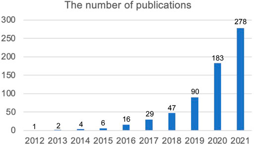
FIGURE 1. The annual article publication trend of “ferroptosis in brain” from 2012 to 2021.
Distribution of countries
Visualization networks of the distribution of countries and organizations were constructed using VOSviewer 1.6. A total of 656 publications on ferroptosis in the brain were completed from 55 different countries. The co-authorship network of 51 countries is shown in Figure 2A, while the rest of the countries not shown in the figure were isolated. Each country was presented as a node in the figure, while the cluster of countries was indicated as the same color. The cooperation strength was stronger when the links between nodes were wider. We summarized the details of the publications, citations, and total link strength of 55 countries in Supplementary Table S2. The top 10 most productive countries are shown in Table 1. The top 3 countries are China (338), the United States (166), and Germany (64). Among them, the citations and total link strength of China were 11846 and 101, which were less than 18038 citations and 134 total link strengths of the United States, respectively. The citations and total link strength of Germany were 5,371 and 85, which ranked only second compared to the United States and China.
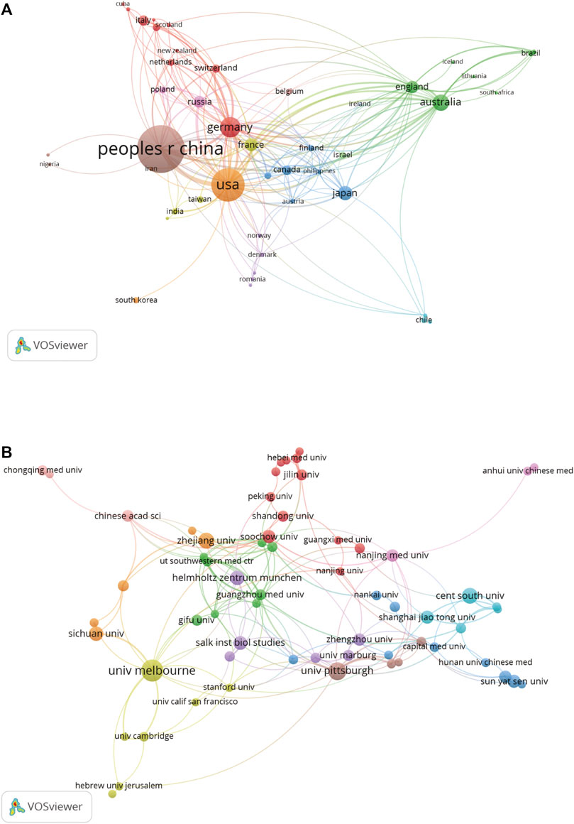
FIGURE 2. Collaborations of countries and institutions. (A) The collaborative relationship between different countries. (B) The co-author analysis of different organizations.
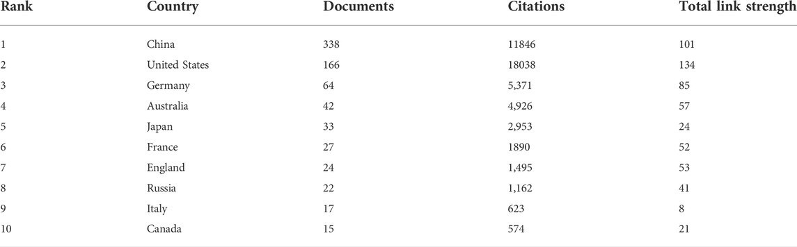
TABLE 1. The top 10 most productive countries.
Contributions from organizations
A total of 879 organizations completed the publication of 656 articles. Complete information regarding publications, citations, and the total link strength of 879 organizations is summarized in Supplementary Table S3. Co-authorship of 63 organizations with at least 5 publications is visualized in Figure 2B. The nodes and lines reflect the documents and cooperation of these organizations. From the visualization network, we can see that the cooperation between these organizations was active. The details of the top 10 most productive organizations are presented in Table 2. Among these organizations, six organizations came from China, two came from the United States, one came from Australia, and another was from Germany. The most productive organization was the University of Melbourne with 34 documents and a total link strength of 100. Next, the number of documents and total link strength came from the University of Pittsburgh in the United States and Central South University of China at 23 and 18 vs. 104 and 18, respectively. Data showed that the Central South University of China had less cooperation with other originations than the University of Melbourne of Australia and the University of Pittsburgh in the United States. In addition, the total citations of six organizations from China (2,510) were lower than those in Australia (4,211) and the United States (5,016). These data indicate that Australia and the United States are predominant in the field of ferroptosis in the brain.
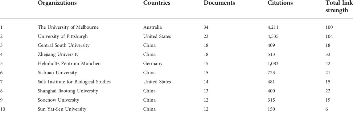
TABLE 2. Top 10 most productive institutions.
The map of co-occurrence and co-cited authors on ferroptosis in the brain
A total of 3,759 researchers contributed to the research of ferroptosis in the brain, and documents, citations, and the total link strength of these authors are summarized in Supplementary Table S4. Among them, the co-authorship network of 1,079 authors was established using VOSviewer (Figure 3A). Close collaborations normally exist in different clusters and are presented as different colors in Figure 5A. Although Marcus Conrad, Hulya Bayir, Wang Jian, and Scott Ayton are at the center, there are also some authors isolated with other authors in the network. Data indicated a lack of cooperation among authors dedicated to the field of ferroptosis in the brain. Table 3 shows the top 10 active authors in the field of ferroptosis in the brain ranked by publication frequency. Ashley I. Bush from the University of Melbourne contributed 20 articles in this field and Marcus Conrad (Chen et al., 2021), Scott Ayton (Li et al., 2022), and Pamela Maher (Dong et al., 2022) ranked after him (Table 3).
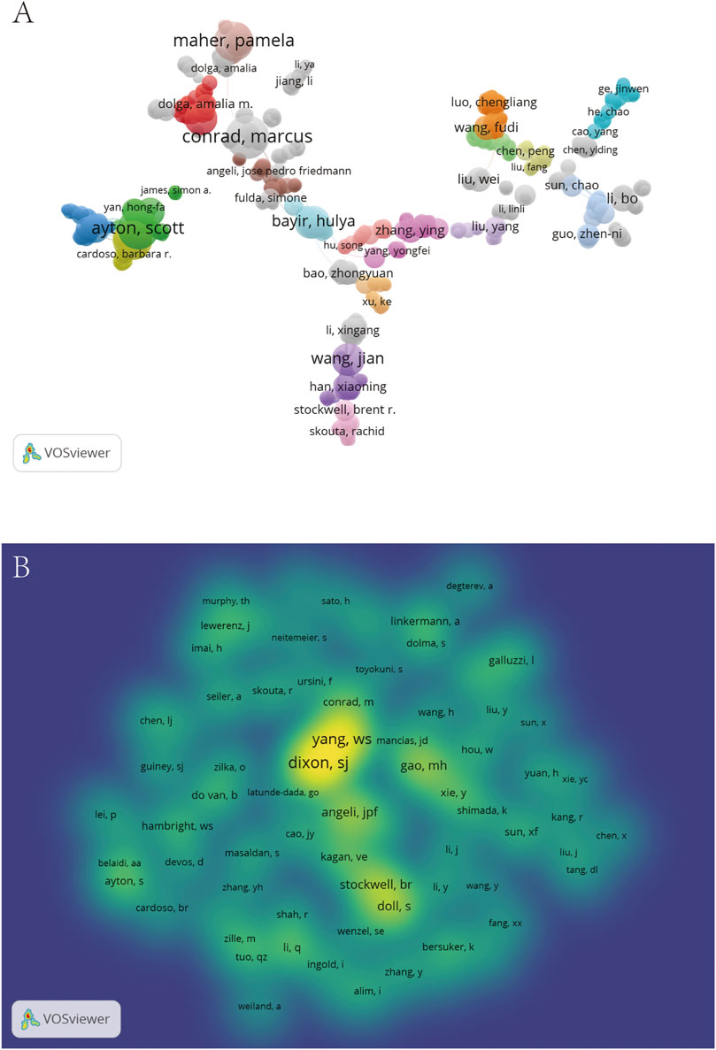
FIGURE 3. Contributions of authors on ferroptosis in the brain. (A) The co-occurrence network of authors. (B) The density map of co-cited authors.
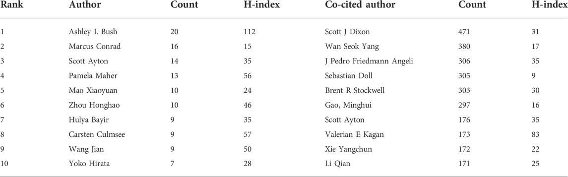
TABLE 3. Top 10 authors and co-cited authors in the field of ferroptosis in the brain.
Among the 24072 co-cited authors, 74 authors had more than 50 co-citations (Supplementary Table S5). The density map of co-cited authors is established in Figure 3B according to color reflected co-cited frequency. The top 10 co-cited authors are listed in Table 4. From the density map, Scott J Dixon and Wan Seok Yang were the most co-cited authors. Details of the complete list of all co-cited authors that could not be shown in the map and table are summarized in Supplementary Table S5.
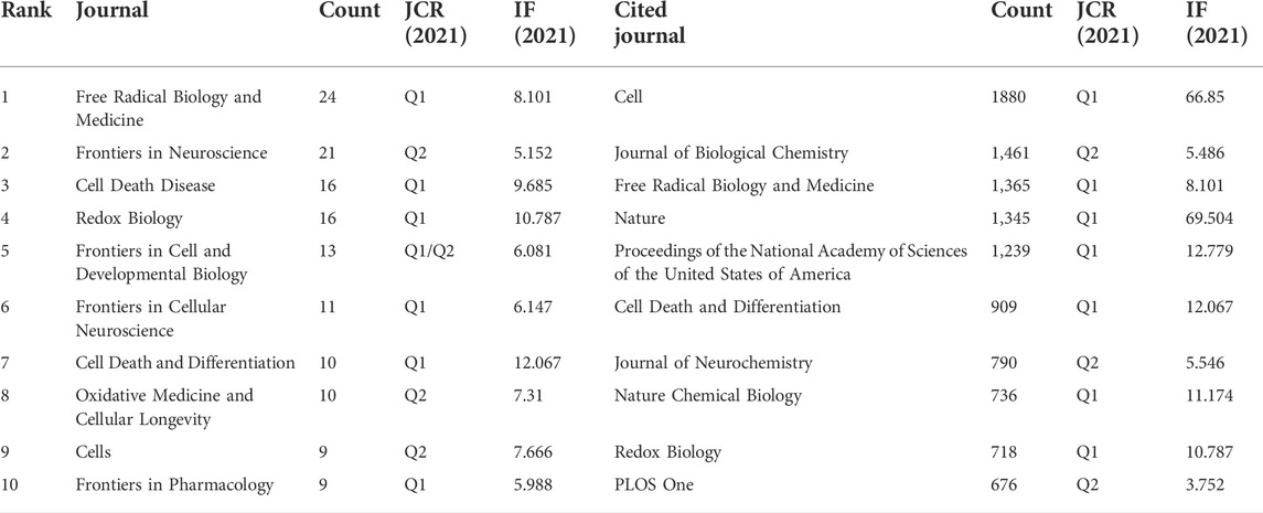
TABLE 4. Top 10 journals and co-cited journals in the field of ferroptosis in the brain.
Journals and Co-Cited journals
All 656 papers in the field of ferroptosis in the brain were published from 313 academic journals (Supplementary Table S6). The top 10 journals published 154 articles in this field and accounted for 23.5% of the number of publications (Table 4). In the top 10 journals, Free Radical Biology and Medicine published the maximum quantity of papers (Zille et al., 2017), followed by Frontiers in Neuroscience (Do Van et al., 2016) and Cell Death Disease (Chen et al., 2021). Seven of the top 10 journals were ranked in Q1 of the WoS-JCR partition.
In 3,669 co-cited sources, 110 had citations of more than 100 (Supplementary Table S7); among them, 19.5% of citations of all cited sources were from top 10 co-cited journals. Cell, Journal of Biological Chemistry, and Free Radical Biology and Medicine had the largest count of citations (Table 4).
The topic distribution of journals was represented as an overlay dual-map using CiteSpace (Figure 4). The left part of Figure 4 represents citing journals and the right part cited journals, while the link between the two parts reflects the citing and cited relationship of journals. Only one primary citation path could be found in Figure 4, which was cited from Molecular/Biology/Genetics journals to Molecular/Biology/Immunology journals.
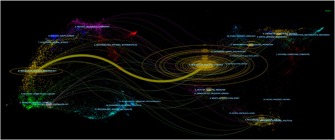
FIGURE 4. The overlay dual-map of journals on ferroptosis in the brain. The left part stands for citing journals and the right part for cited journals. The colored link between the two parts reflects the citing and cited relationship of journals.
Top cited publications and references burst
The top 10 highly cited publications in this field are found in Table 5, and all publications were cited at least 300 times. In these 10 documents, 7 were reviews (70%) and 3 were research articles (30%). An article entitled “Ferroptosis: An Iron-Dependent Form of Nonapoptotic Cell Death” published in Cell by Scott J Dixon et al. in 2012 was the most cited publication. A burst detection of references was usually used to determine references that were frequently cited in a certain period. The top 20 references with the strongest citation bursts were analyzed by CiteSpace in Figure 5. The results show that the first burst started by the paper entitled “Ferroptosis: An Iron-Dependent Form of Nonapoptotic Cell Death” published in Cell by Scott J Dixon et al. in 2012 (strength = 23.65). The strongest citation bursts occurred in an article entitled “Regulation of Ferroptotic Cancer Cell Death by GPX4” published in Cell by Wan Seok Yang et al. in 2014 (strength = 25.41), within the period from 2014 to 2019. Among these 20 references, nine papers had a burst until 2019.
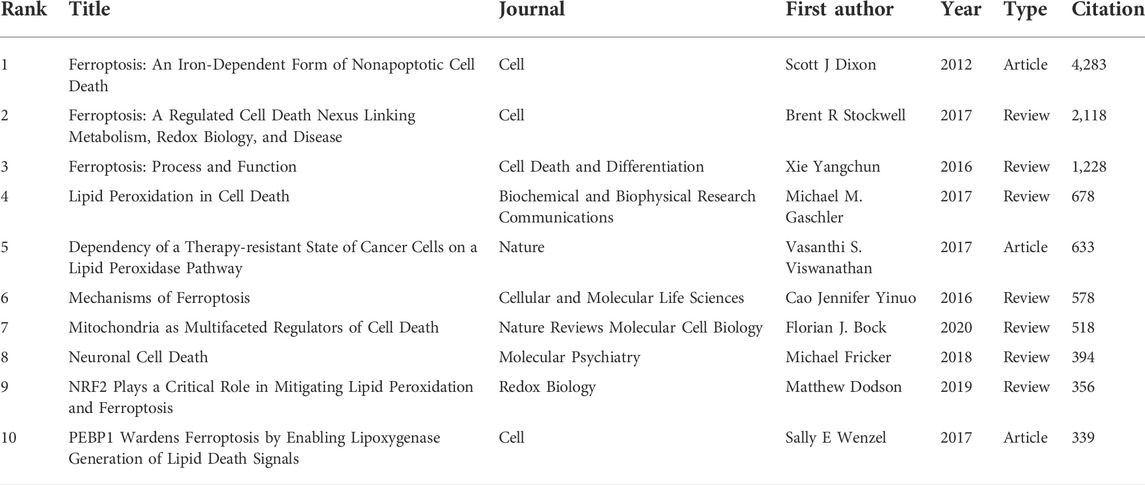
TABLE 5. Top 10 most cited documents in the field of ferroptosis in the brain.
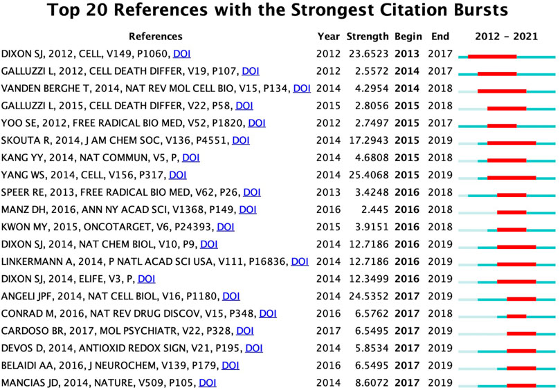
FIGURE 5. The top 20 references with the strongest citation bursts (sorted by the beginning year of the burst).
Analysis of hot research topics and frontiers
The co-occurrence of all keywords involving author keywords and keywords plus of 656 publications were analyzed to investigate hot research topics in the field of ferroptosis in the brain by using VOSviewer. The co-occurrence of 2,723 keywords is summarized in Supplementary Table S8, and 117 keywords appeared at least 10 times. The overlay map of these keywords (≥10 times) was established by VOSviewer (Figure 6). The top 20 keywords appeared at least 40 times and are summarized in Table 6, and “lipid-peroxidation” and “lipid peroxidation” were merged as “lipid peroxidation,” “cell-death” and “cell death” were merged as “cell death,” “Alzheimer’s-disease” and “Alzheimer’s disease” were merged as “Alzheimer’s disease,” “Parkinson’s-disease” and “Parkinson’s disease” were merged as “Alzheimer’s disease” and “glutathione -peroxidase 4,” “gpx4” and “glutathione peroxidase 4” were merged as “glutathione peroxidase 4.” From Table 6, “ferroptosis” (437) was the most cited keyword, followed by “cell death” (247), “oxidative stress” (245), “lipid peroxidation” (198) and “iron” (163).
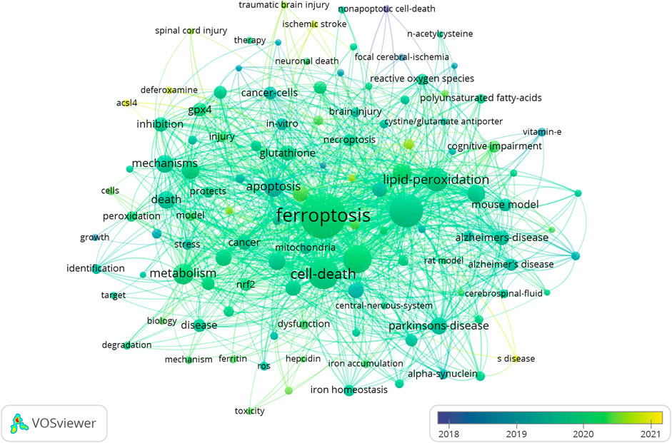
FIGURE 6. The overlay map of keywords established by VOSviewer.
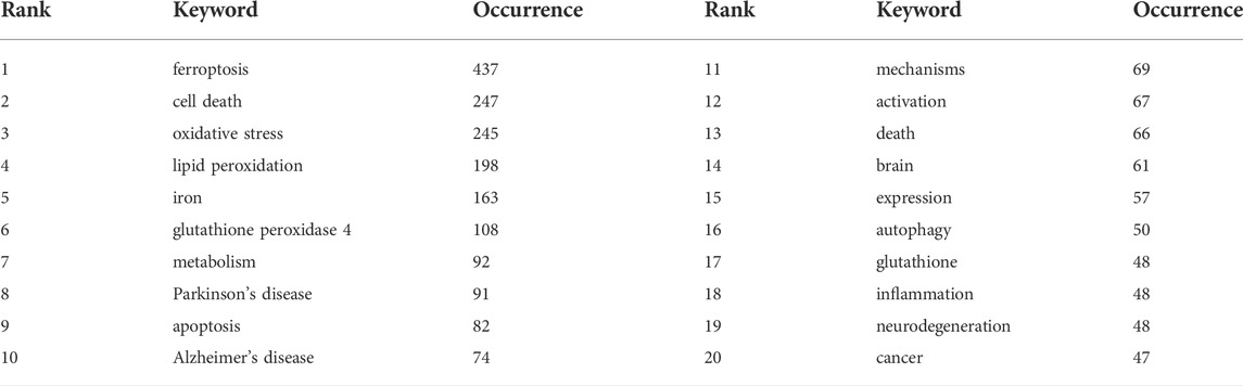
TABLE 6. Top 20 keywords related to ferroptosis in the brain.
Keyword burst analysis was performed to find the frontiers in the current field. As shown in Figure 7, the first burst of keyword “growth” started in 2012 (strength = 2.65) followed by nonapoptotic cell death (strength = 3.78), while the strongest bursts of keywords occurred on “amyloid precursor protein” from 2018 to 2019 (strength = 4.53). In addition, half of them, such as “neurodegenerative disease,” “iron homeostasis” and “reactive oxygen species,” had bursts until 2021 (Figure 7).
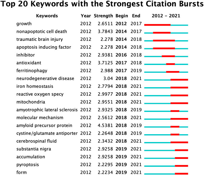
FIGURE 7. The top 20 keywords with the strongest citation bursts (sorted by the beginning year of the burst).
Discussion
Since ferroptosis was first defined in 2012, its role in wide biological processes and various organ damage has been investigated by researchers throughout the world. With the deepening of the research on ferroptosis, it has attracted considerable attention in the field of brain sciences. To better understand the growth trend of this topic, we performed this bibliometric analysis. Several other studies have also focused on the bibliometric analysis of ferroptosis (Chen et al., 2021; Wu et al., 2021; Xiong et al., 2021; Zhang et al., 2021; Zhou et al., 2021; Dong et al., 2022; Li et al., 2022), but these analyses examined the application of ferroptosis throughout all biological research areas without detailed division or concentrating on its role in stroke/cancer. Specifically, no analysis of its future landscape in brain research was performed. In this bibliometric analysis, we identified 656 publications on ferroptosis in the brain through a search of WOSCC from 1 January 2012 to 31 December 2021. It was surprising that more than 50% of the studies on ferroptosis in the brain were produced during the past 2 years (183 in 2021 and 278 in 2022), which indicates the explosion of this topic.
Changes in the annual number of publications in one research area usually reflect the development trend of a topic. Brent R Stockwell’s group proposed the concept of “ferroptosis” in 2012 (1), and this was first article investigating the role of ferroptosis in the brain. Although there were less than 10 papers published per year from 2012 to 2015, the annual number of publications has rapidly increased since 2016 (Figure 1). More than 50 different countries have produced evidence of ferroptosis in the brain (Supplementary Table S2), which reflects extensive global concern on this topic. Specifically, China was the most productive country, followed by the United States, but the number of citations from the United States was far greater (Table 1). These results show that China had more interest in this field than the United States, but the United States had more influence in this field. Similarly, in the top 10 productive institutes, 6 organizations were from China, but the total citation number of these organizations was still less than the University of Melbourne in Australia or the University of Pittsburgh and Salk Institute for Biological Studies in the United States (Table 2). These results suggest that China should further enhance the impact of studies, especially after the dominant tendency of the publication number in this field.
From 2012 to 2021, Free Radical Biology and Medicine and Redox Biology from the JCR 1 region published the highest number of articles in the field of ferroptosis in the brain (Table 4). In the top 10 productive journals, more than half of them were located in the JCR 1 region and all of them had an impact factor greater than 5 (Table 4). Among the top 10 most cited documents (Table 5), 3 were research articles (Dixon et al., 2012; Do Van et al., 2016; Li et al., 2017; Tuo et al., 2017; Viswanathan et al., 2017; Wenzel et al., 2017; Zille et al., 2017; Shen et al., 2018; Alim et al., 2019), while the other seven were reviews (Cao and Dixon, 2016; Xie et al., 2016; Gaschler and Stockwell, 2017; Stockwell et al., 2017; Fricker et al., 2018; Dodson et al., 2019; Bock and Tait, 2020). These papers contributed to the foundation of ferroptosis research in the field of the brain and help researchers understand the basics of this field.
The involvement of ferroptosis in many diseases has been proven (Tang et al., 2021). Especially in recent years, its targetable role in neurological diseases involving neurodegeneration, ischemic lesions and neurol tumors has been gradually revealed (Ashraf et al., 2020; Chen et al., 2020; David et al., 2021; Miao et al., 2022; Peeples and Genaro-Mattos, 2022). Furthermore, the inhibition of ferroptosis presented protective effects in models of cerebral hemorrhage in mice (Li et al., 2017). Iron dysregulation, lipid peroxidation, and mitochondrial abnormalities have been observed in patients with Alzheimer’s disease (AD) (Peña-Bautista et al., 2018). Additionally, iron dysregulation induced the decline of glutathione (GSH) and glutathione peroxidase 4 (GPX4) and ROS accumulation, and these factors together caused changes in AD markers such as amyloid beta peptide and Tau protein (Gleason and Bush, 2021). A close relationship between ferroptosis and Parkinson’s disease was established based on elevated iron levels in the brain (Jiang et al., 2017). Evidence shows that iron chelator treatment alleviated the motor damage of PD patients (Devos et al., 2014) and protected the blood-brain barrier (BBB) of PD model mice (Bar-Am et al., 2015). In addition, ferroptosis occurred in neuronal cells of stroke patients (Liu et al., 2022), and ferroptosis inhibitors reduced the neuronal damage of stroke patients (Alim et al., 2019). By inducing ferroptosis, the treatment effects of glioblastoma were improved in patients with drug resistance (Yakubov et al., 2021). These studies suggest a great prospect for targeting ferroptosis to treat the structural and functional abnormalities of the brain.
The following limitations of this study should not be ignored. First, we searched WOSCC (the most applied database in bibliometric studies) for relevant publications, so any publications excluded from WOSCC were missed. Second, we limited the timeline up to 31 December 2021; hence, any updated studies published during the writing and submission process of this article were also excluded from the analysis. Third, the drawback of VOSviewer and CiteSpace may limit the results of the current bibliometric analysis.
Conclusion
In summary, this study reported the development tendency of ferroptosis research on the brain.
The results indicate that the recent global research on ferroptosis in the field of brain science is exploding. The most productive countries were China, the United States, and Australia. With the development of ferroptosis research in the brain, many keywords related to its mechanisms occurred with high frequency. “Ferroptosis” was the most cited keyword, followed by “cell death,” “oxidative stress,” “lipid peroxidation” and “iron.” “Amyloid precursor protein” was the keyword with the strongest bursts, while “neurodegenerative disease,” “iron homeostasis,” and “reactive oxygen species” had bursts until 2021. These keywords revealed the hotspots and frontiers of ferroptosis research in the brain science field. This research landscape analysis of ferroptosis in the brain will offer a new reference to build on future research.
Data availability statement
The original contributions presented in this study are included in the article/Supplementary Material; further inquiries can be directed to the corresponding authors.
Author contributions
All listed authors have made a substantial, direct, and intellectual contribution to the work and approved it for publication.
Funding
This work was supported by the Natural Science Foundation of China (Nos 82071217 and 82001147), the Natural Science Foundation of Henan Province (No. 202300410353), and the Henan Province Medical Science and Technology Research Project Joint Construction Project (LHGJ20210065).
Conflict of interest
The authors declare that the research was conducted in the absence of any commercial or financial relationships that could be construed as a potential conflict of interest.
Publisher’s note
All claims expressed in this article are solely those of the authors and do not necessarily represent those of their affiliated organizations, or those of the publisher, the editors and the reviewers. Any product that may be evaluated in this article, or claim that may be made by its manufacturer, is not guaranteed or endorsed by the publisher.
Supplementary material
The Supplementary Material for this article can be found online at: https://www.frontiersin.org/articles/10.3389/fphar.2022.1014550/full#supplementary-material
References
Alim, I., Caulfield, J. T., Chen, Y., Swarup, V., Geschwind, D. H., Ivanova, E., et al. (2019). Selenium drives a transcriptional adaptive program to block ferroptosis and treat stroke. Cell 177 (5), 1262e25–1279. doi:10.1016/j.cell.2019.03.032
Ashraf, A., Jeandriens, J., Parkes, H. G., and So, P. W. (2020). Iron dyshomeostasis, lipid peroxidation and perturbed expression of cystine/glutamate antiporter in Alzheimer's disease: Evidence of ferroptosis. Redox Biol. 32, 101494. doi:10.1016/j.redox.2020.101494
Bar-Am, O., Amit, T., Kupershmidt, L., Aluf, Y., Mechlovich, D., Kabha, H., et al. (2015). Neuroprotective and neurorestorative activities of a novel iron chelator-brain selective monoamine oxidase-A/monoamine oxidase-B inhibitor in animal models of Parkinson's disease and aging. Neurobiol. Aging 36 (3), 1529–1542. doi:10.1016/j.neurobiolaging.2014.10.026
Bock, F. J., and Tait, S. W. G. (2020). Mitochondria as multifaceted regulators of cell death. Nat. Rev. Mol. Cell Biol. 21 (2), 85–100. doi:10.1038/s41580-019-0173-8
Cao, J. Y., and Dixon, S. J. (2016). Mechanisms of ferroptosis. Cell. Mol. Life Sci. 73 (11-12), 2195–2209. doi:10.1007/s00018-016-2194-1
Chen, C. (2004). Searching for intellectual turning points: Progressive knowledge domain visualization. Proc. Natl. Acad. Sci. U. S. A. 101 (1), 5303–5310. doi:10.1073/pnas.0307513100
Chen, S., Chen, Y., Zhang, Y., Kuang, X., Liu, Y., Guo, M., et al. (2020). Iron metabolism and ferroptosis in epilepsy. Front. Neurosci. 14, 601193. doi:10.3389/fnins.2020.601193
Chen, Y., Long, T., Xu, Q., and Zhang, C. (2021). Bibliometric analysis of ferroptosis in stroke from 2013 to 2021. Front. Pharmacol. 12, 817364. doi:10.3389/fphar.2021.817364
David, S., Jhelum, P., Ryan, F., Jeong, S. Y., and Kroner, A. (2021). Dysregulation of iron homeostasis in the CNS and the role of ferroptosis in neurodegenerative disorders. Antioxid. Redox Signal. doi:10.1089/ars.2021.0218
Devos, D., Moreau, C., Devedjian, J. C., Kluza, J., Petrault, M., Laloux, C., et al. (2014). Targeting chelatable iron as a therapeutic modality in Parkinson's disease. Antioxid. Redox Signal. 21 (2), 195–210. doi:10.1089/ars.2013.5593
Dixon, S. J., Lemberg, K. M., Lamprecht, M. R., Skouta, R., Zaitsev, E. M., Gleason, C. E., et al. (2012). Ferroptosis: An iron-dependent form of nonapoptotic cell death. Cell 149 (5), 1060–1072. doi:10.1016/j.cell.2012.03.042
Do Van, B., Gouel, F., Jonneaux, A., Timmerman, K., Gelé, P., Pétrault, M., et al. (2016). Ferroptosis, a newly characterized form of cell death in Parkinson's disease that is regulated by PKC. Neurobiol. Dis. 94, 169–178. doi:10.1016/j.nbd.2016.05.011
Dodson, M., Castro-Portuguez, R., and Zhang, D. D. (2019). NRF2 plays a critical role in mitigating lipid peroxidation and ferroptosis. Redox Biol. 23, 101107. doi:10.1016/j.redox.2019.101107
Dong, X., Tan, Y., Zhuang, D., Hu, T., and Zhao, M. (2022). Global characteristics and trends in research on ferroptosis: A data-driven bibliometric study. Oxid. Med. Cell. Longev. 2022, 8661864. doi:10.1155/2022/8661864
Fricker, M., Tolkovsky, A. M., Borutaite, V., Coleman, M., and Brown, G. C. (2018). Neuronal cell death. Physiol. Rev. 98 (2), 813–880. doi:10.1152/physrev.00011.2017
Friedmann Angeli, J. P., Schneider, M., Proneth, B., Tyurina, Y. Y., Tyurin, V. A., Hammond, V. J., et al. (2014). Inactivation of the ferroptosis regulator Gpx4 triggers acute renal failure in mice. Nat. Cell Biol. 16 (12), 1180–1191. doi:10.1038/ncb3064
Gaschler, M. M., and Stockwell, B. R. (2017). Lipid peroxidation in cell death. Biochem. Biophys. Res. Commun. 482 (3), 419–425. doi:10.1016/j.bbrc.2016.10.086
Gleason, A., and Bush, A. I. (2021). Iron and ferroptosis as therapeutic targets in alzheimer's disease. Neurotherapeutics 18 (1), 252–264. doi:10.1007/s13311-020-00954-y
Jiang, H., Wang, J., Rogers, J., and Xie, J. (2017). Brain iron metabolism dysfunction in Parkinson's disease. Mol. Neurobiol. 54 (4), 3078–3101. doi:10.1007/s12035-016-9879-1
Jiang, X., Stockwell, B. R., and Conrad, M. (2021). Ferroptosis: Mechanisms, biology and role in disease. Nat. Rev. Mol. Cell Biol. 22 (4), 266–282. doi:10.1038/s41580-020-00324-8
Li, G., Liang, Y., Yang, H., Zhang, W., and Xie, T. (2022). The research landscape of ferroptosis in cancer: A bibliometric analysis. Front. Cell Dev. Biol. 10, 841724. doi:10.3389/fcell.2022.841724
Li, J., Cao, F., Yin, H.-L., Huang, Z.-J., Lin, Z.-T., Mao, N., et al. (2020). Ferroptosis: Past, present and future. Cell Death Dis. 11 (2), 88. doi:10.1038/s41419-020-2298-2
Li, Q., Han, X., Lan, X., Gao, Y., Wan, J., Durham, F., et al. (2017). Inhibition of neuronal ferroptosis protects hemorrhagic brain. JCI Insight 2 (7), e90777. doi:10.1172/jci.insight.90777
Liu, J., Guo, Z. N., Yan, X. L., Huang, S., Ren, J. X., Luo, Y., et al. (2020). Crosstalk between autophagy and ferroptosis and its putative role in ischemic stroke. Front. Cell. Neurosci. 14, 577403. doi:10.3389/fncel.2020.577403
Liu, Y., Fang, Y., Zhang, Z., Luo, Y., Zhang, A., Lenahan, C., et al. (2022). Ferroptosis: An emerging therapeutic target in stroke. J. Neurochem. 160 (1), 64–73. doi:10.1111/jnc.15351
Miao, Z., Tian, W., Ye, Y., Gu, W., Bao, Z., Xu, L., et al. (2022). Hsp90 induces Acsl4-dependent glioma ferroptosis via dephosphorylating Ser637 at Drp1. Cell Death Dis. 13 (6), 548. doi:10.1038/s41419-022-04997-1
Peeples, E. S., and Genaro-Mattos, T. C. (2022). Ferroptosis: A promising therapeutic target for neonatal hypoxic-ischemic brain injury. Int. J. Mol. Sci. 23 (13), 7420. doi:10.3390/ijms23137420
Peña-Bautista, C., Vigor, C., Galano, J. M., Oger, C., Durand, T., Ferrer, I., et al. (2018). Plasma lipid peroxidation biomarkers for early and non-invasive Alzheimer Disease detection. Free Radic. Biol. Med. 124, 388–394. doi:10.1016/j.freeradbiomed.2018.06.038
Shen, Z., Liu, T., Li, Y., Lau, J., Yang, Z., Fan, W., et al. (2018). Fenton-reaction-acceleratable magnetic nanoparticles for ferroptosis therapy of orthotopic brain tumors. ACS Nano 12 (11), 11355–11365. doi:10.1021/acsnano.8b06201
Stockwell, B. R. (2022). Ferroptosis turns 10: Emerging mechanisms, physiological functions, and therapeutic applications. Cell 185 (14), 2401–2421. doi:10.1016/j.cell.2022.06.003
Stockwell, B. R., Friedmann Angeli, J. P., Bayir, H., Bush, A. I., Conrad, M., Dixon, S. J., et al. (2017). Ferroptosis: A regulated cell death nexus linking metabolism, redox biology, and disease. Cell 171 (2), 273–285. doi:10.1016/j.cell.2017.09.021
Tang, D., Chen, X., Kang, R., and Kroemer, G. (2021). Ferroptosis: Molecular mechanisms and health implications. Cell Res. 31 (2), 107–125. doi:10.1038/s41422-020-00441-1
Tuo, Q. Z., Lei, P., Jackman, K. A., Li, X. L., Xiong, H., Li, X. L., et al. (2017). Tau-mediated iron export prevents ferroptotic damage after ischemic stroke. Mol. Psychiatry 22 (11), 1520–1530. doi:10.1038/mp.2017.171
Viswanathan, V. S., Ryan, M. J., Dhruv, H. D., Gill, S., Eichhoff, O. M., Seashore-Ludlow, B., et al. (2017). Dependency of a therapy-resistant state of cancer cells on a lipid peroxidase pathway. Nature 547 (7664), 453–457. doi:10.1038/nature23007
Wenzel, S. E., Tyurina, Y. Y., Zhao, J., St Croix, C. M., Dar, H. H., Mao, G., et al. (2017). PEBP1 wardens ferroptosis by enabling lipoxygenase generation of lipid death signals. Cell 171 (3), 628e26–641. doi:10.1016/j.cell.2017.09.044
Wu, H., Wang, Y., Tong, L., Yan, H., and Sun, Z. (2021). Global research trends of ferroptosis: A rapidly evolving field with enormous potential. Front. Cell Dev. Biol. 9, 646311. doi:10.3389/fcell.2021.646311
Xie, Y., Hou, W., Song, X., Yu, Y., Huang, J., Sun, X., et al. (2016). Ferroptosis: Process and function. Cell Death Differ. 23 (3), 369–379. doi:10.1038/cdd.2015.158
Xiong, J., Qi, W., Liu, J., Zhang, Z., Wang, Z., Bao, J., et al. (2021). Research progress of ferroptosis: A bibliometrics and visual analysis study. J. Healthc. Eng. 2021, 2178281. doi:10.1155/2021/2178281
Yakubov, E., Eibl, T., Hammer, A., Holtmannspotter, M., Savaskan, N., and Steiner, H. H. (2021). Therapeutic potential of selenium in glioblastoma. Front. Neurosci. 15, 666679. doi:10.3389/fnins.2021.666679
Yang, L., Cao, L.-m., Zhang, X.-j., and Chu, B. (2022). Targeting ferroptosis as a vulnerability in pulmonary diseases. Cell Death Dis. 13 (7), 649. doi:10.1038/s41419-022-05070-7
Zhang, J., Song, L., Xu, L., Fan, Y., Wang, T., Tian, W., et al. (2021). Knowledge domain and emerging trends in ferroptosis research: A bibliometric and knowledge-map analysis. Front. Oncol. 11, 686726. doi:10.3389/fonc.2021.686726
Zhao, L., Zhou, X., Xie, F., Zhang, L., Yan, H., Huang, J., et al. (2022). Ferroptosis in cancer and cancer immunotherapy. Cancer Commun. 42 (2), 88–116. doi:10.1002/cac2.12250
Zhou, Q., Wu, F., Zhao, M., and Yang, M. (2021). Bibliometric evaluation of 2012-2020 publications on ferroptosis in cancer treatment. Front. Cell Dev. Biol. 9, 793347. doi:10.3389/fcell.2021.793347
Keywords: ferroptosis, brain, bibliometric analysis, knowledge map, VOSviewer, CiteSpace
Citation: Miao M, Han Y, Wang Y, Yang Y, Zhu R, Sun M and Zhang J (2022) The research landscape of ferroptosis in the brain: A bibliometric analysis. Front. Pharmacol. 13:1014550. doi: 10.3389/fphar.2022.1014550
Received: 08 August 2022; Accepted: 20 September 2022;
Published: 18 October 2022.
Edited by:
Suganthy Natarajan, Alagappa University, IndiaReviewed by:
M. Ahmed, Phcog.Net, IndiaXiangning Cui, Guang’anmen Hospital, China Academy of Chinese Medical Sciences, China
Copyright © 2022 Miao, Han, Wang, Yang, Zhu, Sun and Zhang. This is an open-access article distributed under the terms of the Creative Commons Attribution License (CC BY). The use, distribution or reproduction in other forums is permitted, provided the original author(s) and the copyright owner(s) are credited and that the original publication in this journal is cited, in accordance with accepted academic practice. No use, distribution or reproduction is permitted which does not comply with these terms.
*Correspondence: Mingyang Sun, bWluZ3lhbmdzdW4xOTg2QDE2My5jb20=; Jiaqiang Zhang, emhhbmdqaXFAenp1LmVkdS5jbg==
†These authors have contributed equally to this work