 Enzo Terreno
 Silvio Aime
95% of researchers rate our articles as excellent or good
Learn more about the work of our research integrity team to safeguard the quality of each article we publish.
Find out more
OPINION article
Front. Pharmacol. , 09 December 2015
Sec. Experimental Pharmacology and Drug Discovery
Volume 6 - 2015 | https://doi.org/10.3389/fphar.2015.00290
This article is part of the Research Topic In Vivo Imaging in Pharmacological Research View all 15 articles
The advent of the molecular imaging era has offered to pharmacologists very powerful tools for drug discovery and development, in vivo evaluation of pharmacokinetic properties, and monitoring drug efficacy (Hargreaves, 2008; Nairne et al., 2015). In fact, molecular imaging technologies provide minimally invasive procedures to visualize, characterize, and quantify biological processes occurring at cellular/subcellular level (Weissleder and Mahmood, 2001), thus overcoming the poor clinical translatability often exhibited by in vitro/ex-vivo experimental models.
The continuous advances in biomedical imaging technologies may significantly boost the development of novel and more effective drugs, and accelerating the selection of lead compounds, with important time and costs benefits for healthcare.
In vivo imaging of drug delivery and release, as well as monitoring of the therapeutic outcomes, represent the base of personalized medicine, thus allowing patients to be successfully addressed to the more effective therapeutic regime.
Overall, the use of molecular imaging procedures aimed at supporting any therapeutic intervention (including surgery) falls within the scopes of theranosis (Lammers et al., 2011).
Focusing on pharmacological therapies, a typical theranostic procedure requires the design of an imaging-traceable agent, whose structure and properties are suitably tailored to the aims of the examination. Imaging drug-delivery allows the assessment of the accumulation of the drug at the biological target, thus helping the selection of the more appropriate treatment. To get accurate information, the imaging agent should have the same physico-chemical properties of the drug. This requirement can be successfully met by labeling pharmaceuticals (organic molecules, peptides, proteins, radiochelates) with PET- or SPECT-traceable radioisotopes, because of the minimal structural perturbation caused by the introduction of commonly used radionuclides (e.g., 18F, 11C, 123I, 68Ga, 111In; Baum et al., 2010; Gains et al., 2011; Gomes et al., 2011; Witzig et al., 2013; Wynendaele et al., 2014).
On the other hand, when the drug is loaded into a nanocarrier, also the other available imaging modalities (CT, MRI, NIRF, US, PAI) can be used to visualize the delivery of the pharmaceutical. The imaging probe can be loaded in the carrier alone or together with the drug. The first option is preferable for drug selection, the second one for monitoring therapies. Among the imaging technologies, MRI is an excellent choice because combines exquisite spatial resolution, no limits in tissue penetration, and a vast portfolio of probes and contrast modalities that allows the design/selection of the best agent for any theranostic application.
MRI contrast agents can be grouped in five classes: T1 agents, T2/T2* agents, CEST agents, 19F-based agents, and hyperpolarized probes (Figure 1).
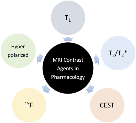
Figure 1. Main classes of MRI Contrast Agents in pharmacological research.
This section intends to provide the reader a brief description of these systems focusing on their main pro and cons with particular reference to the applications in pharmacological research.
T1 agents are mostly represented by paramagnetic metal [Gd(III) or Mn(II)] complexes that enhance the MR water signal (signal brightening) in T1w scans. The main benefits using T1 agents relies on the high versatility of this contrast mechanism that is affected by a large number of factors related to either structural and dynamic characteristics of the agent or biological aspects like the intra-voxel distribution of the probe (e.g., intra/extra-vascular, intra/extra-cellular). A representative example is the use of paramagnetic complexes to visualize the delivery and the release of a drug from liposomes (Viglianti et al., 2006; Hijnen et al., 2014; Rizzitelli et al., 2015). However, such systems displayed a limited sensitivity that makes necessary a local concentration of agent around 10 μM. This drawback can be partially overcome using nanometric materials that can aggregate even millions of contrastographic units, thus reducing significantly the local concentration of contrast agent (here represented by the nanosystem) necessary to generate a detectable T1 contrast.
T2/T2* agents are chemicals, mostly superparamagnetic nanoparticles made of iron oxides, capable to shorten the T2/T2* of water protons much more than T1. Thus, their presence in the MRI image is signaled by a signal loss (darkening). Such nanoparticles show an intrinsic higher sensitivity than T1 agents that justifies their extensive use in MR-molecular imaging procedures, especially for cellular imaging (Srivastava et al., 2015). On the other side, these agents are often considered not to be the candidates of choice for designing smart agents, due to the difficulty to modulate the T2/T2* contrast as a function of the microenvironment characteristics. Furthermore, the signal loss is not desirable when the target site has an intrinsically low signal (e.g., lungs, hemorrhages). However, interesting and promising theranostic applications of iron oxide nanoparticles for the visualization of drug delivery and release have been recently published (Krol et al., 2013; Liu et al., 2013).
The family of CEST agents is constantly growing, and though there are no agents in clinical trials yet, the peculiarities of these systems could open new and interesting future perspectives for MRI agents in pharmacology research. The acronym CEST stands for Chemical Exchange Saturation Transfer and identifies those chemicals that generate a MRI contrast through the transfer, mediated by chemical exchange, of saturated (i.e., irradiated with a frequency specific RF pulse) protons from the donor pool (CEST agent) to the acceptor pool (bulk water).
The most important advantage of using CEST agents is that the contrast can be detected only following the irradiation of the specific NMR resonance of the donor pool. It follows that the detection of the agent is frequency-encoded and this property can be exploited for multiplex imaging or for the design of concentration-independent smart agents, both tasks being very challenging in the case of the above described relaxation agents.
The sensitivity of CEST contrast detection was recognized as an issue since the early days of the development of these agents (Ward et al., 2000). Few mM of the donor pool are necessary. However, in analogy with T1 agents, a large sensitivity gain can be accomplished by recurring to nanosystems. As far as the use of CEST agents in pharmacology, excellent results have been obtained using liposomes as carriers of a huge amount of exchanging water protons (the water molecules entrapped in the nanovesicles) properly shifted by the entrapment of a paramagnetic shift reagent in the inner aqueous compartment. The resulting systems (called LipoCEST, Aime et al., 2005) have been demonstrated to be very promising for imaging drug release at preclinical level (Langereis et al., 2009; Delli Castelli et al., 2010; Castelli et al., 2014).
CEST agents share the frequency-encoded contrast property with agents containing MRI detectable nuclei different from protons. Among them, two classes deserve to be mentioned here because there are compounds already approved for humans or in advanced clinical trials: 19F agents and hyperpolarized probes.
19F nuclei are the most sensitive spins after protons, and, therefore can be detected by MRI without any enrichment. The detection sensitivity is similar to CEST agents (few mM of fluorine atoms). Consequently, 19F agents are almost exclusively represented by nanosystems, among which perfluorocarbon nanoparticles (PFCs) are by far the most commonly used (Jacoby et al., 2013). The important advantage of fluorinated agents over the other class of contrast media stems from the possibility to directly correlate the MR signal to the agent concentration, thus allowing the quantification of targeted biomarkers and/or drugs delivered at the site of interest (Lanza et al., 2002). A commercially available formulation of PFCs will enter soon in clinical phase 1 for labeling and in vivo tracking human adipose-derived stem cells for breast reconstruction [19F Hot Spot MRI of Human Adipose-derived Stem Cells for Breast Reconstruction (CS-1000), ID NCT02035085, source: ClinicalTrials.gov].
This class of MRI agents is by far the most sensitive one, owing to the use of polarization techniques (like dynamic nuclear polarization, DNP, laser optical pumping, para-hydrogen induced polarization) that increase dramatically (up to five order of magnitude) the population difference between the spin energy levels. These agents have some similarity with PET tracer, not only for the excellent sensitivity, but also for the decay of the signal they generate (caused by the return back to the thermal polarization) that occurs on the timescale of the T1 of the polarized spin. Hence, one limitation in the use of hyperpolarized probes is the signal loss over time that requires fast injection and rapid accumulation at the target site.
Hyperpolarized gases (e.g., 3He and 129Xe) are clinically used for imaging the respiratory apparatus (Liu et al., 2014), whereas a 13C hyperpolarized compound (13C pyruvate) is currently in phase 1 clinical trial as metabolic agents for prostate cancer diagnosis (University of California, 2010). Besides cancer, 13C hyperpolarized agents are under intense scrutiny in cardiovascular research (Rider and Tyler, 2013).
The use of hyperpolarized probes for imaging drug delivery is quite limited, mainly due to the time constrain. Hence, their impact in pharmacological research is primarily in monitoring therapy outcome (Laustsen et al., 2014; Park et al., 2014).
A very intriguing combination between hyperpolarized and CEST agents has been proposed using 129Xe-based probes. The contrast arising from these agents (dubbed Hyper-CEST) relies on the reversible binding of hyperpolarized Xe with a macrocyclic host (e.g., cryptophane, cucurbituril; Schröder et al., 2006). The large chemical shift difference between the exchanging free and host-bound species allows the generation of a CEST contrast where the presence of very small amounts of the host-bound Xe can be detected after transferring its saturation to the signal of the free gas. In vitro proof-of-concepts highlighting the great potential and high sensitivity of these agents has been very recently published (Kunth et al., 2015; Schnurr et al., 2015).
In spite of the intrinsic limited sensitivity of NMR/MRI response, several routes have been identified to allow the use of MRI probes in pharmacological studies. The enhanced sensitivity allows to take advantage of the superb spatial and temporal resolution of the MR imaging modality. On this basis, MRI is increasing its competitiveness in the Molecular Imaging arena, allowing the design of innovative experiments that provide a detailed picture of the biological microenvironment at cellular and molecular level. Moreover, functional and molecular MRI investigations imply a level of invasiveness that is definitively low in respect to the commonly used probes for nuclear medicine.
Finally, the use of frequency-encoding agents opens new horizons as they allow the visualization of more targets in the same anatomical region, i.e., they provide the access to multicolor MR images of the kind the biomedical operators are used to deal with in the histological characterization of bioptical specimens.
Both the authors contributed to: (i) the design and organization of the manuscript, (ii) the drafting and critical revising of the article, and (iii) the approval of the final version to be published.
The authors declare that the research was conducted in the absence of any commercial or financial relationships that could be construed as a potential conflict of interest.
Aime, S., Delli Castelli, D., and Terreno, E. (2005). Highly sensitive MRI chemical exchange saturation transfer agents using liposomes. Angew. Chem. Int. Ed. Engl. 44, 5513–5515. doi: 10.1002/anie.200501473
Baum, R. P., Prasad, V., Müller, D., Schuchardt, C., Orlova, A., Wennborg, A., et al. (2010). Molecular imaging of HER2-expressing malignant tumors in breast cancer patients using synthetic 111In- or 68Ga-labeled affibody molecules. J. Nucl. Med. 51, 892–897. doi: 10.2967/jnumed.109.073239
Castelli, D. D., Boffa, C., Giustetto, P., Terreno, E., and Aime, S. (2014). Design and testing of paramagnetic liposome-based CEST agents for MRI visualization of payload release on pH-induced and ultrasound stimulation. J. Biol. Inorg. Chem. 19, 207–214. doi: 10.1007/s00775-013-1042-0
Delli Castelli, D., Dastrù, W., Terreno, E., Cittadino, E., Mainini, F., Torres, E., et al. (2010). In vivo MRI multicontrast kinetic analysis of the uptake and intracellular trafficking of paramagnetically labeled liposomes. J. Control. Release 144, 271–279. doi: 10.1016/j.jconrel.2010.03.005
Gains, J. E., Bomanji, J. B., Fersht, N. L., Sullivan, T., D'souza, D., Sullivan, K. P., et al. (2011). 177Lu-DOTATATE Molecular Radiotherapy for Childhood Neuroblastoma. J. Nucl. Med. 52, 1041–1047. doi: 10.2967/jnumed.110.085100
Gomes, C. M., Abrunhosa, A. J., Ramos, P., and Pauwels, E. K. (2011). Molecular imaging with SPECT as a tool for drug development. Adv. Drug Deliv. Rev. 63, 547–554. doi: 10.1016/j.addr.2010.09.015
Hargreaves, R. J. (2008). The role of molecular imaging in drug discovery and development. Clin. Pharmacol. Ther. 83, 349–353. doi: 10.1038/sj.clpt.6100467
Hijnen, N. M., Langereis, S., and Grüll, H. (2014). Magnetic resonance guided high-intensity focused ultrasound for image-guided temperature-induced drug delivery. Adv. Drug Deliv. Rev. 72, 65–81. doi: 10.1016/j.addr.2014.01.006
Jacoby, C., Temme, S., Mayenfels, F., Benoit, N., Krafft, M. P., Schubert, R., et al. (2013). Probing different perfluorocarbons for in vivo inflammation imaging by 19F MRI: image reconstruction, biological half-lives and sensitivity. NMR Biomed. 27, 261–271. doi: 10.1002/nbm.3059
Krol, S., Macrez, R., Docagne, F., Defer, G., Laurent, S., Rahman, M., et al. (2013). Therapeutic benefits from nanoparticles: the potential significance of nanoscience in diseases with compromise to the blood brain barrier. Chem. Rev. 113, 1877–1903. doi: 10.1021/cr200472g
Kunth, M., Witte, C., Hennig, A., and Schroder, L. (2015). Identification, classification, and signal amplification capabilities of high-turnover gas binding hosts in ultra-sensitive NMR. Chem. Sci. 6, 6069–6075. doi: 10.1039/C5SC01400J
Lammers, T., Aime, S., Hennink, W. E., Storm, G., and Kiessling, F. (2011). Theranostic nanomedicine. Acc. Chem. Res. 44, 1029–1038. doi: 10.1021/ar200019c
Langereis, S., Keupp, J., van Velthoven, J. L., de Roos, I. H., Burdinski, D., Pikkemaat, J. A., et al. (2009). A temperature-sensitive liposomal 1H CEST and 19F contrast agent for MR image-guided drug delivery. J. Am. Chem. Soc. 131, 1380–1381. doi: 10.1021/ja8087532
Lanza, G. M., Yu, X., Winter, P. M., Abendschein, D. R., Karukstis, K. K., Scott, M. J., Chinen, L. K., et al. (2002). Targeted antiproliferative drug delivery to vascular smooth muscle cells with a magnetic resonance imaging nanoparticle contrast agent: implications for rational therapy of restenosis. Circulation 106, 2842–2847. doi: 10.1161/01.CIR.0000044020.27990.32
Laustsen, C., Lipsø, K., Ostergaard, J. A., Nørregaard, R., Flyvbjerg, A., Pedersen, M., et al. (2014). Insufficient insulin administration to diabetic rats increases substrate utilization and maintains lactate production in the kidney. Physiol. Rep. 11:e12233. doi: 10.14814/phy2.12233
Liu, J., Detrembleur, C., Debuigne, A., De Pauw-Gillet, M. C., Mornet, S., Vander Elst, L., et al. (2013). Poly(acrylic acid)-block-poly(vinyl alcohol) anchored maghemite nanoparticles designed for multi-stimuli triggered drug release. Nanoscale 5, 11464–11477. doi: 10.1039/c3nr02861e
Liu, Z., Araki, T., Okajima, Y., Albert, M., and Hatabu, H. (2014). Pulmonary hyperpolarized noble gas MRI: recent advances and perspectives in clinical application. Eur. J. Radiol. 83, 1282–1291. doi: 10.1016/j.ejrad.2014.04.014
Nairne, J., Iveson, P. B., and Meijer, A. (2015). Imaging in drug development. Prog. Med. Chem. 54, 231–280. doi: 10.1016/bs.pmch.2014.10.002
Park, I., Mukherjee, J., Ito, M., Chaumeil, M. M., Jalbert, L. E., Gaensler, K., et al. (2014). Changes in pyruvate metabolism detected by magnetic resonance imaging are linked to DNA damage and serve as a sensor of temozolomide response in glioblastoma cells. Cancer Res. 74, 7115–7124. doi: 10.1158/0008-5472.CAN-14-0849
Rider, O. J., and Tyler, D. J. (2013). Clinical implications of cardiac hyperpolarized magnetic resonance imaging. J. Cardiovasc. Magn. Reson. 15:93. doi: 10.1186/1532-429X-15-93
Rizzitelli, S., Giustetto, P., Cutrin, J. C., Delli Castelli, D., Boffa, C., Ruzza, M., et al. (2015). Sonosensitive theranostic liposomes for preclinical in vivo MRI-guided visualization of doxorubicin release stimulated by pulsed low intensity non-focused ultrasound. J. Control. Release 202, 21–30. doi: 10.1016/j.jconrel.2015.01.028
Schnurr, M., Sloniec-Myszk, J., Döpfert, J., Schröder, L., and Hennig, A. (2015). Supramolecular assays for mapping enzyme activity by displacement-triggered change in hyperpolarized (129)Xe magnetization transfer NMR spectroscopy. Angew. Chem. Int. Ed. Engl. 54, 13444–13447. doi: 10.1002/anie.201507002
Schröder, L., Lowery, T. J., Hilty, C., Wemmer, D. E., and Pines, A. (2006). Molecular imaging using a targeted magnetic resonance hyperpolarized biosensor. Science 314, 446–449. doi: 10.1126/science.1131847
Srivastava, A. K., Kadayakkara, D. K., Bar-Shir, A., Gilad, A. A., McMahon, M. T., and Bulte, J. W. (2015). Advances in using MRI probes and sensors for in vivo cell tracking as applied to regenerative medicine. Dis. Model Mech. 8, 323–336. doi: 10.1242/dmm.018499
University of California (2010). Hyperpolarized Pyruvate Injection in Subjects with Prostate Cancer, Phase 1 Clinical Trial, ID NCT01229618. Available online at: clinicaltrials.gov
Viglianti, B. L., Ponce, A. M., Michelich, C. R., Yu, D., Abraham, S. A., Sanders, L., et al. (2006). Chemodosimetry of in vivo tumor liposomal drug concentration using MRI. Magn. Reson. Med. 56, 1011–1018. doi: 10.1002/mrm.21032
Ward, K. M., Aletras, A. H., and Balaban, R. S. (2000). A new class of contrast agents for MRI based on proton chemical exchange dependent saturation transfer (CEST). J. Magn. Reson. 143, 79–87. doi: 10.1006/jmre.1999.1956
Weissleder, R., and Mahmood, U. (2001). Molecular imaging. Radiology 219, 316–333. doi: 10.1148/radiology.219.2.r01ma19316
Witzig, T. E., Wiseman, G. A., Maurer, M. J., Habermann, T. M., Micallef, I. N., Nowakowski, G. S., et al. (2013). A phase I trial of immunostimulatory CpG 7909 oligodeoxynucleotide and 90-yttrium ibritumomab tiuxetan radioimmunotherapy for relapsed B-cell non-Hodgkin lymphoma. Am. J. Hematol. 88, 589–593. doi: 10.1002/ajh.23460
Keywords: molecular imaging, MRI, contrast agents, theranosis, pharmacology
Citation: Terreno E and Aime S (2015) MRI Contrast Agents for Pharmacological Research. Front. Pharmacol. 6:290. doi: 10.3389/fphar.2015.00290
Received: 15 September 2015; Accepted: 19 November 2015;
Published: 09 December 2015.
Edited by:
Nicolau Beckmann, Novartis Institutes for BioMedical Research, SwitzerlandReviewed by:
Gustav Strijkers, Academisch Medisch Centrum, NetherlandsCopyright © 2015 Terreno and Aime. This is an open-access article distributed under the terms of the Creative Commons Attribution License (CC BY). The use, distribution or reproduction in other forums is permitted, provided the original author(s) or licensor are credited and that the original publication in this journal is cited, in accordance with accepted academic practice. No use, distribution or reproduction is permitted which does not comply with these terms.
*Correspondence: Silvio Aime, c2lsdmlvLmFpbWVAdW5pdG8uaXQ=
Disclaimer: All claims expressed in this article are solely those of the authors and do not necessarily represent those of their affiliated organizations, or those of the publisher, the editors and the reviewers. Any product that may be evaluated in this article or claim that may be made by its manufacturer is not guaranteed or endorsed by the publisher.
Research integrity at Frontiers
Learn more about the work of our research integrity team to safeguard the quality of each article we publish.