- 1Department of Pediatrics, Guangxi International Zhuang Medicine Hospital, Nanning, China
- 2Guangxi Key Laboratory of Chinese Medicine Foundation Research, Guangxi University of Chinese Medicine, Nanning, China
- 3International Medical Department, Guangxi International Zhuang Medicine Hospital, Nanning, China
- 4Department of Endocrinology, Guangxi International Zhuang Medicine Hospital, Nanning, China
- 5Zhuang Medical College, Guangxi University of Chinese Medicine, Nanning, China
Background: Children with severe adenoviral pneumonia (ADVP) have poor prognosis and high risk of mortality. We performed a meta-analysis to evaluate the association between pretreatment lactate dehydrogenase (LDH) and severity, postinfectious bronchiolitis obliterans (PIBO), and mortality in children with ADVP.
Methods: Relevant observational studies were identified by search of PubMed, Embase, Web of Science, Wanfang, and CNKI databases from inception to August 3, 2022. A random effect model was used to pool the results by incorporating the potential between-study heterogeneity.
Results: Overall, 23 studies with 4,481 children with ADVP were included in this meta-analysis. Results of meta-analysis showed that children with severe ADVP had a significantly higher level of pretreatment LDH as compared to those with non-severe ADVP (standard mean difference [SMD]: 0.51, 95% confidence interval [CI]: 0.36 to 0.66, p < 0.001; I2 = 69%). Besides, pooled results also suggested that the pretreatment LDH was significantly higher in children who developed PIBO as compared to those who did not (SMD: 0.47, 95% CI: 0.09 to 0.84, p = 0.02, I2 = 80%). Finally, results of the meta-analysis also confirmed that a higher pretreatment LDH (>500 IU/L) was a risk factor of increased mortality during hospitalization (odds ratio: 3.10, 95% CI: 1.62 to 5.92, p < 0.001, I2 = 0%). Sensitivity analyses by excluding one dataset at a time showed consistent results.
Conclusion: High pretreatment LDH may be associated with disease severity, development of PIBO, and increased risk of mortality in children with ADVP.
Introduction
Adenovirus belongs to the family Adenoviridae, which is characterized by its non-enveloped and double-stranded DNA properties (1). It has been discovered that there are at least 90 genotypes of human adenovirus, which can be divided into seven species from A to G (2). As a result of adenovirus infection, a variety of illnesses can be acquired, including bronchitis, pneumonia, conjunctivitis, gastroenteritis, and hemorrhagic cystitis (3). In children, adenoviral pneumonia (ADVP) has become an important cause of mortality, particularly for those <5 years and suffering from severe pneumonia (4, 5). According to a statistic report from some southern cities in China in 2018–2019, ADVP accounts for approximately 25% of the overall community acquired pneumonia in children (6). In addition, the early mortality of children with ADVP was reported as high as 50% (7). For children who survived from ADVP, severe complication may develop, such as the postinfectious bronchiolitis obliterans (PIBO) (8). In general, PIBO is the predominant type of bronchiolitis obliterans in children which is characterized of persistent airway obstruction with functional and radiological evidence of small airway involvement after respiratory infection (9). The clinical consequences of PIBO include impaired pulmonary function and even respiratory failure in later life (9). However, clinical parameters that are closely related to the disease severity, development of PIBO and early mortality in children with ADVP remain to be determined (10).
The lactate dehydrogenase (LDH) enzyme plays a vital role in the anaerobic metabolism of the body (11, 12). It is ubiquitously present in all cells and responds to tissue damage in a nonspecific manner. For coronavirus disease 2019 (COVID-19), accumulating evidence suggests that the level of LDH could be used as a maker of disease severity (13). As a result of the reemergence of the adenovirus epidemic in central and southern China in 2018 and 2019, many studies have been conducted to evaluate the prognosis factors associated with ADVP among children, including the clinical significance of serum LDH (14–36). In this meta-analysis, we systematically evaluated the relationships of LDH at baseline with disease severity, risk of PIBO, and in-hospital mortality of children with ADVP. The findings are expected to provide information regarding early risk stratification in children with ADVP.
Materials and methods
The Preferred Reporting Items for Systematic Reviews and Meta-Analyses (PRISMA) statement (37, 38) and the Cochrane's Handbook (39) guideline was followed in the conceiving, conducting, and reporting the study.
Search of databases
Studies were retrieved by search electronic databases including PubMed, Embase, Web of Science, Wanfang, and CNKI (China National Knowledge Infrastructure) databases from inception to August 3, 2022, with combined search terms including (1) “lactate dehydrogenase” OR “LDH”; (2) “adenovirus” OR “adnoviral” OR “ADV”; and (3) “pneumonia” OR “respiratory” OR “infection”. The search was restricted to human studies with no limitation of the publication language. The reference lists of the relevant original and review articles were also manually screened for possible related studies.
Study inclusion and exclusion criteria
We formulated the inclusion criteria according to the aim of the meta-analysis, with the following specified inclusion criteria: (1) designed as observational studies, including the case-control study, cross-sectional study, and cohort study; (2) included children with confirmed diagnosis of ADVP; (3) serum level of LDH was measure at baseline (within 24 h of admission); and (4) reported the association between LDH with at least one of the following outcomes, including the severity of ADVP, the development of PIBO, and the all-cause mortality during hospitalization. We did not apply restriction for the definition of disease severity of ADVP during the selection of the studies. This is mostly because there has been no international consensus regarding the classification according to the severity of ADV-pneumonia. However, because all of the retrieved studies were performed in China and the severity of ADVP was evaluated according to the Chinese guidelines, we therefore used these criteria in our meta-analysis accordingly. Specifically, a severe ADVP was diagnosed based on the criteria of the 2019 Chinese Guideline for the Diagnosing and Management of Children with Community-Acquired Pneumonia (40) if any of the following criteria were met: (1) Overall poor health condition of a patient. (2) A conscious disturbance. (3) Cyanosis or tachypnea [age <2 months: respiratory rate (RR) 60 breaths/minute; age 2 months to 1 year: RR 50 breaths/minute; age 1–5 years: RR 40 breaths/minute; and age >5 years: RR 30 breaths/minute)], intermittent apnea, or oxygen saturation of <92%. (4) Persistent hyperpyrexia or ultrahyperpyrexia for more than 5 days. (5) Dehydration or food refusal. (6) A chest x-ray or CT reveals pulmonary infiltration of at least two-thirds of the lung, pneumothorax, lung necrosis, or lung abscess on one side. (7) Extrapulmonary complications. Diagnosis of PIBO was consistent with the criteria applied in previous studies (41): (1) continuous or repeated coughing and wheezing for 6 weeks after the infection; (2) evidence of obstructive pulmonary disease on computed tomography (CT) examination imaging such as hyperventilation, atelectasis, bronchial wall thickening, bronchiectasis and mosaic perfusion, and air trapping (two fixed radiologists performed imaging reports); (3) excluding other chronic pulmonary diseases such as tuberculosis, cystic fibrosis, bronchopulmonary congenital dysplasia, and primary immune deficiency. Only studies published as full-length articles were included. Reviews, studies not including children with ADVP, studies not evaluating pretreatment LDH, or studies not reporting the outcome of interest were excluded.
Data collection and quality assessing
The literature search, data collection, and study quality assessment were independently conducted by two authors separately. If discrepancies occurred, a third author was contacted for discussion and reaching the consensus. We collected data regarding study information, design, diagnosis of the children, age, timing of LDH measurement, follow-up durations, and outcomes reported. Study quality was assessed via the Newcastle–Ottawa Scale (42) with scoring regarding the criteria for participant selection, comparability of the groups, and the validity of the outcomes. The scale ranged between 1 and 9 stars, with larger number of stars presenting higher study quality.
Statistical analyses
The differences of LDH levels admission between children with severe and non-severe ADVP, and between those who developed and did not develop PIBO were summarized as standard mean difference (SMD) and the corresponding 95% confidence interval (CI). The association between high LDH at baseline and the risk of all-cause mortality during hospitalization in children with ADVP was summarized as odds ratio (OR) and 95% CI. Using the 95% CIs or p values, data of OR and the standard error (SE) could be calculated, and a subsequent logarithmical transformation was conducted to keep stabilized variance and normalized distribution. Between study heterogeneity was estimated with the Cochrane's Q test and the I2 statistic (43), with I2 > 50% reflecting the significant heterogeneity. A random-effect model was applied to combine the results by incorporating the influence of heterogeneity (39). Sensitivity analysis by excluding one dataset at a time was performed to evaluate the influence of individual study on the meta-analysis results (44). For meta-analyses with adequate datasets (at least ten) the publication bias was estimated based on the visual judgement of the symmetry of the plots, supplemented with the Egger's regression asymmetry test (45). The RevMan (Version 5.1; Cochrane Collaboration, Oxford, United Kingdom) and Stata software (version 12.0; Stata Corporation, College Station, TX) were applied for these analyses.
Results
Literature search
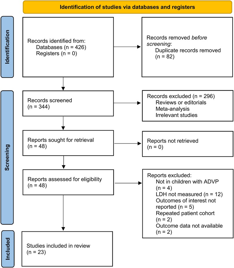
The flowchart of literature search and study inclusion was displayed in Figure 1. In summary, 426 records were obtained in the initial database search, and 82 duplications were removed. Subsequently, 296 studies were further removed after screening with titles and abstracts, largely because they were not relevant to the objective of the meta-analysis. Finally, 48 studies underwent full-text review, and 25 of them were excluded for the reasons listed in Figure 1. Accordingly, 23 studies were available for the meta-analysis (14–36).
Study characteristics
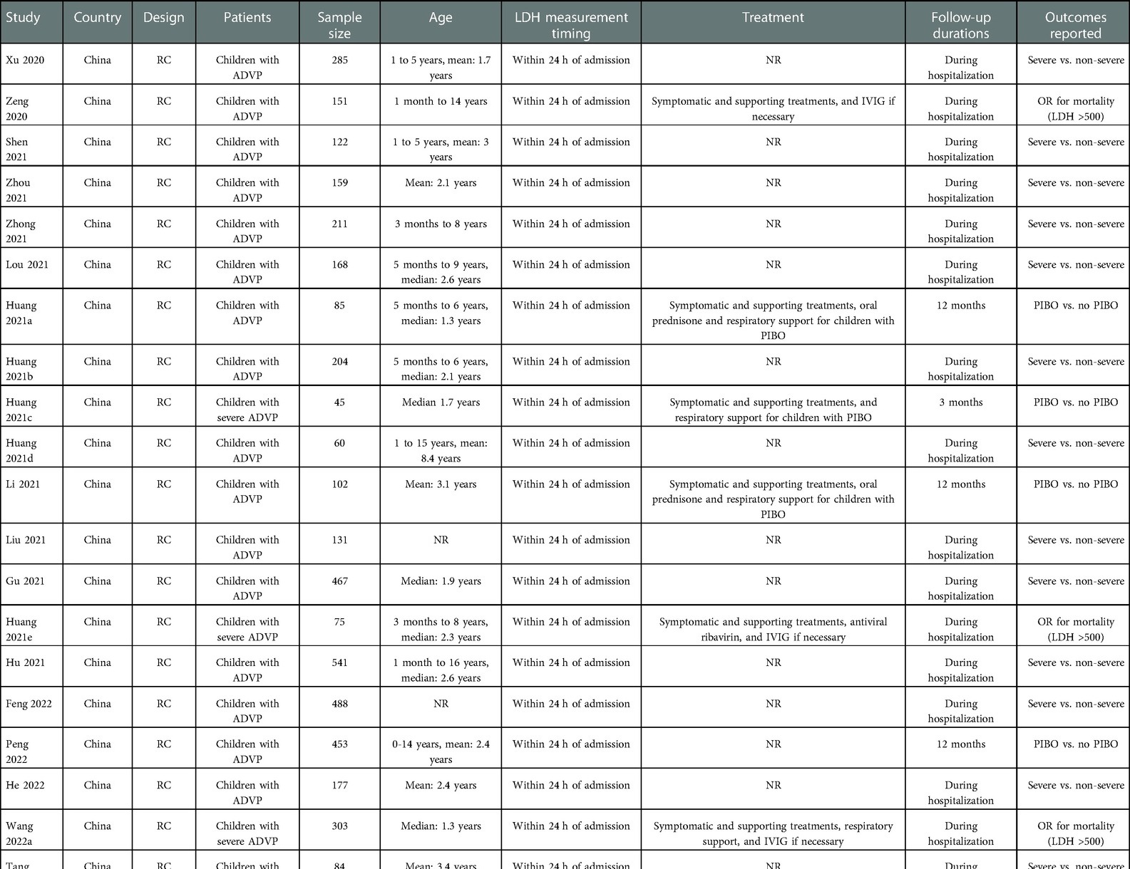
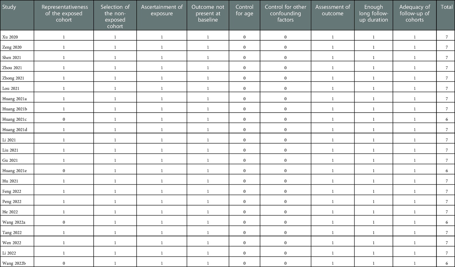
Overall, 23 studies (14–36) involving 4,481 children with confirmed diagnosis of ADVP were included in the meta-analysis. Generally, children with confirmed diagnosis of ADVP without known immunodeficiency or history of receiving immunosuppressive agents were included in these studies. The characteristics of the included studies are displayed in Table 1. Briefly, these studies were all retrospective cohort studies from China, and published between 2020 and 2022. The serum level of LDH was measured within 24 h of admission for all the included patients. For all the included studies, clinically diagnosed pneumonia with nasopharyngeal swab positive for adenovirus nucleic acid, serum adenovirus-specific IgM antibody positive, or detected adenovirus nucleic acid sequences in bronchoalveolar lavage fluid (BALF) and metagenomics next generation sequencing (mNGS) in severity cases were all considered as the confirmation of the diagnosis of ADVP. The treatments during hospitalization were only reported in eight studies (15, 18, 20, 22, 23, 34–36), which included symptomatic and supporting treatments, oral prednisone and intravenous steroids, intravenous immunoglobulin, antiviral ribavirin, and respiratory support if necessary (Table 1). The follow-up durations were within hospitalization in most of the included studies, while others were with a follow-up duration of 1 month (36), 3 months (20, 35), and 12 months (18, 23, 32). As for the outcomes reported, 14 studies reported the difference of LDH level at baseline between children with severe and non-severe ADVP (14, 16, 17, 19, 21, 24–31, 33), 6 studies reported the difference of LDH level at baseline between children who developed and did not develop PIBO (18, 20, 23, 32, 35, 36), and three studies reported the association between LDH admission and in-hospital mortality of children with ADVP (15, 22, 34). The NOS of the included studies were all six to seven stars, suggesting moderate to good study quality (Table 2).
Meta-analysis results
Pooled results of 14 studies (14, 16, 17, 19, 21, 24–31, 33) with a random-effect model showed that children with severe ADVP had a significantly higher level of pretreatment LDH as compared to those with non-severe ADVP (SMD: 0.51, 95% CI: 0.36 to 0.66, p < 0.001; I2 = 69%; Figure 2A). Sensitivity analyses by excluding one study at a time showed similar results (SMD: 0.47 to 0.54, p all < 0.05).
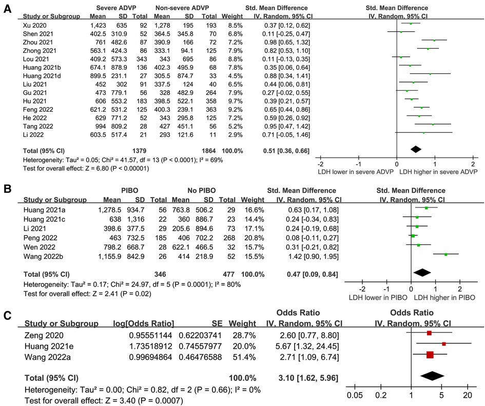
Figure 2. Forest plots for the meta-analyses of the role of LDH in children with ADVP. (A), meta-analysis for the difference of serum LDH between children with severe and non-severe ADVP; (B), meta-analysis for the difference of serum LDH between children who developed and did not develop PIBO; and (C), meta-analysis for the association between high LDH (> 500 IU/L) and in-hospital mortality risk in children with ADVP.
Besides, pooled results of 6 studies (18, 20, 23, 32, 35, 36) indicated that the pretreatment LDH was significantly higher in children who developed PIBO as compared to those who did not (SMD: 0.47, 95% CI: 0.09 to 0.84, p = 0.02, I2 = 80%; Figure 2B). Sensitivity analysis by omitting one study at a time also showed consistent results (SMD: 0.23 to 0.57, p all < 0.05).
Finally, pooling the results of three studies (15, 22, 34) showed that a higher pretreatment LDH (>500 IU/L) was a risk factor of increased mortality during hospitalization (OR: 3.10, 95% CI: 1.62 to 5.92, p < 0.001, I2 = 0%; Figure 2C). Sensitivity analyses by excluding one study at a time showed consistent results (OR: 2.67 to 3.58, p all < 0.05).
Publication bias
Figure 3 displays the funnel plots for the meta-analysis of the difference of the LDH between children with severe and non-severe ADVP. Visual inspection revealed symmetry of the plots, reflecting a low risk of publication biases. The Egger's regression tests also indicated low risk of publication biases (p = 0.31). The publication biases for the meta-analyses of the other two outcomes were difficult to estimate because less than 10 studies were included.
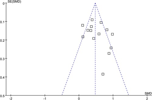
Figure 3. Funnel plots for the publication bias underlying the meta-analysis for the difference of serum LDH between children with severe and non-severe ADVP.
Discussion
In this study, by pooling the results of eligible observational studies, results of the meta-analysis showed that children with severe ADVP had a significantly higher level of baseline LDH as compared to those with non-severe ADVP. Besides, the serum level of LDH was also higher in children who developed PIBO as compared to those who did not develop PIBO within 12 months. Finally, a high pretreatment LDH > 500 IU/L may be a predictor of early mortality of children with ADVP during hospitalization. Collectively, results of the meta-analysis suggest that for children with ADVP, a higher LDH level at admission is associated with the severity of the disease, risk for the development of PIBO, and the all-cause mortality during hospitalization.
To the best of our knowledge, this is the first meta-analysis regarding the role of LDH in the disease severity evaluation and prognostic prediction in children with ADVP. The strengths of the meta-analysis include extensive literature search to retrieve eligible studies from both the English and Chinses databases, comprehensive evaluation of three outcomes such as disease severity, development of PIBO, and risk of all-cause mortality, as well as the performance of sensitivity analyses to indicate the robustness of the findings. Previous studies have shown that several lung conditions affect serum LDH levels (46), and patients with severe adenovirus respiratory tract infections have elevated LDH levels (47). In addition, a recent study showed that the LDH level was the associated factor to predict the types of pneumonia, which was significantly higher in children with ADVP as compared to those with bacterial pneumonia (48). In this meta-analysis, we further confirmed that higher baseline LDH may be a marker of disease severity and predictor of PIBO and in-hospital mortality in children with ADVP, which further supported the role of LDH as an important marker for the diagnosis and clinical management of ADVP. From the clinical perspective, serum LDH could be easily measured at admission in the routine biochemical blood test for these children, which reinforced the potential clinical significance of LDH for children with ADVP.
The mechanisms underlying the association between high LDH and the disease severity and poor prognosis of children with ADVP remain not fully determined. Almost all organ system cells contain the enzyme LDH, which catalyzes the conversion of pyruvate and lactate, with simultaneous conversion of NADH and NAD + (49). Five separate isozymes exist in humans, including LDH-1 in cardiomyocytes, LDH-2 in reticuloendothelial system, LDH-3 in pneumocytes, LDH-4 in kidneys and pancreas, and LDH-5 in liver and striated muscle (49). Besides of its role as a traditional marker of cardiac injury, infections and tissue trauma could also lead to increased LDH, that result in increased lactate trigger metalloprotease activation and increase macrophage-mediated angiogenesis (50). Considering that lung tissue (isozyme 3) contains LDH, children with severe ADVP can be expected to have higher levels of LDH in their bloodstreams. However, there is no evidence that the different LDH isoenzymes contribute to the LDH elevation observed in severe ADVP. Besides, accumulating evidence suggests that elevated LDH may be a maker of over-activated inflammation of pulmonary tissue (46) and compromised immune response, which are both associated with poor prognosis of patients with severe infection (12). Studies are needed in the future to determine the accurate mechanisms underlying the relationships of high LDH with disease severity and poor prognosis of ADVP.
Our study has limitations. First, all of the studies included in the meta-analysis were performed in China, which may primarily due to the reemergence of epidemic of the adenovirus infection in China in 2018–2019. Therefore, studies are needed to validate the potential role of LDH in ADVP in children from other countries. Besides, the severity of ADVP was evaluated according to the Chinese guidelines in this study. Correlations of LDH with more practical parameters for evaluating the severity of ADVP should be investigated in the future, such as the odds of mechanical ventilations, incidence of multi-organ failure, and the proportions of patients requiring extracorporeal membrane oxygenation etc. Second, significant heterogeneity was observed for the meta-analyses of the differences of LDH between severe and non-severe ADVP, and between children who developed and did not develop PIBO. These heterogeneities may be explained by the potential differences of adenoviral type, antiviral treatments, and related comorbidities etc. among the included studies. However, we were unable to determine the exact source of heterogeneity because the above characteristics mentioned were rarely reported in the original studies. Moreover, comorbidities potentially affecting serum LDH level may confound the results. Similarly, co-infection of the pathogens which may affect LDH may also confound the results, such as the co-infections of Pneumocystis jirovecii (51). In addition, all the included studies were retrospective, which were associated with the risks of recall and selection biases. Therefore, large-scale prospective studies are needed to validate our findings. Finally, the normal range for total LDH is 160 to 450 IU/L in newborns, 60 to 170 IU/L in children, and 140 to 280 units/L IU/L in adults (52). However, the optimal cutoff of LDH at baseline to predict the clinical severity and poor prognosis in children with ADVP remains unknown. Studies are warranted in the future in this regard.
To sum up, results of the meta-analysis indicate that for children with ADVP, a higher LDH level at admission was associated with the severity of the disease, risk for the development of PIBO, and the all-cause mortality during hospitalization. These findings support that serum LDH may be applied as potential marker of disease severity and poor prognosis for children with ADVP. Studies may be considered to evaluate whether measuring LDH for predicting disease severity could optimize the treatments for children with severer ADVP.
Data availability statement
The original contributions presented in the study are included in the article/Supplementary Material, further inquiries can be directed to the corresponding author.
Author contributions
MZ designed the study. MZ and YZ performed literature search, study quality evaluation, and data collection. MZ, XM, and XW performed statistical analyses and interpreted the results. MZ drafted the manuscript. All authors critically revised the manuscript and approved the submission. All authors contributed to the article and approved the submitted version.
Funding
This study was supported by 2018 Guangxi University Young and Middle-aged Teachers Basic Ability Improvement Project (2018KY0302), Key project of Guangxi International Zhuang Medical Hospital in 2021 (GZ2021012), and Self-funded project of Guangxi Administration of Traditional Chinese Medicine in 2021 (20210036). This study was supported by Guangxi Key Discipline of Traditional Chinese Medicine (No.GZXK-Z-20-62); Guangxi TCM External Treatment Demonstration Base Project (Guangxi TCM Development [2019] No.14), and Guangxi International Zhuang Medical Hospital Green Seedling Program Top Talent Project.
Conflict of interest
The authors declare that the research was conducted in the absence of any commercial or financial relationships that could be construed as a potential conflict of interest.
Publisher's note
All claims expressed in this article are solely those of the authors and do not necessarily represent those of their affiliated organizations, or those of the publisher, the editors and the reviewers. Any product that may be evaluated in this article, or claim that may be made by its manufacturer, is not guaranteed or endorsed by the publisher.
References
1. Greber UF, Suomalainen M. Adenovirus entry: stability, uncoating, and nuclear import. Mol Microbiol. (2022) 118(4):309–320. doi: 10.1111/mmi.14909
2. Nemerow GR, Stewart PL, Reddy VS. Structure of human adenovirus. Curr Opin Virol. (2012) 2(2):115–21. doi: 10.1016/j.coviro.2011.12.008
3. Radke JR, Cook JL. Human adenovirus infections: update and consideration of mechanisms of viral persistence. Curr Opin Infect Dis. (2018) 31(3):251–6. doi: 10.1097/QCO.0000000000000451
4. Pscheidt VM, Gregianini TS, Martins LG, Veiga A. Epidemiology of human adenovirus associated with respiratory infection in southern Brazil. Rev Med Virol. (2021) 31(4):e2189. doi: 10.1002/rmv.2189
5. Shieh WJ. Human adenovirus infections in pediatric population - an update on clinico-pathologic correlation. Biomed J. (2022) 45(1):38–49. doi: 10.1016/j.bj.2021.08.009
6. Mao NY, Zhu Z, Zhang Y, Xu WB. Current status of human adenovirus infection in China. World J Pediatr. (2022) 18(8):533–7. doi: 10.1007/s12519-022-00568-8
7. Li M, Han XH, Liu LY, Yao HS, Yi LL. Epidemiological characteristics, clinical characteristics, and prognostic factors of children with atopy hospitalised with adenovirus pneumonia. BMC Infect Dis. (2021) 21(1):1051. doi: 10.1186/s12879-021-06741-0
8. Colom AJ, Teper AM. Post-infectious bronchiolitis obliterans. Pediatr Pulmonol. (2019) 54(2):212–9. doi: 10.1002/ppul.24221
9. Jerkic SP, Brinkmann F, Calder A, Casey A, Dishop M, Griese M, et al. Postinfectious bronchiolitis obliterans in children: diagnostic workup and therapeutic options: a workshop report. Can Respir J. (2020) 2020:5852827. doi: 10.1155/2020/5852827
10. Biserni GB, Scarpini S, Dondi A, Biagi C, Pierantoni L, Masetti R, et al. Potential diagnostic and prognostic biomarkers for adenovirus respiratory infection in children and young adults. Viruses. (2021) 13(9):1885. doi: 10.3390/v13091885
11. Gupta GS. The lactate and the lactate dehydrogenase in inflammatory diseases and Major risk factors in COVID-19 patients. Inflammation. (2022) 45(6):2091–2123. doi: 10.1007/s10753-022-01680-7
12. Van Wilpe S, Koornstra R, Den Brok M, De Groot JW, Blank C, De Vries J, et al. Lactate dehydrogenase: a marker of diminished antitumor immunity. Oncoimmunology. (2020) 9(1):1731942. doi: 10.1080/2162402X.2020.1731942
13. Henry BM, Aggarwal G, Wong J, Benoit S, Vikse J, Plebani M, et al. Lactate dehydrogenase levels predict coronavirus disease 2019 (COVID-19) severity and mortality: a pooled analysis. Am J Emerg Med. (2020) 38(9):1722–6. doi: 10.1016/j.ajem.2020.05.073
14. Xu N, Chen P, Wang Y. Evaluation of risk factors for exacerbations in children with adenoviral pneumonia. Biomed Res Int. (2020) 2020:4878635. doi: 10.1155/2020/4878635
15. Zeng XN, LI L, Chen Q, Zhu XH, Wu AM, Hu CL. Risk factors of the poor prognosis in children with adenoviral pneumonia. Jiangxi Med J. (2020) 55(8):1100–4. doi: 10.3969/j.issn.1006-2238.2020.08.042
16. Gu Y, Huang RW, Wang M, Tang CH, Li P, Duan J, et al. [Epidemiological characteristics of adenovirus infection in hospitalized children with acute respiratory tract infection in Kunming during 2019]. Zhonghua Er Ke Za Zhi. (2021) 59(9):772–6. doi: 10.3760/cma.j.cn112140-20210319-00231
17. Hu Q, Zheng YJ, Wang JW, Hong X, Wang W, Chen JH, et al. Clinical feature analysis of 541 children with adenovirus pneumonia. Chin J Appl Clin Pediatr. (2021) 36(16):1230–4. doi: 10.3760/cma.j.cn101070-20200418-00671
18. (a) Huang AF, Ma YC, Wang F, Li YN. Clinical analysis of adenovirus postinfectious bronchiolitis obliterans and nonadenovirus postinfectious bronchiolitis obliterans in children. Lung India. (2021) 38(2):117–21. doi: 10.4103/lungindia.lungindia_374_20
19. (b) Huang X, Yi Y, Chen X, Wang B, Long Y, Chen J, et al. Clinical characteristics of 204 children with human adenovirus type 7 pneumonia identified by whole genome sequencing in liuzhou, China. Pediatr Infect Dis J. (2021) 40(2):91–5. doi: 10.1097/INF.0000000000002925
20. (c) Huang BY, Jiang SH, Liang YQ, Chen XQ. The causes of bronchiolitis obliterans in children with severe adenovirus pneumonia. J Trop Med. (2021) 21(6):770–3.
21. (d) Huang DM, Deng JR, Xiao XB. Study on the correlation between serum LDH, PLT, PCT and leukocyte levels and severity of adenovirus infection in children. Pract Clin J Integr Trad Chin West Med. (2021) 21(21):120–2. doi: 10.13638/j.issn.1671-4040.2021.21.058
22. (e) Huang H, Chen Y, Ma LY, Yan MM, Deng Y, Zhang WD, et al. [Analysis of the clinical features and the risk factors of severe adenovirus pneumonia in children]. Zhonghua Er Ke Za Zhi. (2021) 59(1):14–9. doi: 10.3760/cma.j.cn112140-20200704-00687
23. Li XL, He W, Shi P, Wang LB, Zheng HM, Wen YJ, et al. Risk factors of bronchiolitis obliterans after adenovirus pneumonia: a nested case-control study. Chin J Evid Based Pediatr. (2021) 2021(16):233–6. doi: 10.3969/j.issn.1673-5501.2021
24. Liu XP, Wang YM. Analysis of risk factors and diagnostic value of LDH in children with severe adenovirus pneumonia. Med Innov China. (2021) 18(06):68–72. doi: 10.3969/j.issn.1674-4985.2021.06.016
25. Lou Q, Zhang SX, Yuan L. Clinical analysis of adenovirus pneumonia with pulmonary consolidation and atelectasis in children. J Int Med Res. (2021) 49(2):300060521990244. doi: 10.1177/0300060521990244
26. Shen Y, Zhou Y, Ma C, Liu Y, Wei B. Establishment of a predictive nomogram and its validation for severe adenovirus pneumonia in children. Ann Palliat Med. (2021) 10(7):7194–204. doi: 10.21037/apm-21-675
27. Zhong H, Dong X. Analysis of clinical characteristics and risk factors of severe adenovirus pneumonia in children. Front Pediatr. (2021) 9:566797. doi: 10.3389/fped.2021.566797
28. Zhou ZY, Chen Q, Li L, Dong Q, Lin YG, Ke JW. Diagnostic value of three items of myocardial enzyme spectrum in children with severe adenovirus pneumonia. J Chongqing Med Univ. (2021) 46(8):998–1000. doi: 10.13406/j.cnki.cyxb.002837
29. Feng K, Gou Y, Wang WJ. Predictive value of Serum lactate dehydrogenase on the severity of adenovirus pneumonia in children. J Chengde Med Univ. (2022) 39(1):22–5. 1004-687901-0022-04
30. He Y, Liu P, Xie L, Zeng S, Lin H, Zhang B, et al. Construction and verification of a predictive model for risk factors in children with severe adenoviral pneumonia. Front Pediatr. (2022) 10:874822. doi: 10.3389/fped.2022.874822
31. Li J, Sun XR, Fan YL, Li ZK. Clinical features of adenovirus pneumonia in children. Chin J Women Child Health Res. (2022) 33(1):141–5. doi: 10.3969/j.issn.1673-5293.2022.01.024
32. Peng L, Liu S, Xie T, Li Y, Yang Z, Chen Y, et al. Predictive value of adenoviral load for bronchial mucus plugs formation in children with adenovirus pneumonia. Can Respir J. (2022) 2022:9595184. doi: 10.1155/2022/9595184
33. Tang YC, Liao J, Huang XY, Yu ZW. Comparative of laboratory findings in children with adenovirus type 3 and type 7 pneumonia. J Trop Med. (2022) 22(5):644–7. doi: 10.3969/j.issn.1672-3619.2022.05.010
34. (a) Wang X, Tan X, Li Q. The difference in clinical features and prognosis of severe adenoviral pneumonia in children of different ages. J Med Virol. (2022) 94(7):3303–11. doi: 10.1002/jmv.27680
35. (b) Wang W, Chen JH, Xie G, Li ZC, He YX, Wang WJ. Risk factors of postinfectious bronchiolitis obliterans after severe adenovirus pneumonia. Chin Pediatr Emerg Med. (2022) 29(8):611–4. doi: 10.3760/cma.j.issn.1673-4912.2022.08.008
36. Wen YZ, Pan ZW, Lin YH. Analysis of related factors of bronchiolitis obliterans in children with adenovirus pneumonia. Med Innov China. (2022) 19(18):126–30. doi: 10.3969/j.issn.1674-4985.2022.18.031
37. Page MJ, Moher D, Bossuyt PM, Boutron I, Hoffmann TC, Mulrow CD, et al. PRISMA 2020 Explanation and elaboration: updated guidance and exemplars for reporting systematic reviews. Br Med J. (2021) 29:372. doi: 10.1136/bmj.n160
38. Page MJ, McKenzie JE, Bossuyt PM, Boutron I, Hoffmann TC, Mulrow CD, et al. The PRISMA 2020 statement: an updated guideline for reporting systematic reviews. Br Med J. (2021) 29:372. doi: 10.1136/bmj.n71
39. Higgins J, Thomas J, Chandler J, Cumpston M, Li T, Page M, Welch V. Cochrane Handbook for Systematic Reviews of Interventions version 6.2. The Cochrane Collaboration. 2021; www.training.cochrane.org/handbook
40. Chang Q, Chen HL, Wu NS, Gao YM, Yu R, Zhu WM. Prediction model for severe Mycoplasma pneumoniae pneumonia in pediatric patients by admission laboratory indicators. J Trop Pediatr. (2022) 68(4):6651464. doi: 10.1093/tropej/fmac059
41. Kurland G, Michelson P. Bronchiolitis obliterans in children. Pediatr Pulmonol. (2005) 39(3):193–208. doi: 10.1002/ppul.20145
42. Wells GA, Shea B, O'Connell D, Peterson J, Welch V, Losos M, Tugwell P. The Newcastle-Ottawa Scale (NOS) for assessing the quality of nonrandomised studies in meta-analyses. 2010; http://www.ohri.ca/programs/clinical_epidemiology/oxford.asp
43. Higgins JP, Thompson SG. Quantifying heterogeneity in a meta-analysis. Stat Med. (2002) 21(11):1539–58. doi: 10.1002/sim.1186
44. Patsopoulos NA, Evangelou E, Ioannidis JP. Sensitivity of between-study heterogeneity in meta-analysis: proposed metrics and empirical evaluation. Int J Epidemiol. (2008) 37(5):1148–57. doi: 10.1093/ije/dyn065
45. Egger M, Davey Smith G, Schneider M, Minder C. Bias in meta-analysis detected by a simple, graphical test. Br Med J. (1997) 315(7109):629–34. doi: 10.1136/bmj.315.7109.629
46. Drent M, Cobben NA, Henderson RF, Wouters EF, van Dieijen-Visser M. Usefulness of lactate dehydrogenase and its isoenzymes as indicators of lung damage or inflammation. Eur Respir J. (1996) 9(8):1736–42. doi: 10.1183/09031936.96.09081736
47. Lai CY, Lee CJ, Lu CY, Lee PI, Shao PL, Wu ET, et al. Adenovirus serotype 3 and 7 infection with acute respiratory failure in children in Taiwan, 2010-2011. PLoS One. (2013) 8(1):e53614. doi: 10.1371/journal.pone.0053614
48. Liu Y, Shen Y, Wei B. The clinical risk factors of adenovirus pneumonia in children based on the logistic regression model: correlation with lactate dehydrogenase. Int J Clin Pract. (2022) 2022:3001013. doi: 10.1155/2022/3001013
49. Hsu PP, Sabatini DM. Cancer cell metabolism: warburg and beyond. Cell. (2008) 134(5):703–7. doi: 10.1016/j.cell.2008.08.021
50. Feng Y, Xiong Y, Qiao T, Li X, Jia L, Han Y. Lactate dehydrogenase A: a key player in carcinogenesis and potential target in cancer therapy. Cancer Med. (2018) 7(12):6124–36. doi: 10.1002/cam4.1820
51. Borstnar S, Lindic J, Tomazic J, Kandus A, Pikelj A, Prah J, et al. Pneumocystis jirovecii pneumonia in renal transplant recipients: a national center experience. Transplant Proc. (2013) 45(4):1614–7. doi: 10.1016/j.transproceed.2013.02.107
Keywords: adenoviral pneumonia, lactate dehydrogenase, severity, postinfectious bronchiolitis obliterans, mortality, meta-analysis
Citation: Zou M, Zhai Y, Mei X and Wei X (2023) Lactate dehydrogenase and the severity of adenoviral pneumonia in children: A meta-analysis. Front. Pediatr. 10:1059728. doi: 10.3389/fped.2022.1059728
Received: 1 October 2022; Accepted: 31 December 2022;
Published: 26 January 2023.
Edited by:
Cristina Calvo, Hospital Infantil La Paz, SpainReviewed by:
Phuc Huu Phan, Vietnam National Children's Hospital, VietnamCarlos Daniel Grasa, University Hospital La Paz, Spain
© 2023 Zou, Zhai, Mei and Wei. This is an open-access article distributed under the terms of the Creative Commons Attribution License (CC BY). The use, distribution or reproduction in other forums is permitted, provided the original author(s) and the copyright owner(s) are credited and that the original publication in this journal is cited, in accordance with accepted academic practice. No use, distribution or reproduction is permitted which does not comply with these terms.
*Correspondence: Min Zou em91bWluZzkyMzIwQDIxY24uY29t
Specialty Section: This article was submitted to Pediatric Infectious Diseases, a section of the journal Frontiers in Pediatrics
 Min Zou
 Yang Zhai2,3