- 1Department for Orthopedic Surgery and Traumatology, University Hospital Bonn, Bonn, Germany
- 2Medical Service of the Health Funds (MDK), Cologne, Germany
- 3Department for Orthopedic Surgery and Traumatology, University Hospital Cologne, Cologne, Germany
Introduction: The method of infant hip sonography according to Graf is used for general hip screening in Serbia, Austria, and Germany and is considered the gold standard. In other countries, such as the USA, however, it is not well accepted and is claimed to lead to high costs and overtreatment. The aim of this study was to investigate how many of the mentioned sources in a recent review article contained sonograms that met the quality criteria as taught in Graf's ultrasound courses.
Methods: A systematic review published by Sakkers et al. was analyzed in terms of addressing the quality criteria of Graf’s method. Studies that were suitable by title, abstract, manuscript, that contained an image of sonographic hip examination, and that were accessible were included into analysis.
Results: Within the described review, there were 22 papers on the Graf method. Of these, 10 contained hip sonograms and were applicable for final analysis. The quality criteria according to Graf were not fulfilled within 5 of these 10 papers. Within these papers, there are examples of schematic sonograms that do not correspond with the quality criteria either.
Conclusion: Skepticism regarding the Graf method may be based on user errors and insufficient application of the Graf quality assessment algorithm, which results in high intra- and inter-observer variations. Based on these findings, a working group was initiated to evaluate further work according to the same procedure (currently approximately 130 papers).
Introduction
The method of infant hip sonography according to Graf is established for general hip screening in Serbia, Austria, and Germany and is considered the gold standard.
Among doctors trained in Germany and working in the field of pediatrics, pediatric orthopedics, or radiology, there is not the slightest doubt about the importance and validity of the general ultrasound screening of the infant hip according to Graf. Accordingly, a statement such as “(…) improvement (…) is still under debate” (1) in the central organ of the German medical profession led to indignant letters to the editor from committed colleagues (2). On the other hand, the question arises why, approximately 40 years after the description of the Graf method and more than a quarter of a century (1996 in Germany) after the establishment of a general hip screening, the evaluation of the method in the international literature sometimes appears to be very different from studies from Germany or Austria.
The authors therefore asked themselves whether the different evaluation of the Graf method is due to a different definition/application. To test this assumption, the following hypothesis was made:
Do the papers listed in a current and international systematic review (3) correspond to the quality criteria of the Graf Method as taught in courses?
The aforementioned review paper states verbatim:
“The fact that even the unstable and dislocated hips do relatively well without treatment in a substantial percentage probably contributes to the fact that all studies on US screening of hips for detection of relevant DDH in order to improve outcomes of treatment are rated as substantially underpowered” (3).
Methods
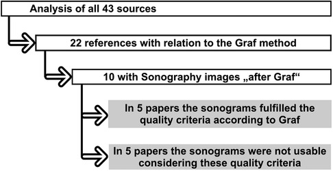
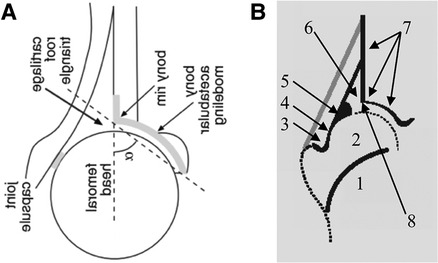
To check the quality criteria, all the original papers cited in the review were examined and those that dealt with ultrasound screening according to Graf were selected. The selected papers were searched (Figure 1) for ultrasound images and these were examined by two independent doctors experienced in sonography with regard to the quality criteria after Graf: Checklist I (anatomical identification) and Checklist II (usability check) (Figure 2) (7).

Figure 1. Overview of systematic evaluation of the original articles used by Sakkers et al. (3). 10 articles included sonograms, only 5 articles fulfilled the quality criteria according to Graf.

Figure 2. Schematic of the systematic evaluation of hip sonograms of different original articles (A). The original diagram from (5) was rotated by 90° and mirrored around the transversal axis for better clarity (ultrasound from the left, upright image). (B) Scheme of anatomical identification (Checklist I) from a course manual for the ultrasound course according to Graf (6). The numbers describe: 1, ChB (chrondro-osseous border); 2, femoral head; 3, synovial fold; 4, joint capsule; 5, labrum; 6, cartilage; 7, bony roof; 8, bony rim (turning point).
The alignment of sonograms in either lying or standing upright positions and performance of the ultrasound from either the left or the right side was not taken into account as error.
Results
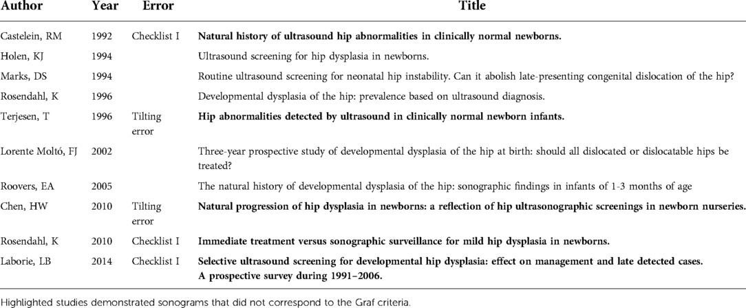
The analysis of the 43 listed sources is shown in Figure 1. Of these 43 sources, 22 dealt with the Graf method. Of these, 10 contained sonograms “according to Graf” (Table 1).
Five articles showed sonograms that met the quality criteria according to Graf (Checklists I and II). The other five papers contained at least one or more sonograms that did not meet the quality criteria.
In the five papers that did not meet the quality criteria, the anatomical identification specified in Checklist I was impossible in the sonograms of three papers. In the other two papers, the sonograms did not meet the usability check (Checklist II). Tilting errors were present.
Surprisingly, in the two most recent studies (from 2010 and 2014) (4, 5), in which one or more sonograms did not fulfil the quality criteria according to Graf, schematic representations of the anatomical structures were also found, which also did not correspond to Checklist I (anatomical identification).
Figure 2 shows an example of the analysis of the most recent of the 10 cited papers with sonograms.
The evaluation of the sonograms was not accurate according to Graf. For instance, no chondro-osseous border was shown in the schematics of both studies (4, 5).
A comparison of the three schematic figures shows that with regard to point 1 (ChB) and point 3 (Synovial Fold), Checklist I is not fulfilled.
Discussion
It is unclear whether the work by Sakkers et al. analyzed in this paper can be considered representative of the international literature. However, with its 22 cited papers dealing with the Graf method, it can certainly be seen as a representative cross-section. Half of the original papers with sonograms did not meet the defined quality criteria according to Graf, at least in one image. This fact suggests that these quality criteria are not given the necessary importance in the teaching of this examination method. This coincides with the diagrams, some of which do not contain anatomical structures that meet the quality criteria.
However, this shows that the insufficient sonograms are not a result of cursorily performed sonographic examinations, but a deficit of knowledge about the method.
The fact that the work by Graf cited by Sakkers et al. dates from 1980 should be seen in the same context. At that time, the ultrasound method taught today according to Graf had not yet been developed in its current form and the quality criteria (Checklist I—anatomical identification; Checklist II—brewability test), which have now been obligatory for decades, had not yet been defined.
Conclusion
The statements made by Sakkers et al., as far as they are based on data of the Graf method, must be doubted, as some of the underlying studies do not consider the defined criteria of this method. Based on these findings, a working group was initiated to evaluate the sonograms of further papers (currently approximately 130 papers, Part II).
Data availability statement
The original contributions presented in the study are included in the article/Supplementary Material. Further inquiries can be directed to the corresponding author/s.
Author contributions
SP and SW contributed to the study conception and design. Material preparation, data collection and analysis were performed by all authors. The first draft of the manuscript was written by SW and all authors commented on previous versions of the manuscript. All authors contributed to the article and approved the submitted version.
Conflict of interest
The authors declare that the research was conducted in the absence of any commercial or financial relationships that could be construed as a potential conflict of interest.
Publisher's note
All claims expressed in this article are solely those of the authors and do not necessarily represent those of their affiliated organizations, or those of the publisher, the editors and the reviewers. Any product that may be evaluated in this article, or claim that may be made by its manufacturer, is not guaranteed or endorsed by the publisher.
References
1. Yagdiran A, Zarghooni K, Semler JO, Eysel P. Hip pain in children. Dtsch Arztebl Int. (2020) 117(5):72. doi: 10.3238/arztebl.2020.0072
2. Hien NM. Correspondence (letter to the editor): the importance of hip ultrasound. Dtsch Arztebl Int. (2020) 117(35-36):600. doi: 10.3238/arztebl.2020.0600b
3. Sakkers R, Pollet V. The natural history of abnormal ultrasound findings in hips of infants under six months of age. J Child Orthop. (2018) 12(4):302–7. doi: 10.1302/1863-2548.12.180056
4. Rosendahl K, Dezateux C, Fosse KR, Aase H, Aukland SM, Reigstad H, et al. Immediate treatment versus sonographic surveillance for mild hip dysplasia in newborns. Pediatrics. (2010) 125(1):e9–e16. doi: 10.1542/peds.2009-0357
5. Laborie LB, Markestad TJ, Davidsen H, Brurås KR, Aukland SM, Bjørlykke JA, et al. Selective ultrasound screening for developmental hip dysplasia: effect on management and late detected cases. A prospective survey during 1991–2006. Pediatr Radiol. (2014) 44(4):410–24. doi: 10.1007/s00247-013-2838-3
Keywords: Graf, developmental dysplasia of the hip, hip, ultrasound, dysplasia
Citation: Ossendorff R, Placzek S, Bornemann R and Walter SG (2022) Four decades of the Graf method in screening for developmental dysplasia of the hip (part I): Rightly the gold standard or of dubious benefit?. Front. Pediatr. 10:1002696. doi: 10.3389/fped.2022.1002696
Received: 25 July 2022; Accepted: 12 October 2022;
Published: 18 November 2022.
Edited by:
Federico Canavese, Centre Hospitalier Regional et Universitaire de Lille, FranceReviewed by:
Christian-Dominik Peterlein, Auguste-Viktoria-Klinik, Germany© 2022 Ossendorff, Placzek, Bornemann and Walter. This is an open-access article distributed under the terms of the Creative Commons Attribution License (CC BY). The use, distribution or reproduction in other forums is permitted, provided the original author(s) and the copyright owner(s) are credited and that the original publication in this journal is cited, in accordance with accepted academic practice. No use, distribution or reproduction is permitted which does not comply with these terms.
*Correspondence: Sebastian G. Walter c2ViYXN0aWFud2FsdGVyMDFAZ21haWwuY29t
Specialty Section: This article was submitted to Pediatric Orthopedics, a section of the journal Frontiers in Pediatrics
 Robert Ossendorff
 Sonja Placzek
 Rahel Bornemann1
 Sebastian G. Walter