 Qiao Li
 Yang Tang
 Guang-Xu Tu
 Cheng Chen
94% of researchers rate our articles as excellent or good
Learn more about the work of our research integrity team to safeguard the quality of each article we publish.
Find out more
MINI REVIEW article
Front. Oncol. , 25 February 2025
Sec. Molecular and Cellular Oncology
Volume 15 - 2025 | https://doi.org/10.3389/fonc.2025.1521988
Centromere protein H (CENP-H) is an important component of a functional centromere. Studies have demonstrated that CENP-H is overexpressed in renal cell, gastric, hypopharyngeal squamous cell, nasopharyngeal, endometrial, lung, cervical, esophageal, liver, colorectal, oral squamous cell, breast, and tongue carcinomas. CENP-H overexpression is positively correlated with a poor prognosis, pathological stage, T stage, and lymph node metastasis in patients with the above carcinomas. CENP-H can promote cancer growth and metastasis through PI3K/AKT, survivin, and mitochondrial apoptosis signaling mechanisms, and it can be regulated by long non-coding ribonucleic acid (lncRNA) plasmacytoma variant translocation 1 (PVT1)/miR-612, Sp1, or Sp3. This review aims to summarize the expression of CENP-H, the relationship between CENP-H expression and prognostic features, growth and metastasis of cancer in patients, as well as the mechanism of CENP-H in cancer. It also proposes a new candidate molecule for treating patients with cancer.
Cancer biomarkers are specific molecules or cells detected in tissue and blood samples when cancer develops inside the body. The levels of these biomarkers vary in a patient’s body during the early stages of cancer growth. They can, therefore, be used to aid early cancer screening and diagnosis. Detecting these markers allows timely identification of potential cancer risk in a patient. During cancer treatment, analyzing the level of these biomarkers can help monitor the disease progression and the therapeutic effect, as well as evaluate the prognosis of patients with cancer and formulate more appropriate treatment plans (1–4). For instance, Chen et al., observed that gamma-glutamyl transferase 5 (GGT5) was highly expressed in gastric cancer (GC) tissues, which was associated with poor prognosis and clinical staging of patients with GC. Downregulating GGT5 expression inhibited the proliferation and migration of GC cells by targeting the PI3K/AKT pathway (1). Such markers can be used to aid cancer diagnosis and assess patient outcomes.
The centromere-centrosome complex plays a key role in mitosis (5, 6). Centromere protein H (CENP-H) can bind to themselves, centromere protein A (CENP-A), centromere protein B (CENP-B), or centromere protein C (CENP-C) to form protein polymers localized to the centromere plate. Studies have confirmed that CENP-H is overexpressed in renal cell carcinoma (RCC), GC, hypopharyngeal squamous cell carcinoma, nasopharyngeal carcinoma, endometrial carcinoma (EC), lung cancer (LC), cervical cancer, esophageal cancer (ESCA), liver cancer, colorectal cancer (CRC), oral squamous cell carcinoma (OSCC), breast cancer (BC), and tongue cancer. CENP-H overexpression has been positively correlated to the poor prognosis, pathological stage, T stage and lymph node metastasis of patients with various cancers (7–26). Quan et al., reported that CENP-H protein and messenger ribonucleic acid (mRNA) expression levels were significantly increased in GC cells. Interference with CENP-H expression could inhibit GC cell growth, proliferation, and clonal formation by inhibiting survivin expression (8, 9). Li et al., observed that inhibition of CENP-H in lung adenocarcinoma (LUAD) cells reduced their migration, proliferation, and invasion and increased their sensitivity to cisplatin (23). The present review summarizes the roles and mechanisms of CENP-H in cancer and its clinical value. It also proposes a new candidate molecule for treating patients with cancer.
Studies have uncovered that CENP-H is overexpressed in various cancer tissues in the Table 1 (7–22). Specifically, CENP-H is overexpressed in tissues of RCC, GC, hypopharyngeal squamous cell carcinoma, nasopharyngeal carcinoma, EC, LC, cervical cancer, ESCA, liver cancer, CRC, OSCC, BC, and tongue cancer (7–22). In addition, CENP-H is overexpressed in cells in RCC (A704, 786-O, and ACHN), GC (AGS, MGC803, BGC823, HGC27, MKN45, MKN28, and SGC7901), nasopharyngeal carcinoma (6-10B, C666, CNE1, CNE2, SUNE-1, 5-8F, and SGC7901), LC (95D, A549, H1299, hLAMP, and PAa), ESCA (108CA and KYSE140), liver cancer (Hep3B, HepG2, HHCC, SMMC7721, and MHHC97H), BC (MCF10A, MCF7, MDA231 and MDAMB435S), and tongue cancer (TSCCa and Tca8113) (7–9, 11, 13, 16–18, 21, 22). These studies collectively suggest that CENP-H is abnormally overexpressed in pan-cancer and acts as a carcinogenic factor.
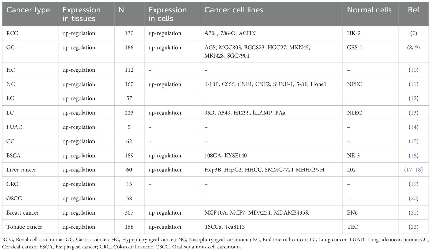
Table 1. CENP-H expression in cancer tissues and cells.
The processes of growth and metastasis are essential to cancer. Inhibiting these processes will inhibit cancer progression and improve the prognosis of patients with cancer. Inhibiting CENP-H can delay cancer growth and metastasis, and promote the sensitivity of cancer cells to cisplatin (25). CENP-H overexpression promotes cancer cell proliferation in RCC (ACHN and 786-O), GC (AGS, HGC27, and GES-1), hypopharyngeal squamous cell carcinoma (FaDu), EC (Ishikawa and HEC-1A), LC (A549 and DDP), liver cancer (Hep3B), CRC (HCT116, RKO, and LoVo), and tongue cancer (Tca8113) (7–13, 18, 19, 22–25). CENP-H was found to inhibit cell apoptosis in RCC (ACHN and 786-O) cells, hypopharyngeal squamous cell carcinoma (FaDu) cells, EC (Ishikawa and HEC-1A) cells, and liver cancer (Hep3B) cells (7, 10, 12, 18). In addition, CENP-H promoted the cell cycle transition in EC (Ishikawa and HEC-1A) cells and tumor formation in nude mice with liver cancer (Hep3B) (12, 18). CENP-H can also promote cancer cell metastasis and sensitivity to cisplatin, which in turn affects cancer progression (Table 2). In particular, CENP-H can promote the invasion and migration of EC (Ishikawa and HEC-1A) and LC (A549 and DDP) cells (12, 23). It can also promote cisplatin resistance in LUAD (A549 and DDP) cells and facilitate CRC cells’ resistance to radiation therapy and rapamycin (23, 25).
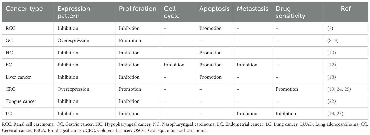
Table 2. In vitro functional characterization of CENP-H in cancer.
CENP-H can participate in cancer cell growth and metastasis through multiple signaling mechanisms. In GC and tongue cancer cells, CENP-H overexpression can increase survivin expression, thereby promoting cancer cell growth (9, 22). On the contrary, inhibiting CENP-H expression in LUAD cells can delay cell growth and metastasis by downregulating the expression of phosphorylated AKT (p-AKT), phosphorylated extracellular signal-regulated kinase (p-ERK), and phosphorylated p38 (p-p38) and promoting the sensitivity of LUAD cells to cisplatin (23). Inhibiting CENP-H expression can upregulate the expression of caspase-3 and B-cell lymphoma-2-associated X (Bax) protein and inhibit the expression of B-cell lymphoma-2 (Bcl-2) and Ki-67 proteins in liver cancer cells (18). In addition, CENP-H may be involved in the proliferation and apoptosis of liver cancer cells through the mitochondrial apoptosis pathway (18). CENP-H can promote CRC progression and modulate response to rapamycin by inhibiting the mechanistic target of rapamycin (mTOR) signaling pathway through interaction with Golgi phosphoprotein 3 (GOLPH3) (25).
CENP-H has also been observed to be regulated by carcinogenic and tumor suppressor factors (9, 12, 26). Specifically, CENP-H promotes EC cell proliferation, migration, and invasion, and it inhibits apoptosis, which can be reversed by overexpression of miR-612. Long non-coding ribonucleic acid (lncRNA) plasmacytoma variant translocation 1 (PVT1) can promote the malignant progression of EC through the miR-612/CENP-H signaling axis (12). The expression of specificity protein 1 (Sp1) and Sp3 is significantly increased in nasopharyngeal carcinoma cells. Inhibiting the activities of Sp1 or Sp3 can reduce the level of CENP-H, thus affecting the growth of nasopharyngeal carcinoma (26). In summary, CENP-H is involved in cancer growth and metastasis through PI3K/AKT, survivin, and mitochondrial apoptosis signaling mechanisms and can be regulated by lncRNA PVT1/miR-612, Sp1, or Sp3.
Patients with cancer who exhibit overexpression of some carcinogenic factors often present a poor prognosis (Table 3). CENP-H overexpression has been positively associated with poor overall survival (OS) in patients with RCC, GC, nasopharyngeal cancer, EC, LC, cervical cancer, esophageal squamous cell carcinoma (ESCC), liver cancer, BC, and tongue cancer (7, 9, 11–13, 15–17, 20–22), and is significantly associated with short relapse-free survival in patients with hypopharyngeal squamous cell carcinoma (10). CENP-H overexpression has also been positively correlated with pathological stage, T stage, and lymph node metastasis of patients with cancer, and is significantly correlated with pathological stage and T stage in patients with RCC, GC, hypopharyngeal squamous cell carcinoma, nasopharyngeal carcinoma, EC, LC, cervical cancer, ESCC, liver cancer, OSCC, BC, and tongue carcinoma. It is also significantly correlated with the expression of the proliferation factor Ki-67 in patients with GC, LC, OSCC, and BC (7, 9–13, 15–17, 20–22). These results suggest that CENP-H overexpression helps predict poor prognosis in patients with such cancers.
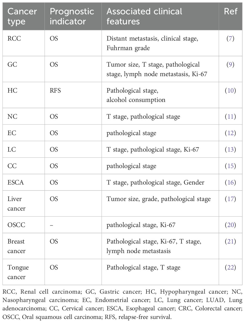
Table 3. CENP-H expression is significantly correlated with the prognosis of patients with cancer.
CENP-H plays a role in organizing and stabilizing centromeres during cell mitosis. CENP-H is overexpressed in RCC, GC, hypopharyngeal squamous cell carcinoma, nasopharyngeal carcinoma, EC, LC, cervical cancer, ESCA, liver cancer, CRC, OSCC, BC, and tongue cancer. This overexpression is correlated with the prognosis, pathological stage, T stage, and lymph node metastasis of patients with such cancers. CENP-H can promote cancer growth and metastasis through PI3K/AKT, Survivin, and mitochondrial apoptosis signaling mechanisms, and it can be regulated by lncRNA PVT1/miR-612, Sp1, or Sp3. Presently, studies on CENP-H are not comprehensive in cancer. First, the interaction between CENP-H and other proteins can be extensively researched to uncover its intrinsic mechanism in regulating cell division. Second, the roles and signaling pathways of CENP-H in normal cells is still unknown, which is also worth exploring in the future. Finally, the structural and functional information of CENP-H can be utilized to develop new drugs to treat cancer. Overall, further studies of CENP-H will contribute to a deeper understanding of the mechanisms of cell division regulation and may provide new candidate molecules and therapeutic targets for disease treatment.
QL: Data curation, Writing – original draft. YT: Writing – review & editing. J-BZ: Writing – review & editing. HH: Writing – review & editing. G-XT: Writing – review & editing. CC: Conceptualization, Data curation, Validation, Writing – review & editing.
The author(s) declare financial support was received for the research, authorship, and/or publication of this article. This study was funded by Zunyi City Joint Fund (Zun Shi Ke He HZ Word (2024) No. 178), Science and Technology Fund of Guizhou Provincial Health Commission (No. gzwjk2024-379), and Science and Technology Project of Guizhou Province (No.LC[2024]099).
The authors declare that the research was conducted in the absence of any commercial or financial relationships that could be construed as a potential conflict of interest.
The author(s) declare that no Generative AI was used in the creation of this manuscript.
All claims expressed in this article are solely those of the authors and do not necessarily represent those of their affiliated organizations, or those of the publisher, the editors and the reviewers. Any product that may be evaluated in this article, or claim that may be made by its manufacturer, is not guaranteed or endorsed by the publisher.
1. Chen W, Yang F, Shen H, Xu J, Chen J, Zhang Z, et al. GGT5 as a promising prognostic biomarker and its effects on tumor cell progression in gastric cancer. Transl Cancer Res. (2024) 13:4459–73. doi: 10.21037/tcr-23-2222
2. Xiang QM, Jiang N, Liu YF, Wang YB, Mu DA, Liu R, et al. Overexpression of SH2D1A promotes cancer progression and is associated with immune cell infiltration in hepatocellular carcinoma via bioinformatics and in vitro study. BMC Cancer. (2023) 23:1005. doi: 10.1186/s12885-023-11315-1
3. Zhang D, Liang P, Xia B, Wu J, Hu X. Comprehensive pan-cancer analysis of ZNF337 as a potential diagnostic, immunological, and prognostic biomarker. BMC Cancer. (2024) 24:987. doi: 10.1186/s12885-024-12703-x
4. Wu CY, Liu Z, Luo WM, Huang H, Jiang N, Du ZP, et al. Downregulation of DIP2B as a prognostic marker inhibited cancer proliferation and migration and was associated with immune infiltration in lung adenocarcinoma via CCND1 and MMP2. Heliyon. (2024) 10:e32025. doi: 10.1016/j.heliyon.2024.e32025
5. Sugata N, Li S, Earnshaw WC, Yen TJ, Yoda K, Masumoto H, et al. Human CENP-H multimers colocalize with CENP-A and CENP-C at active centromere–kinetochore complexes. Hum Mol Genet. (2000) 9:2919–26. doi: 10.1093/hmg/9.19.2919
6. Zhang W, Mao JH, Zhu W, Jain AK, Liu K, Brown JB, et al. Centromere and kinetochore gene misexpression predicts cancer patient survival and response to radiotherapy and chemotherapy. Nat Commun. (2016) 7:12619. doi: 10.1038/ncomms12619
7. Wu X, Lin Y, Shi L, Huang Y, Lai C, Wang Y, et al. Upregulation of centromere protein H is associated with progression of renal cell carcinoma. J Mol Histol. (2015) 46:377–85. doi: 10.1007/s10735-015-9635-2
8. Quan T, He B, Liu T, Li W, Wu S, Jiang Q, et al. Role of centromere protein H in human gastric cancer cell proliferation. Nan Fang Yi Ke Da Xue Xue Bao. (2012) 32:265–9.
9. He WL, Li YH, Yang DJ, Song W, Chen XL, Liu FK, et al. Combined evaluation of centromere protein H and Ki-67 as prognostic biomarker for patients with gastric carcinoma. Eur J Surg Oncol. (2013) 39:141–9. doi: 10.1016/j.ejso.2012.08.023
10. Wang JX, Zhang YY, Yu XM, Jin T, Pan XL. Role of centromere protein H and Ki67 in relapse-free survival of patients after primary surgery for hypopharyngeal cancer. Asian Pac J Cancer Prev. (2012) 13:821–5. doi: 10.7314/apjcp.2012.13.3.821
11. Liao WT, Song LB, Zhang HZ, Zhang X, Zhang L, Liu WL, et al. Centromere protein H is a novel prognostic marker for nasopharyngeal carcinoma progression and overall patient survival. Clin Cancer Res. (2007) 13:508–14. doi: 10.1158/1078-0432.CCR-06-1512
12. Cong R, Kong F, Ma J, Li Q, Yang H, Ma X. The PVT1/miR-612/CENP-H/CDK1 axis promotes Malignant progression of advanced endometrial cancer. Am J Cancer Res. (2021) 11:1480–502.
13. Liao WT, Wang X, Xu LH, Kong QL, Yu CP, Li MZ, et al. Centromere protein H is a novel prognostic marker for human nonsmall cell lung cancer progression and overall patient survival. Cancer. (2009) 115:1507–17. doi: 10.1002/cncr.24128
14. Wang Y, Chen J, Meng W, Zhao R, Lin W, Mei P, et al. A five-gene expression signature of centromeric proteins with prognostic value in lung adenocarcinoma. Transl Cancer Res. (2023) 12:273–86. doi: 10.21037/tcr-22-2166
15. Weng MY, Li L, Hong SJ, Feng SY. Clinical significance of CENP-H expression in uterine cervical cancer. Cancer Biol Med. (2012) 9:192–6. doi: 10.7497/j.issn.2095-3941.2012.03.007
16. Guo XZ, Zhang G, Wang JY, Liu WL, Wang F, Dong JQ, et al. Prognostic relevance of Centromere protein H expression in esophageal carcinoma. BMC Cancer. (2008) 8:233. doi: 10.1186/1471-2407-8-233
17. Lu G, Shan T, He S, Ren M, Zhu M, Hu Y, et al. Overexpression of CENP-H as a novel prognostic biomarker for human hepatocellular carcinoma progression and patient survival. Oncol Rep. (2013) 30:2238–44. doi: 10.3892/or.2013.2675
18. Lu G, Hou H, Lu X, Ke X, Wang X, Zhang D, et al. CENP-H regulates the cell growth of human hepatocellular carcinoma cells through the mitochondrial apoptotic pathway. Oncol Rep. (2017) 37:3484–92. doi: 10.3892/or.2017.5602
19. Tomonaga T, Matsushita K, Ishibashi M, Nezu M, Shimada H, Ochiai T, et al. Centromere protein H is up-regulated in primary human colorectal cancer and its overexpression induces aneuploidy. Cancer Res. (2005) 65:4683–9. doi: 10.1158/0008-5472.CAN-04-3613
20. Shigeishi H, Higashikawa K, Ono S, Mizuta K, Ninomiya Y, Yoneda S, et al. Increased expression of CENP-H gene in human oral squamous cell carcinomas harboring high-proliferative activity. Oncol Rep. (2006) 16:1071–5. doi: 10.3892/or.16.5.1071
21. Liao WT, Feng Y, Li ML, Liu GL, Li MZ, Zeng MS, et al. Overexpression of centromere protein H is significantly associated with breast cancer progression and overall patient survival. Chin J Cancer. (2011) 30:627–37. doi: 10.5732/cjc.010.10599
22. Liao WT, Yu CP, Wu DH, Zhang L, Xu LH, Weng GX, et al. Upregulation of CENP-H in tongue cancer correlates with poor prognosis and progression. J Exp Clin Cancer Res. (2009) 28:74. doi: 10.1186/1756-9966-28-74
23. Li Q, Huang X, Li QY, Tang Y, Liang LB, Luo Q, et al. CENPH overexpression promotes the progression, cisplatin resistance, and poor prognosis of lung adenocarcinoma via the AKT and ERK/P38 pathways. Am J Cancer Res. (2023) 13:1682–97.
24. Song Y, Deng Z, Sun H, Zhao Y, Zhao R, Cheng J, et al. Predicting tumor repopulation through the gene panel derived from radiation resistant colorectal cancer cells. J Transl Med. (2023) 21:390. doi: 10.1186/s12967-023-04260-x
25. Wu W, Wu F, Wang Z, Di J, Yang J, Gao P, et al. CENPH inhibits rapamycin sensitivity by regulating GOLPH3-dependent mTOR signaling pathway in colorectal cancer. J Cancer. (2017) 8:2163–72. doi: 10.7150/jca.19940
Keywords: CENP-H, cancer, poor prognosis, biomarker, lncRNA
Citation: Li Q, Tang Y, Zuo J-B, Han H, Tu G-X and Chen C (2025) CENP-H as a new prognostic biomarker for tumors: a real-world literature review. Front. Oncol. 15:1521988. doi: 10.3389/fonc.2025.1521988
Received: 03 November 2024; Accepted: 07 February 2025;
Published: 25 February 2025.
Edited by:
Katsumi Kitagawa, The University of Texas Health Science Center at San Antonio, United StatesReviewed by:
Yohei Niikura, Nanjing University, ChinaCopyright © 2025 Li, Tang, Zuo, Han, Tu and Chen. This is an open-access article distributed under the terms of the Creative Commons Attribution License (CC BY). The use, distribution or reproduction in other forums is permitted, provided the original author(s) and the copyright owner(s) are credited and that the original publication in this journal is cited, in accordance with accepted academic practice. No use, distribution or reproduction is permitted which does not comply with these terms.
*Correspondence: Cheng Chen, Y2hlbmNoZW5nQHptdS5lZHUuY24=
Disclaimer: All claims expressed in this article are solely those of the authors and do not necessarily represent those of their affiliated organizations, or those of the publisher, the editors and the reviewers. Any product that may be evaluated in this article or claim that may be made by its manufacturer is not guaranteed or endorsed by the publisher.
Research integrity at Frontiers
Learn more about the work of our research integrity team to safeguard the quality of each article we publish.