- 1Department of Oncology, Yantai Yuhuangding Hospital of Qingdao University, Yantai, China
- 2Department of Gynecology, Haiyang Maternal and Child Health Hospital, Yantai, China
- 3Department of Pathology, Yantai Yuhuangding Hospital of Qingdao University, Yantai, China
Aim: We aimed to analyze the clinico-pathological and molecular features of mesonephric-like adenocarcinoma (MLA) to enhance understanding of this tumor type.
Methods: This is the first case of MLA occurring in the retroperitoneum of a male patient. Clinico-pathological and molecular characteristics were analyzed, and the relevant literature was reviewed.
Results: A 65-year-old elderly male was admitted to the hospital with mild bilateral dull pain in the lumbar region for more than 1 month, accompanied by a feeling of dysuria. CT tomography revealed a retroperitoneal tumor. While tumor immuno-histochemistry was positive for CK, CK7, Vimentin, PAX-8, CD10, GATA-3, EMA, and CR to varying degrees, it was negative for P53, WT-1, HMB45, MelanA, CD117, DOG-1, CD34, S-100, ER, PR, AR, CEA, α-inhibin and TTF-1. Ki67 index was <10% in most areas and was approximately 30% in the hotspot areas in the glandular ductal region. Molecular detection (Next-generation sequencing method, 425-gene panel from NanjingShihe Gene Biotechnology Co., Ltd. for targeted DNA enrichment): No clinically significant variants detected. The final pathological diagnosis was a retroperitoneal malignant tumor consistent with a well-moderately differentiated MLA.
Conclusion: MLA in the retroperitoneum of men has not been reported yet. The diverse morphology and unclear molecular characteristics of this tumor mandate careful diagnosis for good clinical outcomes.
1 Introduction
Mesonephric-like adenocarcinoma (MLA) is a newly defined group that encompasses specific types of adenocarcinomas of the ovary and uterine corpus (1), which has been included in the 2020 World Health Organization (WHO) classification of tumors of the female genitalia (2). Studies have shown that these tumors predominantly occur in females, and no reports of cases in males exist. We report a case of male retroperitoneal MLA, and present clinico-pathological features and molecular characterization in the patient in an attempt to enhance understanding of this tumor, in conjunction with an extensive literature review.
2 Case description
2.1 Patient details and initial diagnosis
Informed consent has been obtained from the participant and the study was approved by Ethics Committee of Yantai Yuhuangding Hospital. The patient was a 65 years old male, with bilateral lumbar pain for more than one month. The pain was described as mild and dull, and was accompanied by a feeling of incomplete urination. The patient had no history of hematuria, increased urinary frequency, urgency, urinary pain, fever, or fatigue. The patient was admitted to an outside hospital and underwent urological ultrasound, which showed 1:abnormal development of both kidneys, hydronephrosis with multiple stones in the left kidney. 2: Multiple cystic structures below the right kidney and peripheral solid nodules. 3: Enlarged prostate with calcified foci, which was not treated, and the patient came to our hospital for further treatment. His routine blood tests were as follows: WBC: 8.09X10/L, lymphocytes: 10.9%, and neutrophils: 6.59 X10/L. The tumor indices were: glycogen antigen-125:6.48 U/ml, carcinoembryonic antigen: 2.24 ng/ml, prostate-specific antigen: 8.80 ng/ml, and squamous cell carcinoma-related antigen: 0.6 ng/ml.
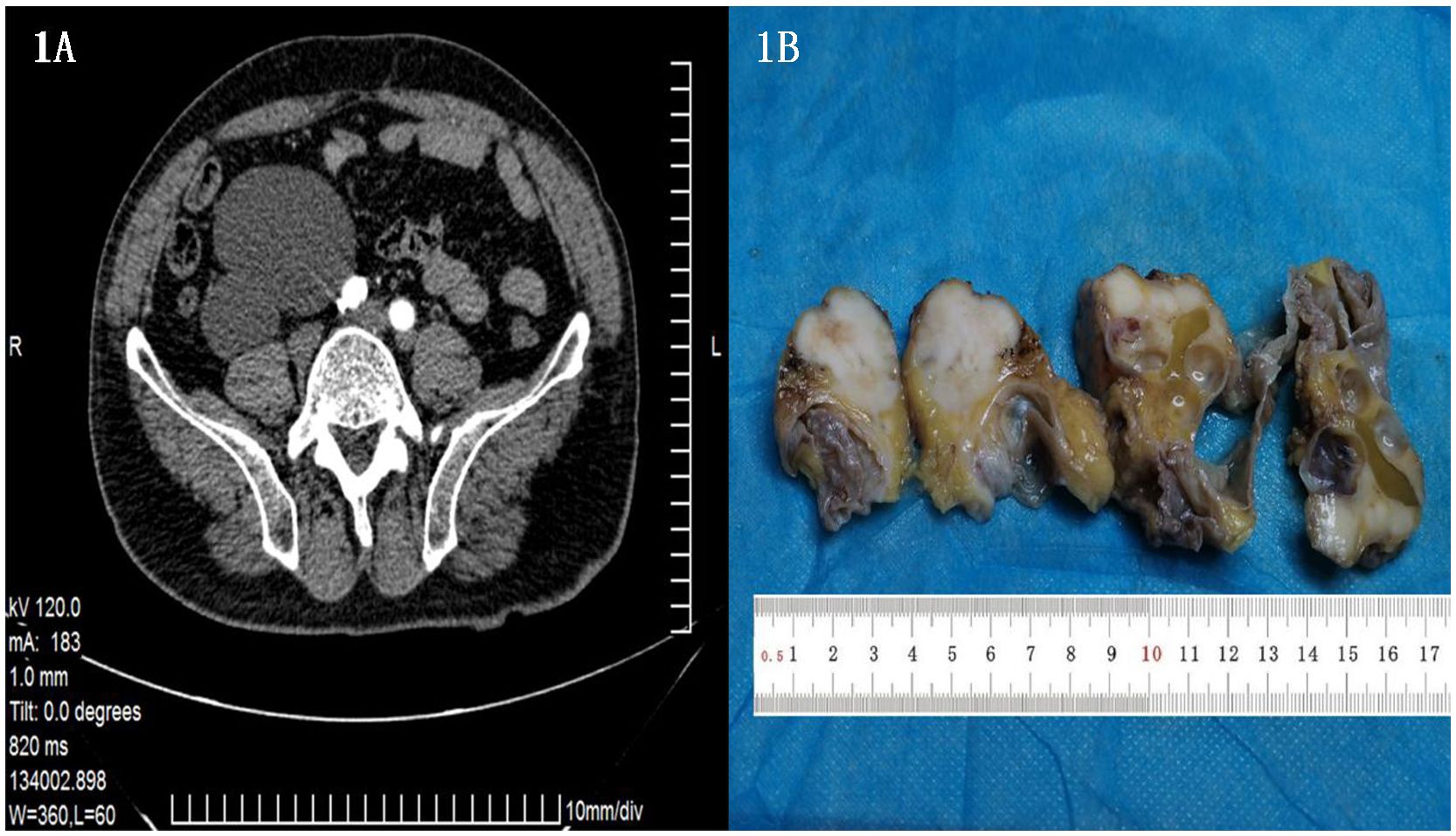
Contrast-enhanced CT revealed a cystic solid occupancy below the right kidney with clear borders. The mass was located approximately in front of the right psoas major muscle, medial right common iliac artery, and lateral of the ascending colon. The tumor measured approximately 8.1×7.7×12.2 cm. While no obvious enhancements were evident in the arterial phase of the enhanced scan and in the cystic portion, a slight enhancement was observed in the solid part in the venous and delayed phases. (Figure 1A, arrow indicates tumor).

Figure 1. (A) Enhanced CT showing a cystic solid space occupying lesion under the right kidney, with a clear boundary measuring approximately 8.1 × 7.7 × 12.2 cm. (B) A single cystic solid tumor measuring 7*6*4 cm was evident in the retroperitoneum. The cystic area had a diameter of 3 cm, with a smooth inner wall of 0.1 cm thickness. The solid area had diameter of 4 cm, and a cut section was found to be gray white in color, hard, and possessed an intact capsule.
2.2 Pathological and molecular characterization
The excised tumor was a single cystic-solid mass measuring 7*6*4 cm, with the cystic area having a diameter of 3 cm, and a smooth inner wall of 0.1 cm thickness. The solid area had diameter of 4 cm, with a grayish-white and hard cut surface and an intact peritoneum (Figure 1B).
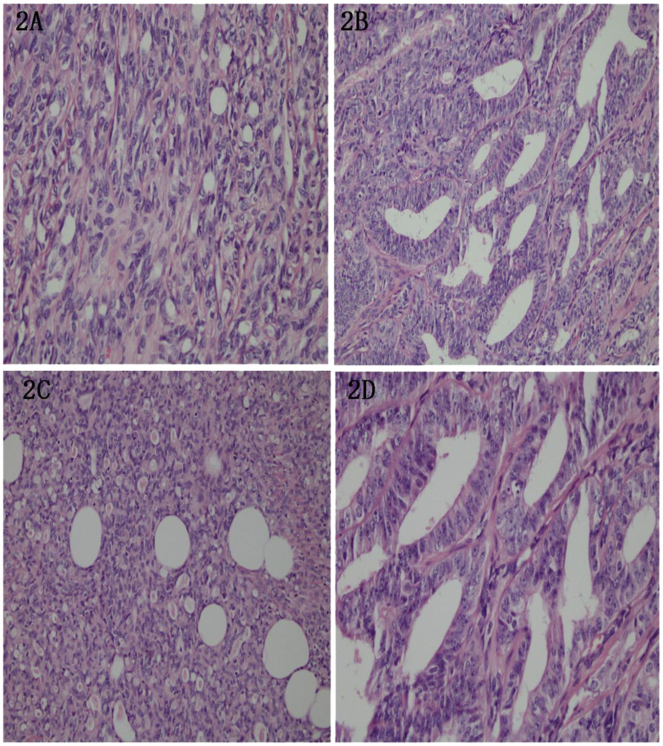
The tumor demonstrated a diverse patho-histological patterns, with spindle-shaped, glandular, and tubular growths. The tubular epithelium was cuboidal or flattened, and eosinophilic material deposition was observed. Additionally, few nuclear divisions were visible with a frequency of 2-10/HPF. Neural invasion was evident, and interstitial fibrotic tissue proliferation was accompanied by hyaline degeneration (Figure 2). The majority of the tumor areas display low-grade morphology, with focal regions showing intermediate-grade features.

Figure 2. (A) Tumor cells showing bidirectional differentiation, of which some were spindle shaped. HE, (X200). (B) Tumor cells showing glandular tubular and tubular growth. HE, (X100). (C) The tubule epithelium was cuboidal or flat. HE, (X100). (D) Pink matter and few mitotic events with a frequency of 2-10/HPS in the cavity. Nerve invasion was evident, along with interstitial fibrous tissue hyperplasia with hyaline degeneration. HE, (X100).
Immuno-histochemical analysis revealed the following results: CK (diffuse +), CK7 (+, with some adenotubular-like structures weakly +), Vimentin (+, adenotubular +, and few remaining weakly +), PAX-8 (adenotubular weakly +, and the rest diffusely strongly +), CD10 (luminal margins +), GATA-3 (foci +), EMA (+, luminal margins predominantly +, and most +), CR (some +); P53 (-, nonsense mutation); PMS2(+), MLH1(+), MSH2(+), MSH6(+); WT- 1 (–), HMB45 (–), MelanA (–), CD117 (–), DOG-1 (–), CD34 (–), S-100 (–), ER (–), PR (–), AR (–), CEA (–), inhibin (–), TTF-1 (–), and Ki67 positivity of <10% in most regions, and of approximately 30% within hotspots in glandular ductal regions. (Figure 3).

Figure 3. (A) Tumor cells were diffusely positive for CK, EnVision method. (X200). (B) Tumor cells were positive for CK7, and part of adenoid structure was weakly positive. EnVision method. (X200). (C) Tumor cells and the glandular tube were positive for Vimentin, and the rest was weakly positive. EnVision method. (X200). (D) Tumor cells were positive for EMA, mainly lumen margin positive, mostly positive. EnVision method. (X200). (E) Tumor cells were partially positive for CR. EnVision method. (X200). (F) Tumor cells were positive for CD10 at the lumen margin. EnVision method. (X200). (G) Tumor cells of the glandular duct were weakly positive for Pax-8, and the rest were diffusely strongly positive. EnVision method. (X200). (H) Tumor cells were GATA-3 foci positive. EnVision method. (X200). (I) The Ki-67 proliferation index in most areas of tumor cells was less than 10%, and about 30% in the hot spots in glandular areas. EnVision method. (X200).
Molecular detection (Next-generation sequencing method): No clinically significant variants detected.
Based on the morphology, surgical, and immuno-histochemical findings, a final pathological diagnosis of a retroperitoneal malignant tumor consistent with a well-moderately differentiated MLA was made.
3 Diagnostic assessment
Based on the imaging findings, a preoperative diagnosis of retroperitoneal mass (right infrarenal) was made. After clinical improvement of relevant examinations, laparoscopic resection of the retroperitoneal mass was performed. The mass was located in the posterior aspect of the colonic appendix with intact peritoneum, which was completely removed surgically. The patient had suffered from allergic asthma for 15 years, and denied any history of surgical trauma, family genetics or related diseases.
The patient did not undergo any post-operative treatment, such as, radiotherapy or chemotherapy. He has remained tumor-free on follow up for 21 months until now.
4 Discussion
The mesonephric and Mullerian ducts travel in parallel during the embryonic period, and form the epididymis, vas deferens, seminal vesicles, and a portion of the prostate and urethra in males. The mesonephric ducts undergo degeneration in females due to lack of testosterone (1–7), and adults only carry remnants of the nonfunctional mesonephric ducts that are usually located in the median ovary, broad ligament, or lateral wall of the cervix, and rarely in the vagina or body of the uterus with low probability of malignancy. Mesonephric adenocarcinoma (MA) is thus a rare malignancy of the female genital tract, usually located in the cervix and vagina that originates from the embryonic remnants of the mesonephric tubules and ducts, and accounts for less than 1% of all gynecologic malignancies (8). Within this context, MA of the female upper genital tract is referred to as mesonephric-like adenocarcinoma (MLA), on account of unproven association with mesonephric remnants (2, 9). Reports have suggested that mesonephric-like adenocarcinoma (MLA) originates from mullerian ducts, and is not associated with mesonephric remnants (10). While cases of MLA have been documented in females, no reports on male patients exist. Literature review revealed that mesonephric ductal remnants may be located not only in the female reproductive system, but also in the retroperitoneum, adjacent to the kidneys, posterior to the colon, and near the head or tail of the pancreas (11, 12).
The present study is the first report of a retroperitoneal MLA in a 65-year-old elderly male who presented at the clinic with mild bilateral lumbar dullness and pain that was accompanied by dysuria. Imaging revealed a cystic-solid retroperitoneal occupancy with clear borders, which was consistent with the findings of Koh et al., who reported that MLA manifests as either mixed solid and cystic masses, or pure solid masses in imaging studies (9), with the typical presentation often being suggestive of an MLA tumor. MLAs exhibit a variety of patho-histological patterns (13), including tubular, adenotubular, papillary, reticular, solid, glomerulonephric, gonadotubular/trabecular, sieve, and osteosarcomatous growths, with the possibility of combinations of these patterns within a single tumor. The common spindle-like osteosarcoma-like differentiation pattern includes tubular and glandular tubular growths. The tubules usually comprise small round tubules that grow in a back-to-back or diffusely infiltrating pattern with cuboidal or flattened epithelium and eosinophilic material deposition was observed. Although adenoductal structures consist of large glands or papillary-like formations, the tumor cells generally have mild to moderate cytological atypia, which was also consistent with the pathological observations made in the present case. The tumor foci in two of the ovarian MLA tumors reported by Koh et. al, could be interpreted as severe cytokinetic polymorphisms with scattered areas of extensive coagulative tumor necrosis (9). The 2-10/HPF nuclear schizophrenic pixels observed in the present case were slightly lower tumor than the numbers reported in literature ranging within 3-50/HPF. While mesonephric ductal hyperplasia is often present in the periphery of most tumors with peripheral nerve invasion (14), it was not evident in our case despite the presence of peripheral nerve invasion.
The MLA immune-phenotypes that have been reported in the literature (9, 13, 15) are predominantly positive for PAX-8, CD10 (luminal rim expression), TTF1, and/or GATA3, with reverse reciprocal staining patterns for GATA-3 and TTF-1. Additionally, these tumors displaying non-diffuse P16 immuno-reactivity and wild-type P53 immuno-staining patterns, with negative results for WT-1, ER, PR, AR, CEA, and inhibin along with a ki-67 positivity index of 50% (15–17). Immuno-histochemical analysis revealed that while CK, CK7, Vimentin, PAX-8, CD10 and GATA-3, EMA, CR were positively expressed to varying degrees; P53(-, nonsense mutation); WT-1, HMB45, MelanA, CD117, DOG-1, CD34, S-100, ER, PR, AR, CEA, inhibin, and TTF-1 were negatively expressed in the present case. Further, Ki67 positivity of <10% in most regions, and of approximately 30% within the hotspots of the glandular ductal region was evident. The results mostly concurred with reports in literature, including negative results for TTF-1 (6, 7, 13). The only discrepancy with reported studies was that the Ki-67 positivity index in our case was lower than that previously reported (15). MLA is known to harbor unique genomic alterations, including, high-frequency KRAS gene mutations and increased chromosome 1q numbers (4, 5, 18), with a mutation rate of 12/16 (75%) in the KRAS gene (19). Molecular detection (Next-generation sequencing method): No clinically significant variants detected. Despite negative molecular test results, the tumor in this case was positive for all the three immuno-histochemical markers, including, PAX-8, CD10, and GATA-3 that effectively diagnose tumors of mesonephric ductal remnants and Müllerian ductal origin (10, 20–27).
In view of the clinical features, histological findings, and immuno-histochemical results, a pathological diagnosis of retroperitoneal MLA was established. Slight discrepancies between our results and those in literature on MLA in the female genitalia were evident in the context of the relatively low mitotic count and Ki67 positivity index in our case. Additionally, the patient did not undergo any postoperative treatment and had a good prognosis at the last follow-up, which categories his tumor as a low-grade malignant cancer. This is in marked contrast to the aggressive clinical course of MLA in females that has a high recurrence rate and distant metastasis (1, 9, 13, 28–30). MLAs may therefore not be exclusively associated (1) with the patient’s gender, (2) tumor site, (3) known molecular associations, and specific pathogenesis. (4) The justification of categorizing of a few well-moderately differentiated malignant tumors as MLA is thus an open question. Given that these tumors are rare, and this is the first case involving a male patient with an MLA located in the retroperitoneum, several more data points are essential to reach a conclusive consensus.
The low incidence, diverse histological patterns, and lack of specific ultrastructural features, make the pathological diagnosis of MLA difficult. A rare occurrence in a male at a specific site, as in our case, made the diagnosis even more challenging. MLAs are included in the differential diagnoses of a multitude of clinical lesions, and patho-morphology, immuno-histochemistry, and molecular testing results in tandem aid its distinction from other tumors, including, mesangial tubular remnants and hyperplasia, malignant mesothelioma, plasmacytoid carcinoma, clear cell carcinoma, and mesenchymal stromal tumors.
To summarize, MLA are rare and can occur in men. It is often misdiagnosed by clinico-pathologists owing to the lack of specific clinical manifestations and diverse pathological patterns. Conclusive diagnosis therefore mandates extensive sampling, careful observation of histological images, and a keen search for characteristic clues, in combination with immuno-histochemical and molecular tests and other auxiliary assessments. MLAs in women are described as a morphologically highly differentiated and aggressive malignancy, that require extensive treatment and adjuvant therapy, including radiotherapy and/or chemotherapy post-surgical resection in early stage patients. Further, patients with MLA harboring KRAS/NRAS gene mutations may be treated with RAS gene-targeted inhibitors. However, currently consensus recommendations for treatment regimens are lacking. Herein, we have brought forth for the first time a case of a male MLA patient, and consequently propose the concept of low-grade malignant MLA that is in sharp contrast to the low-intermediate grade female MLA tumors. Further validation of this concept via long-term follow-up and analysis of a larger number of cases is essential.
Data availability statement
The datasets presented in this study can be found in online repositories. The names of the repository/repositories and accession number(s) can be found in the article/supplementary material.
Ethics statement
The studies involving humans were approved by Committee of Yantai Yuhuangding Hospital. The studies were conducted in accordance with the local legislation and institutional requirements. The participants provided their written informed consent to participate in this study. Written informed consent was obtained from the individual(s) for the publication of any potentially identifiable images or data included in this article.
Author contributions
PY: Writing – original draft, Writing – review & editing. YL: Writing – original draft, Writing – review & editing. JT: Writing – review & editing, Investigation, Methodology. BH: Writing – original draft, Writing – review & editing. DS: Writing – original draft, Writing – review & editing.
Funding
The author(s) declare that no financial support was received for the research, authorship, and/or publication of this article.
Acknowledgments
We would like to extend our sincere gratitude to Yantai Yuhuangding Hospital for providing a reliable case and we are also grateful to the participant who generously shared the time and information for this study. Finally, we appreciate the editorial team and reviewers for their insightful comments and suggestions, which significantly improved the quality of this manuscript.
Conflict of interest
The authors declare that the research was conducted without any commercial or financial relationships that could be construed as a potential conflict of interest.
Publisher’s note
All claims expressed in this article are solely those of the authors and do not necessarily represent those of their affiliated organizations, or those of the publisher, the editors and the reviewers. Any product that may be evaluated in this article, or claim that may be made by its manufacturer, is not guaranteed or endorsed by the publisher.
References
1. Roma AA, Goyal A, Yang B. Differential expression patterns of G A T A 3 in uterine mesonephric and nonmesonephric lesions. Int J Gynecol Pathol. (2015) 34:480. doi: 10.1097/PGP.0000000000000167
2. Howitt BE, Emori MM, Drapkin R, Gaspar C, Barletta JA, Nucci MR, et al. G A T A 3 is a sensitive and specific marker of benign and Malignant mesonephric lesions in the lower female genital tract. Am J Surg Pathol. (2015) 39:1411–9. doi: 10.1097/PAS.0000000000000471
3. Houghton O, Jamison J, Wilson R, Carson J, McCluggage WG. p16 Immunoreactivity in unusual types of cervical adenocarcinoma does not reflect human papillomavirus infection. Histopathology. (2010) 57:342–50. doi: 10.1111/j.1365-2559.2010.03632.x
4. McCluggage WG. Recent Developments in Non-HPV-related adenocarcinomas of the lower female genital tract and their precursors. Adv Anat Pathol. (2016) 23:58–69. doi: 10.1097/PAP.0000000000000095
5. McCluggage WG. New development in endocervical glandular lesions. Histopathology. (2013) 62:138–60. doi: 10.1111/his.2012.62.issue-1
6. Silver SA, Devouassoux-Shisheboran M, Mezzetti TP, Tavassoli FA. Mesonephric Adenocarcinomas of the uterine cervix: a study of 11 cases with immunohistochemical findings. Am J Surg Pathol. (2001) 25:379–87. doi: 10.1097/00000478-200103000-00013
7. Yap OWS, Hendrickson MR, Teng NNH, Kapp DS. Mesonephric adenocarcinoma of the cervix: A case report and review of the literature. Gynecol Oncol. (2006) 103:1155–8. doi: 10.1016/j.ygyno.2006.08.031
8. Kezlarian B, Muller S, Werneck Krauss Silva V, Gonzalez C, Fix DJ, Park KJ, et al. Cytologic features of upper gynecologic tract adenocarcinomas exhibiting mesonephric-like differentiation. Cancer Cytopathol. (2019) 127:521–8. doi: 10.1002/cncy.v127.8
9. Ando H, Watanabe Y, Ogawa M, Tamura H, Deguchi T, Ikeda K, et al. Mesonephric adenocarcinoma of the uterine corpus with intracystic growth completely confined to the myometrium: A case report and literature review. Diagn Pathol. (2017) 12:63. doi: 10.1186/s13000-017-0655-y
10. Kim SS, Nam JH, Kim G-E, Choi YD, Choi C, Park CS. Mesonephric adenocarcinoma of the uterine corpus: A case report and diagnostic pitfall. Int J Surg Pathol. (2016) 24:153–8. doi: 10.1177/1066896915611489
11. Puljiz M, Danolić D, Kostić L, Alvir I, Tomica D, Mamić I, et al. Mesonephric adenocarcinoma of endocervix with lobular mesonephric hyperplasia: Case report. Acta Clin Groat. (2016) 55:326–30. doi: 10.20471/acc.2016.55.02.22
12. Yeo MK, Choi SY, Kim M, Kim KH, Suh KS. Malignant mesonephric tumor of the cervix with an initial manifestation as pulmonary metastasis: Case report and review of the literature. Eur J Gynaecol Oncol. (2016) 37:270–7. doi: 10.1097/00000478-199510000-00006
13. Iris M, Gerhard K, Jacobs VR. Mesonephric adenocarcinoma of the vagina: Diagnosis and multimodal treatment of a rare rumor and analysis of worldwide experience. Strahlentherapie Und Onkol. (2016) 192:668–71. doi: 10.1007/s00066-016-1004-x
14. Bifulco G, Mandato VD, Mignogna C, Giampaolino P, Di Spiezio Sardo A, De Cecio R, et al. A case of mesonephric adenocarcinoma of the vagina with a 1-yearfollow-up. Int J Gynecol Cancer. (2010) 18:1127–31. doi: 10.1111/j.1525-1438.2007.01143.x
15. Roma AA. Mesonephric carcinosarcoma involving uterinecervix and vagina: Report of 2 cases with immunohistochemicalpositivity For PAX 2, PAX8, and GATA -3. Int J Gynecol Pathol. (2014) 33:624–9. doi: 10.1097/PGP.0000000000000088
16. Wu HX, Zhang L, Cao WF, Hu YJ, Liu YX. Mesonephric adenocarcinoma of the uterine corpus. Int J Clin Exp Pathol. (2014) 7:7012–9. doi: 10.4103/IJPM.IJPM_298_20
17. Montagut C, Mármol M, Rey V, Ordi J, Pahissa J, Rovirosa A, et al. Activity of chemotherapy with carboplatin plus paclitaxel in a recurrent mesonephric adenocarcinoma of the uterine corpus. Gynecol Oncol. (2003) 90:458–61. doi: 10.1016/S0090-8258(03)00228-2
18. Akkawi R, Valente AL, Badawy SZ. Large mesonephric cyst with acute adnexal torsion in a teenage girl. J Pediatr Adolesc Gynecol. (2012) 25:e143–5. doi: 10.1016/j.jpag.2012.06.009
19. Mirkovic J, Sholl LM, Garcia E, Lindeman N, MacConaill L, Hirsch M, et al. Targeted genomic profiling reveals recurrent KRAS mutations and gain of chromosome 1q in mesonephric carcinomas of the female genital tract. Mod Pathol. (2015) 28:1504–14. doi: 10.1038/modpathol.2015.103
20. Meguro S, Yasuda M, Shimizu M, Kurosaki A, Fujiwara K. Mesonephric adenocarcinoma with a sarcomatous component, a notable subtype of cervical carcinosarcoma: a case report and review of the literature. Diagn Pathol. (2013) 8:74. doi: 10.1186/1746-1596-8-74
21. Zhang L, Cai ZJ, Ambelil MJ, Conyers J, Zhu H. Mesonephric adenocarcinoma of the uterine corpus: report of 2 cases and review of the literature. Inter J Gynecol Pathol. (2019) 38:224–9. doi: 10.1097/PGP.0000000000000493
22. Shoeir S, Balachandran AA, Wang J. Sultan A H.Mesonephric adenocarcinoma of the vagina masquerading as a suburethral cyst. BMJ Case Rep. (2018) pii:bcr-2018-224758. doi: 10.1136/bcr-2018-224758
23. Na K, Kim HS. Clinicopathologic and molecular characteristics of mesonephric adenocarcinoma arising from the uterine body. Am J Surg Pathol. (2019) 43:12–25. doi: 10.1097/PAS.0000000000000991
24. Mirkovic J, Mcfarland M, Garcia E, Sholl LM, Lindeman N, MacConaill L, et al. Targeted genomic profiling reveals recurrent KRAS mutations in mesonephric-like adenocarcinomas of the female genital tract. Am J Surg Pathol. (2017) 42(2):227–33. doi: 10.1097/PAS.0000000000000958
25. Marquette A, Moerman P, Vergote I, Amant F. Second case of uterine mesonephric adenocarcinoma. Int J Gynecol Cancer. (2006) 16:1450–4. doi: 10.1136/ijgc-00009577-200605000-00079
26. Wani Y, Notohara K, Tsukayama C. Mesonephric adenocarcinoma of the uterine corpus:a case report and review of the literature. Int J Gynecol Pathol. (2008) 27:346–52. doi: 10.1097/PGP.0b013e318166067f
27. Ordi J, Nogales FF, Palacin A, Márquez M, Pahisa J, Vanrell JA, et al. Mesonephric adenocarcinoma of the uterine corpus:CD10 expression as evidence of mesonephric differentiation. Am J Surg Pathol. (2001) 25:1540–5. doi: 10.1097/00000478-200112000-00011
28. Kenny SL, Mcbride HA, Jamison J, McCluggage WG. Mesonephric adenocarcinomas of the uterine cervix and corpus: HPV-negative neoplasms that are commonly PAX8, CA125, and HMGA2 positive and that may be immunoreactive TTF1 and hepatocyte nuclear factor 1-β. Am J Surg Pathol. (2012) 36:799. doi: 10.1097/PAS.0b013e31824a72c6
29. Cavalcanti MS, Schultheis AM, Ho C, Wang L, DeLair DF, Weigelt B, et al. Mixed mesonephric adenocarcinoma and high-grade neuroendocrine carcinoma of the uterine cervix: case description of a previously entity with insight into its molecular pathogenesis. Int J Gynecol Pathol. (2017) 36(1):76–89. doi: 10.1097/PGP.0000000000000306
Keywords: male, retroperitoneum, MLA, pathological characteristics, literature review
Citation: Hu B, Liu Y, Tang J, Yang P and Sun D (2024) Case report: The first known case of male retroperitoneal mesonephric-like adenocarcinoma. Front. Oncol. 14:1433563. doi: 10.3389/fonc.2024.1433563
Received: 31 May 2024; Accepted: 30 September 2024;
Published: 28 October 2024.
Edited by:
Ulrich Ronellenfitsch, University Hospital Halle, GermanyReviewed by:
Stefano Restaino, Ospedale Santa Maria della Misericordia di Udine, ItalyGiuseppe Angelico, Agostino Gemelli University Polyclinic (IRCCS), Italy
David Gibbons, St. Vincent’s University Hospital, Ireland
Copyright © 2024 Hu, Liu, Tang, Yang and Sun. This is an open-access article distributed under the terms of the Creative Commons Attribution License (CC BY). The use, distribution or reproduction in other forums is permitted, provided the original author(s) and the copyright owner(s) are credited and that the original publication in this journal is cited, in accordance with accepted academic practice. No use, distribution or reproduction is permitted which does not comply with these terms.
*Correspondence: Ping Yang, eTE1NzUzNTUxNjY2QDE2My5jb20=; Di Sun, NjM1MjQ3MDNAcXEuY29t
†These authors have contributed equally to this work
 Baohong Hu1†
 Ping Yang