- 1Department of Neurosurgery, West China Hospital, Sichuan University, Chengdu, China
- 2Department of Intensive Care Unit, Fourth People’s Hospital of Sichuan Province, Chengdu, China
Background: Cases of lateral ventricular ectopic schwannomas (LVES) are extremely rare, with only 23 cases reported thus far. This study aimed to obtain a better understanding of the disease.
Methods: We reported a rare case of LVES, in which the patient was admitted to our hospital, and reviewed the relevant literature on LVES to summarize and analyze the clinical manifestations, pathologies, imaging features and progress.
Results: Of the 23 patients, LVES was more common in men (74%, 17/23) than in women and was mostly located on the right side (78%, 18/23). The average age at clinical presentation was 28 years, with an age range between 8 and 68 years. Moreover, most cases were histologically benign, except in one case of malignancy. In all the benign cases, there were 2 cases of subtotal resection, but no recurrence was found during follow-up.
Conclusions: The origin of LVES could be the tumor transformation of autonomic nerve tissue in the perivascular choroid plexus. For lateral ventricle tumors,which are rare benign lesions with good prognosis after surgical resection, LVES should be considered in the differential diagnosis. Moreover, whether LVES could be considered for gamma knife treatment, similar to a small acoustic neuromas,requires further investigation.
1. Introduction
Schwannomas originate from the myelin sheath of peripheral nerves and are mostly benign, accounting for approximately 8% of central nervous system (CNS) tumors. Vestibular schwannoma is the most common, but schwannomas occurring in the brain ventricle or parenchyma are extremely uncommon (1, 2). Ectopic schwannomas (ES) refer to schwannomas occurring in the brain parenchyma or ventricles and are rare in the lateral ventricle (3). The first case of lateral ventricular ES(LVES)was reported by David in 1965 (4), and thus far, only 23 cases have been reported in the English literature. Of these, only one case of malignant biological behavior was reported (5).
Herein, we report a case of right LVES, which was misdiagnosed as a meningioma before surgery. To obtain a better understanding of LVES, this study reviewed the relevant literature on LVES to summarize and analyze the clinical manifestations, pathologies, imaging features and progress.
2. Case presentation
2.1. Preoperative examination
A 22-year-old Han Chinese woman presented with paroxysmal dizziness, fatigue, nausea and vomiting for 6 years, and the symptoms had worsened over the prior few months. The patient had no previous medical history and no family genetic history of related diseases, and there were no obvious abnormalities on physical examination and laboratory tests, such as blood cell analysis, blood coagulation function, blood biochemistry test, plasmic electrolyte test, cranial nerves examination, motor function and sensory function. Magnetic resonance imaging (MRI) revealed a heterogeneously contrast-enhancing, irregular, lobulated lesion (3.0 cm × 2.5 cm × 2.5 cm in size) at the posterior horn of the right lateral ventricle, and the lesion was intimately related to the choroid plexus. The mass was hypointense on T1-weighted images and iso-hyperintense on T2-weighted images (Figures 1A–D). A diagnosis of lateral ventricle meningioma was made before surgery.
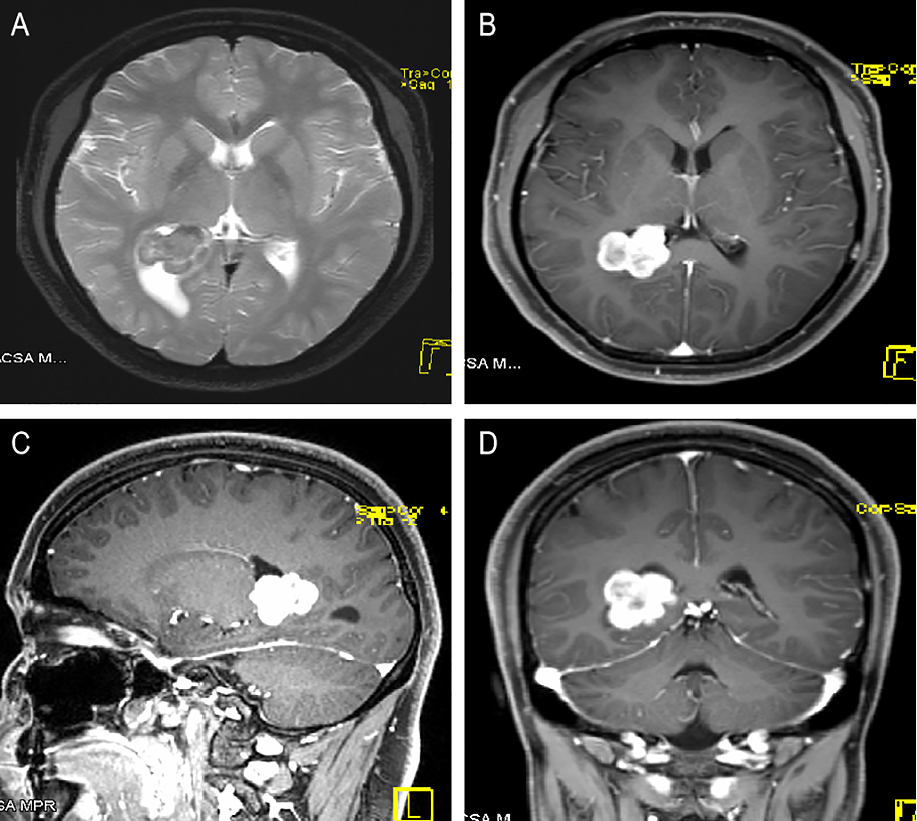
Figure 1 A 22-year-old woman with right LVES. (A) Axial T2WI scan showed an approximately 3.0 cm × 2.5 cm × 2.5 cm irregular cystic-solid lesion at the posterior horn of the right lateral ventricle, intimately related to the choroid plexus, with mild peritumoral edema. The solid components exhibited low signa on T2WI, and the cystic components presented hyperintense signals on T2WI. (B–D) Axial, sagittal and coronal contrast-enhanced T1WI showed heterogeneous and apparent enhancement of the solid part of the mass but no obvious enhancement in the cystic part.
2.2. Surgical treatment
The patient underwent surgery with the right temporal-occipital craniotomy approach. The intraoperative findings showed that the lesion was irregular and hard and measured 3.0×3.0×2.7 cm in size, and the lesion was closely attached to the choroid plexus. The diagnosis of meningioma was confirmed according to the intraoperative findings.
2.3. Postoperative diagnosis and follow-up
The lesion was proven to be a schwannoma by pathological analysis (Figure 2A). In terms of immunohistochemical staining, the tumor cells were positive for S-100 (Figure 2B), vimentin and Ki-67 (1%) and negative for glial fibrillary acidic protein (GFAP) and epithelial membrane antigen (EMA). The postoperative axial and coronal MRI scan revealed that the lesion had been completely resected (Figures 3A, B). The patient developed mild depression during the follow-up. The prognosis was goodat the 14-month follow-up.
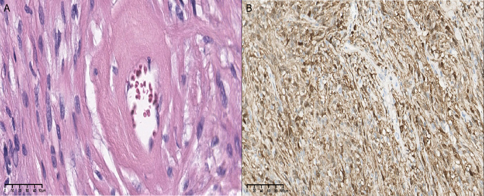
Figure 2 (A) Cells are regular, round or spindle shaped, with clear or eosinophilic cytoplasm (H&E×40). (B) Diffuse expression of the S-100 protein with immunohistochemistry (×20).
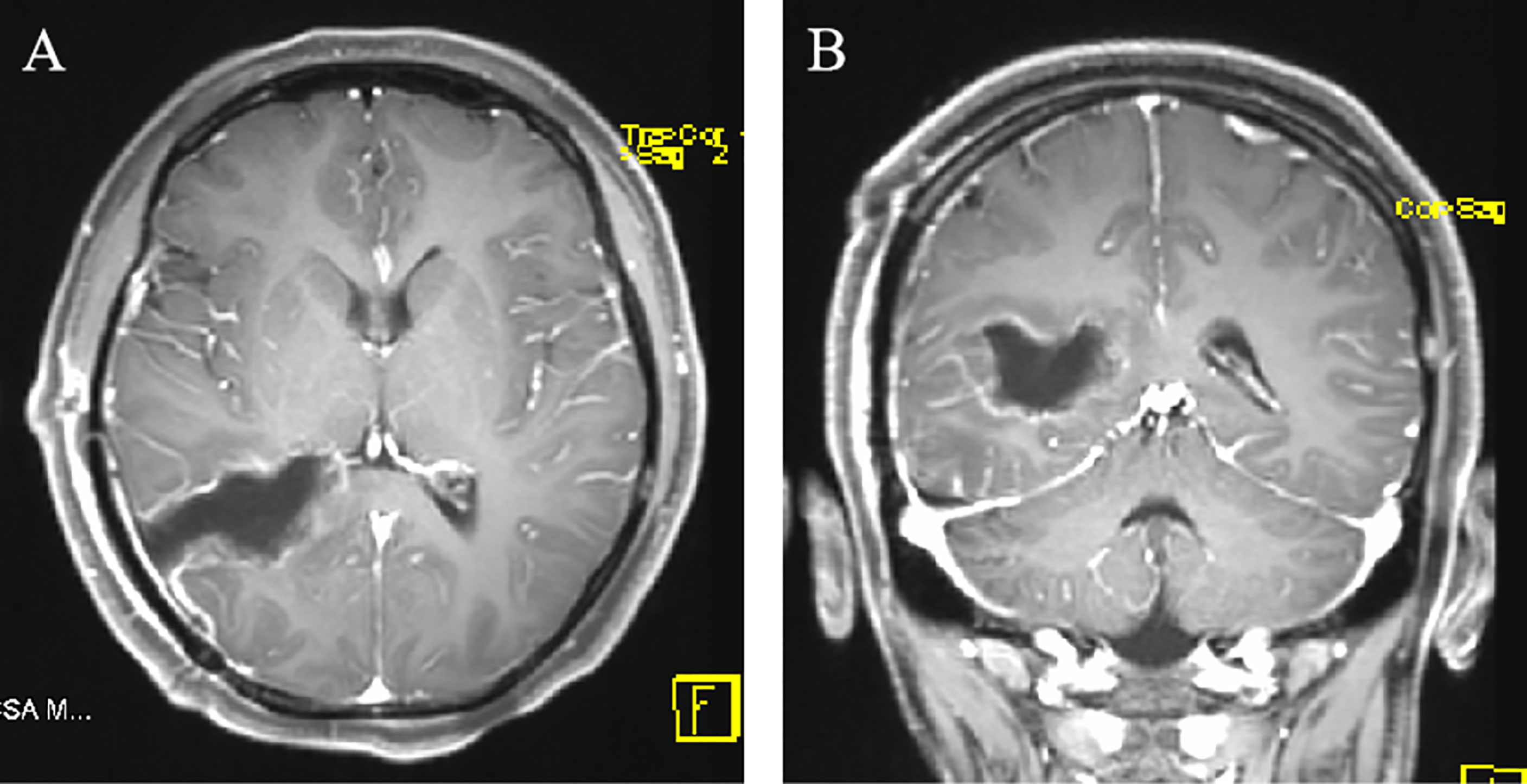
Figure 3 (A, B) Postoperative axial and coronal MRI showing that no parts of the right lateral ventricle lesion remained after resection by a temporal-occipital craniotomy.
3. Discussion
Schwannomas are benign tumors that arise from nerve sheath cells and are commonly found in the head, neck, and limbs. Intracranial schwannomas account for approximately 8% of CNS tumors. Schwannomas occurring in the brain ventricle or parenchyma are extremely rare (1, 2). LVES is extremely rare, with only 23 cases reported thus far (6).
Although the origin of ES remains unknown, four theories have been proposed: (1) tumor transformation of the autonomic nerve tissue in the perivascular choroid plexus; (2) the transformation of multipotent mesenchymal cells into Schwann cells after tissue injury; (3) the transformation of ectopic fragments of neural crest cells into tumors in the ventricular system during abnormal embryonic development; and (4) the possibility of transformation of mesoderm-derived mesenchymal leptomeningeal cells in the brain into Schwann cells (7–10). In our case, ES was at the posterior horn of the right lateral ventricle, which was closely associated with the choroid plexus during the operation. The tumor transformation of autonomic nerve tissue originating from the perivascular choroid plexus is a reasonable explanation of our case.
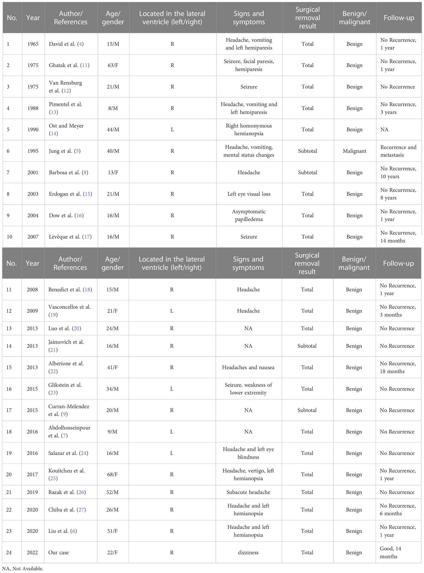
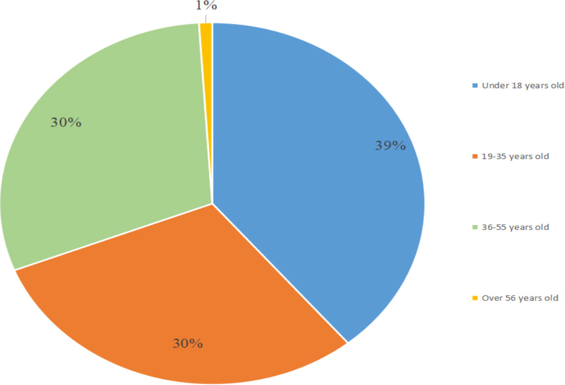
Of the 23 cases (Table 1) (4–8, 11–27), LVES was more common in men (74%, 17/23) than in women and was mostly located on the right side (78%, 18/23). The average age at clinical presentation is 28 years, with an age range between 8 and 68 years (Figure 4). Moreover, most cases are histologically benign, except in one case of malignancy, in which the patient developed recurrence and metastasis of the tumor (5). The main clinical symptoms of the patient who had malignancy were headache and vomiting, which are similar to the symptoms of most of the reported benign intraventricular schwannomas. Meanwhile, the patient was 40 years old, while we found that 16 of the 22 benign patients were younger than 40 years old. Therefore, malignancy should be suspected in older patients with intraventricular tumors. In addition, the patient was rehospitalized seven months after the first surgery and presented with severe headache and vertigo. Brain MRI revealed tumor recurrence and metastasis. The rapid clinical course was different from the reported cases of benign tumors.
Surgical resection is considered to be curative. Most patients have good results after surgical removal of the tumor (Table 1). Of the 21 benign cases reported in previous studies, there was no recurrence of LVES during long-term follow-up after surgery. In our case, the patient, with a completely excised tumor, also had a good outcome after 1 year of follow-up. However, if schwannomas can be accurately identified preoperatively, can gamma knife therapy be considered for small tumors such as small acoustic neuromas?
These tumors cause symptoms that depend on where they are located. The most common symptoms are headache and epilepsy. Of the 23 previously reported cases, except for 4 cases where the symptoms were not recorded, 12 of the patients presented primarily with symptoms such as headache, which may be caused by the mass effect of the tumor (7). In our patient, daily dizziness of gradually increasing intensity, associated with fatigue, nausea and vomiting, was the main clinical manifestation. However, we do not think the patient’s symptoms had much to do with the tumor, or even that the patient’s tumor was an accidental discovery. Unfortunately, the patient was not given more in-depth investigation to support our hypothesis. A review of the clinical features of LVES is shown in Table 1.
Immunohistochemical staining is indispensable for the diagnosis of ES. Sometimes it is difficult to distinguish it from meningioma visually and microscopically. S-100 and vimentin are typically positively expressed, while GFAP and EMA are often negatively expressed (7, 25). Through a literature review, we found that some cases appeared as misdiagnoses based on the preoperative and intraoperative frozen section, and the misdiagnoses included ependymoma, cystic astrocytoma, cystic meningioma, hemangioblastoma, fibroblastic meningioma, papilloma and choroid plexus carcinoma (8, 14, 16, 18, 21, 27). In addition, the majority of the 23 cases were diagnosed as LVES based on the pathological findings. Microscopically, the tumor cells can be divided into two types. Antoni A region: cells are often arranged in fusiform; and Antoni B region: cells are often arranged as palisade patterns.
In terms of imaging features, LVES has specific characteristics. MRI is the best diagnostic tool for these tumors because it can be used to determine the location of the ventricles and the relationship between the tumor and choroid plexus.Combined with this case and related literature, these characteristics are summarized as follows. Cystic changes: cystic and solid changes are characteristic of this disease. The cystic part is mostly manifested as low signal intensity on T1WI and high signal intensity on T2WI, while the solid part is often characterized by slightly low signal intensity on T1WI and high signal intensity on T2WI.Moreover, contrast-enhanced MRI showed significant enhancement in the solid but not cystic areas. Edema: peritumoral edema is considered characteristic of benign schwannomas. ES is characterized by peritumoral edema of different degrees (23). Calcification: A previous study reported that part of LVES may exhibit calcification (28), which is helpful for the diagnosis of these tumors to some extent. Among the MRI findings of these 23 LVES cases, cystic changes and edema were more common. Cystic changes were found in 12 patients, and edema was found in 11 patients. Calcification was observed in only 4 patients. In our case, the patient presented with cystic and solid changes and mild edema around the lateral ventricle, and these findings are consistent with previous literature. In addition, whether tumors showing lobulated changes are more likely to be schwannomas is worth considering in future cases.
4. Conclusion
The origin of LVES could be the tumor transformation of autonomic nerve tissue in the perivascular choroid plexus. Lateral ventricle tumors are rare benign lesions with good prognosis after surgical resection, and LVES should be considered in the differential diagnosis. Moreover, whether ES could be considered for gamma knife treatment, such as a small acoustic neuroma, requires further investigation.
Author contributions
All authors contributed to the diagnosis and treatment of the patient. YuL and YX drafted the work and wrote the manuscript. HZ and JZ edited the manuscript, substantively revised it, and approved the re-submitted version. HL and XH provide substantial help to the writing of the article. XH and YuL made substantial contributions to the treatment and diagnosis of the patient. All authors contributed to the article and approved the submitted version.
Funding
This work was supported by National Natural Science Foundation of China grant number 81801186, Science and Technology Department of Sichuan Province grant number 2020YFQ0009 and Outstanding Subject Development 135 Project of West China Hospital, Sichuan University grant number ZY2016102.
Conflict of interest
The authors declare that the research was conducted in the absence of any commercial or financial relationships that could be construed as a potential conflict of interest.
Publisher’s note
All claims expressed in this article are solely those of the authors and do not necessarily represent those of their affiliated organizations, or those of the publisher, the editors and the reviewers. Any product that may be evaluated in this article, or claim that may be made by its manufacturer, is not guaranteed or endorsed by the publisher.
References
1. Ambekar S, Devi BI, Maste P, Chickabasaviah Y. Frontal intraparenchymal schwannoma–case report and review of literature. Br J Neurosurg (2009) 23:86–9. doi: 10.1080/02688690802562663
2. Messing-Junger AM RM, Reifen berger G. A 21-year-old female with a third ventricular tumor. Brain Pathol (2006) 16:87–8. doi: 10.1111/j.1750-3639.2006.tb00566.x
3. Mussi A, Rhoton A. Telovelar approach to the fourth ventricle: Microsurgical anatomy. J Neurosurg (2000) 92:812–23. doi: 10.3171/jns.2000.92.5.0812
4. David M, Guyot J, Ballivet J, Sachs M. Schwannoid tumor of the lateral ventricle. Neurochirurgie (1965) 11:578–81.
5. Jung J, Shin H, Chi J, Park I, Kim E, Han J. Malignant intraventricular schwannoma. Case Rep J Neurosurg (1995) 82:121–4. doi: 10.3171/jns.1995.82.1.0121
6. Liu X, Deng J, Xue C, Li S, Zhou J. Ectopic schwannoma of the lateral ventricle: Case report and review of the literature. Acta Neurol Belg. (2020) 121:801–5. doi: 10.1007/s13760-020-01553-6
7. Abdolhosseinpour H, Vahedi P, Saatian M, Entezari A, Tarimani-Zamanabadi M, Tubbs R. Intraventricular schwannoma in a child. literature review and case illustration. Childs Nerv Syst (2016) 32:1135–40. doi: 10.1007/s00381-015-2986-x
8. Barbosa M, Rebelo O, Barbosa P, Gonçalves J, Fernandes R. Cystic intraventricular schwannoma: Case report and review of the literature. Neurocirugia (Astur). (2001) 12:56–60. doi: 10.1016/S1130-1473(01)70719-1
9. Curran-Melendez S, Fukui M, Bivin W, Bivin W, Oliver-Smith D. An intraventricular schwannoma with associated hydrocephalus and ventricular entrapment: A case report. J Neurol Surg Rep (2015) 76:e32–36. doi: 10.1055/s-0034-1395493
10. Hodges T, Karikari I, Nimjee S, Tibaleka J, Cummings T, Radhakrishnan S, et al. Fourth ventricular schwannoma: Identical clinicopathologic features as schwann cell-derived schwannoma with unique etiopathologic origins. Case Rep Med (2011), 165954. doi: 10.1155/2011/165954
12. Van Rensburg M, Proctor N, Danziger J, Orelowitz M. Temporal lobe epilepsy due to an intracerebral schwannoma: Case report. J Neurol Neurosurg Psychiatry (1975) 38:703–9. doi: 10.1136/jnnp.38.7.703
13. Pimentel J, Tavora L, Cristina M, Antunes J. Intraventricular schwannoma. Childs Nerv Syst (1988) 4:373–5. doi: 10.1007/BF00270615
14. Ost A, Meyer R. Cystic intraventricular schwannoma: A case report. AJNR Am J Neuroradiol (1990) 11:1262–4.
15. Erdogan E, Ongürü O, Bulakbasi N, Baysefer A, Gezen F, Timurkaynak E. Schwannoma of the lateral ventricle: Eight-year follow-up and literature review. Minim Invasive Neurosurg (2003) 46:50–3. doi: 10.1055/s-2003-37969
16. Dow G, Hussein A, Robertson IJ. Supratentorial intraventricular schwannoma. Br J Neurosurg (2004) 18:561–2. doi: 10.1080/02688690400012632
17. Lévêque M, Gilliard C, Godfraind C, Ruchoux M, Gustin T. Intraventricular schwannoma: A case report. Neurochirurgie (2007) 53:383–6. doi: 10.1016/j.neuchi.2007.06.005
18. Benedict W, Brown H, Sivarajan G, Prabhu V. Intraventricular schwannoma in a 15-year-old adolescent: A case report. Childs Nerv Syst (2008) 24:529–32. doi: 10.1007/s00381-007-0556-6
19. Vasconcellos L, Santos A, Veiga J, Schilemann I, Lancellotti C. Supratentorial intraventricular schwannoma of the choroid plexus. Arq Neuropsiquiatr. (2009) 67:1100–2. doi: 10.1590/S0004-282X2009000600027
20. Luo W, Ren X, Chen S, Liu H, Sui D, Lin S, et al. Intracranial intraparenchymal and intraventricular schwannomas: Report of 18 cases. Clin Neurol Neurosurg (2013) 115:1052–7. doi: 10.1016/j.clineuro.2012.10.029
21. Jaimovich R, Jaimovich S, Arakaki N, Sevlever G. Supratentorial intraventricular solitary schwannoma. Case Rep literature review. Childs Nerv Syst (2013) 29:499–504. doi: 10.1007/s00381-012-1977-4
22. Alberione F, Welter D, Peralta B, Schulz J, Asmus H, Brennan W. Intraventricular schwannoma of the choroid plexus. case report and review of the literature. Neurocirugia (Astur). (2013) 24:272–6. doi: 10.1016/j.neucir.2012.02.007
23. Glikstein R, Biswas A, Mohr G, Albrecht S. Supratentorial paraventricular schwannoma. Neuroradiol J (2015) 28:46–50. doi: 10.15274/nrj-2014-10104
24. Salazar M, Tena Suck M, Rembao Bojórquez D, Rembao Bojórquez D, Salinas Lara C. Intraventricular neurilemmoma (schwannoma): Shall GFAP immunostaining be regarded as a histogenetical tag or as a mere histomimetical trait? Case Rep Pathol (2016), 2494175. doi: 10.1155/2016/2494175
25. Kouitcheu R, Melot A, Diallo M, Troude L, Appay R. Intraventricular schwannoma: Case report and review of literature. Neurochirurgie (2017) 64:310–5. doi: 10.1016/j.neuchi.2018.01.010
26. Razak A, O’Reilly G, Highley R, Hussain M. Case report of intraventricular schwannoma. Br J Neurosurg (2019) 33:96–8. doi: 10.1080/02688697.2017.1297380
27. Chiba R, Akiyama Y, Kimura Y, Yokoyama R, Mikuni N. Diagnosis of a rare intraventricular schwannoma. World Neurosurg (2020) 134:145–9. doi: 10.1016/j.wneu.2019.09.137
Keywords: lateral ventricular, ectopic schwannomas, clinical manifestation, pathology, imaging feature
Citation: Li Y, Yang X, Zhou H, Zheng J, Hui X, Li H and Liu Y (2023) Lateral ventricle ectopic schwannoma: Case report and literature review. Front. Oncol. 13:1090509. doi: 10.3389/fonc.2023.1090509
Received: 05 November 2022; Accepted: 04 January 2023;
Published: 24 January 2023.
Edited by:
Karl-Michael Schebesch, University of Regensburg, GermanyReviewed by:
Kajari Bhattacharya, Tata Memorial Hospital, IndiaZiwei Yang, Nanchang University, China
Copyright © 2023 Li, Yang, Zhou, Zheng, Hui, Li and Liu. This is an open-access article distributed under the terms of the Creative Commons Attribution License (CC BY). The use, distribution or reproduction in other forums is permitted, provided the original author(s) and the copyright owner(s) are credited and that the original publication in this journal is cited, in accordance with accepted academic practice. No use, distribution or reproduction is permitted which does not comply with these terms.
*Correspondence: Yanhui Liu, bHlobWVkMDUxNEAxNjMuY29t
†These authors have contributed equally to this work and share first authorship
 Yujian Li1†
 Xiang Yang
 Jun Zheng
 Hao Li
 Yanhui Liu