- 1Department of Plastic and Reconstructive Surgery, Seoul National University College of Medicine, Seoul National University Bundang Hospital, Seoul, Republic of Korea
- 2Department of Pathology, Seoul National University College of Medicine, Seoul, Republic of Korea
Although epidermoid cysts are frequently seen as benign lesions, they are highly uncommon to develop into cancerous lesions. A 36-year-old man with a cystic mass present on his left flank since childhood presented to our department. Based on the patient’s medical history and abdominal computed tomography scan, we excised the lesion under the suspicion of an epidermoid cyst. Histopathological evaluation revealed the presence of poorly differentiated carcinoma with squamoid and basaloid differentiation, which showed a strong possibility of carcinoma arising from an epidermal cyst. Next-generation sequencing using TruSight oncology 500 assay showed copy number variation of ATM and CHEK1 genes.
Introduction
Epidermoid cysts are the most common benign skin lesion with a high prevalence, compromising about 85-90% of all excised subcutaneous cysts (1). However, malignant transformations of epidermoid cysts have been rarely reported, with the transformation rate to squamous cell carcinoma (SCC) from 0.011 to 0.045% (2). Due to the rarity of the phenomenon, some cases of malignant transformationa have been reported in the skin, the abdominal cavity, and the intracranial space; however, the etiology or the risk factors are not well known yet (3, 4). We report a case of poorly differentiated carcinoma with squamoid and basaloid differentiation arising from a suspected epidermoid cyst, diagnosed using immunohistochemistry and next-generation sequencing using TruSight™ Oncology 500 (TSO500). This study was approved beforehand by the Institutional Review Board of Seoul National University Bundang Hospital (IRB No. B-2208-773-702) and performed in accordance with the principles of the Declaration of Helsinki. All patients provided written informed consent for publication and use of their images.
Case report
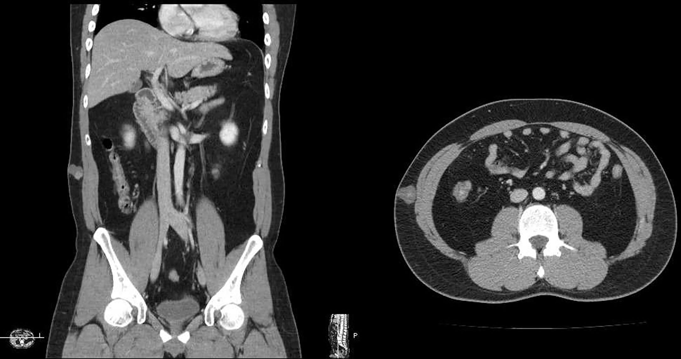
A 36-year-old male visited our clinic with a flank mass that has existed since childhood and has rapidly grown in size over six months. On the computed tomography (CT) scan, a 2.2cm cystic mass in the subcutaneous tissue was observed (Figure 1). An excisional biopsy was performed under the initial clinical impression of epidermoid cyst. Following the pathological diagnosis of malignancy, further wide excision was performed with a 2cm margin from the previous scar and deep to the deep fascia of the external oblique muscle to obtain clear surgical margins.
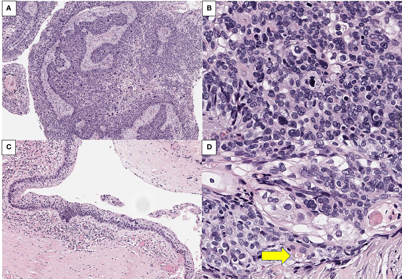
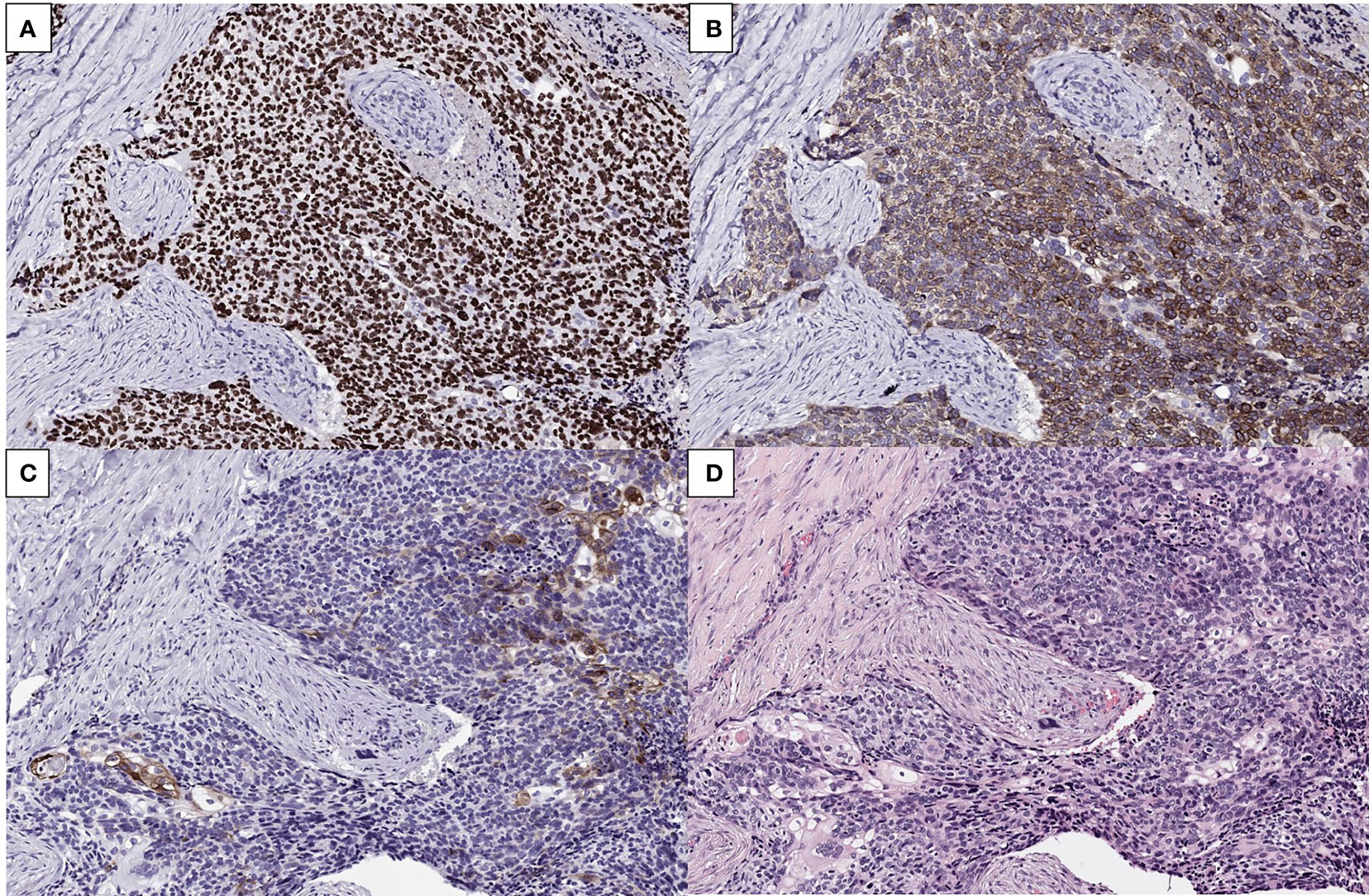
Microscopically, a malignant lesion with an infiltrative margin was observed. The cells had a high nuclear to cytoplasm ratio and often contained clear vacuoles in their cytoplasm. The nuclei showed severe nuclear atypia and partial basaloid features (Figures 2A, B). A lesion corresponding to squamous cell carcinoma in situ was also observed at the edge of the main lesion, but no connection with normal skin epithelium was observed (Figure 2C). In some areas, cells with clear squamous differentiation were observed and cells containing keratin pearl were also noted (Figure 2D). In immunohistochemical staining, the cells of the lesion showed diffuse strong positivity for p63 and bcl-2 while epithelial membrane antigen (EMA) was positive only in well differentiated squamous cells (Figure 3). Taken together, the pathological diagnosis was poorly differentiated carcinoma with squamous and basal differentiation.

Figure 2 Representative H&E images of the tumor. (A) An infiltrative tumor with connection to cystic lining with high grade squamous intraepithelial lesion (x 100). (B) the tumor cells showed severe nuclear atypia and clear, vacuolar cytoplasm (x 800). (C) adjacent squamous cell carcinoma in situ lesion with no direct connection to normal skin epithelium (x 200). (D) cells with clear squamous differentiation and keratin pearl (arrow, x800).

Figure 3 (A, B) Immunohistochemistry showing diffuse strong positivity of the tumor cells for p63 (A) and bcl-2 protein (B) (×200). (C, D) EMA stain revealing focal positivity in well differentiated cells (C) and corresponding H&E image (D) (x200).
As the tumor was situated in the subcutaneous tissue and no direct connection to overlying skin was observed on gross and histological findings, metastatic carcinoma could not be ruled out. Further systemic evaluation including whole-body fluorodeoxyglucose-positron emission tomography (FDG-PET), bronchoscopy, esophagogastroduodenoscopy, and colonoscopy did not reveal any other cancerous lesions. Also, since the flank mass was present since childhood, the possibility of the tumor being a metastatic lesion was very low. Considering the histopathological features, clinical examination results and past medical history, the final diagnosis was likely to be primary poorly differentiated carcinoma with squamous and basal cell differentiation arising from an epidermoid cyst.
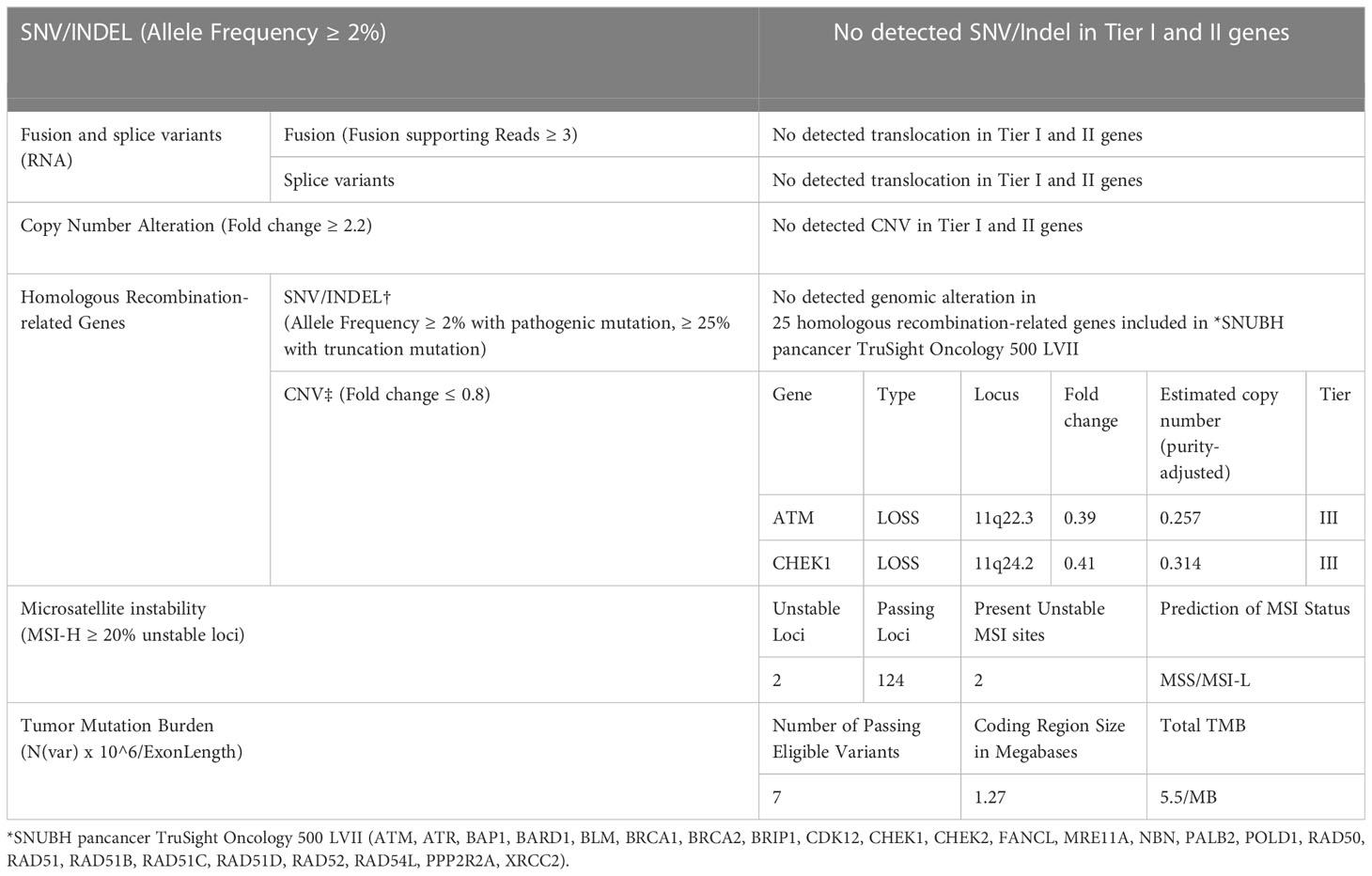
Since TNM staging system is not available for the tumor, NGS using TSO500 assay was performed to determine if additional postoperative treatments such as adjuvant chemotherapy or immunotherapy were applicable (Supplementary Table 1). There was no detected translocation or genomic alteration on Tier I and II genes, but gene fold changes were identified as copy number variation in homologous recombination-related genes ATM and CHEK1. The total tumor mutation burden was 5.5/MB (Table 1). No additional postoperative treatments were performed. No recurrence or metastasis was observed until the two-year follow-up visit.
Discussion
Epidermoid cysts, commonly seen as benign intradermal or subcutaneous tumors, are defined as cutaneous cystic mass appearing on the whole body with epidermis-like epithelial walls (5). Though epidermoid cysts grow slowly and are mostly benign, malignant transformations have rarely been reported. Possible malignant tumors that can develop from epidermoid cysts include SCC, basal cell carcinoma, Paget’s disease, Bowen’s disease, mycosis fungoides, Merkel cell carcinoma, and malignant melanoma. Locations of previously reported malignant transformations include the skin, the intracranial, and the abdominal cavity (3, 4). SCC is the most commonly reported transformation among the abovementioned malignancies, with a rate of 0.011%–0.045% (2, 6).. A cohort study of 41 cases from 1976 to 2018 showed that the most common site of SCC transformation was head and neck (54.8%), and the mean time from occurrence to diagnosis was 92.6 months (7).
The etiology of malignant transformation of the epidermoid cyst has not yet been determined. The risk factors for cutaneous SCC, such as radiation, immunosuppression, scars, and chronic inflammation, are also considered risk factors for this transformation (6). Clinically, pain, fast growth, overlaying skin abnormalities, and continuous drainage are some warning signs of neoplastic progression (7).
In the present case, TSO500 assay was performed to characterize the tumor and assist in determining the need for adjuvant therapy. At our institution, SNV/Indel, fusion or splice variants (RNA), copy number variation(CNV), twenty-five homologous recombination-related genes, microsatellite instability, and tumor mutation burden (TMB) are reported. Interestingly, CNV with fold change below 0.8 of ATM and CHEK1 homologous recombination-related genes were identified in our case.
The activation of ATR-Chk1 and ATM-Chk2 pathways are essential in DNA damage response and cell cycle checkpoints. The ATR-Chk1 pathway, in particular, manages a broad spectrum of DNA abnormalities (8, 9). Furthermore, ATM loss at the cellular level is associated with chromosomal instability and radiosensitivity (10). The activation of ATM and downstream checkpoint kinases Chk1 has a major role in eradicating precancerous cells. The mutations identified in the present case may have promoted tumor growth and cell proliferation (11–13).
Both ATM gene and CHEK1 gene mutations have been found to be related to various cancers through previous studies. ATM gene is one of the most frequently aberrant genes in sporadic cancers, especially hematologic malignancies, according to next-generation sequencing studies (14). Numerous ATM mutations have been identified and linked to a moderate risk of BC occurrence (15). In the case of CHEK1 gene, the association with endometrial and colorectal cancer is known (16), and the association with solid tumor is also being studied (17).
To the best of our knowledge, this is the first report of NGS on carcinoma originating from an epidermoid cyst. Due to the low incidence of such malignant transformations, the mechanism behind it has been difficult to research. With the increased availability of NGS and other sequencing modalities, more data can be collected to elucidate our understanding of these malignant transformations and to assist in finding new adjuvant immunotherapies for these entities.
Data availability statement
The datasets presented in this study can be found in online repositories. The names of the repository/repositories and accession number(s) can be found in the article/Supplementary Material.
Ethics statement
Written informed consent was obtained from the individual(s) for the publication of any potentially identifiable images or data included in this article.
Author contributions
WC, JP, and BK contributed to conception and design of the study. WC wrote the first draft of the manuscript. WC and JP wrote sections of the manuscript. SS selected proper pathologic image and interpreting those images. All authors contributed to the article and approved the submitted version.
Conflict of interest
The authors declare that the research was conducted in the absence of any commercial or financial relationships that could be construed as a potential conflict of interest.
Publisher’s note
All claims expressed in this article are solely those of the authors and do not necessarily represent those of their affiliated organizations, or those of the publisher, the editors and the reviewers. Any product that may be evaluated in this article, or claim that may be made by its manufacturer, is not guaranteed or endorsed by the publisher.
Supplementary material
The Supplementary Material for this article can be found online at: https://www.frontiersin.org/articles/10.3389/fonc.2023.1017624/full#supplementary-material
References
1. Kirkham N, Elder D, Elenitsas R, Johnson B, Murphy G, Lever W. Lever's histopathology of the skin. Chapter. (2005) 29:831.
2. Anton-Badiola I, San Miguel-Fraile P, Peteiro-Cancelo A, Oritz-Rey J. Squamous cell carcinoma arising on an epidermal inclusion cyst: a case presentation and review of the literature. Actas dermo-sifiliograficas. (2010) 101(4):349–53. doi: 10.1016/S1578-2190(10)70646-6
3. Rajan A, Nair A, Srinivasan S. Malignant transformation of retroperitoneal epidermoid cyst into squamous cell carcinoma: A case report. J Radiol Case Rep (2019) 13(3):25–32.
4. Vellutini EA, de Oliveira MF, Ribeiro AP, Rotta JM. Malignant transformation of intracranial epidermoid cyst. Br J Neurosurg (2014) 28(4):507–9. doi: 10.3109/02688697.2013.869552
5. Hoang VT, Trinh CT, Nguyen CH, Chansomphou V, Chansomphou V, Tran TTT. Overview of epidermoid cyst. Eur J Radiol Open (2019) 6:291–301. doi: 10.1016/j.ejro.2019.08.003
6. Lopez-Rios F, Rodriguez-Peralto JL, Castano E, Benito A. Squamous cell carcinoma arising in a cutaneous epidermal cyst: case report and literature review. Am J dermatopathology. (1999) 21(2):174–7. doi: 10.1097/00000372-199904000-00012
7. Frank E, Macias D, Hondorp B, Kerstetter J, Inman JC. Incidental squamous cell carcinoma in an epidermal inclusion cyst: A case report and review of the literature. Case Rep Dermatol (2018) 10(1):61–8. doi: 10.1159/000487794
8. Casper AM, Nghiem P, Arlt MF, Glover TW. ATR regulates fragile site stability. Cell. (2002) 111(6):779–89. doi: 10.1016/S0092-8674(02)01113-3
9. Heffernan TP, Simpson DA, Frank AR, Heinloth AN, Paules RS, Cordeiro-Stone M, et al. An ATR- and Chk1-dependent s checkpoint inhibits replicon initiation following UVC-induced DNA damage. Mol Cell Biol (2002) 22(24):8552–61. doi: 10.1128/MCB.22.24.8552-8561.2002
10. Uhrhammer N, Fritz E, Boyden L, Meyn MS. Human fibroblasts transfected with an ATM antisense vector respond abnormally to ionizing radiation. Int J Mol Med (1999) 4(1):43–7. doi: 10.3892/ijmm.4.1.43
11. Gorgoulis VG, Vassiliou LV, Karakaidos P, Zacharatos P, Kotsinas A, Liloglou T, et al. Activation of the DNA damage checkpoint and genomic instability in human precancerous lesions. Nature. (2005) 434(7035):907–13. doi: 10.1038/nature03485
12. Ismail F, Ikram M, Purdie K, Harwood C, Leigh I, Storey A. Cutaneous squamous cell carcinoma (SCC) and the DNA damage response: pATM expression patterns in pre-malignant and malignant keratinocyte skin lesions. PloS One (2011) 6(7):e21271. doi: 10.1371/journal.pone.0021271
13. Zhang Y, Hunter T. Roles of Chk1 in cell biology and cancer therapy. Int J cancer. (2014) 134(5):1013–23. doi: 10.1002/ijc.28226
14. Forbes SA, Beare D, Gunasekaran P, Leung K, Bindal N, Boutselakis H, et al. COSMIC: exploring the world's knowledge of somatic mutations in human cancer. Nucleic Acids Res (2015) 43(Database issue):D805–11. doi: 10.1093/nar/gku1075
15. Rotman G, Shiloh Y. ATM: from gene to function. Hum Mol Genet (1998) 7(10):1555–63. doi: 10.1093/hmg/7.10.1555
16. Bertoni F, Codegoni AM, Furlan D, Tibiletti MG, Capella C, Broggini M. CHK1 frameshift mutations in genetically unstable colorectal and endometrial cancers. Genes Chromosomes cancer. (1999) 26(2):176–80. doi: 10.1002/(SICI)1098-2264(199910)26:2<176::AID-GCC11>3.0.CO;2-3
Keywords: epidermoid cyst, squamous epithelium, next-generation sequencing, DNA copy number variation, malignant transformation
Citation: Choi W, Park JK-h, Song SG and Kim B-k (2023) Next-generation sequencing study on poorly differentiated carcinoma derived from a thirty-year-old epidermoid cyst: A case report. Front. Oncol. 13:1017624. doi: 10.3389/fonc.2023.1017624
Received: 24 August 2022; Accepted: 21 March 2023;
Published: 03 April 2023.
Edited by:
Omer Faruk Bayrak, Yeditepe University, TürkiyeReviewed by:
Ana Paula Nunes Alves, Federal University of Ceara, BrazilJoshua Aaron Cuoco, Carilion Clinic, United States
Copyright © 2023 Choi, Park, Song and Kim. This is an open-access article distributed under the terms of the Creative Commons Attribution License (CC BY). The use, distribution or reproduction in other forums is permitted, provided the original author(s) and the copyright owner(s) are credited and that the original publication in this journal is cited, in accordance with accepted academic practice. No use, distribution or reproduction is permitted which does not comply with these terms.
*Correspondence: Joseph Kyu-hyung Park, am9zZXBocGFyazg1QGdtYWlsLmNvbQ==; Baek-kyu Kim, cGxhc3JlY29uQHNudWJoLm9yZw==
†These authors have contributed equally to this work and share first authorship
 Woosuk Choi
 Joseph Kyu-hyung Park
 Seung Geun Song2†
