- 1Department of Oncology-Pathology, Karolinska Institutet, Stockholm, Sweden
- 2Breast Center, Theme Cancer, Karolinska University Hospital, Stockholm, Sweden
- 3Department of Oncology, Faculty of Medicine and Health, Örebro University, Örebro, Sweden
Purpose: High levels of tumor-infiltrating lymphocytes (TILs) are associated with better outcomes in early breast cancer and higher pathological response rates to neoadjuvant chemotherapy especially in the triple-negative (TNBC) and HER2+ subtypes. However, the dynamic changes in TILs levels after neoadjuvant treatment (NAT) are less studied. This systematic review and meta-analysis aimed to investigate the patterns and role of TILs dynamics change in early breast cancer patients receiving NAT.
Methods: Medline, Embase, Web of Science Core Collection and PubMed Central databases were searched for eligible studies. Data were extracted independently by two researchers and discordances were resolved by a third. Pooled TILs rates pre- & post-treatment (overall and per subtype), pooled rates of ΔTILs and direction of change after NAT as well as correlation of ΔTILs with survival outcomes were generated in the outcome analysis.
Results: Of 2116 identified entries, 34 studies fulfilled the criteria and provided adequate data for the outcomes of interest. A decreased level of TILs was observed after NAT in paired samples across all subtypes. The effect of NAT on TILs was most prominent in TNBC subtype with a substantial change, either increase or decrease, in 79.3% (95% CI 61.7-92.6%) of the patients as well as in HER2+ disease (14.4% increased vs 46.2% decreased). An increase in ΔTILs in TNBC was associated with better disease-free/relapse-free survival in pooled analysis (univariate HR = 0.59, 95% CI: 0.37–0.95, p = 0.03).
Conclusion: This meta-analysis illustrates the TILs dynamics during NAT for breast cancer and indicates prognostic implications of ΔTILs in TNBC. The potential clinical utility of the longitudinal assessment of TILs during neoadjuvant therapy warrants further validation.
Introduction
Breast cancer (BC) is the most frequently diagnosed cancer and the leading cause of cancer-related death among women worldwide (1). While neoadjuvant and adjuvant chemotherapy clearly improve patient outcomes, clinical-pathologic factors and available gene signatures failed to demonstrate validated predictive value for chemotherapy benefit (2, 3). We have previously shown that immune-related gene expression is both prognostic and predictive for chemotherapy benefit in early and advanced BC (4–8). However, using immune gene signatures in the clinical routine is complex due to the lack of standardized and prospectively validated methods and the lack of estimations on potential health impact and costs.
A simple-to-use and widely available immune biomarker is the number of tumor-infiltrating lymphocytes (TILs) on hematoxylin eosin (H&E) stained tissue sections. It has been previously described that high TIL infiltration at diagnosis was strongly associated with a better response to neoadjuvant chemotherapy (NACT) (9). Several subsequent studies reported the positive predictive and prognostic value of primary TILs both in the neoadjuvant and adjuvant setting, especially for TNBC and HER2+ tumors (10–12). In order to mitigate interobserver variability, the International Immuno-Oncology Biomarker Working Group has established guidelines for the standardized evaluation of TILs (13, 14). The latest edition of the WHO classification of tumors has introduced TILs as an important prognostic marker (15) whereas some currently on-going prospective trials include TILs as a pre-specified stratification factor in TNBC and HER2+ patients receiving neoadjuvant treatment (16, 17).
The current evidence on TILs is mainly based on a cross sectional evaluation where the level of TILs is assessed once, usually before any systemic treatment is administered. However, a dynamic, longitudinal evaluation of immunological markers may give us better understanding of the mechanisms that govern the host response to tumor and be a potential source of clinically useful biomarkers. Some studies have investigated both pre-treatment and post-treatment TILs in paired tissues during neoadjuvant chemotherapy and the association of TILs change with prognosis, with conflicting results.
The aim of the present meta-analysis was to gather the current evidence on TIL dynamics following neoadjuvant therapy and investigate the magnitude and direction of TILs changes as well as their correlation with therapy prediction and survival outcomes.
Methods
Search strategy and study selection
A comprehensive literature search was conducted by the Karolinska Institutet University Library in May 2020 and updated in September 2021. The following four databases were searched: Medline (Ovid), Embase (embase.com), Web of Science Core Collection and PubMed Central. The MeSH (Medical Subject Headings) terms used were: Breast Neoplasms, Lymphocytes, Tumor-Infiltrating, CD3 Complex, Neoadjuvant Therapy, Chemotherapy, Adjuvant. The MeSH terms for searching Medline (Ovid) were adapted in accordance with corresponding vocabulary in Embase. Databases were searched from inception. The detailed search strategies are provided in Supplementary Data.
Studies included in our meta-analysis were restricted to English and fulfilled at least one of the following criteria (1): Stromal TILs evaluated in paired human breast cancer tumor samples before and after neoadjuvant chemotherapy, targeted and/or endocrine therapy (2); TILs evaluated in paired human breast cancer tumor samples before and during neoadjuvant chemotherapy targeted and/or endocrine therapy; (3) relationship between ΔTILs levels and short-/long-term prognosis in non-pCR and pCR cases; (4) relationship between ΔTILs levels and pCR (for pCR patient cases, TILs were measured on tissue scar or tumor bed area). TILs could have been reported as continuous or categorical variables and assessed on H&E slides, regardless of methods used, including manual evaluation or digital image analysis. If both intra-tumoral and stromal TILs were evaluated, only stromal TILs information was included for analysis. ΔTILs is defined as change in median/mean lymphocyte density between pre- and post-treatment samples; ΔTILs was either reported in the articles or calculated manually in articles with relevant data.
Studies were excluded if they met at least one of the following criteria: (1) reviews, commentaries, editorials, conference abstracts, protocols, case reports, qualitative research, or letters; (2) duplicate publications/entries; (3) full text not published in English. Study selection was performed independently by two investigators (Y. Zhu and E. Tzoras) and consensus was reached in all eligible studies.
Data extraction
Two investigators (Y. Zhu and E. Tzoras) independently extracted the data to a predefined form and a third investigator (I. Zerdes) resolved any discrepancies. The concordance rate between the two investigators was 86%. Data collected from each study included: first author’s last name, journal name, year of publication, country where the study was conducted, type of study (retrospective/prospective), enrolment dates, number of evaluable patients before NAT, number of evaluable patients after NAT, number of patients with matched- paired samples, tissue used for analysis (tissue microarrays, whole-tissue sections), method used for analysis, threshold for positivity/high expression of stromal TILs, median/mean TILs level before NAT, median/mean TILs level after NAT, ΔTILs mean change, absolute number of patients with increased/decreased/unchanged TILs, % TILs in matched pre- and post-NAT samples and change-ΔTILs if reported, characteristics of study cohort, follow-up time; outcomes (pCR and time-to-event endpoints) within all patients and whenever possible within different breast cancer subtypes including both univariate and multivariate results.
Quality assessment
Two investigators (Y. Zhu and E. Tzoras) independently assessed each eligible study for methodological quality using the 20-item REMARK checklist (18) and the discrepancies were resolved by a third investigator (I. Zerdes). The REMARK checklist consists of 20 items to report in tumor marker prognostic studies evaluating several aspects of study quality from scientific rationale and result interpretation to study design and methodology used. Each of the 20 items listed in REMARK was scored with 0 (not defined or inadequate defined or not applicable), 1 (incomplete or unclear defined), or 2 (clearly defined) for each eligible study, with a maximum score of 40. No studies were excluded based on quality control.
Statistical analysis
High and low TILs were defined according to cut-offs described in each article for articles reported TILs as a categorical variable.
For analyses of pooled expression of TILs in matched breast cancer patients in studies presented TILs as categorical variable, a random-effects model was used to calculate the pooled high-level TILs and corresponding 95% confidence interval (CI) pre- vs. post-treatment for different breast cancer subtypes (HER2-positive, TNBC, luminal, not specified [contain studies recruit all BC patients without limitation of molecular subtype]). An overall effect estimate was thereafter calculated using Odds Ratio (OR) with 95% CI through the DerSimonian and Laird method (19).
For pooled analyses of difference in TILs expression pre- vs. post-treatment when TILs were presented as continuous variables, standardized mean differences (SMD) with 95% CI were calculated for each study and then pooled to present a measure of the effect size of the difference in TILs in pre- and post- treatment groups.
For the comparisons of time-to-event variables based on the direction of TILs changes, a meta-analysis was performed first by transforming the Hazard ratios (HRs) and their errors into their log counterparts, and then using the inverse variance method for transforming back into the HR scale. If adequate data from time-to-event variables were unavailable for direct extraction from the primary studies, data were extracted according to the method described by Tierney et al. (20). A pooled analysis was performed only if at least three primary studies presented adequate data for analyses.
The presence of statistical heterogeneity among the studies was addressed by using the Q statistics, and the magnitude of heterogeneity by using the I² statistic. A p-value < 0.10 or a I² value of greater than 50% was considered as substantial statistical heterogeneity. Considering the substantial clinical heterogeneity among eligible studies, all meta-analyses except the one with time-to-event variable as outcome of interest were performed using random-effects models. The presence of publication bias was evaluated qualitatively using a funnel plot.
All reported p values are two sided. Analyses were conducted on RevMan 5.3 (Review Manager, Version 5.3; The Cochrane Collaboration, 2014) and on StatsDirect (StatsDirect Ltd. StatsDirect statistical software. http://www.statsdirect.com. England: StatsDirect Ltd. 2013).
Results
Study characteristics
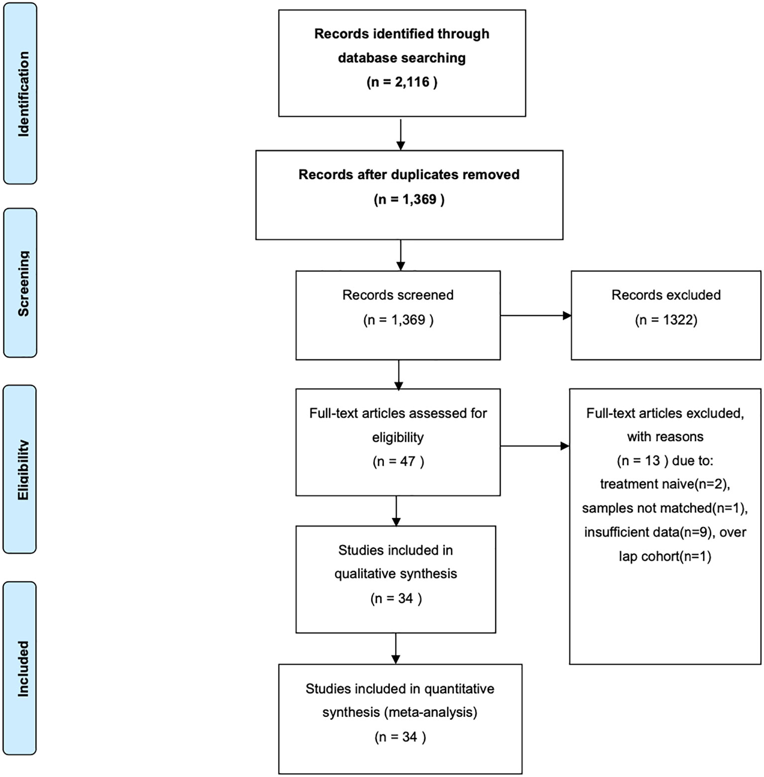
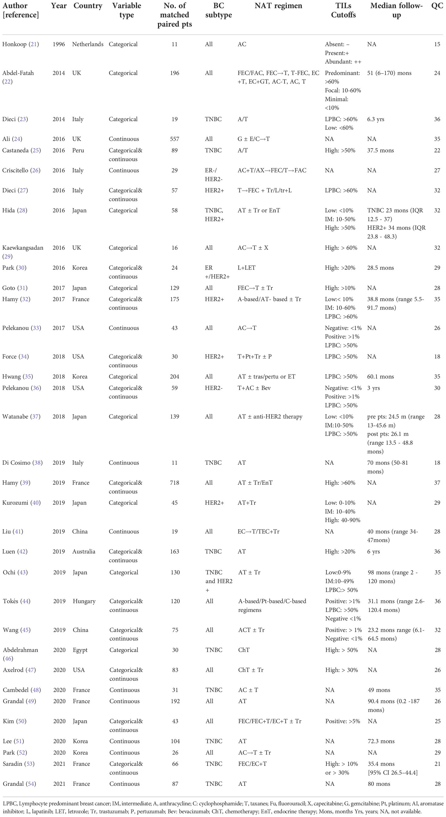
The flow diagram of study selection for the study-level meta-analysis is shown in Figure 1. The initial search identified 2,116 entries, or 1,369 entries following deduplication. Through exclusion by reading the title and/or abstract, 47 possibly eligible studies were retrieved as full text; In total, 34 studies fulfilled the inclusion criteria and were included for various meta-analytic questions. All 34 studies were included for pooled pre- and post-TILs change direction analysis (separate analysis for TILs as categorical variable [14 studies] and continuous variable [21 studies]); 26 studies reporting matched paired breast cancer patients were included for pooled rates of ΔTILs. 4 studies reported survival information and were included for pooled HR analysis of ΔTILs and prognosis association. The detailed characteristics of eligible studies are presented in Table 1.
Quality of eligible studies, between-study heterogeneity and assessment of publication bias
All eligible studies for the meta-analysis were retrospective. The median number of study quality score was 29 (range: 15-37) out of a maximum score of 40. Substantial between-study heterogeneity was noted among eligible studies regarding the breast cancer subtypes, treatment regimens used, variable types used to report TILs level, and the follow-up period. The risk of publication bias for the pooled estimates was visually assessed by funnel plots. With reservation due the low number of primary studies in some pooled estimates, no evidence of asymmetry was observed in funnel plots implying a lower risk for publication bias (Supplementary Figure 1).
Pooled TILs expression before and after neoadjuvant treatment
The number of studies and patient cases with available information on TILs as categorical variable across BC subtypes, as well as the pooled rates of high-level TILs are presented in Table 2. The proportion of cases classified as high-level TILs decreased post-treatment across BC subtypes, although no pooled analysis was possible for the Luminal subtype due to the low number of studies (Not specified: pooled OR [95% CI] = 1.60 [95% CI: 1.12-2.30]; HER2-positive: pooled OR [95% CI] =1.88 [0.87-4.08); TNBC: pooled OR [95% CI] =1.05 [0.41-2.68]. Difference in pooled rates of TILs pre- vs. post-treatment was statistically significant for the “not specified” subgroup.

Table 2 Pooled expression of TILs pre- vs. post-treatment in matched breast cancer patients in studies presented TILs as categorical variable.
Furthermore, twenty-one studies reported TILs as continuous variable. Number of studies, cases, pooled standardized mean difference (SMD) and I2 in four subgroups were summarized in Supplementary Table 1. Positive SMD values were seen in the HER2+, TNBC and not specific subgroups while no pooled analysis was done for the Luminal subtype due to only two studies have available data. Forest plots on pooled SMD pre- and post-treatment in studies with each BC subtype are shown in Figures 2A–C. Although the magnitude of pooled effect sizes is not statistically significant, numerically higher TILs expression at pre-treatment compared to post-treatment was seen in all three subgroups.

Figure 2 Forest-plots on Standardized mean difference (SMD) of Tumor-Infiltrating Lymphocytes (TILs) pre- and post-treatment per breast cancer subtype (A) not specified (B) HER2-positive (C) Triple-negative breast cancer [TNBC] and (D) Disease-free survival [DFS]/Recurrence-free survival [RFS] according to TILs change in TNBC subtype.
Pooled ΔTILs rates after neoadjuvant therapy in early BC patients
Studies evaluating ΔTILs in matched pre-treatment and surgical samples from the same patients were also included in this analysis from all/unspecified (10 studies; n=1758), luminal (1 study; n=106), HER2-positive (6 studies; n=414), TNBC (9 studies; n=483) subtypes. Change in TILs following neoadjuvant therapy was bi-directional mainly in TNBC cases, whereas TILs mostly decreased post-therapy in the mixed and HER2-positive populations (Table 3).

Table 3 Pooled rates of ΔTILs before and after NAT in matched-paired tissue samples in early breast cancer patients.
Prognostic implications of TILs change after neoadjuvant therapy
Pooled HRs from univariate analyses for disease-free survival (DFS) or Recurrence-free survival (RFS) for TNBC patients from 4 eligible studies are presented in Figure 2D. For this outcome, we considered different definitions of DFS or RFS as similar and analyzed within the same meta-analysis. Two studies defined RFS as the time from the date of primary surgery until the date of disease recurrence (31, 47), one study defined RFS as time from diagnosis to locoregional recurrence, distant metastasis, or death from any cause (43) whereas no clear definition was described in one study (51). Increased ΔTILs was associated with better DFS/RFS with a pooled HR of 0.59 (95% CI: 0.37 – 0.95, p = 0.03). Because of data paucity, meta-analysis in other BC subtypes, or pooled HR from multivariate analyses, could not be performed.
Studies assessing on-treatment TILs and correlation with pCR
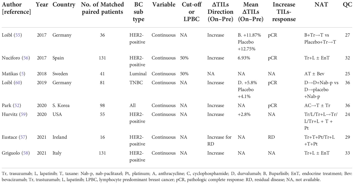
Eight prospective studies retrospectively assessed TILs before and during neoadjuvant treatment were identified from the systematic literature review. Within these 8 studies, 5 included HER2-positive breast cancer patients that received HER2-targeted therapy with or without chemotherapy (55–59), 1 study included TNBC patients who received combination of immunotherapy and chemotherapy (60), 1 study included hormone receptor positive breast cancer patients received chemotherapy combined with anti-angiogenesis therapy (5) and 1 study included non-specific patients received chemotherapy with or without HER2-trageted therapy (52) (Table 4). On-treatment TILs counts uniformly increased compared with baseline status. With the exception of one study (57), increased TILs between pre- and on-treatment biopsies were positively associated with pCR status. Pooled analyses were not possible due to inadequate number of studies per breast cancer subtype and heterogeneity among the eligible studies.

Table 4 Studies assessed the expression of on-treatment TILs in matched-paired early breast cancer patients receiving neoadjuvant therapy.
Discussion
This meta-analysis summarizes the current evidence on pooled TILs levels in matched paired tissues before and after NAT in breast cancer patients and presents data related to dynamic changes of TILs during NAT. Higher TILs expression at pre-treatment compared to post-treatment was seen across all BC subgroups with consistent results in studies reported TILs as categorical or continuous variable types, with a more distinct decreasing trend seen in HER2-positive subgroup. By pooling data from around 450 TNBC patients, we also reported a positive correlation of increased ΔTILs with improved survival, though confounding bias cannot be excluded.
Our study provides some interesting insights on how TILs could be potentially used to better optimize neoadjuvant treatment mainly for TNBC and HER2+ patients.
First, a trend towards decreased TILs after NAT was observed in all pooled analyses irrespectively breast cancer subtype. Although this trend is small and not statistically significant in most of the analyses, the consistency of the decreased trend across all breast cancer subtypes implies a potential true effect. Since all included studies except for one (30) used neoadjuvant regimens containing at least one chemotherapeutic agent, the decreased TILs seen in our findings may be driven by the treatment effect of cytotoxic chemotherapy, which is generally considered to be immunosuppressive (61). Considering the diversity of chemotherapeutic agents used in eligible studies, no firm conclusion can be made on how different chemotherapeutic agents could affect immune response. In fact, some recent studies suggest that different chemotherapeutic agents might have distinct effect on immune cell surface marker expression (62) whereas some third-generation cytotoxic drugs such as pemetrexed can potentiate immunogenic tumor cell death and enhance T cell–mediated immunity in mice models (63).
Notably, the magnitude of decreased TILs seemed to be numerically larger in HER2-positive breast cancer implying a potential synergistic interaction between HER2-targeted therapy and chemotherapy regarding pattern of TIL changes over time. However, the variation in treatment combinations across eligible studies and the complex interplay between immune system and tumor in HER2-positive breast cancer preclude any firm conclusion.
Second, a potential prognostic role of dynamic TILs changes in patients with TNBC was seen. In fact, increased TILs during NACT seemed to be associated with better prognosis. Although this pooled analysis is prone to confounding bias since it was based on results from univariate rather than multivariate analyses, these findings trigger some interesting hypotheses. Currently, the presence of residual disease after NACT is the only well-established approach to optimize postoperative treatment strategy in patients with TNBC. Recently, TILs have been confirmed as having a strong prognostic value in early TNBC patients treated with chemotherapy (64)but also in early TNBC patients without chemotherapy where high TILs could identify a subset of patients with an excellent prognosis able to de-escalation strategies (65). According to our findings, increased ΔTILs after NACT might serve as an additional potential biomarker for de-escalation by defining a subgroup of patients with better prognosis and should be further validated in future studies.
Our meta-analysis has several limitations that should be considered when interpreting the results. First, all studies were retrospective with limited sample size, thus influencing the quality of current evidence and the generalizability of our findings. Second, all our pooled analyses were based on study-level results rather on individual patient data. Another limitation that deserves attention is the lack of current methodological standards for post-NAC TILs enumeration in residual cancers and pCR tumor specimens which is a source of potential methodological heterogeneity among eligible studies. In addition, TILs after NACT were counted only on residual disease in some studies whereas other counted TILs even in stroma from patients with pCR. Furthermore, some studies used TMAs that only represent small portion of tissue, which might introduce bias in heterogeneous tumors. Another source of heterogeneity among eligible studies was the various therapeutic regimens used as NACT. Considering the high between-study heterogeneity, we actively chose to use random effects model for pooled analyses as an effort to reduce the impact of heterogeneity on the pooled analyses.
Despite these caveats, our meta-analysis offers some new insights on the potential role of dynamic TILs changes after NACT in breast cancer patients that might be of clinical significance upon confirmation in further studies. In summary, we found a decreased trend in TILs through all BC subtypes after neoadjuvant treatment that might be more evident in HER2-positive breast cancer. Increased TILs might be of prognostic significance in patients with TNBC and might serve as a biomarker to identify patients with better prognosis where de-escalation strategies might be applied. Overall, dynamic TILs change evaluation on hematoxylin–eosin slides might perform as a versatile and cost-effective biomarker for breast cancer patients, specifically for HER2-positive and TNBC patients. Establishing international methodological standards on how TILs should be evaluated in residual disease and in surgical specimens with pCR is essential to be able to further validate the potential role of dynamic TILs changes after neoadjuvant therapy in future studies.
Dynamic evaluation of TILs may be of particular interest in patients with early-stage TNBC who are treated with neoadjuvant chemotherapy combined with immune checkpoint inhibitors (ICIs). The response to PD(L)-1 inhibition seems independent of PD-L1 status in the neoadjuvant setting (66), while in the GeparNuevo trial, TILs at baseline were predictive for pCR in TNBC patients receiving neoadjuvant chemotherapy with or without durvalumab, with no significant interaction between TILs and treatment arms (67). Specific chemotherapeutics may induce a stronger immunogenicity (68) and additionally, immunotherapy can induce the migration of TILs from stroma to tumor nests (67). Longitudinal evaluation of TILs might stand as an easy method to help better understand the interactions between cytotoxic and ICI and guide their combination and sequence, if prognostic correlations can be demonstrated. Further evaluating the relative proportion of specific immune subpopulations as well as the spatial organization of the immune infiltrate, including tertiary lymphoid structures may also add to the information provided by TILs enumeration and could be the future focus of the research field.
Data availability statement
The original contributions presented in the study are included in the article/Supplementary Material. Further inquiries can be directed to the corresponding author.
Author contributions
Conception and design: TF, IZ, AM, YZ; Acquisition of data: IZ, ET, YZ; Data analysis: AV, IZ, ET, YZ; Writing, review, and/or revision of the manuscript: all authors; Study supervision: IZ, AV, TF. All authors contributed to the article and approved the submitted version.
Funding
This work was supported by grants from; the Swedish Cancer Society (grant number CAN 2018/846 and Senior Clinical Investigator award CAN 2017/1043) and the Cancer Society in Stockholm (174113) to TF; by a grant from the Swedish Breast Cancer Association to TF and IZ; AM is supported by the Swedish Cancer Society (Junior Clinical Investigator award, grant number 21 0277 JCIA 01); JB’s research group receives funding from the Stockholm region, the Swedish Cancer Society, the funds at Radiumhemmet, the Swedish Research Council, the Knut and Alice Wallenberg fund.
Acknowledgments
We would like to thank Carl Gornitzki, Emma-Lotta Säätelä and Sabina Gillsund, librarians at Karolinska Institutet University Library, for their assistance in the search design. Part of this study was presented (online publication-only) at the American Society of Clinical Oncology (ASCO) Annual Meeting 2022.
Conflict of interest
JB receives research funding from Merck paid to Karolinska Institutet and from Amgen, Bayer, Pfizer, Roche and Sanofi-Aventis paid to Karolinska University Hospital. No personal payments. Payment from UpToDate for a chapter in breast cancer prediction paid to Asklepios Medicine HB. TF: institutional research grants from Roche and Pfizer, institutional fees from Roche, Pfizer and Astra Zeneca and personal fees from Affibody, Novartis, Pfizer, Roche, Exact Sciences, Veracyte and UpToDate. AM: consultancy to Veracyte no financial or other compensation, AV: institutional research grant from Roche.
The remaining authors declare that the research was conducted in the absence of any commercial or financial relationships that could be construed as a potential conflict of interest.
Publisher’s note
All claims expressed in this article are solely those of the authors and do not necessarily represent those of their affiliated organizations, or those of the publisher, the editors and the reviewers. Any product that may be evaluated in this article, or claim that may be made by its manufacturer, is not guaranteed or endorsed by the publisher.
Supplementary material
The Supplementary Material for this article can be found online at: https://www.frontiersin.org/articles/10.3389/fonc.2022.999843/full#supplementary-material
References
1. Sung H, Ferlay J, Siegel RL, Laversanne M, Soerjomataram I, Jemal A, et al. Global cancer statistics 2020: Globocan estimates of incidence and mortality worldwide for 36 cancers in 185 countries. CA Cancer J Clin (2021) 71(3):209–49. doi: 10.3322/caac.21660
2. Group EBCTC. Comparisons between different polychemotherapy regimens for early breast cancer: Meta-analyses of long-term outcome among 100 000 women in 123 randomised trials. Lancet (2012) 379(9814):432–44. doi: 10.1016/S0140-6736(11)61625-5
3. Kalinsky K, Barlow WE, Gralow JR, Meric-Bernstam F, Albain KS, Hayes DF, et al. 21-gene assay to inform chemotherapy benefit in node-positive breast cancer. New Engl J Med (2021) 385(25):2336–47. doi: 10.1056/NEJMoa2108873
4. Foukakis T, Lovrot J, Matikas A, Zerdes I, Lorent J, Tobin N, et al. Immune gene expression and response to chemotherapy in advanced breast cancer. Br J Cancer (2018) 118(4):480–8. doi: 10.1038/bjc.2017.446
5. Matikas A, Lövrot J, Ramberg A, Eriksson M, Lindsten T, Lekberg T, et al. Dynamic evaluation of the immune infiltrate and immune function genes as predictive markers for neoadjuvant chemotherapy in hormone receptor positive, Her2 negative breast cancer. Oncoimmunology (2018) 7(9):e1466017. doi: 10.1080/2162402x.2018.1466017
6. Matikas A, Zerdes I, Lovrot J, Richard F, Sotiriou C, Bergh J, et al. Prognostic implications of pd-L1 expression in breast cancer: Systematic review and meta-analysis of immunohistochemistry and pooled analysis of transcriptomic data. Clin Cancer Res (2019) 25(18):5717–26. doi: 10.1158/1078-0432.Ccr-19-1131
7. Matikas A, Zerdes I, Lovrot J, Sifakis E, Richard F, Sotiriou C, et al. Pd-1 protein and gene expression as prognostic factors in early breast cancer. ESMO Open (2020) 5(6):e001032. doi: 10.1136/esmoopen-2020-001032
8. Zerdes I, Sifakis EG, Matikas A, Chretien S, Tobin NP, Hartman J, et al. Programmed death-ligand 1 gene expression is a prognostic marker in early breast cancer and provides additional prognostic value to 21-gene and 70-gene signatures in estrogen receptor-positive disease. Mol Oncol (2020) 14(5):951–63. doi: 10.1002/1878-0261.12654
9. Denkert C, Loibl S, Noske A, Roller M, Muller BM, Komor M, et al. Tumor-associated lymphocytes as an independent predictor of response to neoadjuvant chemotherapy in breast cancer. J Clin Oncol (2010) 28(1):105–13. doi: 10.1200/JCO.2009.23.7370
10. Adams S, Gray RJ, Demaria S, Goldstein L, Perez EA, Shulman LN, et al. Prognostic value of tumor-infiltrating lymphocytes in triple-negative breast cancers from two phase iii randomized adjuvant breast cancer trials: Ecog 2197 and ecog 1199. J Clin Oncol (2014) 32(27):2959–66. doi: 10.1200/JCO.2013.55.0491
11. Denkert C, von Minckwitz G, Darb-Esfahani S, Lederer B, Heppner BI, Weber KE, et al. Tumour-infiltrating lymphocytes and prognosis in different subtypes of breast cancer: A pooled analysis of 3771 patients treated with neoadjuvant therapy. Lancet Oncol (2018) 19(1):40–50. doi: 10.1016/s1470-2045(17)30904-x
12. Loi S, Sirtaine N, Piette F, Salgado R, Viale G, Van Eenoo F, et al. Prognostic and predictive value of tumor-infiltrating lymphocytes in a phase iii randomized adjuvant breast cancer trial in node-positive breast cancer comparing the addition of docetaxel to doxorubicin with doxorubicin-based chemotherapy: Big 02-98. J Clin Oncol (2013) 31(7):860–7. doi: 10.1200/JCO.2011.41.0902
13. Salgado R, Denkert C, Demaria S, Sirtaine N, Klauschen F, Pruneri G, et al. The evaluation of tumor-infiltrating lymphocytes (Tils) in breast cancer: Recommendations by an international tils working group 2014. Ann Oncol (2015) 26(2):259–71. doi: 10.1093/annonc/mdu450
14. Hendry S, Salgado R, Gevaert T, Russell PA, John T, Thapa B, et al. Assessing tumor infiltrating lymphocytes in solid tumors: A practical review for pathologists and proposal for a standardized method from the international immuno-oncology biomarkers working group: Part 2: Tils in melanoma, gastrointestinal tract carcinomas, non-small cell lung carcinoma and mesothelioma, endometrial and ovarian carcinomas, squamous cell carcinoma of the head and neck, genitourinary carcinomas, and primary brain tumors. Adv anatomic Pathol (2017) 24(6):311.
15. Board WCoTE. Who classification of breast tumours: Who classification of tumours, volume 2. World Health Organization (2019).
16. Lusho S, Durando X, Bidet Y, Molnar I, Kossai M, Bernadach M, et al. Perception trial protocol: Comparison of predictive and prognostic capacities of neutrophil, lymphocyte, and platelet counts and tumor-infiltrating lymphocytes in triple negative breast cancer. Medicine (2020) 99(50).
18. Altman DG, McShane LM, Sauerbrei W, Taube SE. Reporting recommendations for tumor marker prognostic studies (Remark): Explanation and elaboration. BMC Med (2012) 10(1):1–39.
19. DerSimonian R, Laird N. Meta-analysis in clinical trials. Controlled Clin trials (1986) 7(3):177–88. doi: 10.1016/0197-2456(86)90046-2
20. Tierney JF, Stewart LA, Ghersi D, Burdett S, Sydes MR. Practical methods for incorporating summary time-to-Event data into meta-analysis. Trials (2007) 8(1):1–16. doi: 10.1186/1745-6215-8-16
21. Honkoop AH. Effects of chemotherapy on pathologic and biologic characteristics of locally advanced breast cancer. (1996).
22. Abdel-Fatah TM, McArdle SE, Johnson C, Moseley PM, Ball GR, Pockley AG, et al. Hage (Ddx43) is a biomarker for poor prognosis and a predictor of chemotherapy response in breast cancer. Br J Cancer (2014) 110(10):2450–61. doi: 10.1038/bjc.2014.168
23. Dieci MV, Criscitiello C, Goubar A, Viale G, Conte P, Guarneri V, et al. Prognostic value of tumor-infiltrating lymphocytes on residual disease after primary chemotherapy for triple-negative breast cancer: A retrospective multicenter study. Ann Oncol (2014) 25(3):611–8. doi: 10.1093/annonc/mdt556
24. Ali HR, Dariush A, Provenzano E, Bardwell H, Abraham JE, Iddawela M, et al. Computational pathology of pre-treatment biopsies identifies lymphocyte density as a predictor of response to neoadjuvant chemotherapy in breast cancer. Breast Cancer Res (2016) 18(1):21. doi: 10.1186/s13058-016-0682-8
25. Castaneda CA, Mittendorf E, Casavilca S, Wu Y, Castillo M, Arboleda P, et al. Tumor infiltrating lymphocytes in triple negative breast cancer receiving neoadjuvant chemotherapy. World J Clin Oncol (2016) 7(5):387–94. doi: 10.5306/wjco.v7.i5.387
26. Criscitiello C, Bayar MA, Curigliano G, Symmans FW, Desmedt C, Bonnefoi H, et al. A gene signature to predict high tumor-infiltrating lymphocytes after neoadjuvant chemotherapy and outcome in patients with triple-negative breast cancer. Ann Oncol (2018) 29(1):162–9. doi: 10.1093/annonc/mdx691
27. Dieci MV, Prat A, Tagliafico E, Pare L, Ficarra G, Bisagni G, et al. Integrated evaluation of Pam50 subtypes and immune modulation of pcr in Her2-positive breast cancer patients treated with chemotherapy and Her2-targeted agents in the cherlob trial. Ann Oncol (2016) 27(10):1867–73. doi: 10.1093/annonc/mdw262
28. Hida AI, Sagara Y, Yotsumoto D, Kanemitsu S, Kawano J, Baba S, et al. Prognostic and predictive impacts of tumor-infiltrating lymphocytes differ between triple-negative and Her2-positive breast cancers treated with standard systemic therapies. Breast Cancer Res Treat (2016) 158(1):1–9. doi: 10.1007/s10549-016-3848-2
29. Kaewkangsadan V, Verma C, Eremin JM, Cowley G, Ilyas M, Eremin O. Crucial contributions by T lymphocytes (Effector, regulatory, and checkpoint inhibitor) and cytokines (Th1, Th2, and Th17) to a pathological complete response induced by neoadjuvant chemotherapy in women with breast cancer. J Immunol Res (2016) 2016:4757405. doi: 10.1155/2016/4757405
30. Park JH, Kang MJ, Ahn JH, Kim JE, Jung KH, Gong G, et al. Phase ii trial of neoadjuvant letrozole and lapatinib in Asian postmenopausal women with estrogen receptor (Er) and human epidermal growth factor receptor 2 (Her2)-positive breast cancer [Neo-all-in]: Highlighting the tils, er expressional change after neoadjuvant treatment, and fes-pet as potential significant biomarkers. Cancer Chemother Pharmacol (2016) 78(4):685–95. doi: 10.1007/s00280-016-3107-6
31. Goto W, Kashiwagi S, Asano Y, Takada K, Takahashi K, Hatano T, et al. Predictive value of improvement in the immune tumour microenvironment in patients with breast cancer treated with neoadjuvant chemotherapy. ESMO Open (2018) 3(6):e000305. doi: 10.1136/esmoopen-2017-000305
32. Hamy AS, Pierga JY, Sabaila A, Laas E, Bonsang-Kitzis H, Laurent C, et al. Stromal lymphocyte infiltration after neoadjuvant chemotherapy is associated with aggressive residual disease and lower disease-free survival in Her2-positive breast cancer. Ann Oncol (2017) 28(9):2233–40. doi: 10.1093/annonc/mdx309
33. Pelekanou V, Carvajal-Hausdorf DE, Altan M, Wasserman B, Carvajal-Hausdorf C, Wimberly H, et al. Effect of neoadjuvant chemotherapy on tumor-infiltrating lymphocytes and pd-L1 expression in breast cancer and its clinical significance. Breast Cancer Res (2017) 19(1):91. doi: 10.1186/s13058-017-0884-8
34. Force J, Howie LJ, Abbott SE, Bentley R, Marcom PK, Kimmick G, et al. Early stage Her2-positive breast cancers not achieving a pcr from neoadjuvant trastuzumab- or pertuzumab-based regimens have an immunosuppressive phenotype. Clin Breast Cancer (2018) 18(5):410–7. doi: 10.1016/j.clbc.2018.02.010
35. Hwang HW, Jung H, Hyeon J, Park YH, Ahn JS, Im YH, et al. A nomogram to predict pathologic complete response (Pcr) and the value of tumor-infiltrating lymphocytes (Tils) for prediction of response to neoadjuvant chemotherapy (Nac) in breast cancer patients. Breast Cancer Res Treat (2019) 173(2):255–66. doi: 10.1007/s10549-018-4981-x
36. Pelekanou V, Barlow WE, Nahleh ZA, Wasserman B, Lo YC, von Wahlde MK, et al. Tumor-infiltrating lymphocytes and pd-L1 expression in pre- and posttreatment breast cancers in the swog S0800 phase ii neoadjuvant chemotherapy trial. Mol Cancer Ther (2018) 17(6):1324–31. doi: 10.1158/1535-7163.MCT-17-1005
37. Watanabe T, Hida AI, Inoue N, Imamura M, Fujimoto Y, Akazawa K, et al. Abundant tumor infiltrating lymphocytes after primary systemic chemotherapy predicts poor prognosis in estrogen receptor-Positive/Her2-Negative breast cancers. Breast Cancer Res Treat (2018) 168(1):135–45. doi: 10.1007/s10549-017-4575-z
38. Di Cosimo S, Appierto V, Silvestri M, Pruneri G, Vingiani A, Perrone F, et al. Targeted-gene sequencing to catch triple negative breast cancer heterogeneity before and after neoadjuvant chemotherapy. Cancers (Basel) (2019) 11(11). doi: 10.3390/cancers11111753
39. Hamy AS, Bonsang-Kitzis H, De Croze D, Laas E, Darrigues L, Topciu L, et al. Interaction between molecular subtypes and stromal immune infiltration before and after treatment in breast cancer patients treated with neoadjuvant chemotherapy. Clin Cancer Res (2019) 25(22):6731–41. doi: 10.1158/1078-0432.CCR-18-3017
40. Kurozumi S, Inoue K, Matsumoto H, Fujii T, Horiguchi J, Oyama T, et al. Prognostic utility of tumor-infiltrating lymphocytes in residual tumor after neoadjuvant chemotherapy with trastuzumab for Her2-positive breast cancer. Sci Rep (2019) 9(1):1583. doi: 10.1038/s41598-018-38272-1
41. Liu J, Xu Y, Yu M, Liu Z, Xu Y, Ma G, et al. Increased stromal infiltrating lymphocytes are associated with circulating tumor cells and metastatic relapse in breast cancer patients after neoadjuvant chemotherapy. Cancer Manag Res (2019) 11:10791–800. doi: 10.2147/CMAR.S220327
42. Luen SJ, Salgado R, Dieci MV, Vingiani A, Curigliano G, Gould RE, et al. Prognostic implications of residual disease tumor-infiltrating lymphocytes and residual cancer burden in triple-negative breast cancer patients after neoadjuvant chemotherapy. Ann Oncol (2019) 30(2):236–42. doi: 10.1093/annonc/mdy547
43. Ochi T, Bianchini G, Ando M, Nozaki F, Kobayashi D, Criscitiello C, et al. Predictive and prognostic value of stromal tumour-infiltrating lymphocytes before and after neoadjuvant therapy in triple negative and Her2-positive breast cancer. Eur J Cancer (2019) 118:41–8. doi: 10.1016/j.ejca.2019.05.014
44. Tokes AM, Rusz O, Cserni G, Toth E, Rubovszky G, Tokes T, et al. Influence of mutagenic versus non-mutagenic pre-operative chemotherapy on the immune infiltration of residual breast cancer. Acta Oncol (2019) 58(11):1603–11. doi: 10.1080/0284186X.2019.1633015
45. Wang Q, Xiang Q, Yu L, Hu T, Chen Y, Wang J, et al. Changes in tumor-infiltrating lymphocytes and vascular normalization in breast cancer patients after neoadjuvant chemotherapy and their correlations with dfs. Front Oncol (2019) 9:1545. doi: 10.3389/fonc.2019.01545
46. Abdelrahman AE, Rashed HE, MostafaToam OA, Abdelhamid MI, Matar I. Clinicopathological significance of the immunologic signature (Pdl1, Foxp3+ tregs, tils) in early stage triple-negative breast cancer treated with neoadjuvant chemotherapy. Ann Diagn Pathol (2021) 51:151676. doi: 10.1016/j.anndiagpath.2020.151676
47. Axelrod ML, Nixon MJ, Gonzalez-Ericsson PI, Bergman RE, Pilkinton MA, McDonnell WJ, et al. Changes in peripheral and local tumor immunity after neoadjuvant chemotherapy reshape clinical outcomes in patients with breast cancer. Clin Cancer Res (2020) 26(21):5668–81. doi: 10.1158/1078-0432.CCR-19-3685
48. Campedel L, Blanc-Durand P, Bin Asker A, Lehmann-Che J, Cuvier C, De Bazelaire C, et al. Prognostic impact of stromal immune infiltration before and after neoadjuvant chemotherapy (Nac) in triple negative inflammatory breast cancers (Tnibc) treated with dose-dense dose-intense nac. Cancers (Basel) (2020) 12(9). doi: 10.3390/cancers12092657
49. Grandal B, Evrevin C, Laas E, Jardin I, Rozette S, Laot L, et al. Impact of brca mutation status on tumor infiltrating lymphocytes (Tils), response to treatment, and prognosis in breast cancer patients treated with neoadjuvant chemotherapy. Cancers (Basel) (2020) 12(12). doi: 10.3390/cancers12123681
50. Kim R, Kawai A, Wakisaka M, Sawada S, Shimoyama M, Yasuda N, et al. Immune correlates of the differing pathological and therapeutic effects of neoadjuvant chemotherapy in breast cancer. Eur J Surg Oncol (2020) 46(1):77–84. doi: 10.1016/j.ejso.2019.09.146
51. Lee H, Lee M, Seo JH, Gong G, Lee HJ. Changes in tumor-infiltrating lymphocytes after neoadjuvant chemotherapy and clinical significance in triple negative breast cancer. Anticancer Res (2020) 40(4):1883–90. doi: 10.21873/anticanres.14142
52. Park YH, Lal S, Lee JE, Choi YL, Wen J, Ram S, et al. Chemotherapy induces dynamic immune responses in breast cancers that impact treatment outcome. Nat Commun (2020) 11(1):6175. doi: 10.1038/s41467-020-19933-0
53. Sarradin V, Lusque A, Filleron T, Dalenc F, Franchet C. Immune microenvironment changes induced by neoadjuvant chemotherapy in triple-negative breast cancers: The mimosa-1 study. Breast Cancer Res (2021) 23(1):61. doi: 10.1186/s13058-021-01437-4
54. Grandal B, Mangiardi-Veltin M, Laas E, Lae M, Meseure D, Bataillon G, et al. Pd-L1 expression after neoadjuvant chemotherapy in triple-negative breast cancers is associated with aggressive residual disease, suggesting a potential for immunotherapy. Cancers (Basel) (2021) 13(4). doi: 10.3390/cancers13040746
55. Loibl S, de la Pena L, Nekljudova V, Zardavas D, Michiels S, Denkert C, et al. Neoadjuvant buparlisib plus trastuzumab and paclitaxel for women with Her2+ primary breast cancer: A randomised, double-blind, placebo-controlled phase ii trial (Neophoebe). Eur J Cancer (2017) 85:133–45. doi: 10.1016/j.ejca.2017.08.020
56. Nuciforo P, Pascual T, Cortes J, Llombart-Cussac A, Fasani R, Pare L, et al. A predictive model of pathologic response based on tumor cellularity and tumor-infiltrating lymphocytes (Celtil) in Her2-positive breast cancer treated with chemo-free dual Her2 blockade. Ann Oncol (2018) 29(1):170–7. doi: 10.1093/annonc/mdx647
57. Eustace AJ, Madden SF, Fay J, Collins DM, Kay EW, Sheehan KM, et al. The role of infiltrating lymphocytes in the neo-adjuvant treatment of women with Her2-positive breast cancer. Breast Cancer Res Treat (2021) 187(3):635–45. doi: 10.1007/s10549-021-06244-1
58. Griguolo G, Serna G, Pascual T, Fasani R, Guardia X, Chic N, et al. Immune microenvironment characterisation and dynamics during anti-Her2-Based neoadjuvant treatment in Her2-positive breast cancer. NPJ Precis Oncol (2021) 5(1):23. doi: 10.1038/s41698-021-00163-6
59. Hurvitz SA, Caswell-Jin JL, McNamara KL, Zoeller JJ, Bean GR, Dichmann R, et al. Pathologic and molecular responses to neoadjuvant trastuzumab and/or lapatinib from a phase ii randomized trial in Her2-positive breast cancer (Trio-us B07). Nat Commun (2020) 11(1):5824. doi: 10.1038/s41467-020-19494-2
60. Loibl S, Untch M, Burchardi N, Huober J, Sinn BV, Blohmer JU, et al. A randomised phase ii study investigating durvalumab in addition to an anthracycline taxane-based neoadjuvant therapy in early triple-negative breast cancer: Clinical results and biomarker analysis of geparnuevo study. Ann Oncol (2019) 30(8):1279–88. doi: 10.1093/annonc/mdz158
61. Zitvogel L, Apetoh L, Ghiringhelli F, Kroemer G. Immunological aspects of cancer chemotherapy. Nat Rev Immunol (2008) 8(1):59–73. doi: 10.1038/nri2216
62. Rojkó L, Reiniger L, Téglási V, Fábián K, Pipek O, Vágvölgyi A, et al. Chemotherapy treatment is associated with altered pd-L1 expression in lung cancer patients. J Cancer Res Clin Oncol (2018) 144(7):1219–26. doi: 10.1007/s00432-018-2642-4
63. Novosiadly R, Schaer D, Amaladas N, Rasmussen E, Lu ZH, Sonyi A, et al. Pemetrexed enhances anti-tumor efficacy of Pd1 pathway blockade by promoting intra tumor immune response via immunogenic tumor cell death and T cell intrinsic mechanisms. AACR (2018). doi: 10.1158/1538-7445.AM2018-4549
64. Loi S, Drubay D, Adams S, Pruneri G, Francis PA, Lacroix-Triki M, et al. Tumor-infiltrating lymphocytes and prognosis: A pooled individual patient analysis of early-stage triple-negative breast cancers. J Clin Oncol (2019) 37(7):559. doi: 10.1200/JCO.18.01010
65. de Jong VM, Wang Y, Ter Hoeve ND, Opdam M, Stathonikos N, Jóźwiak K, et al. Prognostic value of stromal tumor-infiltrating lymphocytes in young, node-negative, triple-negative breast cancer patients who did not receive (Neo) adjuvant systemic therapy. J Clin oncology: Off J Am Soc Clin Oncol (2022) 2361. doi: 10.1200/JCO.21.01536
66. Schmid P, Cortes J, Pusztai L, McArthur H, Kümmel S, Bergh J, et al. Pembrolizumab for early triple-negative breast cancer. New Engl J Med (2020) 382(9):810–21. doi: 10.1056/NEJMoa1910549
67. Karn T, Denkert C, Weber KE, Holtrich U, Hanusch C, Sinn BV, et al. Tumor mutational burden and immune infiltration as independent predictors of response to neoadjuvant immune checkpoint inhibition in early tnbc in geparnuevo. Ann Oncol (2020) 31(9):1216–22. doi: 10.1016/j.annonc.2020.05.015
68. Denkert C, von Minckwitz G, Brase JC, Sinn BV, Gade S, Kronenwett R, et al. Tumor-infiltrating lymphocytes and response to neoadjuvant chemotherapy with or without carboplatin in human epidermal growth factor receptor 2-positive and triple-negative primary breast cancers. J Clin Oncol (2015) 33(9):983–91. doi: 10.1200/JCO.2014.58.1967
Keywords: tumor-infiltrating lymphocytes (TILs), TILs dynamics, breast cancer, biomarker, neoadjuvant treatment, prognosis
Citation: Zhu Y, Tzoras E, Matikas A, Bergh J, Valachis A, Zerdes I and Foukakis T (2022) Expression patterns and prognostic implications of tumor-infiltrating lymphocytes dynamics in early breast cancer patients receiving neoadjuvant therapy: A systematic review and meta-analysis. Front. Oncol. 12:999843. doi: 10.3389/fonc.2022.999843
Received: 21 July 2022; Accepted: 14 November 2022;
Published: 30 November 2022.
Edited by:
William M. Gallagher, University College Dublin, IrelandReviewed by:
Catherine M. Kelly, Mater Private Hospital, IrelandPelagia G. Tsoutsou, Hôpitaux universitaires de Genève (HUG), Switzerland
Copyright © 2022 Zhu, Tzoras, Matikas, Bergh, Valachis, Zerdes and Foukakis. This is an open-access article distributed under the terms of the Creative Commons Attribution License (CC BY). The use, distribution or reproduction in other forums is permitted, provided the original author(s) and the copyright owner(s) are credited and that the original publication in this journal is cited, in accordance with accepted academic practice. No use, distribution or reproduction is permitted which does not comply with these terms.
*Correspondence: Theodoros Foukakis, dGhlb2Rvcm9zLmZvdWtha2lzQGtpLnNl
†These authors have contributed equally to this work
 Yajing Zhu
 Evangelos Tzoras
 Alexios Matikas
 Jonas Bergh1,2
 Antonios Valachis
 Ioannis Zerdes
 Theodoros Foukakis
