 Haim Werner
 Derek LeRoith
94% of researchers rate our articles as excellent or good
Learn more about the work of our research integrity team to safeguard the quality of each article we publish.
Find out more
REVIEW article
Front. Oncol., 21 November 2022
Sec. Molecular and Cellular Oncology
Volume 12 - 2022 | https://doi.org/10.3389/fonc.2022.1055589
This article is part of the Research TopicHallmark of Cancer: Sustained Proliferative SignallingView all 5 articles
The identification of a series of attributes or hallmarks that are shared by virtually all cancer cells constitutes a true milestone in cancer research. The conceptualization of a catalogue of common genetic, molecular, biochemical and cellular events under a unifying Hallmarks of Cancer idea had a major impact in oncology. Furthermore, the fact that different types of cancer, ranging from pediatric tumors and leukemias to adult epithelial cancers, share a large number of fundamental traits reflects the universal nature of the biological events involved in oncogenesis. The dissection of a complex disease like cancer into a finite directory of hallmarks is of major basic and translational relevance. The role of insulin-like growth factor-1 (IGF1) as a progression/survival factor required for normal cell cycle transition has been firmly established. Similarly well characterized are the biochemical and cellular activities of IGF1 and IGF2 in the chain of events leading from a phenotypically normal cell to a diseased one harboring neoplastic traits, including growth factor independence, loss of cell-cell contact inhibition, chromosomal abnormalities, accumulation of mutations, activation of oncogenes, etc. The purpose of the present review is to provide an in-depth evaluation of the biology of IGF1 at the light of paradigms that emerge from analysis of cancer hallmarks. Given the fact that the IGF1 axis emerged in recent years as a promising therapeutic target, we believe that a careful exploration of this signaling system might be of critical importance on our ability to design and optimize cancer therapies.
The insulin-like growth factors (IGF1, IGF2) constitute one of the best characterized families of signaling molecules (1–4). The role of the IGFs as mediators of the growth hormone (GH)-stimulated incorporation of sulfate into cartilage was demonstrated more than sixty years ago (5). The specific, GH-activated serum factor that was originally termed ‘sulfation factor’ and then ‘somatomedin’ is now accepted as IGF1. The IGFs developed early in evolution, possibly as regulators of cellular proliferation in relation to nutrient availability (6, 7). Circulating IGF1 levels are dependent on liver production, which is tightly controlled by pituitary-derived GH (8, 9). In addition to its classical endocrine role, many extrahepatic tissues (e.g., brain, kidney, stomach, etc) produce measurable quantities of IGF1 (10, 11). Locally synthesized IGF1 exhibits tissue-specific paracrine and autocrine activities (12). Both IGF1 and IGF2 activate a common receptor, the IGF1 receptor (IGF1R), which signals mitogenic, antiapoptotic and pro-survival activities (13, 14). The IGF1R is a cell-surface tyrosine kinase receptor coupled to a number of intracellular second messenger pathways, including the ras-raf-MAPK and PI3K-AKT signaling cascades (15–17). The IGF1R is vital for cell survival, as illustrated by the lethal phenotype of mice in which the IGF1R gene was disrupted by homologous recombination (18–20). IGF2 also interacts with the mitogenic subtype of the insulin receptor (INSR) and thus it is usually more mitogenic than IGF1.
Recent technological developments, including the use of modern genomic and proteomic approaches as well as other high-throughput platforms, are having a huge impact on our understanding of both basic and clinical aspects of the IGF system (21, 22). The unprecedented gain-of-knowledge generated by post-genomic technologies is allowing us to analyze physiological and pathological processes at a level of integration that was, until recently, unthinkable (23, 24). Given that the IGF axis and, particularly, the IGF1R emerged in recent years as promising therapeutic targets in oncology, the identification of signaling networks linked to IGF1 action (‘IGF1 signatures’) is expected to be of major importance on our ability to optimize interventional tools for the manipulation of this endocrine system (25–31). Furthermore, combined omics analyses will most certainly impinge on our capacity to predict responsiveness to selective IGF1R-directed drugs (32, 33).
The dissection of a complex disease like cancer into a well-defined series of shared genetic, molecular, biochemical and cellular events, or ‘hallmarks’, constitutes a true landmark in the history of cancer research. The original catalogue proposed by Hanahan and Weinberg in 2000 included six hallmarks (34). This set of unifying attributes was revised and expanded on a number of occasions to include a number of additional emerging or ‘enabling’ hallmarks (35). For a detailed review of the development of the Hallmarks of Cancer concept the reader is referred to the original publications of the authors (36, 37).
As of today, the set of hallmarks includes the following ten traits: (1) evasion of growth suppressors (2); avoidance of immune destruction; (3) replicative immortality; (4) promotion of inflammation by tumor; (5) activation of invasion and metastasis; (6) induction or accession to vasculature; (7) genome instability and mutation; (8) resistance to cell death; (9) deregulation of cellular metabolism; and (10) sustained proliferative signaling. Additional emerging hallmarks include: (1) unlocking phenotypic plasticity; (2) non-mutational epigenetic reprogramming; (3) senescence; and (4) polymorphic microbiomes (37).
The purpose of the present review is to provide an in-depth analysis of the biology of IGF1 at the light of universal paradigms that emerge from exploration of individual and combined hallmarks. Different aspects of selected hallmarks will be evaluated from the perspective of the IGF1 system. When relevant, the roles of IGF1 and IGF2 in cancer biology will be compared to those of the closely related insulin molecule (38). We believe that this comparison is important from a clinical viewpoint given the vast amount of information linking obesity, hyperinsulinemia and diabetes with cancer initiation and progression (39, 40).
One of the fundamental attributes of cancer cells involves their proficiency to undergo chronic and, essentially, unlimited proliferation (36). Thus, whereas normal cells exhibit tightly regulated growth signals, transformed cells display a largely deregulated signaling capacity. In most cases, this unrestricted behavior results from the ability of cancer cells to synthesize a variety of growth factor ligands and/or their cognate cell-surface receptors. Growth factor independence may also result from constitutive activation of signaling molecules downstream of the receptors. Regardless of the specific event that is directly responsible for supplying this growth signal, the net outcome is identical, i.e., malignantly-transformed cells are able to traverse the cell cycle in the absence of exogenous stimuli in an unopposed manner. In other words, cancer cells exhibit an inherent sustained proliferative potential. In this section we will discuss the role of IGF1 in the context of proliferation signaling.
The ubiquitous nature of the IGF system has prompted the use of multiple experimental models to study its effects both in vitro and in vivo (41). At the cellular level, IGF1 stimulates proliferation and inhibits death in a wide variety of cell types (42). IGF1 stimulates a mitogenic response in primary cultures of cells from various origins as well as in cancer cell lines. Of importance, IGF1 has a fundamental role in stem cells biology (43, 44). IGF1 fits the criteria of a progression factor, i.e., a molecule that is expressly required to traverse the cell cycle (45, 46). Quiescent cells in G0 can be induced to enter G1 by competence factors (e.g., PDGF, FGF). Once in G1, the cells require sub-physiological quantities of IGF1 to evade arrest and to progress through the rest of the cycle (47) (Figure 1). IGF1 can also induce differentiation (48), while antisense oligonucleotides against the IGF1 gene blocked this effect (49).
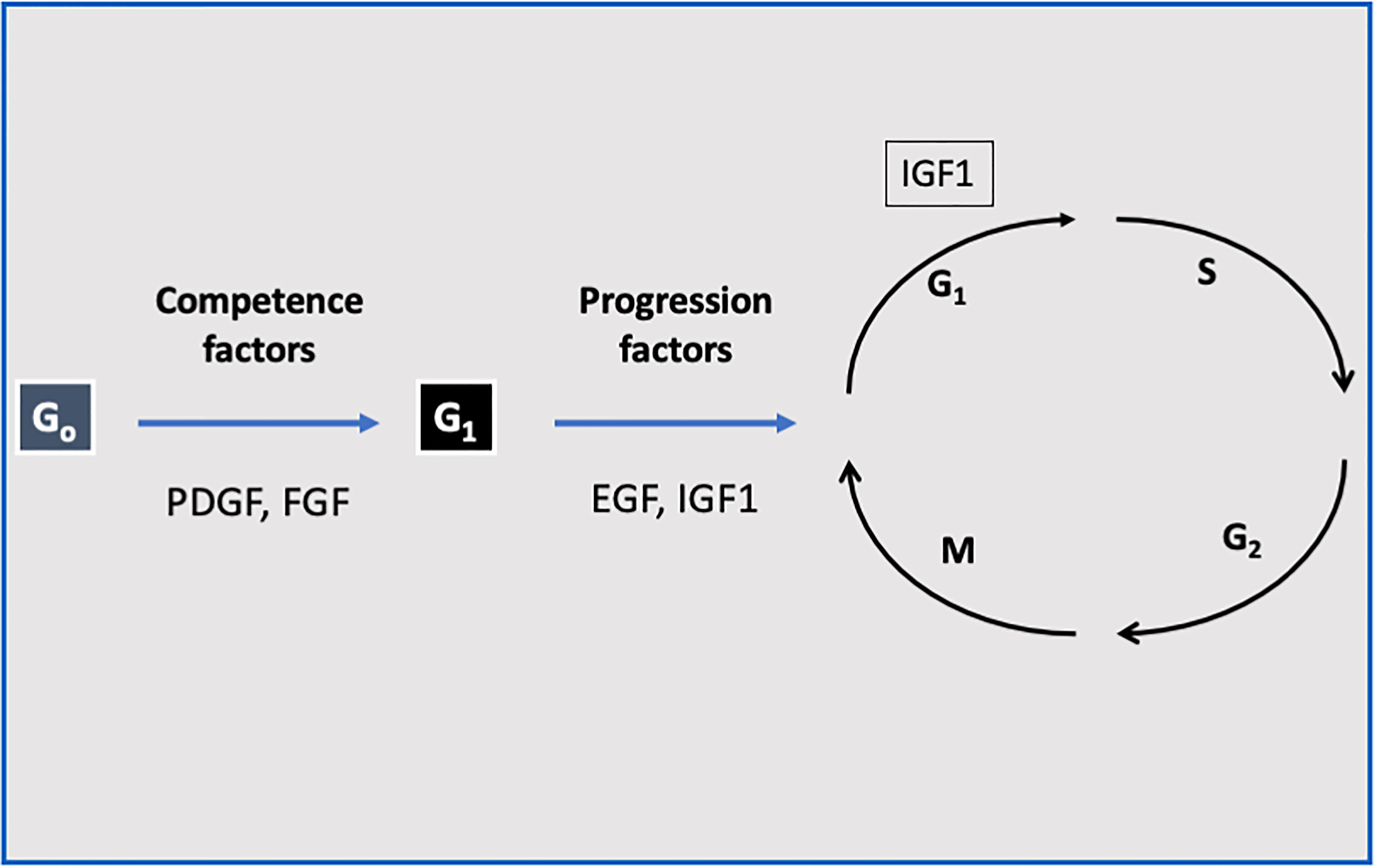
Figure 1 The role of IGF1 as a progression factor. Quiescent cells in G0 can be induced to enter G1 by competence factors such as PDGF and FGF. Once in G1, the cells require sub-physiological quantities of IGF1 or EGF to evade arrest and to progress through the rest of the cycle. Hence, IGF1 fits the criteria of a progression factor.
IGF1 induces a variety of cell- and organ-specific functions, ranging from regulation of hormone synthesis and secretion (50), chemo-attractant migration (51) and neuromodulation (52). IGF1 also participates in cell recognition by the immune system. Thus, glioblastoma cells in which IGF1 expression was disrupted by antisense oligonucleotides generated a strong host response and didn’t form tumors when injected into syngeneic mice (53). In the central nervous system, the IGF1 gene is widely expressed and promotes proliferation, survival and differentiation of neuronal and non-neuronal cells. In rat brain, distinct regions (e.g., cerebellar neurons, retina, sensory and trigeminal ganglia) express high IGF1 mRNA levels during embryonic development while other regions (e.g., midbrain, cerebral cortex, hippocampus) expresses the gene mainly during postnatal growth (54). Up-regulation of IGF1 in the central nervous system is observed 1-7 days after a variety of insults, including hypoxia-ischemia (55), brain contusion (56) and penetrating brain trauma (57). Of notice, IGF1 has been identified as a neurotrophic factor, rescuing neurons from apoptosis (58) and enhancing neuronal growth and myelination (59). The role of IGF1 as an anti-apoptotic factor will be discussed below.
Finally, it is important in this context to discuss the mechanisms associated with IGF1 action (12). Ligand binding to the IGF1R extracellular α-subunits results in conformational changes that induce autophosphorylation of tyrosine residues within the mainly intracellular β-subunits (13). Autophosphorylation stimulates the receptor tyrosine kinase activity and leads to phosphorylation of additional substrates. A number of SH2 domain-containing proteins, or ‘docking proteins’, bind to specific phospho-tyrosine residues in the C-terminal portion of the β-subunit (17). The insulin receptor substrate (IRS) family of proteins and Shc are the best characterized docking proteins. Thus, enzymatic activation of the IGF1R tyrosine kinase domain results in stimulation of an array of intracellular cascades, including the ras-raf-MAPK and PI3K-AKT pathways. Classically, IGF1-induced mitogenesis was primarily attributed to the ras-raf-MAPK pathway whereas the anti-apoptotic effect of IGF1 was thought to be mediated by the PI3K-AKT pathway. Today, it is clear that the situation is, in fact, much more complex. Whereas in the past IGF1R pathways (like growth factor cascades in general) were depicted as linear tracts, it has become increasingly evident that there is a cross-talk between IGF1R and additional cell-surface receptors, including G-proteins, integrins, and others (12).
The capacity of normal adult cells to remain in a post-mitotic, terminally differentiated state is dictated by their ability to respond to a series of secreted, cellular or extracellular growth inhibitors. These antiproliferative signals operate to keep the cells out of the cell cycle and in a quiescent state. One of the prototypical cancer hallmarks refers to the acquired faculty of transformed cells to evade these antigrowth signals (34, 37). As a result, cells might regain a previously repressed mitogenic potential that would allow them to re-enter the cell cycle.
The E2F family of transcription factors plays a key role in regulating the expression of genes involved in the G1/S transition and DNA synthesis (60–62). The retinoblastoma (Rb) and E2F proteins form a complex (Rb-E2F) that undergoes dissociation upon phosphorylation of Rb, with ensuing activation of E2F-dependent transcription and cell cycle progression (63, 64). E2F binds to DNA and regulates the expression of genes involved in cell cycle progression. Microarray analyses of E2F1-induced genes revealed that genes associated with proliferation as well as apoptosis are usually upregulated by E2F1 (65–67). Using transient transfection assays we have demonstrated that E2F1 is a potent inducer of IGF1R gene expression in prostate cancer cells (68). Augmented IGF1R levels correlated with elevated phospho-IGF1R values, suggesting activation of the IGF1R signaling pathway. Deletion analysis indicated that the ability of E2F1 to stimulate IGF1R promoter activity correlated with the number of E2F1 sites present in the various constructs, suggesting a dose-dependent effect of E2F1 binding on IGF1R gene expression. In addition, in vivo analysis of promoter occupancy by chromatin immunoprecipitation assays revealed that E2F1 was specifically recruited to the IGF1R promoter. Combined, data indicate that transcription factor E2F1 is an important regulator of the IGF1R gene. Elevation in IGF1R levels may contribute to the proliferative effects associated with initiation of prostate (and other) cancer (30, 69).
Classically, loss-of-function mutations of tumor suppressor genes or gain-of-function mutations of oncogenes are regarded as critical events in cancer development. In terms of the ‘two-hit hypothesis’ these events fit the criteria of a first, i.e. oncogenic, event. A mechanism of action that is shared by multiple oncogenes involves the transactivation of different growth factors or growth factor receptors, including IGF1R. Activation of IGF1 axis components suits the definition of a second, i.e. permissive, hit. This mode of action is commonly referred to as ‘adoption’ of the IGF1R signaling pathway by oncogenes (Figure 2). For example, pp60src, the protein encoded by the src oncogene of Rous sarcoma virus stimulates the constitutive phosphorylation of the IGF1R tyrosine kinase domain (70). Hence, pp60src alters growth regulation by rendering the cells constitutively subject to a mitogenic signal. Other oncogenes, including c-myb, can transactivate the IGF1R promoter, with enhanced IGF1R gene transcription and biosynthesis (71). In summary, cellular and viral oncogenes require an intact, activated IGF1R signaling pathway in order to elicit their transforming activities (69, 72).
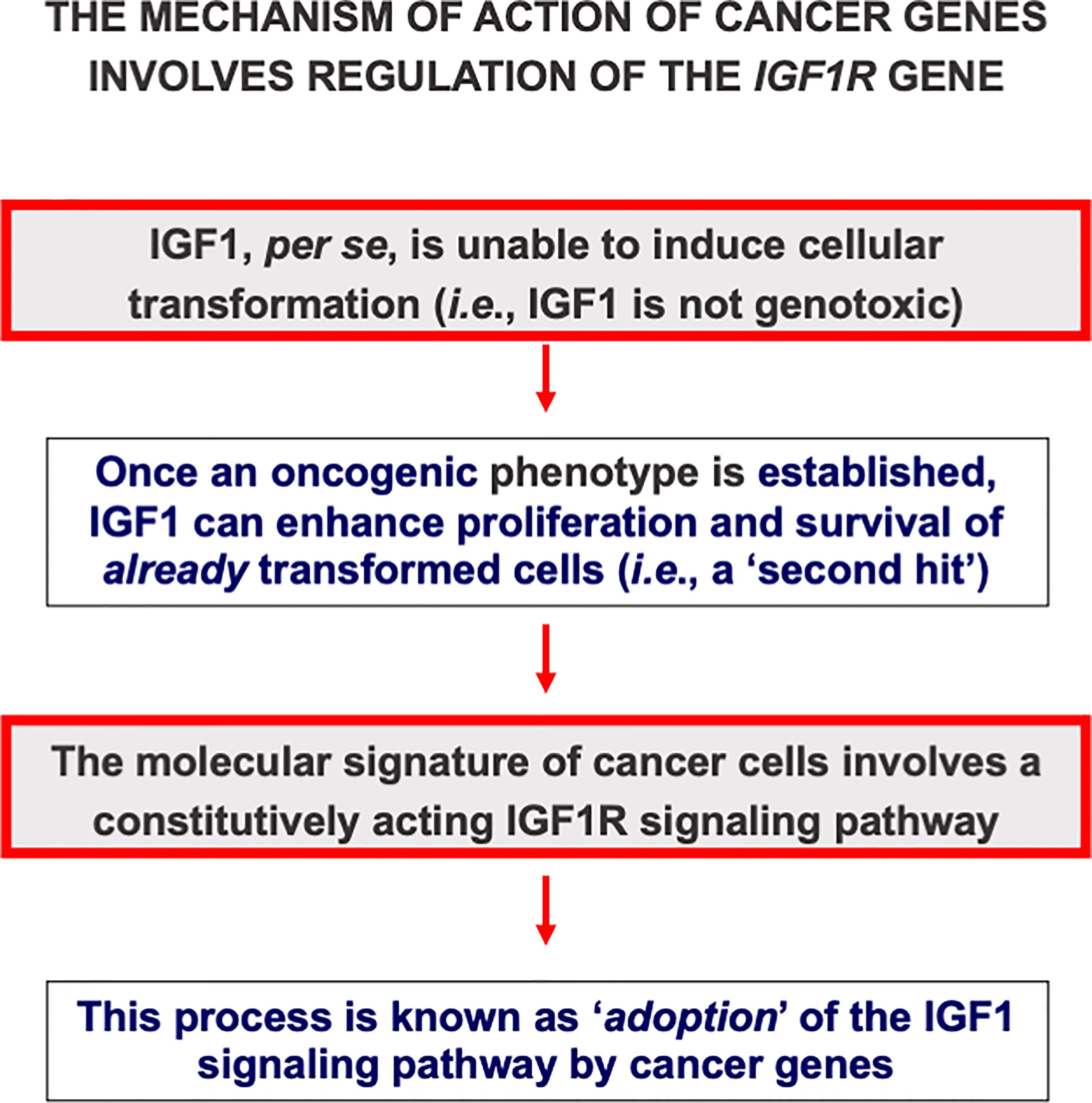
Figure 2 Cancer genes adopt the IGF1 signaling pathway. IGF1 is regarded as a non-genotoxic growth factor, i.e., it is unable, in itself, to induce mutations or transformation. However, once an oncogenic event has occurred, IGF1 can enhance proliferation and survival of already transformed cells. In the context of the ‘two hit hypothesis’, IGF1 action is regarded as a second, or permissive, hit. Multiple cancer genes, including oncogenes and anti-oncogenes, adopt the IGF1 signaling pathway. In agreement with this notion, cells devoid of the IGF1R usually do not undergo transformation.
A quintessential feature of cancer cells involves the acquisition of molecular and genetic means that will allow them not-to-die. The ability of transformed cells to endure is dictated not only by their proliferative potential but also by their capacity to oppose death, particularly apoptosis. As stated by Hanahan and Weinberg in their original report, acquired resistance towards apoptosis is a fundamental hallmark of, most probably, all types of cancer (34). Classically, programmed cell death has been regarded as an ‘altruistic’ mechanism that offers protection to the entire organism by engaging in a self-annihilation program (73). Extensive research over more than fifty years has identified complex biochemical and molecular strategies that are acquired by cancer cells and that are directly responsible for evasion of apoptosis.
Seminal studies from the laboratory of Renato Baserga generated early evidence that the IGF1R exhibits a very potent anti-apoptotic activity in comparison to most other growth factor receptors (74–78). Using a series of IGF1R mutants, O’Connor et al. demonstrated that the domains of the IGF1R required for its anti-apoptotic function are distinct from those required for proliferation or transformation (79). Of notice, IGF1 inhibition of apoptosis occurs in the absence of protein synthesis and, therefore, does not require immediate gene expression (80). Finally, protection from apoptosis is evident in the post-commitment (i.e., mitogen-independent) S-G2-M phases of the cell cycle.
The inherent anti-apoptotic activity of the IGF1R confers upon receptor-expressing cells enhanced survivability, a fundamental property of cancer cells. Consistent with this notion, fibroblasts (R-) derived from IGF1R ‘knock-out’ embryos (the total deficiency of IGF1R is a lethal condition) do not undergo malignant transformation when exposed to oncogenes (81, 82). Reintroduction of a functional receptor renders R- cells susceptible to the transforming activities of these oncogenes. Certain exceptions to this general paradigm have been reported. For instance, transfection of the GTPase-deficient mutant Gα13 resulted in transformation of R- cells. These results indicate that Gα13 can induce cellular transformation through pathways independent of IGF1R (83). Taken together, studies are in agreement with the notion that IGF1R expression and/or activation are fundamental pre-requisites for cancer development (8, 20) (Table 1). It is important to understand, however, that IGF1, per se, is neither genotoxic nor oncogenic. In other words, even supra-pharmacological doses of the hormone cannot induce malignant transformation.
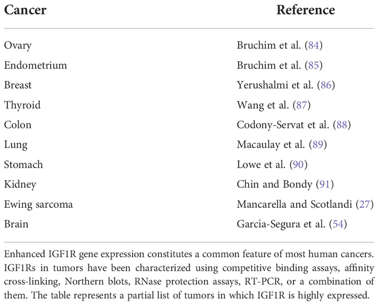
Table 1 IGF1R gene expression in human cancers.
The key role of the mitochondria in the regulation of biochemical and molecular events associated with apoptosis have been described by Green and Reed (92). Recent genomic analyses identified the thioredoxin interacting protein (TXNIP) as a novel target for IGF1 and insulin action (93). TXNIP is a mitochondrial protein that belongs to the α-arrestin family and plays a key role in redox regulation (94–98). TXNIP binds to the catalytic active center of reduced thioredoxin and inhibits its expression and activity (99). TXNIP is highly expressed in lymphoblastoid cells derived from Laron syndrome patients, a type of congenital IGF1 deficiency. The role of the TXNIP gene as a downstream target for negative regulation by IGF1 was confirmed by studies showing that IGF1 (or insulin) treatment led to marked reductions in TXNIP levels in cultured cells. Furthermore, transfection studies revealed that the effect of IGF1 on TXNIP gene expression was mediated at the level of transcription (93). We envision a scenario in which IGF1 could inhibit apoptosis by down-regulating TXNIP at the transcriptional level. Independent of redox regulation, TXNIP also functions as a regulator of glucose metabolism (100) and its levels are increased in diabetes (101).
Genome instability and mutation were categorized as an enabling cancer hallmark that serves as a pre-requisite for some (possibly most) of the previously described hallmarks (36). The biological rationale for this trait relies on the recognition that specific mutations might confer upon certain cell populations distinctive advantages that could, eventually, facilitate their selective expansion and dominance.
Tumor suppressor p53 is a transcription factor that typically accumulates in the cell in response to DNA damage (102). Mutation of the p53 gene is the most common event in human cancer (103, 104). When hyperphosphorylated, p53 arrests cell cycle progression at the G1 phase. The p53 pathway is activated in response to different stress signals, including DNA damage and telomere shortening, hypoxia, heat and cold shock, inflammation and activation of oncogenes by mutations (105, 106). These various strains bear the potential to decrease the fidelity of cell cycle progression and DNA replication, thus leading to increased mutation rates (107, 108). As alluded to above, accumulation of mutations constitutes an early event in malignant transformation. p53-mediated cell cycle arrest enables damaged DNA to be repaired before the replicative phase of the cell cycle (104, 109). Alternatively, p53 can elicit an apoptotic program. Of relevance, evidence gathered in recent years indicate that, in addition to its well established ability to control cell cycle progression, p53 activation has a major impact on metabolic processes, including glucose transport (110) and obesity (111).
Extensive molecular and genetic analyses revealed that the mechanism of action of wild-type p53 involves transcriptional suppression of the IGF1R gene (112). Gain-of-function, or loss-of-function, mutations of p53 in tumor cells seem to disrupt its inhibitory activity, generating oncogenic molecules capable of transactivating the IGF1R gene. Because p53 is a potent inducer of apoptosis, we assume that the effect of this molecule on apoptosis is mediated, at least in part, via suppression of the IGF1R promoter. Lack of IGF1R inhibition by mutant p53 molecules may help expand cancer cell populations that are otherwise destined to die (113). The ubiquitin ligase Mdm2 is of major importance in regulation of p53 activity (114) and IGF1 was shown to induce p53 degradation in an Mdm2-dependent manner (115). Girnita et al. have shown that Mdm2 physically associates with IGF1R and causes its ubiquitination and degradation (116). Mdm2 serves as a ligase in ubiquitination of IGF1R and thereby causes its degradation by the proteasome system. Consequently, by sequestering Mdm2 in the cell nuclei, the level of p53 may indirectly influence the expression of IGF1R. This function of Mdm2 and p53 constitutes a potential mechanism for the regulation of IGF1R and cell growth. As an operational outcome to these studies, IGF1R was identified as a molecular determinant for response to p53 reactivation therapy in conjunctival melanoma (117).
The characterization of the mechanisms responsible for regulation of the IGF1 pathway by p53, as described above, led us to formulate an hypothesis aimed at offering a generalized paradigm for regulation of IGF1R expression by different tumor suppressors (69, 113, 118). While tumor suppressors might differ in their organ-specific expression, mechanisms of activation, type of tumors involved and other parameters, they share the IGF1R pathway as a common response path. This unifying model may explain the involvement of the IGF1 axis in genome instability and mutations.
The breast and ovarian cancer susceptibility gene (BRCA1) is a transcription factor involved in DNA damage repair, cell growth and apoptosis (119, 120). Mutations of the BRCA1 gene are detected in a significant proportion of families with inherited breast and/or ovarian cancer (121, 122). Transfection of a BRCA1 expression vector in breast cancer cells led to a marked reduction in endogenous IGF1R levels and promoter activity (123–125). In contrast, a mutant BRCA1 gene encoding a truncated version of the molecule (del185AG, a mutation with a high incidence among Ashkenazi Jews) had no effect on IGF1R expression. Hence, activation of BRCA1 in response to DNA damage, oxidative stress, or other cellular insults, may lead to a reduction in IGF1R levels and IGF1 action. Suppression of the IGF1 pathway is expected to prevent from cells from engaging in mitosis.
The ability to grow new blood vessels, or angiogenesis, represents an important capability that neoplasms develop to increase in size and, ultimately, to endure (126). This feature is critically required by the proliferating cell in order to obtain nutrients and oxygen (127). The labeling of sustained angiogenesis as a hallmark of cancer reflects the universal nature of this trait (34). The process of angiogenesis is tightly regulated by secreted growth factors and their receptors as well as by cellular and extracellular adhesion molecules (i.e., integrins, cadherin, etc). The single most important protein that epitomizes the angiogenic process is vascular endothelial growth factor (VEGF) (128, 129).
The physiological role of VEGF is to induce the formation of new blood vessels during embryonic development and to restore injured vessels (130). Members of the VEGF family (VEGF-A, -B, -C, -D, and placenta growth factor, PGF) are produced by many types of cancer, being the expression of VEGF-A usually correlated with metastatic potential. VEGF-A enhances migration and mitosis of endothelial cells, stimulates matrix metalloproteinase activity and augments integrin αvβ3 activity (131). The VEGF-A gene is regarded as an hypoxia-inducible gene, being its transcription regulated by hypoxia-inducible factor-α (HIF-α) (132). In hypoxia (low oxygen pressure), the absence of oxygen-dependent hydroxylation of HIF-α prolines allows HIF-α subunits to accumulate, dimerize and translocate to the nucleus, triggering transcription of VEGF-A and other hypoxia-inducible genes (133). The IGF axis enhances the hypoxic response by activation of Akt signaling, leading to stabilization of HIF-α and upregulation of VEGF-A (134, 135).
The von Hippel-Lindau gene product (pVHL, the substrate recognition component of an E3 ubiquitin ligase complex) has an important role in the oxygen-dependent proteolysis of HIF-α (136). Inactivation of VHL in clear cell renal cell cancer (CC-RCC) allows normoxic accumulation of HIF-α subunits, leading to constitutive expression of the angiogenic VEGF-A gene. We have previously identified a new hypoxia-independent role for VHL in suppressing IGF1R transcription and mRNA stability (137). IGF1R levels were higher in CC-RCC cells harboring a mutant inactive VHL than in isogenic cells expressing a wild-type VHL. Hence, mutant VHL leads to IGF1R upregulation, an event typically associated with renal tumorigenesis. Taken together, data indicate a functional interplay between the IGF1R and VEGF signaling pathways (138). Dysregulation of specific components in these paths might lead to sustained angiogenesis, an important hallmark of cancer.
The ability of tumor cells to invade adjacent tissues and to colonize remote sites in the body, (i.e., metastasis), is responsible for the vast majority of deaths from cancer (139). This cancer hallmark depends, to a large extent, on hallmarks described above (34). The processes of invasion and metastasis have been extensively investigated over the years from both basic and translational angles. Research led to the identification of molecules and signaling pathways that are directly involved in pathological changes at the interface between malignant cells and the microenvironment. Most of the clinically-relevant changes can be ascribed to proteins involved in cell-cell adhesion, including integrin, cadherin and others (140–143).
Accumulating experimental and epidemiological evidence provide support to the idea that obesity is an important risk factor for cancer (39, 144, 145). A number of mechanisms by which obesity contributes to tumor progression have been described. Obesity is associated with systemic hyperinsulinemia as well as differences in circulating IGFs, adipokines and cytokines (146). The contribution of these molecules to proliferative and cell-survival events has been well documented (147). The increase in adipose tissue in the tumor microenvironment is a source of lipids that can be used by tumors for metabolism and as structural and signaling molecules. In this context, cholesterol was shown to affect gene expression of the jun family in colon cancer cells, leading to potentially pathogenic signaling events (148).
Obesity also leads to changes in the extracellular matrix, adipose stromal cells and immune cells, creating a cancer-permissive microenvironment. The reader is referred to a recent comprehensive review article by Vella et al. that provides a thorough analysis of the interplay between estrogens, insulin/IGFs and their receptors, and stroma (149). Regarding the mechanisms of action responsible for these interactions, the estrogen receptor-α (ERα) has been identified as a potent transactivator of the IGF1R gene (30, 150, 151). Furthermore, GC-rich sequences in the proximal IGF1R promoter region were required for this effect. Impaired interactions between ERα and zinc-finger proteins may lead to aberrant IGF1R expression in breast cancer cells. Dysregulated expression and availability of IGFs are regarded as key regulators of metastasis (152). Recent genomic analyses identified the nephronectin (NPNT) gene as a downstream target for IGF1 action (153). NPNT is an intracellular and secreted extracellular matrix protein with important roles in kidney development (154, 155). NPNT interacts with α8β1 integrin through its central linker segment. NPNT expression correlated with poor prognosis in breast cancer and was shown to promote metastasis via its integrin-binding motif (156, 157). Our analyses identified NPNT as the top down-regulated gene in Laron syndrome cells, a condition associated with diminished IGF1 levels (158).
Finally, constitutive activation of the IGF1R was shown to affect lineage differentiation during mammary tumorigenesis (159). Constitutive IGF1R activation promoted tumors with mixed histology and multiple cell lineages. In these tumors, IGF1R expanded the luminal-progenitor population while influencing myoepithelial differentiation. Combined, the capacity to affect lineage differentiation may promote heterogeneous mammary tumors and might have translational implications.
As alluded to above, the classical model of IGF1 action involves the ligand-induced phosphorylation of IGF1R, a heterotetrameric cell-surface tyrosine kinase receptor, with ensuing activation of cytoplasmic signaling cascades. The recent identification of nuclear IGF1R translocation provides an additional level of biological complexity by allowing a typical transmembrane receptor to function in a discrete, membrane-bound cellular environment (160–162). Using cell fractionation techniques and confocal microscopy it was shown that the cell-surface IGF1R undergoes modification by the small ubiquitin-like modifier protein (SUMO-1), with subsequent translocation to the nucleus (163, 164). SUMOylation sites on lysine residues within the tyrosine kinase domain are conserved among a variety of homologues from different species. Mutagenesis of these sites arrested nuclear import and gene activation (165).
Lysosomal and endocytic pathways that are mainly involved in IGF1R and INSR degradation were shown to be also responsible for nuclear translocation of the receptors (162). Importin-β, an important player in nuclear translocation, was shown to coimmunoprecipitate with IGF1R (164). In addition, use of the clathrin-dependent endocytosis inhibitor dansylcadaverine abrogated IGF1R nuclear import (162, 166). The capacity of IGF1R to interact with DNA was investigated by chromatin immunoprecipitation (ChIP)-seq assays. Analyses showed that the vast majority (~80%) of IGF1R-enriched regions were intergenic (i.e., distal from any annotated gene), while ~6% of these regions were located in introns and ~6% in exons (165). Hence, data is consistent with the notion that IGF1R may bind to enhancer regions and function as a transcriptional activator.
What are the clinical implications of nuclear IGF1R translocation? The impact of nuclear IGF1R on tumor aggressiveness can be deduced from the fact that inhibition of nuclear IGF1R import correlated with a reduced proliferative potential (167, 168). Aleksic and colleagues reported that nuclear IGF1R was present in renal cancer cells, preinvasive breast lesions and non-malignant tissues with a high proliferative index (162). Moreover, nuclear IGF1R staining correlated with an adverse prognosis in renal cancer. Similarly, nuclear IGF1R localization in alveolar rhabdomyosarcoma was associated with an aggressive phenotype. Finally, immunohistochemical analyses identified IGF1R staining in 47 out of 53 pediatric gliomas (169). Ten out of the 47 cases exhibited nuclear staining. IGF1R staining was mostly non-nuclear in low-grade tumors, while nuclear expression was predominant in high-grade gliomas. Survival was significantly longer in patients with gliomas having non-nuclear IGF1R localization than in patients with nuclear IGF1R. Taken together, data indicate that intracellular IGF1R distribution may help in stratifying pediatric glioma patients.
Following our analysis of cancer hallmarks from the perspective of the IGF1 signaling pathway, it is relevant to question what was the rationale behind the identification of IGF1R as a therapeutic target. Three main lines of research over the past 25 years led to the concept that the IGF1 axis and, in particular, the IGF1R is a potential goal for pharmacological (or other) intervention (1): during oncogenic transformation a “primitive” pattern of IGF1R expression is established, leading to enhanced IGF1R levels. A similar developmental trend is exhibited by IGF2, which is produced by most cancer cells and, usually, constitutes the main ligand in tumors (170, 171). These observations led to the dogma that IGF1R expression is a critical requirement for establishment of a tumor (15). This paradigm, however, is not necessarily true in every type of cancer. Thus, whereas IGF1R overexpression is a common trait of most pediatric and other solid tumors (e.g., brain, kidney), a more complex pattern of expression is seen in adult epithelial tumors (e.g., breast, prostate) (172–174); (2) further support to the notion that IGF1R might constitute a rational therapeutic target in oncology was provided by studies showing that cells deprived of the receptor, in their vast majority, do not undergo oncogenic transformation (81); and (3) the identification of endocrine IGF1 as a risk factor in multiple neoplasias generated the “critical mass” needed to proceed with clinical trials against the IGF1 axis (175). IGF1R-directed therapies are aimed at:
● inhibiting cancer cell survival and proliferation;
● reversing tumor growth and metastasis development; and
● sensitizing to chemotherapy, radiotherapy and biological therapies.
Different strategies have been developed to target the IGF system in vitro and in animal models (25, 27, 28, 176–178). However, three main strategies progressed to clinical trials (1): antibodies that target the IGF1R and induce its internalization and degradation; (2) small molecule IGF1R tyrosine kinase inhibitors; and (3) neutralizing antibodies that target the IGF1 and IGF2 ligands (179, 180). Unfortunately, most clinical trials led to disappointing results and it is nowadays clear that a combined approach aimed against the IGF axis along with additional pathway/s should lead to a better outcome (181). A recent pre-clinical study provided evidence that co-treatment of breast cancer cells with AEW541 (a selective IGF1R inhibitor) along with gemcitabine (a chemotherapeutic drug) improved the treatment efficiency (31). The degree of synergy achieved, as expressed in combination index values, was very strong. Finally, cell cycle analyses suggested that the synergism was derived, at least in part, from AEW541-induced G1 arrest and gemcitabine-induced S arrest.
Some of the IGF1R antibodies developed in recent years were shown to cross-react with the INSR leading to hyperglycemia. The potential effect of IGF1R antibodies on INSR signaling is of special concern given that these antibodies can alter INSR function, leading to insulin resistance and adverse effects on glucose and carbohydrate metabolism. On the other hand, INSR targeting could be potentially beneficial because inhibition of the INSR, in addition to IGF1R inhibition, might increase the effective anti-tumoral activity (182, 183). Hence, one of the critical challenges in the field is to define whether future trials should be limited to IGF1R or, alternatively, targeting tools should be implemented also against INSR.
In summary, we believe that the negative outcomes of recent clinical trials, despite the obvious disappointment, were critically analyzed and important lessons were learned (26, 184, 185). Among other possible causes, a retrospective evaluation points out at a fierce competition between pharma companies as one of the reasons that hampered the development of efficient IGF1R drugs. These ‘battles’ led to the design of badly planned trials that, for the most part, were conducted on unselected patients. Therefore, it is of critical importance to identify predictive markers that could assist in selecting patients who might benefit from these treatments. Furthermore, biomarkers are also needed to monitor patient’s response to therapy. We believe that a rational and integrated use of omics platforms may certainly help identifying ‘IGF1 signatures’ that correlate with better clinical outcomes.
The question whether INSR is a druggable target in cancer is a matter of controversy. As mentioned in the previous section, a critical challenge is to define whether future trials should be limited to IGF1R or, alternatively, should target also INSR. Given the important role of insulin and INSR in cancer biology, mainly breast and endometrial tumors, it is expected that future efforts will embrace also the development of INSR-directed molecules.
As stated above, most available evidence indicates that selective IGF1R inhibitors along with chemotherapy or other biological drug (i.e., combination therapy) usually lead to a better outcome than monotherapy. The enhancement of the therapeutic effect stems from the fact that IGF1R therapy and chemotherapy are aimed against different phases or targets of the cellular machinery. Hence, whereas chemicals mostly induce DNA damage, IGF1R inhibitors specifically target the survival machinery of the cell.
Elucidation of the interplay between the IGF1 axis and additional pathways, including oncogenes and anti-oncogenes, will have a major impact on our understanding of basic molecular oncology processes as well as on our ability to design and optimize cancer therapies. A summary of the involvement of the IGF1 system in Cancer Hallmarks is presented in Table 2. The potential of new drugs, alone or in combination with chemotherapy or other biological agents, needs to be investigated in randomized studies. Finally, lessons from the field of personalized medicine will be implemented in IGF1R targeting.
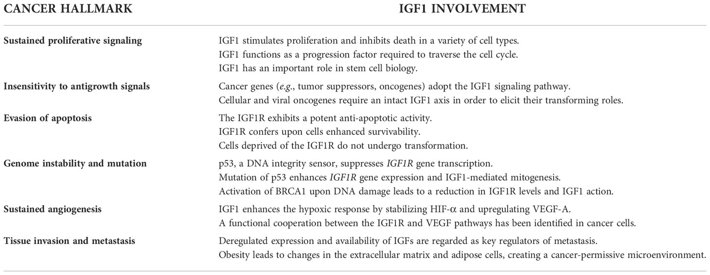
Table 2 Hallmarks of cancer and the IGF1 signaling pathway.
HW and DL: substantial contributions to the conception and design of the work and interpretation of data for the work; drafting the work and revising it critically for important intellectual content; final approval of the version to be published; and agreement to be accountable for all aspects of the work in ensuring that questions related to the accuracy or integrity of any part of the work are appropriately investigated and resolved. Both authors contributed to the article and approved the submitted version.
Work in the laboratory of HW is supported by grants from the Israel Science Foundation, US-Israel Binational Science Foundation, the Israel Cancer Association and the Recanati Foundation (Tel Aviv University). Work in the laboratory of DL is supported by NCI grant R01CA128799.
HW is the incumbent of the Lady Davis Chair in Biochemistry.
The authors declare that the research was conducted in the absence of any commercial or financial relationships that could be construed as a potential conflict of interest.
All claims expressed in this article are solely those of the authors and do not necessarily represent those of their affiliated organizations, or those of the publisher, the editors and the reviewers. Any product that may be evaluated in this article, or claim that may be made by its manufacturer, is not guaranteed or endorsed by the publisher.
1. Yakar S, Adamo ML. Insulin-like growth factor 1 physiology: lessons from mouse models. Endocrinol Metab Clin North Am (2012) 41:231–47. doi: 10.1016/j.ecl.2012.04.008
2. Yakar S, Werner H, Rosen CJ. Insulin-like growth factors: actions on the skeleton. J Mol Endocrinol (2018) 61:T115–T37. doi: 10.1530/JME-17-0298
3. LeRoith D, Yakar S. Mechanisms of disease: metabolic effects of growth hormone and insulin-like growth factor-1. Nat Clin Pract Endocrinol Metab (2007) 3:302–10. doi: 10.1038/ncpendmet0427
4. LeRoith D, Bondy C, Yakar S, Liu J-L, Butler A. The somatomedin hypothesis: 2001. Endocr Rev (2001) 22:53–74. doi: 10.1210/edrv.22.1.0419
5. Salmon WD, Daughaday WH. A hormonally controlled serum factor which stimulates sulfate incorporation by cartilage. vitro J Lab Clin Med (1957) 49:825–36.
6. Bentov I, Werner H. IGF, IGF receptor and overgrowth syndromes. Ped Endocrinol Rev (2004) 1:352–60.
7. Holly JM, Perks CM. Insulin-like growth factor physiology: what we have learned from human studies. Endocrinol Metab Clin North Am (2012) 41:249–63. doi: 10.1016/j.ecl.2012.04.009
8. Werner H, Laron Z. Role of the GH-IGF1 system in progression of cancer. Mol Cell Endocrinol (2020) 518:111003. doi: 10.1016/j.mce.2020.111003
9. Lupu F, Terwilliger JD, Lee K, Segre GV, Efstratiadis A. Roles of growth hormone and insulin-like growth factor 1 in mouse postnatal growth. Dev Biol (2001) 229:141–62. doi: 10.1006/dbio.2000.9975
10. LeRoith D. Clinical relevance of systemic and local IGF-I: lessons from animal models. Ped Endocrinol Rev (2008) 5:739–43.
11. Bentov I, Werner H. Insulin-like growth factor-I. In: Kastin A, editor. Handbook of biologically active peptides, vol. pp . San Diego: Elsevier Press (2006). p. 1385–92.
12. Werner H, LeRoith D. New concepts in regulation and function of the insulin-like growth factors: implications for understanding normal growth and neoplasia. Cell Mol Life Sci (2000) 57:932–42. doi: 10.1007/PL00000735
13. LeRoith D, Werner H, Beitner-Johnson D, Roberts CT Jr. Molecular and cellular aspects of the insulin-like growth factor I receptor. Endocr Rev (1995) 16:143–63 doi: 10.1210/edrv-16-2-143
14. Takahashi SI, Perks CM. The role of the IGF/Insulin-IGFBP axis in normal physiology and disease. Front Endocrinol (2022) 13:892140. doi: 10.3389/fendo.2022.892140
15. Baserga R. The insulin-like growth factor I receptor: a key to tumor growth? Cancer Res (1995) 55:249–52.
16. De Meyts P, Whittaker J. Structural biology of insulin and IGF1 receptors: implications for drug design. Nat Rev Drug Discovery (2002) 1:769–83. doi: 10.1038/nrd917
17. Klammt J, Pfaffle R, Werner H, Kiess W. IGF signaling defects as causes of growth failure and IUGR. Trends Endocrinol Metab (2008) 19:197–205. doi: 10.1016/j.tem.2008.03.003
18. Baker J, Liu J-P, Robertson EJ, Efstratiadis A. Role of insulin-like growth factors in embryonic and postnatal growth. Cell (1993) 75:73–82. doi: 10.1016/S0092-8674(05)80085-6
19. Liu J-P, Baker J, Perkins AS, Robertson EJ, Estratiadis A. Mice carrying null mutations of the genes encoding insulin-like growth factor I (Igf-1) and type 1 IGF receptor (Igf1r). Cell (1993) 75:59–72. doi: 10.1016/S0092-8674(05)80084-4
20. Baserga R. The decline and fall of the IGF-I receptor. J Cell Physiol (2013) 28:675–9. doi: 10.1002/jcp.24217
21. Domené S, Domené HM. Genetic mutations in the GH/IGF axis. Ped Endocrinol Rev (2018) 16:39–62. doi: 10.17458/per.vol16.2018.dd.geneticmutationsghigf
22. Sarfstein R, Yeheskel A, Sinai-Livne T, Pasmanik-Chor M, Werner H. Systems analysis of insulin and IGF1 receptors networks in breast cancer cells identifies commonalities and divergences in expression patterns. Front Endocrinol (2020) 11:435. doi: 10.3389/fendo.2020.00435
23. Domené S, Scaglia PA, Gutiérrez ML, Domené HM. Applying bioinformatic platforms, in vitro, and in vivo functional assays in the characterization of genetic variants in the GH/IGF pathway affecting growth and development. Cells (2021) 10:2063. doi: 10.3390/cells10082063
24. Ayyadevara S, Ganne A, Hendrix RD, Balasubramaniam M, Shmookler Reis RJ, Barger SW. Functional assessments through novel proteomics approaches: Application to insulin/IGF signaling in neurodegenerative diseases. J Neurosci Meth (2019) 319:40–6. doi: 10.1016/j.jneumeth.2018.11.005
25. Simpson A, Petnga W, Macaulay VM, Weyer-Czernilofsky U, Bogenrieder T. Insulin-like growth factor (IGF) pathway targeting in cancer: Role of the IGF axis and opportunities for future combination studies. Target Oncol (2017) 12:571–97. doi: 10.1007/s11523-017-0514-5
26. King H, Aleksic T, Haluska P, Macaulay VM. Can we unlock the potential of IGF-1R inhibition in cancer therapy? Cancer Treat Rev (2014) 40:1096–105. doi: 10.1016/j.ctrv.2014.07.004
27. Mancarella C, Scotlandi K. IGF system in sarcomas: a crucial pathway with many unknowns to exploit for therapy. J Mol Endocrinol (2018) 61:T45–60. doi: 10.1530/JME-17-0250
28. Girnita L, Worrall C, Takahashi S, Seregard S, Girnita A. Something old, something new and something borrowed: emerging paradigm of insulin-like growth factor type 1 receptor (IGF-1R) signaling regulation. Cell Mol Life Sci (2014) 71:2403–27. doi: 10.1007/s00018-013-1514-y
29. LeRoith D, Helman LJ. The new kid on the block(ade) of the IGF-1 receptor. Cancer Cell (2004) 5:403. doi: 10.1016/S1535-6108(04)00093-5
30. Sarfstein R, Maor S, Reizner N, Abramovitch S, Werner H. Transcriptional regulation of the insulin-like growth factor-1 receptor in breast cancer. Mol Cell Endocrinol (2006) 252:241–6. doi: 10.1016/j.mce.2006.03.018
31. Sinai-Livne T, Pasmanik-Chor M, Cohen Z, Tsarfaty I, Werner H, Berger R. Proteomic analysis of combined IGF1 receptor targeted therapy and chemotherapy identifies signatures associated with survival in breast cancer patients. Oncotarget (2020) 11:1515–30. doi: 10.18632/oncotarget.27566
32. Bruchim I, Attias Z, Werner H. Targeting the IGF1 axis in cancer proliferation. Exp Opin Ther Targets (2009) 13:1179–92. doi: 10.1517/14728220903201702
33. Werner H, Bruchim I. Basic and clinical significance of IGF-1-induced signatures in cancer. BMC Med (2010) 8:2. doi: 10.1186/1741-7015-8-2
34. Hanahan D, Weinberg RA. The hallmarks of cancer. Cell (2000) 100:57–70. doi: 10.1016/s0092-8674(00)81683-9
35. Lazebnik Y. What are the hallmarks of cancer? Nat Rev Cancer (2010) 10:232–3. doi: 10.1038/nrc2827
36. Hanahan D, Weinberg RA. Hallmarks of cancer: the next generation. Cell (2011) 144:646–74. doi: 10.1016/j.cell.2011.02.013
37. Hanahan D. Hallmarks of cancer: new dimensions. Cancer Discovery (2022) 12:31–46. doi: 10.1158/2159-8290.CD-21-1059
38. De Meyts P, Palsgaard J, Sajid W, Theede AM, Aladdin H. Structural biology of insulin and IGF-1 receptors. Novartis Found Symp (2004) 262:160–71. doi: 10.1002/0470869976.ch10
39. Gallagher EJ, LeRoith D. Obesity and diabetes: the increased risk of cancer and cancer-related mortality. Physiol Rev (2015) 95:727–48. doi: 10.1152/physrev.00030.2014
40. Rosenzweig SA. The continuing evolution of insulin-like growth factor signaling. F1000Res (2020) 9:F1000. doi: 10.12688/f1000research.22198.1
41. Jones JI, Clemmons DR. Insulin-like growth factors and their binding proteins: biological actions. Endocr Rev (1995) 16:3–24. doi: 10.1210/edrv-16-1-3
42. Macaulay VM. Insulin-like growth factors and cancer. Br J Cancer (1992) 65:311–20. doi: 10.1038/bjc.1992.65
43. Malaguarnera R, Belfiore A. The emerging role of insulin and insulin-like growth factor signaling in cancer stem cells. Front Endocrinol (Lausanne) (2014) 5:10. doi: 10.3389/fendo.2014.00010
44. Bendall SC, Stewart MH, Menendez P, George D, Vijayaragavan K, Werbowetski-Ogilvie T, et al. IGF and FGF cooperatively establish the regulatory stem cell niche of pluripotent human cells in vitro. Nature (2007) 448:1015–23. doi: 10.1038/nature06027
45. Moschos SJ, Mantzoros CS. The role of the IGF system in cancer: from basic to clinical studies and clinical applications. Oncology (2002) 63:317–32. doi: 10.1159/000066230
46. Baserga R. Oncogenes and the strategy of growth factors. Cell (1994) 79:927–30. doi: 10.1016/0092-8674(94)90023-X
47. Lu K, Campisi J. Ras proteins are essential and selective for the action of insulin-like growth factor 1 late in the G1 phase of the cell cycle in BALB/c murine fibroblasts. Proc Natl Acad Sci USA (1992) 89:3889–93. doi: 10.1073/pnas.89.9.3889
48. Florini JR, Ewton DZ, Coolican SA. Growth hormone and the insulin-like growth factor system in myogenesis. Endocr Rev (1996) 17:481–517. doi: 10.1210/edrv-17-5-481
49. Florini JR, Ewton DZ, Roof SL. IGF-I stimulates terminal myogenic differentiation by induction of myogenin gene expression. Mol Endocrinol (1991) 5:718–24. doi: 10.1210/mend-5-5-718
50. Giudice L. Insulin-like growth factors and ovarian follicular development. Endocr Rev (1992) 13:641–69. doi: 10.1210/edrv-13-4-641
51. Tapson VF, Boni-Schnetzler M, Pilch PF, Center DM, Berman JS. Structural and functional characterization of the human T lymphocyte receptor for insulin-like growth factor I in vitro. J Clin Invest (1988) 82:950–7. doi: 10.1172/JCI113703
52. Castro-Alamancos MA, Torres-Aleman I. Long-term depression of glutamate-induced gamma-aminobutyric acid release in cerebellum by insulin-like growth factor I. Proc Natl Acad Sci USA (1993) 90:7386–90. doi: 10.1073/pnas.90.15.7386
53. Trojan J, Johnson TR, Rudin SD, Ilan J, Tykocinski ML, Ilan J. Treatment and prevention of rat glioblastoma by immunogenic C6 cells expressing antisense insulin-like growth factor I RNA. Science (1993) 259:94–7. doi: 10.1126/science.8418502
54. García-Segura LM, Pérez J, Pons S, Rejas MT, Torres-Alemán I. Localization of insulin-like growth factor I (IGF-i)-like immunoreactivity in the developing and adult rat brain. Brain Res (1991) 560:167–74. doi: 10.1016/0006-8993(91)91228-S
55. Klempt M, Klempt ND, Gluckman P. Hypoxia and hypoxia/ischemia affect the expression of insulin-like growth factor binding protein 2 in the developing rat brain. Brain Res Mol Brain Res (1993) 17:55–61. doi: 10.1016/0169-328X(93)90021-G
56. Nordqvist AC, Holmin S, Nilsson M, Mathiesen T, Schalling M. MK-801 inhibits the cortical increase in IGF-1, IGFBP-2 and IGFBP-4 expression following trauma. Neuroreport (1997) 8:455–60. doi: 10.1097/00001756-199701200-00016
57. Walter HJ, Berry M, Hill DJ, Logan A. Spatial and temporal changes in the insulin-like growth factor (IGF) axis indicate autocrine/paracrine actions of IGF-I within wounds of the rat brain. Endocrinology (1997) 138:3024–34. doi: 10.1210/endo.138.7.5284
58. Zheng WH, Kar S, Doré S, Quirion R. Insulin-like growth factor-1 (IGF-1): a neuroprotective trophic factor acting via the akt kinase pathway. J Neural Transm Suppl (2000) 60:261–72. doi: 10.1007/978-3-7091-6301-6_17
59. Feldman EL, Sullivan KA, Kim B, Russell JW. Insulin-like growth factors regulate neuronal differentiation and survival. Neurobiol Dis (1997) 4:201–14. doi: 10.1006/nbdi.1997.0156
60. Dyson N. The regulation of E2F by pRB-family proteins. Genes Dev (1998) 12:2245–62. doi: 10.1101/gad.12.15.2245
61. Cam H, Dynlacht BD. Emerging roles for E2F: beyond the G1/S transition and DNA replication. Cancer Cell (2003) 3:311–6. doi: 10.1016/S1535-6108(03)00080-1
62. DeGregori J, Johnson DG. Distinct and overlapping roles for E2F family members in transcription, proliferation and apoptosis. Curr Mol Med (2006) 6:739–48. doi: 10.2174/1566524010606070739
63. Weinberg RA. The retinoblastoma protein and cell cycle control. Cell (1995) 81:323–30. doi: 10.1016/0092-8674(95)90385-2
64. Stanelle J, Putzer BM. E2F1-induced apoptosis: turning killers into therapeutics. Trends Mol Med (2006) 12:177–85. doi: 10.1016/j.molmed.2006.02.002
65. Ma Y, Croxton R, Moorer RLJ, Cress WD. Identification of novel E2F1-regulated genes by microarray. Arch Biochem Biophys (2002) 399:212–24. doi: 10.1006/abbi.2002.2761
66. Stanelle J, Stiewe T, Theseling CC, Peter M, Putzer BM. Gene expression changes in response to E2F1 activation. Nucleic Acids Res (2002) 30:1859–67. doi: 10.1093/nar/30.8.1859
67. Muller H, Bracken AP, Vernell R, Moroni MC, Christians F, Grassilli E, et al. E2Fs regulate the expression of genes involved in differentiation, development, proliferation, and apoptosis. Genes Dev (2001) 15:267–85. doi: 10.1101/gad.864201
68. Schayek H, Bentov I, Rotem I, Pasmanik-Chor M, Ginsberg D, Plymate SR, et al. Transcription factor E2F1 is a potent transactivator of the insulin-like growth factor-I receptor gene. Growth Hormone IGF Res (2010) 20:68–72. doi: 10.1016/j.ghir.2009.08.001
69. Werner H. Tumor suppressors govern insulin-like growth factor signaling pathways: implications in metabolism and cancer. Oncogene (2012) 31:2703–14. doi: 10.1038/onc.2011.447
70. Peterson JE, Jelinek T, Kaleko M, Siddle K, Weber MJ. C phosphorylation and activation of the IGF-I receptor in src-transformed cells. J Biol Chem (1994) 269:27315–21. doi: 10.1016/S0021-9258(18)46987-6
71. Reiss K, Ferber A, Travali S, Porcu P, Phillips PD, Baserga R. The protooncogene c-myb increases the expression of insulin-like growth factor I and insulin-like growth factor I receptor messenger RNAs by a transcriptional mechanism. Cancer Res (1991) 51:5997–6000.
72. Werner H, Maor S. The insulin-like growth factor-I receptor gene: a downstream target for oncogene and tumor suppressor action. Trends Endocrinol Metab (2006) 17:236–42. doi: 10.1016/j.tem.2006.06.007
73. Kaczanowski S. Apoptosis: its origin, history, maintenance and the medical implications for cancer and aging. Phys Biol (2016) 13:031001. doi: 10.1088/1478-3975/13/3/031001
74. Hongo A, Yumet G, Resnicoff M, Romano G, O'Connor R, Baserga R. Inhibition of tumorigenesis and induction of apoptosis in human tumor cells by the stable transfection of a myristylated COOH terminus of the insulin-like growth factor-I receptor. Cancer Res (1998) 58:2477–84.
75. Resnicoff M, Abraham D, Yutanawiboonchai W, Rotman HL, Kajstura J, Rubin R, et al. The insulin-like growth factor I receptor protects tumor cells from apoptosis in vivo. Cancer Res (1995) 55:2463–9.
76. Resnicoff M, Burgaud J-L, Rotman HL, Abraham D, Baserga R. Correlation between apoptosis, tumorigenesis, and levels of insulin-like growth factor I receptors. Cancer Res (1995) 55:3739–41.
77. Prisco M, Hongo A, Rizzo MG, Sacchi A, Baserga R. The insulin-like growth factor I receptor as a physiologically relevant target of p53 in apoptosis caused by interleukin-3 withdrawal. Mol Cell Biol (1997) 17:1084–92. doi: 10.1128/MCB.17.3.1084
78. Baserga R, Hongo A, Rubini M, Prisco M, Valentinis B. The IGF-I receptor in cell growth, transformation and apoptosis. Biochim Biophys Acta (1997) 1332:F105–F26. doi: 10.1016/S0304-419X(97)00007-3
79. O'Connor R, Kauffmann-Zeh A, Liu Y, Lehar S, Evan GI, Baserga R, et al. Identification of domains of the insulin-like growth factor I receptor that are required for protection from apoptosis. Mol Cell Biol (1997) 17:427–35. doi: 10.1128/MCB.17.1.427
80. Harrington EA, Bennett MR, Fanidi A, Evan GI. C-myc-induced apoptosis in fibroblasts is inhibited by specific cytokines. EMBO J (1994) 13:3286–95. doi: 10.1002/j.1460-2075.1994.tb06630.x
81. Sell C, Rubini M, Rubin R, Liu J-P, Efstratiadis A, Baserga R. Simian virus 40 large tumor antigen is unable to transform mouse embryonic fibroblasts lacking type 1 insulin-like growth factor receptor. Proc Natl Acad Sci USA (1993) 90:11217–21. doi: 10.1073/pnas.90.23.11217
82. Sell C, Dumenil G, Deveaud C, Miura M, Coppola D, DeAngelis T, et al. Effect of a null mutation of the insulin-like growth factor I receptor gene on growth and transformation of mouse embryo fibroblasts. Mol Cell Biol (1994) 14:3604–12. doi: 10.1128/mcb.14.6.3604-3612.1994
83. Liu J-L, Blakesley VA, Gutkind JS, LeRoith D. The constitutively active mutant Ga13 transforms mouse fibroblast cells deficient in insulin-like growth factor-I receptor. J Biol Chem (1997) 272:29438–42. doi: 10.1074/jbc.272.47.29438
84. Bruchim I, Werner H. Targeting IGF-1 signaling pathways in gynecologic malignancies. Exp Opin Targets (2013) 3:307–20. doi: 10.1517/14728222.2013.749863
85. Bruchim I, Sarfstein R, Werner H. The IGF hormonal network in endometrial cancer: functions, regulation and targeting approaches. Front Endocrinol (2014) 5:76. doi: 10.3389/fendo.2014.00076
86. Yerushalmi R, Gelmon KA, Leung S, Gao D, Cheang M, Pollak M, et al. Insulin-like growth factor receptor (IGF1R) in breast cancer subtypes. Breast Cancer Res Treat (2012) 132:131–42. doi: 10.1007/s10549-011-1529-8
87. Wang Z, Chakravarty G, Kim SY, Yazici YD, Younes MN, Jasser SA, et al. Growth-inhibitory effects of human anti-insulin-like growth factor-I receptor antibody (A12) in an orthotopic nude mouse model of anaplastic thyroid carcinoma. Clin Cancer Res (2006) 12:4755–65. doi: 10.1158/1078-0432.CCR-05-2691
88. Codony-Servat J, Cuatrecasas M, Asensio E, Montironi C, Martínez-Cardús A, Marín-Aguilera M, et al. Nuclear IGF-1R predicts chemotherapy and targeted therapy resistance in metastatic colorectal cancer. Br J Cancer (2017) 117:1777–86. doi: 10.1038/bjc.2017.279
89. Macaulay VM, Everard MJ, Teale JD, Trott P, Van Wyk JJ, Smith IE, et al. Autocrine function for insulin-like growth factor I in human small cell lung cancer cell lines and fresh tumor cells. Cancer Res (1990) 50:2511–7.
90. Lowe WL Jr, Adamo M, Werner H, Roberts CT Jr, LeRoith D. Regulation by fasting of rat insulin-like growth factor and its receptor. effects on gene expression and binding. J Clin Inv (1989) 84:619–26. doi: 10.1172/JCI114207
91. Chin E, Bondy CA. Insulin-like growth factor system gene expression in the human kidney. J Clin Endocrinol Metab (1992) 75:962–8. doi: 10.1210/jcem.75.3.1381376
92. Green DR, Reed JC. Mitochondria and apoptosis. Science (1998) 281:1309–12. doi: 10.1126/science.281.5381.1309
93. Nagaraj K, Lapkina-Gendler L, Sarfstein R, Gurwitz D, Pasmanik-Chor M, Laron Z, et al. Identification of thioredoxin-interacting protein (TXNIP) as a downstream target for IGF1 action. Proc Natl Acad Sci USA (2018) 115:1045–50. doi: 10.1073/pnas.1715930115
94. Patwari P, Higgins LJ, Chutkow WA, Yoshioka J, Lee RT. The interaction of thioredoxin with txnip: Evidence for formation of a mixed disulfide by disulfide exchange. J Biol Chem (2006) 281:21884–91. doi: 10.1074/jbc.M600427200
95. Chen KS, DeLuca HF. Isolation and characterization of a novel cDNA from HL-60 cells treated with 1,25-dihydroxyvitamin d-3. Biochim Biophys Acta - Gene Struct Expr (1994) 1219:26–32. doi: 10.1016/0167-4781(94)90242-9
96. Takeuchi J, Hirota K, Itoh T, Shinkura R, Kitada K, Yodoi J, et al. Thioredoxin inhibits tumor necrosis factor- or interleukin-1-induced NF-kappaB activation at a level upstream of NF-kappaB-inducing kinase. Antioxid Redox Signal (2000) 2:83–92. doi: 10.1089/ars.2000.2.1-83
97. Shah A, Xia L, Goldberg H, Lee KW, Quaggin SE, Fantus IG. Thioredoxin-interacting protein mediates high glucose-induced reactive oxygen species generation by mitochondria and the NADPH oxidase, Nox4, in mesangial cells. J Biol Chem (2013) 288:6835–48. doi: 10.1074/jbc.M112.419101
98. Junn E, Han SH, Im JY, Yang Y, Cho EW, Um HD, et al. Vitamin D3 up-regulated protein 1 mediates oxidative stress via suppressing the thioredoxin function. J Immunol (2000) 164:6287–95. doi: 10.4049/jimmunol.164.12.6287
99. Hui ST, Andres AM, Miller AK, Spann NJ, Potter DW, Post NM, et al. Txnip balances metabolic and growth signaling via PTEN disulfide reduction. Proc Natl Acad Sci USA (2008) 105:3921–6. doi: 10.1073/pnas.0800293105
100. Spindel ON, World C, Berk BC. Thioredoxin interacting protein: redox dependent and independent regulatory mechanisms. Antioxid Redox Signal (2012) 16:587–96. doi: 10.1089/ars.2011.4137
101. Thielen LA, Chen J, Jing G, Moukha-Chafiq O, Xu G, Jo S, et al. Identification of an anti-diabetic, orally available small molecule that regulates TXNIP expression and glucagon action. Cell Metab (2020) 32:353–65. doi: 10.1016/j.cmet.2020.07.002
102. Oren M. p53: The ultimate tumor suppressor gene? FASEB J (1992) 6:3169–76. doi: 10.1096/fasebj.6.13.1397838
103. Bieging KT, Mello SS, Attardi LD. Unravelling mechanisms of p53-mediated tumour suppression. Nat Rev Cancer (2014) 14:359–70. doi: 10.1038/nrc3711
104. Harris CC, Hollstein M. Clinical implications of the p53 tumor suppressor gene. New Engl J Med (1993) 329:1318–27. doi: 10.1056/NEJM199310283291807
105. Levine AJ. p53, the cellular gatekeeper for growth and division. Cell (1997) 88:323–31. doi: 10.1016/S0092-8674(00)81871-1
106. Kruiswijk F, Labuschagne CF, Vousden KH. p53 in survival, death and metabolic health: a lifeguard with a licence to kill. Nat Rev Mol Cell Biol (2015) 16:393–405. doi: 10.1038/nrm4007
107. Vogelstein B, Kinzler KW. Cancer genes and the pathways they control. Nat Med (2004) 10:789–99. doi: 10.1038/nm1087
108. Solomon H, Madar S, Rotter V. Mutant p53 gain of function is interwoven into the hallmarks of cancer. J Pathol (2011) 225:475–8. doi: 10.1002/path.2988
109. Kern SE, Kinzler KW, Bruskin A, Jarosz D, Friedman P, Prives C, et al. Identification of p53 as a sequence-specific DNA-binding protein. Science (1991) 252:1708–11. doi: 10.1126/science.2047879
110. Schwartzenberg-Bar-Yoseph F, Armoni M, Karnieli E. The tumor suppressor p53 down-regulates glucose transporters GLUT1 and GLUT4 gene expression. Cancer Res (2004) 64:2627–33. doi: 10.1158/0008-5472.CAN-03-0846
111. Molchadsky A, Ezra O, Amendola PG, Krantz D, Kogan-Sakin I, Buganim Y, et al. p53 is required for brown adipogenic differentiation and has a protective role against diet-induced obesity. Cell Death Differ (2013) 20:774–83. doi: 10.1038/cdd.2013.9
112. Werner H, Karnieli E, Rauscher FJ III, LeRoith D. Wild type and mutant p53 differentially regulate transcription of the insulin-like growth factor I receptor gene. Proc Natl Acad Sci USA (1996) 93:8318–23. doi: 10.1073/pnas.93.16.8318
113. Werner H, Sarfstein R, LeRoith D, Bruchim I. Insulin-like growth factor 1 signaling axis meets p53 genome protection pathways. Front Oncol (2016) 6:159. doi: 10.3389/fonc.2016.00159
114. Lakin ND, Jackson SP. Regulation of p53 in response to DNA damage. Oncogene (1999) 18:7644–55. doi: 10.1038/sj.onc.1203015
115. Heron-Milhavet L, LeRoith D. Insulin-like growth factor I induces MDM2-dependent degradation of p53 via the p38 MAPK pathway in response to DNA damage. J Biol Chem (2002) 277:15600–6. doi: 10.1074/jbc.M111142200
116. Girnita L, Girnita A, Larsson O. Mdm2-dependent ubiquitination and degradation of the insulin-like growth factor-I receptor. Proc Natl Acad Sci USA (2003) 100:8247–52. doi: 10.1073/pnas.1431613100
117. Song D, Cismas S, Crudden C, Trocme E, Worrall C, Suleymanova N, et al. IGF-1R is a molecular determinant for response to p53 reactivation therapy in conjunctival melanoma. Oncogene (2022) 41:600–11. doi: 10.1038/s41388-021-02111-x
118. Werner H, Bruchim I. IGF-1 and BRCA1 signalling pathways in familial cancer. Lancet Oncol (2012) 13:e537–44. doi: 10.1016/S1470-2045(12)70362-5
119. Miki Y, Swensen J, Shattuck-Eidens D, Futreal PA, Harshman K, Tavtigian S, et al. A strong candidate for the breast and ovarian cancer susceptibility gene BRCA1. Science (1994) 266:66–71. doi: 10.1126/science.7545954
120. Holt JT, Thompson ME, Szabo C, Robinson-Benion C, Arteaga CL, King MC, et al. Growth retardation and tumour inhibition by BRCA1. Nat Gen (1996) 12:298–301. doi: 10.1038/ng0396-298
121. Futreal PA, Liu Q, Shattuck-Eidens D, Cochran C, Harshman K, Tavtigian S, et al. BRCA1 mutations in primary breast and ovarian carcinomas. Science (1994) 266:120–2. doi: 10.1126/science.7939630
122. Wang Q, Zhang H, Fishel R, Greene MI. BRCA1 and cell signaling. Oncogene (2000) 19:6152–8. doi: 10.1038/sj.onc.1203974
123. Maor SB, Abramovitch S, Erdos MR, Brody LC, Werner H. BRCA1 suppresses insulin-like growth factor-I receptor promoter activity: potential interaction between BRCA1 and Sp1. Mol Gen Metab (2000) 69:130–6. doi: 10.1006/mgme.1999.2958
124. Abramovitch S, Glaser T, Ouchi T, Werner H. BRCA1-Sp1 interactions in transcriptional regulation of the IGF-IR gene. FEBS Lett (2003) 541:149–54. doi: 10.1016/S0014-5793(03)00315-6
125. Abramovitch S, Werner H. Functional and physical interactions between BRCA1 and p53 in transcriptional regulation of the IGF-IR gene. Horm Metab Res (2003) 35:758–62. doi: 10.1055/s-2004-814154
126. Hanahan D, Folkman J. Patterns and emerging mechanisms of the angiogenic switch during tumorigenesis. Cell (1996) 86:353–64. doi: 10.1016/S0092-8674(00)80108-7
127. Folkman J. Antiangiogenesis in cancer therapy–endostatin and its mechanisms of action. Exp Cell Res (2006) 312:594–607. doi: 10.1016/j.yexcr.2005.11.015
128. Barr MP, Bouchier-Hayes DJ, Harmey JJ. Vascular endothelial growth factor is an autocrine survival factor for breast tumour cells under hypoxia. Int J Oncol (2008) 32:41–8. doi: 10.3892/ijo.32.1.41
129. Shaik F, Cuthbert GA, Homer-Vanniasinkam S, Muench SP, Ponnambalam S, Harrison MA. Structural basis for vascular endothelial growth factor receptor activation and implications for disease therapy. Biomolecules (2020) 10:1673. doi: 10.3390/biom10121673
130. Wang X, Bove AM, Simone G, Ma B. Molecular bases of VEGFR-2-Mediated physiological function and pathological role. Front Cell Dev Biol (2020) 8:599281. doi: 10.3389/fcell.2020.599281
131. Kieran MW, Kalluri R, Cho YJ. The VEGF pathway in cancer and disease: responses, resistance, and the path forward. Cold Spring Harb Perspect Med (2012) 2:a006593. doi: 10.1101/cshperspect.a006593
132. Harris AL. Hypoxia - a key regulatory factor in tumor growth. Nat Rev Cancer (2002) 2:38–47. doi: 10.1038/nrc704
133. Semenza GL. Regulation of mammalian O2 homeostasis by hypoxia-inducible factor 1. Annu Rev Cell Dev Biol (1999) 15:551–78. doi: 10.1146/annurev.cellbio.15.1.551
134. Zelzer E, Levy Y, Kahana C, Shilo BZ, Rubinstein M, Cohen B. Insulin induces transcription of target genes through the hypoxia-inducible factor HIF-1alpha/ARNT. EMBO J (1998) 17:5085–94. doi: 10.1093/emboj/17.17.5085
135. Fukuda R, Hirota K, Fan F, Jung YD, Ellis LM, Semenza GL. Insulin-like growth factor 1 induces hypoxia-inducible factor 1-mediated vascular endothelial growth factor expression, which is dependent on MAP kinase and phosphatidylinositol 3-kinase signaling in colon cancer cells. J Biol Chem (2002) 277:38205–11. doi: 10.1074/jbc.M203781200
136. Conaway RC, Conaway JW. The von hippel-lindau tumor suppressor complex and regulation of hypoxia-inducible transcription. Adv Cancer Res (2002) 85:1–12. doi: 10.1016/S0065-230X(02)85001-1
137. Yuen JSP, Cockman ME, Sullivan M, Protheroe A, Turner GDH, Roberts IS, et al. The VHL tumor suppressor inhibits expression of the IGF1R and its loss induces IGF1R upregulation in human clear cell renal carcinoma. Oncogene (2007) 26:6499–508. doi: 10.1038/sj.onc.1210474
138. De Francesco EM, Sims AH, Maggiolini M, Sotgia F, Lisanti MP, Clarke RB. GPER mediates the angiocrine actions induced by IGF1 through the HIF-1α/VEGF pathway in the breast tumor microenvironment. Breast Cancer Res (2017) 19:129. doi: 10.1186/s13058-017-0923-5
139. Sporn MB. The war on cancer: a review. Ann N Y Acad Sci (1997) 833:137–46. doi: 10.1111/j.1749-6632.1997.tb48599.x
140. Tobi D, Krashin E, Davis PJ, Cody V, Ellis M, Ashur-Fabian O. Three-dimensional modeling of thyroid hormone metabolites binding to the cancer-relevant αvβ3 integrin: In-silico based study. Front Endocrinol (2022) 13:895240. doi: 10.3389/fendo.2022.895240
141. Seraya-Bareket C, Weisz A, Shinderman-Maman E, Teper-Roth S, Stamler D, Arbib N, et al. The identification of nuclear αvβ3 integrin in ovarian cancer: non-paradigmal localization with cancer promoting actions. Oncogenesis (2020) 9:69. doi: 10.1038/s41389-020-00254-2
142. Bonfil RD, Fridman R, Mobashery S, Cher ML. Are matrix metalloproteinases relevant therapeutic targets for prostate cancer bone metastasis? Curr Oncol (2008) 15:188–92. doi: 10.3747/co.v15i4.216
143. Wasinski B, Sohail A, Bonfil RD, Kim S, Saliganan A, Polin L, et al. Discoidin domain receptors, DDR1b and DDR2, promote tumour growth within collagen but DDR1b suppresses experimental lung metastasis in HT1080 xenografts. Sci Rep (2020) 10:2309. doi: 10.1038/s41598-020-59028-w
144. Calle EE, Kaaks R. Overweight, obesity and cancer: epidemiological evidence and proposed mechanisms. Nat Rev Cancer (2004) 4:579–91. doi: 10.1038/nrc1408
145. Lorincz AM, Sukumar S. Molecular links between obesity and breast cancer. Endocr-Relat Cancer (2006) 13:279–92. doi: 10.1677/erc.1.00729
146. Gallagher EJ, LeRoith D. Obesity and cancer. Cancer Metastasis Rev (2022) 41:463–64. doi: 10.1007/s10555-022-10049-z
147. Holly JMP, Biernacka K, Perks CM. The neglected insulin: IGF-II, a metabolic regulator with implications for diabetes, obesity, and cancer. Cells (2019) 8:1207. doi: 10.3390/cells8101207
148. Scheinman EJ, Rostoker R, Leroith D. Cholesterol affects gene expression of the jun family in colon carcinoma cells using different signaling pathways. Mol Cell Endocrinol (2013) 374:101–7. doi: 10.1016/j.mce.2013.04.011
149. Vella V, De Francesco EM, Lappano R, Muoio MG, Manzella L, Maggiolini M, et al. Microenvironmental determinants of breast cancer metastasis: focus on the crucial interplay between estrogen and insulin/insulin-like growth factor signaling. Front Cell Dev Biol (2020) 8:608412. doi: 10.3389/fcell.2020.608412
150. Maor S, Mayer D, Yarden RI, Lee AV, Sarfstein R, Werner H, et al. Estrogen receptor regulates insulin-like growth factor-I receptor gene expression in breast tumor cells: involvement of transcription factor Sp1. J Endocrinol (2006) 191:605–12. doi: 10.1677/joe.1.07016
151. Werner H, Shalita-Chesner M, Abramovitch S, Idelman G, Shaharabani-Gargir L, Glaser T. Regulation of the insulin-like growth factor-I receptor gene by oncogenes and antioncogenes: implications in human cancer. Mol Gen Metab (2000) 71:315–20. doi: 10.1006/mgme.2000.3044
152. Farabaugh S, Boone D, Lee A. Role of IGF1R in breast cancer subtypes, stemness, and lineage differentiation. Front Endocrinol (Lausanne) (2015) 6:59. doi: 10.3389/fendo.2015.00059
153. Sarfstein R, Lapkina-Gendler L, Nagaraj K, Laron Z, Werner H. Identification of nephronectin as a new target for IGF1 action. Eur J Cancer (2020) 141:115–27. doi: 10.1016/j.ejca.2020.09.034
154. Brandenberger R, Schmidt A, Linton J, Wang D, Backus C, Denda S, et al. Identification and characterization of a novel extracellular matrix protein nephronectin that is associated with integrin alpha8beta1 in the embryonic kidney. J Cell Biol (2001) 154:447–58. doi: 10.1083/jcb.200103069
155. Linton JM, Martin GR, Reichardt LF. The ECM protein nephronectin promotes kidney development via integrin alpha8beta1-mediated stimulation of gdnf expression. Development (2007) 134:2501–9. doi: 10.1242/dev.005033
156. Steigedal TS, Toraskar J, Redvers RP, Valla M, Magnussen SN, Bofin AM, et al. Nephronectin is correlated with poor prognosis in breast cancer and promotes metastasis via its integrin-binding motifs. Neoplasia (2018) 20:387–400. doi: 10.1016/j.neo.2018.02.008
157. Dilmac S, Erin N, Demir N, Tanriover G. Nephronectin is decreased in metastatic breast carcinoma and related to metastatic organs. Pathol Oncol Res (2018) 24:679–88. doi: 10.1007/s12253-017-0289-0
158. Werner H, Sarfstein R, Nagaraj K, Laron Z. Laron syndrome research paves the way for new insights in oncological investigation. Cells (2020) 9:2446. doi: 10.3390/cells9112446
159. Farabaugh SM, Litzenburger BC, Elangovan A, Pecar G, Walheim L, Atkinson JM, et al. IGF1R constitutive activation expands luminal progenitors and influences lineage differentiation during breast tumorigenesis. Dev Biol (2020) 463:77–87. doi: 10.1016/j.ydbio.2020.04.007
160. Sarfstein R, Pasmanik-Chor M, Yeheskel A, Edry L, Shomron N, Warman N, et al. Insulin-like growth factor-I receptor (IGF-IR) translocates to nucleus and autoregulates IGF-IR gene expression in breast cancer cells. J Biol Chem (2012) 287:2766–76. doi: 10.1074/jbc.M111.281782
161. Sarfstein R, Werner H. Nuclear insulin and insulin-like growth factor-1 receptors: a novel paradigm in signal transduction. Endocrinology (2013) 154:1672–9. doi: 10.1210/en.2012-2165
162. Aleksic T, Chitnis MM, Perestenko OV, Gao S, Thomas PH, Turner GD, et al. Type 1 insulin-like growth factor receptor translocates to the nucleus of human tumor cells. Cancer Res (2010) 70:6412–9. doi: 10.1158/0008-5472.CAN-10-0052
163. Warsito D, Sjostrom S, Andersson S, Larsson O, Sehat B. Nuclear IGF1R is a transcriptional co-activator of LEF1/TCF. EMBO Rep (2012) 13:244–50. doi: 10.1038/embor.2011.251
164. Packham S, Warsito D, Lin Y, Sadi S, Karlsson R, Sehat B, et al. Nuclear translocation of IGF1R via p150glued and an importin-β/RanBP2- dependent pathway in cancer cells. Oncogene (2015) 34:2227–38. doi: 10.1038/onc.2014.165
165. Sehat B, Tofigh A, Lin Y, Trocmé E, Liljedahl U, Lagergren J, et al. SUMOylation mediates the nuclear translocation and signaling of the IGF-1 receptor. Sci Signal (2010) 3:108–19. doi: 10.1126/scisignal.2000628
166. Solomon-Zemler R, Sarfstein R, Werner H. Nuclear insulin-like growth factor-1 receptor (IGF1R) displays proliferative and regulatory activities in non-malignant cells. PloS One (2017) 12:e0185164. doi: 10.1371/journal.pone.0185164
167. Solomon-Zemler R, Pozniak Y, Geiger T, Werner H. Identification of nucleolar protein NOM1 as a novel nuclear IGF1R-interacting protein. Mol Gen Metab (2019) 126:259–65. doi: 10.1016/j.ymgme.2019.01.002
168. Aleksic T, Gray N, Wu X, Rieunier G, Osher E, Mills J, et al. Nuclear IGF1R interacts with regulatory regions of chromatin to promote RNA polymerase II recruitment and gene expression associated with advanced tumor stage. Cancer Res (2018) 78:3497–509. doi: 10.1158/0008-5472.CAN-17-3498
169. Clément F, Martin A, Venara M, Calcagno MDL, Mathó C, Maglio S, et al. Type 1 IGF receptor localization in paediatric gliomas: significant association with WHO grading and clinical outcome. Horm Cancer (2018) 9:205–14. doi: 10.1007/s12672-018-0328-7
170. Werner H, Sarfstein R, Bruchim I. Investigational IGF1R inhibitors in early stage clinical trials for cancer therapy. Exp Opin Inv Drugs (2019) 28:1101–12. doi: 10.1080/13543784.2019.1694660
171. Christofori G, Naik P, Hanahan D. A second signal supplied by insulin-like growth factor II in oncogene-induced tumorigenesis. Nature (1994) 369:414–8. doi: 10.1038/369414a0
172. Schnarr B, Strunz K, Ohsam J, Benner A, Wacker J, Mayer D. Down-regulation of insulin-like growth factor-I receptor and insulin receptor substrate-1 expression in advanced human breast cancer. Int J Cancer (2000) 89:506–13. doi: 10.1002/1097-0215(20001120)89:6<506::AID-IJC7>3.0.CO;2-F
173. Schayek H, Haugk K, Sun S, True LD, Plymate SR, Werner H. Tumor suppressor BRCA1 is expressed in prostate cancer and control IGF1-r gene transcription in an androgen receptor-dependent manner. Clin Cancer Res (2009) 15:1558–65. doi: 10.1158/1078-0432.CCR-08-1440
174. Hellawell GO, Turner GD, Davies DR, Poulsom R, Brewster SF, Macaulay VM. Expression of the type 1 insulin-like growth factor receptor is up-regulated in primary prostate cancer and commonly persists in metastatic disease. Cancer Res (2002) 62:2942–50.
175. Renehan AG, Zwahlen M CM, O'Dwyer ST, Shalet SM, Egger M. Insulin-like growth factor-I, IGF binding protein-3, and cancer risk: systematic review and meta-regression analysis. Lancet (2004) 363:1346–53. doi: 10.1016/S0140-6736(04)16044-3
176. Osher E, Macaulay VM. Therapeutic targeting of the IGF axis. Cells (2019) 8:E895. doi: 10.3390/cells8080895
177. Macaulay VM, Middleton MR, Eckhardt SG, Rudin CM, Juergens RA, Gedrich R, et al. Phase I dose-escalation study of linsitinib (OSI-906) and erlotinib in patients with advanced solid tumors. Clin Cancer Res (2016) 22:2897–907. doi: 10.1158/1078-0432.CCR-15-2218
178. Crudden C, Girnita A, Girnita L. Targeting the IGF-1R: the tale of the tortoise and the hare. Front Endocrinol (Lausanne) (2015) 6:64. doi: 10.3389/fendo.2015.00064
179. Beckwith H, Yee D. Were the IGF signaling inhibitors all bad? Mol Endocrinol (2015) 29:1549–57. doi: 10.1210/me.2015-1157
180. Yee D. Insulin-like growth factor receptor inhibitors: baby or the bathwater? J Natl Cancer Inst (2012) 104:975–81. doi: 10.1093/jnci/djs258
181. Rieunier G, Wu X, Macaulay VM, Lee AV, Weyer-Czernilofsky U, Bogenrieder T. Bad to the bone: the role of the insulin-like growth factor axis in osseous metastasis. Clin Cancer Res (2019) 25:3479–35. doi: 10.1158/1078-0432.CCR-18-2697
182. Hofmann F, Garcia-Echeverria C. Blocking the insulin-like growth factor-I receptor as a strategy for targeting cancer. Drugs Discovery Today (2005) 10:1041–7. doi: 10.1016/S1359-6446(05)03512-9
183. Belfiore A, Malaguarnera R. The insulin receptor and cancer. Endocr Relat Cancer (2011) 18:R125–47. doi: 10.1530/ERC-11-0074
184. Bulatowicz JJ, Wood TL. Activation versus inhibition of IGF1R: a dual role in breast tumorigenesis. Front Endocrinol (2022) 13:911079. doi: 10.3389/fendo.2022.911079
Keywords: insulin-like growth factor-1 (IGF1), IGF1 receptor (IGF1R), cancer hallmarks, cell cycle, apoptosis, p53, tumor suppressors
Citation: Werner H and LeRoith D (2022) Hallmarks of cancer: The insulin-like growth factors perspective. Front. Oncol. 12:1055589. doi: 10.3389/fonc.2022.1055589
Received: 28 September 2022; Accepted: 07 November 2022;
Published: 21 November 2022.
Edited by:
Fares Al-Ejeh, Qatar Biomedical Research Institute, QatarReviewed by:
Patricia Alejandra Pennisi, CONICET Centro de Investigaciones Endocrinológicas “Dr. César Bergadá” (CEDIE), ArgentinaCopyright © 2022 Werner and LeRoith. This is an open-access article distributed under the terms of the Creative Commons Attribution License (CC BY). The use, distribution or reproduction in other forums is permitted, provided the original author(s) and the copyright owner(s) are credited and that the original publication in this journal is cited, in accordance with accepted academic practice. No use, distribution or reproduction is permitted which does not comply with these terms.
*Correspondence: Haim Werner, aHdlcm5lckBwb3N0LnRhdS5hYy5pbA==
Disclaimer: All claims expressed in this article are solely those of the authors and do not necessarily represent those of their affiliated organizations, or those of the publisher, the editors and the reviewers. Any product that may be evaluated in this article or claim that may be made by its manufacturer is not guaranteed or endorsed by the publisher.
Research integrity at Frontiers
Learn more about the work of our research integrity team to safeguard the quality of each article we publish.