
95% of researchers rate our articles as excellent or good
Learn more about the work of our research integrity team to safeguard the quality of each article we publish.
Find out more
CORRECTION article
Front. Oncol. , 29 September 2022
Sec. Cancer Imaging and Image-directed Interventions
Volume 12 - 2022 | https://doi.org/10.3389/fonc.2022.1040637
 Diana Yuzhakova1*†
 Elena Kiseleva1†
 Marina Shirmanova1
 Vladislav Shcheslavskiy1,2*
 Daria Sachkova1,3Ludmila Snopova1Evgeniya Bederina1
 Maria Lukina1,4Varvara Dudenkova1Gaukhar Yusubalieva5,6Tatyana Belovezhets7Daria Matvienko7
 Vladimir Baklaushev5,6
A corrigendum on
Highly invasive fluorescent/bioluminescent patient-derived orthotopic model of glioblastoma in mice
by Yuzhakova D, Kiseleva E, Shirmanova M, Shcheslavskiy V, Sachkova D, Snopova L, Bederina E, Lukina M, Dudenkova V, Yusubalieva G, Belovezhets T, Matvienko D and Baklaushev V (2022). 12:897839. doi: 10.3389/fonc.2022.897839
In the published article, there was an error in the order for Figure 7 and Figure 8 as published. The images from Figure 7 and Figure 8 were interchanged, while the Figure legends were in the right places. The corrected Figure 7 and Figure 8 appear below.
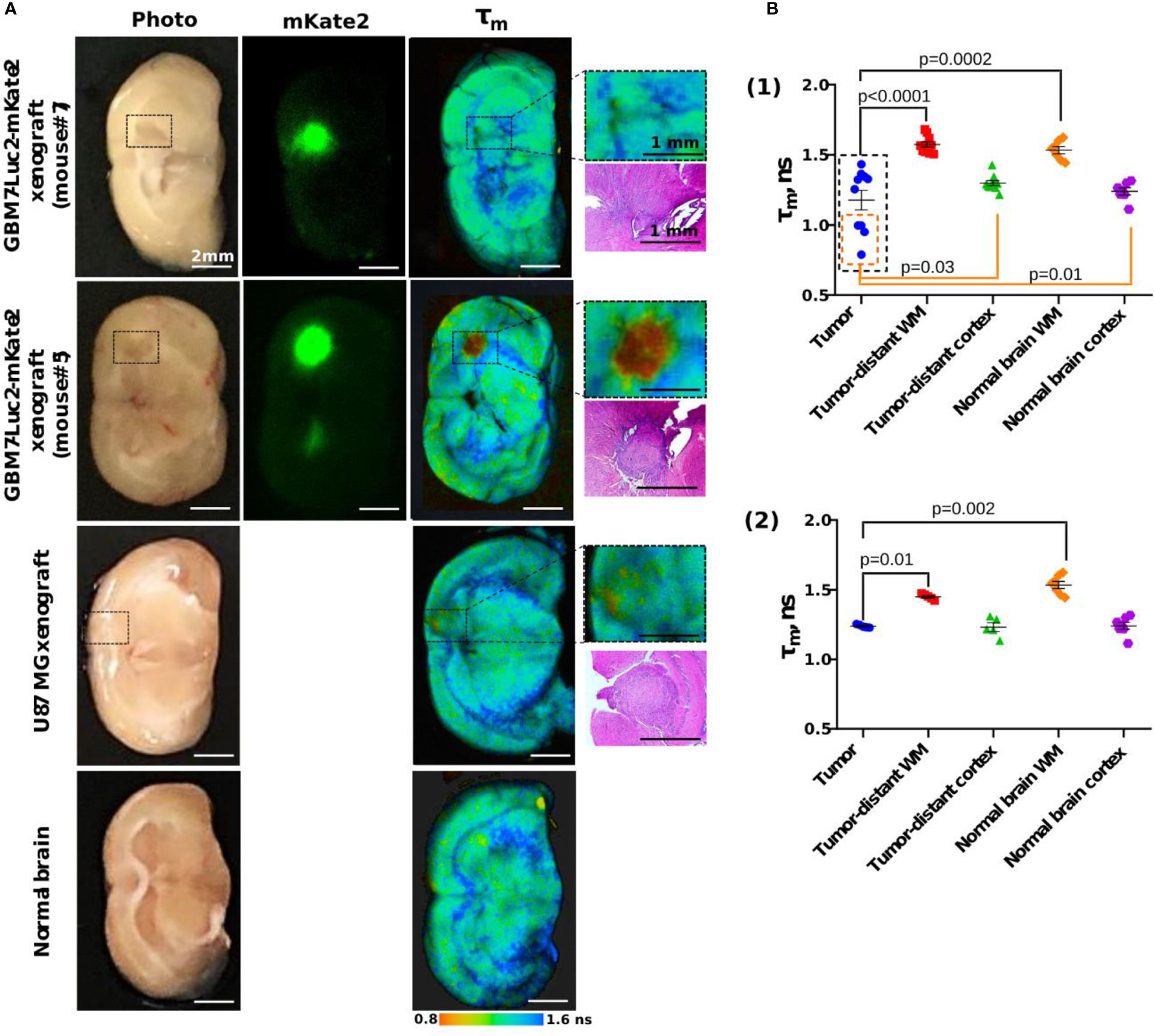
Figure 7 Macro-FLIM of human GBM xenografts and normal brain. (A) Representative autofluorescence time-resolved images of GBM7-Luc2-mKate2 xenografts, U87 MG xenograft and normal mouse brain without tumor. Enlarged regions with a tumor are indicated by the black squares on the lower-magnification panel. Corresponding H&E stained section is presented under each enlarged region. (B) Quantification of the mean fluorescence lifetime tm in NAD(P)H spectral channel in (1) dual-labeled human GBM xenografts and (2) U87 MG xenografts and normal brain. Scatter dot plot displays the measurements for individual animals (dots) and the mean and SEM (horizontal lines). WM is a white matter.
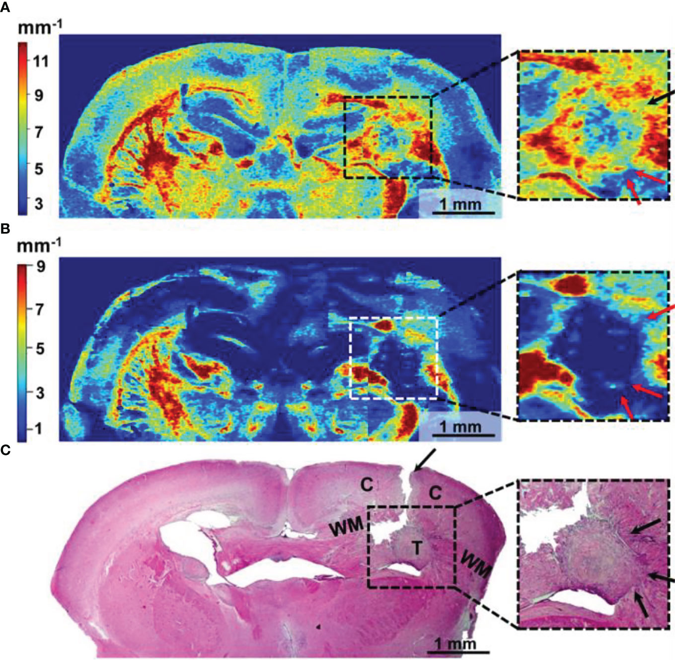
Figure 8 Wide-field OCT color-coded maps of the mouse brain with GBM7-Luc2-mKate2 tumor (A, B) and corresponding histology (C). Color-coded maps based on two optical coefficients calculation: attenuation in co-channel (Attco-) (A) and in cross-channel (Attcross-) (B). Perifocal areas of high cancer density are marked with arrows (see enlarged fragments). T, tumor; C, cortex; WM, white matter.
The authors apologize for this error and state that this does not change the scientific conclusions of the article in any way.
All claims expressed in this article are solely those of the authors and do not necessarily represent those of their affiliated organizations, or those of the publisher, the editors and the reviewers. Any product that may be evaluated in this article, or claim that may be made by its manufacturer, is not guaranteed or endorsed by the publisher.
Keywords: glioblastoma (GBM), primary cell line, patient-derived xenograft (PDX), fluorescence imaging, FLIM (fluorescence lifetime imaging microscopy)
Citation: Yuzhakova D, Kiseleva E, Shirmanova M, Shcheslavskiy V, Sachkova D, Snopova L, Bederina E, Lukina M, Dudenkova V, Yusubalieva G, Belovezhets T, Matvienko D and Baklaushev V (2022) Corrigendum: Highly invasive fluorescent/bioluminescent patient-derived orthotopic model of glioblastoma in mice. Front. Oncol. 12:1040637. doi: 10.3389/fonc.2022.1040637
Received: 09 September 2022; Accepted: 15 September 2022;
Published: 29 September 2022.
Edited and Reviewed by:
Ellen Ackerstaff, Memorial Sloan Kettering Cancer Center, United StatesCopyright © 2022 Yuzhakova, Kiseleva, Shirmanova, Shcheslavskiy, Sachkova, Snopova, Bederina, Lukina, Dudenkova, Yusubalieva, Belovezhets, Matvienko and Baklaushev. This is an open-access article distributed under the terms of the Creative Commons Attribution License (CC BY). The use, distribution or reproduction in other forums is permitted, provided the original author(s) and the copyright owner(s) are credited and that the original publication in this journal is cited, in accordance with accepted academic practice. No use, distribution or reproduction is permitted which does not comply with these terms.
*Correspondence: Diana Yuzhakova, eXV6aGFrb3ZhLWRpYW5hQG1haWwucnU=; Vladislav Shcheslavskiy, dmlzQGJlY2tlci1oaWNrbC5kZQ==
†These authors have contributed equally to this work
Disclaimer: All claims expressed in this article are solely those of the authors and do not necessarily represent those of their affiliated organizations, or those of the publisher, the editors and the reviewers. Any product that may be evaluated in this article or claim that may be made by its manufacturer is not guaranteed or endorsed by the publisher.
Research integrity at Frontiers
Learn more about the work of our research integrity team to safeguard the quality of each article we publish.