94% of researchers rate our articles as excellent or good
Learn more about the work of our research integrity team to safeguard the quality of each article we publish.
Find out more
MINI REVIEW article
Front. Synaptic Neurosci. , 06 May 2022
Volume 14 - 2022 | https://doi.org/10.3389/fnsyn.2022.891740
This article is part of the Research Topic Subcellular Computations and Information Processing View all 5 articles
Binaural coincidence detection is the initial step in encoding interaural time differences (ITDs) for sound-source localization. In birds, neurons in the nucleus laminaris (NL) play a central role in this process. These neurons receive excitatory synaptic inputs on dendrites from both sides of the cochlear nucleus and compare their coincidences at the soma. The NL is tonotopically organized, and individual neurons receive a pattern of synaptic inputs that are specific to their tuning frequency. NL neurons differ in their dendritic morphology along the tonotopic axis; their length increases with lower tuning frequency. In addition, our series of studies have revealed several frequency-dependent refinements in the morphological and biophysical characteristics of NL neurons, such as the amount and subcellular distribution of ion channels and excitatory and inhibitory synapses, which enable the neurons to process the frequency-specific pattern of inputs appropriately and encode ITDs at each frequency band. In this review, we will summarize these refinements of NL neurons and their implications for the ITD coding. We will also discuss the similarities and differences between avian and mammalian coincidence detectors.
A neuron is a computational unit in the brain. It receives and integrates synaptic inputs at dendrites and the soma, and generates action potentials in the axon, thereby communicating with other neurons. These neuronal compartments show huge diversity among neuronal types and brain regions. They include variations in length, thickness, branching patterns of dendrites, distributions of ion channels as well as excitatory and inhibitory synapses, all of which finely tune the computation of individual neurons and play a key role in determining the function of neural circuits and the brain (Shepherd, 2004).
In the auditory system, sound information is processed after the sound is divided into each frequency component in the cochlea and converted into a sequence of action potentials in the auditory nerve, where the frequency is represented as the selective activity in the fibers tuned to a specific frequency (characteristic frequency, CF), phase as the timing of action potentials phase-locked to the sound wave, and intensity as the number of action potentials in the fibers. Accordingly, the processing of sound in central auditory circuits is operated for each frequency separately by neurons with different CFs, which are arranged topographically in circuits (tonotopy). These results imply that the temporal profiles of synaptic input to auditory neurons differ substantially among tonotopic regions. To ensure precise signal processing across intensities in each tonotopic region, auditory neurons show multiple frequency-specific refinements in the morphological and biophysical characteristics of dendrites and other neuronal compartments. Such refinements have been studied extensively in brainstem circuits that are involved in the calculation of the interaural time difference (ITD) for sound localization in birds, primarily chickens that have excellent sound localization ability (Krumm et al., 2022).
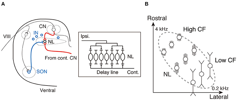
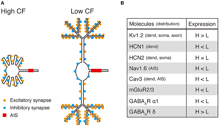
ITD information is extracted at the 3rd-order brainstem auditory nucleus, the nucleus laminaris (NL) in birds and the medial superior olive (MSO) in mammals, where synaptic inputs from both ears first converge (Klumpp and Eady, 1956; Moiseff and Konishi, 1981; Carr and Konishi, 1990; Overholt et al., 1992; Grothe, 2003). The extraction of ITDs is explained using the classical Jeffress model (Jeffress, 1948), which is based on an array of coincidence detectors innervated by delay lines. Delay lines create systematic delays in the arrival of action potentials within the array, while coincidence detectors generate action potentials when they receive simultaneous synaptic inputs from both ears. In the NL of the chicken, projection fibers from the contralateral cochlear nucleus form delay lines, and NL neurons act as coincidence detectors, which allows the encoding of each ITD as the location of the most active neuron within the nucleus (Figure 1A). The NL is organized tonotopically (Rubel and Parks, 1975), with the CF of neurons increasing from caudo-lateral to rostro-medial direction for the audible frequency range (0.2–4 kHz), and ITDs are processed by frequency-specific neurons (Figure 1B). Accurate coincidence detection in each tonotopic region is a prerequisite for ITD coding (Kuba, 2007). Importantly, NL neurons receive synaptic inputs at a rate corresponding to their CF from each side of the cochlear nucleus during ongoing sound (Funabiki et al., 2011), which is attributed to the convergence of phase-locked inputs from multiple afferents on the neurons, implying that the oscillation frequency of converging synaptic inputs reaches a few kHz for neurons with higher CFs (>1 kHz). Thus, rapid synaptic and membrane responses are expected to be particularly important for neurons with higher CF to integrate binaural signals during ongoing sound. In contrast, for neurons with low CF (<1 kHz), the requirement for speed may be smaller because of the relatively long period of sound waves (Slee et al., 2010); however, it is still critical to maintain integration for wide intensity ranges by adjusting the size of synaptic responses. To meet these requirements and accomplish ITD coding across frequencies, NL neurons are differentiated along the tonotopic axis, which is represented by dendritic morphology, potassium and other active conductance channels, and distributions of excitatory and inhibitory synapses (Figure 2). In the following section, we summarize these differentiations and explain how they optimize frequency-dependent ITD coding in the NL. We will also discuss the similarities and differences between NL and MSO neurons.

Figure 1. Organization of interaural time difference (ITD) coding circuits in the nucleus laminaris (NL) of the chicken. (A) Brainstem auditory circuit of the chicken. Red lines indicate the excitatory projections from the bilateral CN to the NL. Blue symbols and line indicate inhibitory sources (SON and IN) and projection from the SON to the NL, respectively. A schematic drawing of the ITD coding circuit within the NL is shown in the box (right). CN, cochlear nucleus; IN, interneuron; VIII, auditory nerve; SON, superior olivary nucleus. (B) Tonotopic organization of the NL.

Figure 2. Summary of tonotopic refinements in chick NL neurons. (A) Tonotopic differences in excitatory and inhibitory synapses (location and density) and AIS (location and length) are shown in schematic drawings of neurons with high and low characteristic frequency (CF). (B) Location of ion channels and receptors and their tonotopic differences in expression are listed.
NL neurons extend their dendrites in the ventral and dorsal directions and receive excitatory synaptic inputs from each ear on different sides of the dendrites. A model study estimated that this segregation of bilateral inputs is crucial for preventing the generation of action potentials by unilateral inputs (Agmon-Snir et al., 1998). Notably, the length of dendrites differs along the tonotopic axis in the NL; it increases with decreasing CF (Figures 1B, 2A) (Smith and Rubel, 1979; Kuba et al., 2005). Neurons with higher CF have multiple short and thick dendrites, which ensure fast and large EPSPs at the soma by making the entire cell electrically compact and are favorable for the ITD coding of high-frequency sounds. In contrast, neurons with low CF have a smaller number of long dendrites with many distal thin branches, which is compatible with the above model, and their functional role in ITD coding will be discussed in a later section.
In the MSO of the gerbil, dendrites are also long, as in NL neurons with a low CF; however, they are composed of a thick trunk and mostly lack branches (Rautenberg et al., 2009), which reduces dendritic filtering and ensures a fast EPSP time course (Mathews et al., 2010). However, these characteristics might have large variations because MSO neurons with many thin dendritic branches are also observed in gerbils (Bondy et al., 2021) and other mammalian species (Schwartz, 1977; Henkel and Brunso-Bechtold, 1990; Smith, 1995).
Kv1.2 channels, which mediate the low-voltage-activated dendrotoxin-sensitive K+ current, are the major source of conductance activated at rest in NL neurons. These channels are distributed in the soma and dendrites, while they are slightly skewed to the soma (Kuba et al., 2005). A similar distribution of Kv1 channels has been observed in the MSO neurons of the gerbil (Mathews et al., 2010). The expression level of Kv1.2 channels differs among tonotopic regions; it is higher in neurons with higher CF (Figure 2B) (Kuba et al., 2005; Hamlet et al., 2014). Because these channels are strongly activated by EPSPs because of their rapid activation kinetics, they accelerate the falling phase of EPSPs. Thus, the rich expression of Kv1.2 channels together with the faster kinetics of EPSCs accelerates the EPSP time course and ensures the accurate ITD coding in the neurons (Kuba et al., 2005; Slee et al., 2010). In contrast, neurons with low CF show relatively low expression of Kv1.2 channels and slow kinetics of EPSCs, which underlies the relatively slow EPSP time course in the neurons.
Hyperpolarization-activated cyclic nucleotide-gated (HCN) channels, which mediate inward current during membrane hyperpolarization, are preferentially distributed at dendrites and are the other major source of conductance at rest in NL neurons (Biel et al., 2009). Two subtypes, HCN1 and HCN2, are expressed in NL, and their expression levels differ among tonotopic regions; HCN1 expression increases toward lower CFs, whereas HCN2 expression is less graded along the tonotopic axis (Figure 2B) (Yamada et al., 2005). HCN1 channels are activated substantially at rest because of their positive voltage dependence, thereby increasing resting conductance at the dendrites of neurons with low CF. In cortical and hippocampal pyramidal neurons, dendritic HCN channels deactivate during EPSPs and ensure a uniform EPSP time course, irrespective of the input location (Magee, 1998; Williams and Stuart, 2000). In auditory neurons, basal activation of HCN channels has been proposed to accelerate the EPSP time course, although its kinetics is slow (Golding et al., 1995; Yamada et al., 2005). Thus, HCN1 channels in NL neurons with low CF might contribute to adjusting the EPSP time course at dendrites. However, HCN2 channels have more negative voltage dependence and their contribution to the resting conductance is small. Interestingly, noradrenaline causes a depolarizing shift in the voltage dependence of HCN2 channels through the elevation of intracellular cAMP in neurons with high CF (Yamada et al., 2005). This modulation causes membrane depolarization, accelerates the EPSP time course, and improves coincidence detection. Monoaminergic modulation has been reported in the MSO, where serotonin causes a negative shift in the activation voltage of axonal HCN1 channels and increases neuronal excitability (Ko et al., 2016). Therefore, the arousal system of animals may have the potential to regulate the acuity of sound localization with attention.
Nav1.6 channels are the major subtype of Na+ channels in NL neurons (Kuba et al., 2014). These channels are not distributed at the dendrites and the soma but accumulate at the axon initial segment (AIS) (Kuba et al., 2006), which is the neuronal compartment that mediates the generation of action potentials (Kole and Stuart, 2012). The AIS of NL neurons differs in length and distance from the soma systematically along the tonotopic axis; it is shorter and located more distally toward the high CF (Figure 2A). This differentiation in AIS distribution is crucial for NL neurons to maintain membrane excitability and maximize their response to ITD during ongoing inputs in each tonotopic region (Kuba et al., 2006). In particular, in neurons with high CF, high-rate synaptic inputs lead to plateau depolarization at the soma due to strong temporal summation, which causes inactivation of Na+ channels at the AIS and reduces neuronal excitability. However, this effect is relieved by separating the AIS from the soma, which attenuates depolarization at the AIS via the filtering effects of the axon. This emphasizes the importance of adjusting the distribution of Na+ channels in the AIS for frequency-dependent ITD coding in NL. A tonotopic difference in AIS length has been reported in MSO neurons of the baboon (Kim et al., 2021), but not in the gerbil (Bondy et al., 2021).
T-type Ca2+ channels (Cav3 channels) are characterized by their rapid and transient activation around resting potentials (Perez-Reyes, 2003) and mediate augmentation of EPSPs at dendrites in various neurons (Huguenard, 1996; Gillessen and Alzheimer, 1997; Magee and Carruth, 1999; Cain and Snutch, 2010). In the NL, Cav3 channels are distributed at the dendrites of neurons with low CF and augment EPSPs (Figure 2B) (Fukaya et al., 2018). However, this augmentation of EPSPs is limited to the initial phase during repetitive inputs because of the progression of inactivation, suggesting their minimal contributions to dendritic integration and the ITD tuning for ongoing sound. Therefore, Cav3 channels may function as a source of Ca2+, which maintains the dendritic structure of neurons through calcium-dependent kinase pathways (Lohmann and Wong, 2005; Redmond and Ghosh, 2005; Nourbakhsh and Yadav, 2021). This might be compatible with the observation that gerbil MSO neurons, which have a rather simple dendritic morphology, lack these channels during the late postnatal period (Franzen et al., 2020). Cav3 channels are also localized in the AIS of NL neurons (Fukaya et al., 2018). The roles of these channels in AIS are unknown, but they may contribute to the reorganization of the AIS structure (Kuba, 2012; Akter et al., 2020).
Excitatory synapses are distributed specifically in the dendrites of NL neurons. The pattern of synapse distribution within dendrites critically affects the computation of neurons, and the effects depend on the morphology and biophysical characteristics of the dendrites. For example, in pyramidal neurons, dendrites have strong active conductances, such as Na+ channels, Ca2+ channels, and NMDA receptors, and clustering of synapses amplifies EPSPs and causes supralinear integration by activating these active conductances (Johnston and Narayanan, 2008; Larkum and Nevian, 2008). In NL neurons, in contrast, this amplification does not occur because these active conductances are weak or absent at dendrites (Kuba et al., 2006; Sanchez et al., 2010; Fukaya et al., 2018). We recently revealed that the distribution of excitatory synapses is coupled with the morphology of dendrites in NL neurons and optimizes ITD coding in each tonotopic region (Figure 2A) (Yamada and Kuba, 2021). In neurons with higher CF, excitatory synapses are distributed uniformly along short and thick dendrites, which minimizes the loss of charge during the integration and propagation of EPSPs along dendrites. Thus, this dendritic synapse geometry in neurons with higher CF maintains the size and shape of EPSPs at the soma, thereby improving ITD coding for high-frequency sound. However, in neurons with low CF, excitatory synapses are clustered in the distal thin part of the long dendrites. This implies that the input site is electrically compact because of the thinness of the dendrites and segregation from the soma. This facilitates spatial summation, causes large depolarization, and increases the loss of charge by reducing the driving force of the synaptic current and increasing the shunting K+ current at the input site, which increases the sublinearity of EPSPs at the soma. Since synaptic inputs from each ear are restricted to one side of the dendrites in these neurons, this sublinearity suppresses action potential generation by monaural inputs, while maintaining action potential generation during binaural coincident inputs by preventing saturation of EPSPs, which ensures ITD coding even for strong inputs (Agmon-Snir et al., 1998; Yamada and Kuba, 2021). Notably, this effect does not occur for high-frequency sound (>1 kHz) because extensive temporal summation causes plateau depolarization and suppresses action potential generation, even for binaural coincident inputs. Thus, the long dendrites together with the distal synapse clustering allow fine adjustment of the size of EPSPs in neurons with low CF and work as a mechanism to maintain ITD coding across wide intensity ranges for low-frequency sounds. Activity-dependent regulation during ongoing sound is also prominent in neurons with low CF; activation of metabotropic glutamate receptors (mGluRs) lowers the release probability at presynaptic terminals to reduce synaptic depression (Okuda et al., 2013; Lu et al., 2018) and/or activate the high-threshold Kv channels to follow repetitive firing (Hamlet and Lu, 2016).
In the MSO of the gerbil, excitatory synapses are distributed uniformly along the long dendrites (Couchman et al., 2012; Callan et al., 2021), which would minimize the loss of charge along dendrites and maintain the size and shape of EPSPs at the soma, as in NL neurons with high CF. However, further analyses of the distribution of synapses are needed in other mammalian species, as dendrite morphology differs among species.
GABAergic inhibitory synapses are distributed uniformly in the dendrites and soma in NL neurons. These GABAergic synapses do not cause hyperpolarization and have their effects primarily via shunting conductance; they accelerate the time course and decrease the size of EPSPs (Hyson et al., 1995; Funabiki et al., 1998; Yang et al., 1999; Tang et al., 2009). The density of GABAergic synapses differs along the tonotopic axis; it increases toward a low CF (Figure 2A) (Carr et al., 1989; Code et al., 1989; Nishino et al., 2008). In addition, the composition of GABAA receptors also differs; the expression of the α1 subunit is higher toward lower CFs, making the kinetics of IPSCs faster in neurons with low CF (Figure 2B) (Tang and Lu, 2012; Yamada et al., 2013). Consequently, GABAergic inputs have more prominent effects on the ITD coding of NL neurons with low CF (Nishino et al., 2008), while the tonic conductance of extrasynaptic GABAA receptors containing δ subunits is larger in neurons with higher CF (Figure 2B) (Tang et al., 2011). There are two sources of GABAergic inputs to the NL: neurons in the superior olivary nucleus (SON; Lachica et al., 1994; Yang et al., 1999; Burger et al., 2005) and inhibitory interneurons around the NL (Carr et al., 1989; von Bartheld et al., 1989). These GABAergic inputs are driven by auditory nerve activity and provide feedforward inhibition to NL neurons. Thus, they increase with an increase in sound intensity and work as another mechanism for maintaining ITD coding for intensity ranges for low-frequency sounds. Interestingly, however, the pattern of IPSCs differs between the two inputs: inputs from SON neurons cause plateau-like IPSCs (Yang et al., 1999; Coleman et al., 2011), whereas those from interneurons cause phasic IPSCs phase-locked to stimuli (Yamada et al., 2013). Therefore, both GABAergic inputs accelerate and attenuate EPSPs and suppress action potential generation by monaural inputs, while the effects from the SON are substantial for the entire intensity range, and the effects from interneurons are restricted to weak intensity stimuli. This indicates that these two types of inhibitions cooperate with each other and ensure ITD coding from weak to strong intensity inputs for the neurons with low CF. The uniform distribution of these inhibitory synapses within a cell may avoid their accumulation at dendrites, which would otherwise increase the shunting conductance and make EPSPs too small to induce action potentials, even for binaural coincident inputs. Thus, the distributions of excitatory and inhibitory synapses would be particularly important for NL neurons with low CF in adjusting the shape and size of EPSPs and optimizing ITD coding against intensities.
MSO neurons receive feedforward glycinergic inhibitory inputs from both ears at the dendrites and the soma. Because these inhibitory inputs are precisely time-locked to the excitatory inputs, they directly modulate the shape of EPSPs and shift the timing of their peak. These glycinergic IPSPs have been proposed to shift the optimal ITDs for maximal firing (Brand et al., 2002), but their functional roles remain under debate (Joris and Yin, 2007; Roberts et al., 2013; Franken et al., 2015).
NL neurons of the chicken show multiple differentiations according to their CF. In neurons with high CF, the rapid EPSP time course is primarily shaped by strong Kv1.2 expression and short dendrites. In addition, the uniform distribution of excitatory synapses, together with fewer inhibitory synapses, prevent extensive loss of charge during the activation of large Kv1.2 conductances. In contrast, in neurons with low CF, the EPSP time course is rather slow, in part because of the weak Kv1.2 expression, as well as long and branched dendrites. However, inhibitory synapses are enriched in the neurons, and excitatory synapses are preferentially clustered at the distal thin part of long dendrites. These characteristics enable the neurons to adjust the size of EPSPs in an intensity-dependent manner, prevent saturation of EPSPs, and ensure the dynamic range of ITD coding, which underlies the intensity tolerance of low-frequency spatial hearing.
MSO neurons of the gerbil encode ITDs of low-frequency sounds and possess long dendrites, like NL neurons with low CF. However, they differ in several ways from NL neurons. First, MSO neurons have large Kv1 currents, lack thin dendritic branches, and distribute excitatory synapses uniformly along dendrites, which accelerates the time course of somatic EPSPs, as observed in NL neurons with high CF. Second, they do not show apparent tonotopic variations in these characteristics (Bondy et al., 2021; but see Baumann et al., 2013). Third, glycinergic inhibitory synapses shape the time course of EPSPs, and their contribution to the size of EPSPs remains elusive. The implications of these differences are unknown, but they may reflect a species difference in the strategy for low-frequency ITD coding and/or in head size between animals. In the mammalian lateral superior olive, which is responsible for binaural spatial hearing of higher frequency sounds, neurons show tonotopic variations in their dendritic morphology and expression of Kv1 channels (Sanes et al., 1992; Barnes-Davies et al., 2004). Therefore, detailed analyses of the lateral superior olive and MSO along the tonotopic axis in other mammalian species may provide a more comprehensive understanding of the sound frequency-dependent computation of ITDs in brainstem auditory circuits.
RY and HK wrote the manuscript. Both authors contributed to the article and approved the submitted version.
This work was supported by a grant-in-aid from MEXT for Scientific Research (21H02577 to HK) and grants from the Takeda Science Foundation (to RY and HK).
The authors declare that the research was conducted in the absence of any commercial or financial relationships that could be construed as a potential conflict of interest.
All claims expressed in this article are solely those of the authors and do not necessarily represent those of their affiliated organizations, or those of the publisher, the editors and the reviewers. Any product that may be evaluated in this article, or claim that may be made by its manufacturer, is not guaranteed or endorsed by the publisher.
We thank Editage for English language editing.
Agmon-Snir, H., Carr, C. E., and Rinzel, J. (1998). The role of dendrites in auditory coincidence detection. Nature 393, 268–272. doi: 10.1038/30505
Akter, N., Fukaya, R., Adachi, R., Kawabe, H., and Kuba, H. (2020). Structural and functional refinement of the axon initial segment in avian cochlear nucleus during development. J. Neurosci. 40, 6709–6721. doi: 10.1523/JNEUROSCI.3068-19.2020
Barnes-Davies, M., Barker, M. C., Osmani, F., and Forsythe, I. D. (2004). Kv1 currents mediate a gradient of principal neuron excitability across the tonotopic axis in the rat lateral superior olive. Eur. J. Neurosci. 19, 325–333. doi: 10.1111/j.0953-816X.2003.03133.x
Baumann, V. J., Lehnert, S., Leibold, C., and Koch, U. (2013). Tonotopic organization of the hyperpolarization-activated current (Ih) in the mammalian medial superior olive. Front. Neural Circ. 7, 117. doi: 10.3389/fncir.2013.00117
Biel, M., Wahl-Schott, C., Michalakis, S., and Zong, X. (2009). Hyperpolarization-activated cation channels: from genes to function. Physiol. Rev. 89, 847–885. doi: 10.1152/physrev.00029.2008
Bondy, B. J., Haimes, D. B., and Golding, N. L. (2021). Physiological diversity influences detection of stimulus envelope and fine structure in neurons of the medial superior olive. J. Neurosci. 41, 6234–6245. doi: 10.1523/JNEUROSCI.2354-20.2021
Brand, A., Behrend, O., Marquardt, T., McAlpine, D., and Grothe, B. (2002). Precise inhibition is essential for microsecond interaural time difference coding. Nature 417, 543–547. doi: 10.1038/417543a
Burger, R. M., Cramer, K. S., Pfeiffer, J. D., and Rubel, E. W. (2005). Avian superior olivary nucleus provides divergent inhibitory input to parallel auditory pathways. J. Comp. Neurol. 481, 6–18. doi: 10.1002/cne.20334
Cain, S. M., and Snutch, T. P. (2010). Contributions of T-type calcium channel isoforms to neuronal firing. Channels 4, 475–482. doi: 10.4161/chan.4.6.14106
Callan, A. R., He,ß, M., Felmy, F., and Leibold, C. (2021). Arrangement of excitatory synaptic inputs on dendrites of the medial superior olive. J. Neurosci. 41, 269–283. doi: 10.1523/JNEUROSCI.1055-20.2020
Carr, C. E., Fujita, I., and Konishi, M. (1989). Distribution of GABAergic neurons and terminals in the auditory system of the barn owl. J. Comp. Neurol. 286, 190–207. doi: 10.1002/cne.902860205
Carr, C. E., and Konishi, M. (1990). A circuit for detection of interaural time differences in the brain stem of the barn owl. J. Neurosci. 10, 3227–3246. doi: 10.1523/JNEUROSCI.10-10-03227.1990
Code, R. A., Burd, G. D., and Rubel, E. W. (1989). Development of GABA immunoreactivity in brainstem auditory nuclei of the chick: ontogeny of gradients in terminal staining. J. Comp. Neurol. 284, 504–518. doi: 10.1002/cne.902840403
Coleman, W. L., Fischl, M. J., Weimann, S. R., and Burger, R. M. (2011). GABAergic and glycinergic inhibition modulate monaural auditory response properties in the avian superior olivary nucleus. J. Neurophysiol. 105, 2405–2420. doi: 10.1152/jn.01088.2010
Couchman, K., Grothe, B., and Felmy, F. (2012). Functional localization of neurotransmitter receptors and synaptic inputs to mature neurons of the medial superior olive. J. Neurophysiol. 107, 1186–1198. doi: 10.1152/jn.00586.2011
Franken, T. P., Roberts, M. T., Wei, L., Golding, N. L., and Joris, P. X. (2015). In vivo coincidence detection in mammalian sound localization generates phase delays. Nat. Neurosci. 18, 444–452. doi: 10.1038/nn.3948
Franzen, D. L., Gleiss, S. A., Kellner, C. J., Kladisios, N., and Felmy, F. (2020). Activity-dependent calcium signaling in neurons of the medial superior olive during late postnatal development. J. Neurosci. 40, 1689–1700. doi: 10.1523/JNEUROSCI.1545-19.2020
Fukaya, R., Yamada, R., and Kuba, H. (2018). Tonotopic variation of the T-type Ca2+ current in avian auditory coincidence detector neurons. J. Neurosci. 38, 335–346. doi: 10.1523/JNEUROSCI.2237-17.2017
Funabiki, K., Ashida, G., and Konishi, M. (2011). Computation of interaural time difference in the owl's coincidence detector neurons. J. Neurosci. 31, 15245–15256. doi: 10.1523/JNEUROSCI.2127-11.2011
Funabiki, K., Koyano, K., and Ohmori, H. (1998). The role of GABAergic inputs for coincidence detection in the neurones of nucleus laminaris of the chick. J. Physiol. 508, 851–869. doi: 10.1111/j.1469-7793.1998.851bp.x
Gillessen, T., and Alzheimer, C. (1997). Amplification of EPSPs by low Ni(2+)- and amiloride-sensitive Ca2+ channels in apical dendrites of rat CA1 pyramidal neurons. J. Neurophysiol. 77, 1639–1643. doi: 10.1152/jn.1997.77.3.1639
Golding, N. L., Robertson, D., and Oertel, D. (1995). Recordings from slices indicate that octopus cells of the cochlear nucleus detect coincident firing of auditory nerve fibers with temporal precision. J. Neurosci. 15, 3138–3153. doi: 10.1523/JNEUROSCI.15-04-03138.1995
Grothe, B. (2003). New roles for synaptic inhibition in sound localization. Nat. Rev. Neurosci. 4, 540–550. doi: 10.1038/nrn1136
Hamlet, W. R., Liu, Y. W., Tang, Z. Q., and Lu, Y. (2014). Interplay between low threshold voltage-gated K+ channels and synaptic inhibition in neurons of the chicken nucleus laminaris along its frequency axis. Front. Neural. Circ. 8, 51. doi: 10.3389/fncir.2014.00051
Hamlet, W. R., and Lu, Y. (2016). Intrinsic plasticity induced by group II metabotropic glutamate receptors via enhancement of high-threshold Kv currents in sound localizing neurons. Neuroscience 324, 177–190. doi: 10.1016/j.neuroscience.2016.03.010
Henkel, C. K., and Brunso-Bechtold, J. K. (1990). Dendritic morphology and development in the ferret medial superior olivary nucleus. J. Comp. Neurol. 294, 377–388. doi: 10.1002/cne.902940307
Huguenard, J. R. (1996). Low-threshold calcium currents in central nervous system neurons. Annu. Rev. Physiol. 58, 329–348. doi: 10.1146/annurev.ph.58.030196.001553
Hyson, R. L., Reyes, A. D., and Rubel, E. W. (1995). A depolarizing inhibitory response to GABA in brainstem auditory neurons of the chick. Brain. Res. 677, 117–126. doi: 10.1016/0006-8993(95)00130-I
Jeffress, L. A. (1948). A place theory of sound localization. J. Comp. Physiol. Psychol. 41, 35–39. doi: 10.1037/h0061495
Johnston, D., and Narayanan, R. (2008). Active dendrites: colorful wings of the mysterious butterflies. Trends Neurosci. 31, 309–316. doi: 10.1016/j.tins.2008.03.004
Joris, P., and Yin, T. C. (2007). A matter of time: internal delays in binaural processing. Trends Neurosci. 30, 70–78. doi: 10.1016/j.tins.2006.12.004
Kim, E. J., Nip, K., Blanco, C., and Kim, J. H. (2021). Structural refinement of the auditory brainstem neurons in baboons during perinatal development. Front. Cell Neurosci. 15, 648562. doi: 10.3389/fncel.2021.648562
Klumpp, R. G., and Eady, H. R. (1956). Some measurements of interaural time difference thresholds. J. Acoust. Soc. Am. 28, 859–860. doi: 10.1121/1.1908493
Ko, K. W., Rasband, M. N., Meseguer, V., Kramer, R. H., and Golding, N. L. (2016). Serotonin modulates spike probability in the axon initial segment through HCN channels. Nat. Neurosci. 19, 826–834. doi: 10.1038/nn.4293
Kole, M. H., and Stuart, G. J. (2012). Signal processing in the axon initial segment. Neuron 73, 235–247. doi: 10.1016/j.neuron.2012.01.007
Krumm, B., Klump, G. M., Köppl, C., Beutelmann, R., and Langemann, U. (2022). Chickens have excellent sound localization ability. J. Exp. Biol. 225, jeb243601. doi: 10.1242/jeb.243601
Kuba, H. (2007). Cellular and molecular mechanisms of avian auditory coincidence detection. Neurosci. Res. 59, 370–376. doi: 10.1016/j.neures.2007.08.003
Kuba, H. (2012). Structural tuning and plasticity of the axon initial segment in auditory neurons. J. Physiol. 590, 5571–5579. doi: 10.1113/jphysiol.2012.237305
Kuba, H., Adachi, R., and Ohmori, H. (2014). Activity-dependent and activity-independent development of the axon initial segment. J. Neurosci. 34, 3443–3453. doi: 10.1523/JNEUROSCI.4357-13.2014
Kuba, H., Ishii, T. M., and Ohmori, H. (2006). Axonal site of spike initiation enhances auditory coincidence detection. Nature 444, 1069–1072. doi: 10.1038/nature05347
Kuba, H., Yamada, R., Fukui, I., and Ohmori, H. (2005). Tonotopic specialization of auditory coincidence detection in nucleus laminaris of the chick. J. Neurosci. 25, 1924–1934. doi: 10.1523/JNEUROSCI.4428-04.2005
Lachica, E. A., Rübsamen, R., and Rubel, E. W. (1994). GABAergic terminals in nucleus magnocellularis and laminaris originate from the superior olivary nucleus. J. Comp. Neurol. 348, 403–418. doi: 10.1002/cne.903480307
Larkum, M. E., and Nevian, T. (2008). Synaptic clustering by dendritic signalling mechanisms. Curr. Opin. Neurobiol. 18, 321–331. doi: 10.1016/j.conb.2008.08.013
Lohmann, C., and Wong, R. O. (2005). Regulation of dendritic growth and plasticity by local and global calcium dynamics. Cell Calcium 37, 403–409. doi: 10.1016/j.ceca.2005.01.008
Lu, Y., Liu, Y., and Curry, R. J. (2018). Activity-dependent synaptic integration and modulation of bilateral excitatory inputs in an auditory coincidence detection circuit. J. Physiol. 596, 1981–1997. doi: 10.1113/JP275735
Magee, J. C. (1998). Dendritic hyperpolarization-activated currents modify the integrative properties of hippocampal CA1 pyramidal neurons. J. Neurosci. 18, 7613–7624. doi: 10.1523/JNEUROSCI.18-19-07613.1998
Magee, J. C., and Carruth, M. (1999). Dendritic voltage-gated ion channels regulate the action potential firing mode of hippocampal CA1 pyramidal neurons. J. Neurophysiol. 82, 1895–1901. doi: 10.1152/jn.1999.82.4.1895
Mathews, P. J., Jercog, P. E., Rinzel, J., Scott, L. L., and Golding, N. L. (2010). Control of submillisecond synaptic timing in binaural coincidence detectors by K(v)1 channels. Nat. Neurosci. 13, 601–609. doi: 10.1038/nn.2530
Moiseff, A., and Konishi, M. (1981). Neuronal and behavioral sensitivity to binaural time differences in the owl. J. Neurosci. 1, 40–48. doi: 10.1523/JNEUROSCI.01-01-00040.1981
Nishino, E., Yamada, R., Kuba, H., Hioki, H., Furuta, T., Kaneko, T., et al. (2008). Sound-intensity-dependent compensation for the small interaural time difference cue for sound source localization. J. Neurosci. 28, 7153–7164. doi: 10.1523/JNEUROSCI.4398-07.2008
Nourbakhsh, K., and Yadav, S. (2021). Kinase signaling in dendritic development and disease. Front. Cell Neurosci. 15, 624648. doi: 10.3389/fncel.2021.624648
Okuda, H., Yamada, R., Kuba, H., and Ohmori, H. (2013). Activation of metabotropic glutamate receptors improves the accuracy of coincidence detection by presynaptic mechanisms in the nucleus laminaris of the chick. J. Physiol. 591, 365–378. doi: 10.1113/jphysiol.2012.244350
Overholt, E. M., Rubel, E. W., and Hyson, R. L. (1992). A circuit for coding interaural time differences in the chick brainstem. J. Neurosci. 12, 1698–1708. doi: 10.1523/JNEUROSCI.12-05-01698.1992
Perez-Reyes, E. (2003). Molecular physiology of low-voltage-activated t-type calcium channels. Physiol. Rev. 83, 117–161. doi: 10.1152/physrev.00018.2002
Rautenberg, P. L., Grothe, B., and Felmy, F. (2009). Quantification of the three-dimensional morphology of coincidence detector neurons in the medial superior olive of gerbils during late postnatal development. J. Comp. Neurol. 517, 385–396. doi: 10.1002/cne.22166
Redmond, L., and Ghosh, A. (2005). Regulation of dendritic development by calcium signaling. Cell Calcium 37, 411–416. doi: 10.1016/j.ceca.2005.01.009
Roberts, M. T., Seeman, S. C., and Golding, N. L. (2013). A mechanistic understanding of the role of feedforward inhibition in the mammalian sound localization circuitry. Neuron 78, 923–935. doi: 10.1016/j.neuron.2013.04.022
Rubel, E. W., and Parks, T. N. (1975). Organization and development of brain stem auditory nuclei of the chicken: tonotopic organization of n. magnocellularis and n. laminaris. J. Comp. Neurol. 164, 411–433. doi: 10.1002/cne.901640403
Sanchez, J. T., Wang, Y., Rubel, E. W., and Barria, A. (2010). Development of glutamatergic synaptic transmission in binaural auditory neurons. J. Neurophysiol. 104, 1774–1789. doi: 10.1152/jn.00468.2010
Sanes, D. H., Song, J., and Tyson, J. (1992). Refinement of dendritic arbors along the tonotopic axis of the gerbil lateral superior olive. Dev. Brain Res. 67, 47–55. doi: 10.1016/0165-3806(92)90024-Q
Schwartz, I. R. (1977). Dendritic arrangements in the cat medial superior olive. Neuroscience 2, 81–101. doi: 10.1016/0306-4522(77)90070-7
Shepherd, G. M. (2004). The Synaptic Organization of the Brain. 5th Edn. New York, NY: Oxford University Press.
Slee, S. J., Higgs, M. H., Fairhall, A. L., and Spain, W. J. (2010). Tonotopic tuning in a sound localization circuit. J. Neurophysiol. 103, 2857–2875. doi: 10.1152/jn.00678.2009
Smith, D. J., and Rubel, E. W. (1979). Organization and development of brain stem auditory nuclei of the chicken: dendritic gradients in nucleus laminaris. J. Comp. Neurol. 186, 213–239. doi: 10.1002/cne.901860207
Smith, P. H. (1995). Structural and functional differences distinguish principal from nonprincipal cells in the guinea pig MSO slice. J. Neurophysiol. 73, 1653–1667. doi: 10.1152/jn.1995.73.4.1653
Tang, Z. Q., Dinh, E. H., Shi, W., and Lu, Y. (2011). Ambient GABA-activated tonic inhibition sharpens auditory coincidence detection via a depolarizing shunting mechanism. J. Neurosci. 31, 6121–6131. doi: 10.1523/JNEUROSCI.4733-10.2011
Tang, Z. Q., Gao, H., and Lu, Y. (2009). Control of a depolarizing GABAergic input in an auditory coincidence detection circuit. J. Neurophysiol. 102, 1672–1683. doi: 10.1152/jn.00419.2009
Tang, Z. Q., and Lu, Y. (2012). Two GABAA responses with distinct kinetics in a sound localization circuit. J. Physiol. 590, 3787–3805. doi: 10.1113/jphysiol.2012.230136
von Bartheld, C. S., Code, R. A., and Rubel, E. W. (1989). GABAergic neurons in brainstem auditory nuclei of the chick: distribution, morphology, and connectivity. J. Comp. Neurol. 287, 470–483. doi: 10.1002/cne.902870406
Williams, S. R., and Stuart, G. J. (2000). Site independence of EPSP time course is mediated by dendritic I(h) in neocortical pyramidal neurons. J. Neurophysiol. 83, 3177–3182. doi: 10.1152/jn.2000.83.5.3177
Yamada, R., and Kuba, H. (2021). Dendritic synapse geometry optimizes binaural computation in a sound localization circuit. Sci. Adv. 7, eabh0024. doi: 10.1126/sciadv.abh0024
Yamada, R., Kuba, H., Ishii, T. M., and Ohmori, H. (2005). Hyperpolarization-activated cyclic nucleotide-gated cation channels regulate auditory coincidence detection in nucleus laminaris of the chick. J. Neurosci. 25, 8867–8877. doi: 10.1523/JNEUROSCI.2541-05.2005
Yamada, R., Okuda, H., Kuba, H., Nishino, E., Ishii, T. M., and Ohmori, H. (2013). The cooperation of sustained and phasic inhibitions increases the contrast of ITD-tuning in low-frequency neurons of the chick nucleus laminaris. J. Neurosci. 33, 3927–3938. doi: 10.1523/JNEUROSCI.2377-12.2013
Keywords: interaural time difference, coincidence detection, synapse, dendrite, ion channel
Citation: Yamada R and Kuba H (2022) Cellular Strategies for Frequency-Dependent Computation of Interaural Time Difference. Front. Synaptic Neurosci. 14:891740. doi: 10.3389/fnsyn.2022.891740
Received: 08 March 2022; Accepted: 04 April 2022;
Published: 06 May 2022.
Edited by:
Tomoe Ishikawa, Massachusetts Institute of Technology, United StatesReviewed by:
Yong Lu, Northeast Ohio Medical University, United StatesCopyright © 2022 Yamada and Kuba. This is an open-access article distributed under the terms of the Creative Commons Attribution License (CC BY). The use, distribution or reproduction in other forums is permitted, provided the original author(s) and the copyright owner(s) are credited and that the original publication in this journal is cited, in accordance with accepted academic practice. No use, distribution or reproduction is permitted which does not comply with these terms.
*Correspondence: Rei Yamada, eWFtYWRhQG1lZC5uYWdveWEtdS5hYy5qcA==; Hiroshi Kuba, a3ViYUBtZWQubmFnb3lhLXUuYWMuanA=
Disclaimer: All claims expressed in this article are solely those of the authors and do not necessarily represent those of their affiliated organizations, or those of the publisher, the editors and the reviewers. Any product that may be evaluated in this article or claim that may be made by its manufacturer is not guaranteed or endorsed by the publisher.
Research integrity at Frontiers

Learn more about the work of our research integrity team to safeguard the quality of each article we publish.