
94% of researchers rate our articles as excellent or good
Learn more about the work of our research integrity team to safeguard the quality of each article we publish.
Find out more
BRIEF RESEARCH REPORT article
Front. Mol. Neurosci., 08 January 2025
Sec. Molecular Signalling and Pathways
Volume 17 - 2024 | https://doi.org/10.3389/fnmol.2024.1504424
This article is part of the Research TopicNervous Regeneration and Functional Recovery in the Central and Peripheral Nervous Systems: Diagnostic Methods, Gene/Cell therapies, and InterventionsView all 6 articles
 Abhijit Sreepada1,2†
 Rasul Khasanov3†Enas Zoheer Elkrewi1,3
 Carolina de la Torre4Judith Felcht3
 Ahmad A. Al Abdulqader3,5
 Richard Martel3
 Nicolás Andrés Hoyos-Celis3
 Michael Boettcher3
 Lucas M. Wessel3
 Karl-Herbert Schäfer6*
 María Ángeles Tapia-Laliena3*
Hirschsprung’s disease (HSCR) is characterized by congenital absence of ganglion cells in the gastrointestinal tract, which leads to impaired defecation, constipation and intestinal obstruction. The current diagnosis of HSCR is based on Rectal Suction Biopsies (RSBs), which could be complex in newborns. Occasionally, there is a delay in diagnosis that can increase the risk of clinical complications. Consequently, there is room for new non-invasive diagnostic methods that are objective, more logistically feasible and also deliver a far earlier base for a potential surgical intervention. In recent years, microRNA (miRNA) has come into the focus as a relevant early marker that could provide more insights into the etiology and progression of diseases. Therefore, in the search of a non-invasive HSCR biomarker, we analyzed miRNA expression in urine samples of HSCR patients. Results from 5 HSCR patients using microarrays, revealed hsa-miR-378 h, hsa-miR-210-5p, hsa-miR-6876-3p, hsa-miR-634 and hsa-miR-6883-3p as the most upregulated miRNAs; while hsa-miR-4443, hsa-miR-22-3p, hsa-miR-4732-5p, hsa-miR-3187-5p, and hsa-miR-371b-5p where the most downregulated miRNAs. Further search in miRNAwalk and miRDB databases showed that certainly most of these dysregulated miRNAs identified target HSCR associated genes, such as RET, GDNF, BDNF, EDN3, EDNRB, ERBB, NRG1, SOX10; and other genes implied in neuronal migration and neurogenesis. Finally, we could also validate some of these miRNA changes in HSCR urine by RT-qPCR. Altogether, our analyzed HSCR cohort presents a dysregulated miRNA expression presents that can be detected in urine. Our findings open the possibility of using specific urine miRNA signatures as non-invasive HSCR diagnosis method in the future.
Hirschsprung’s disease (HSCR) (incidence 1/5,000 births) is a congenital gastrointestinal disorder caused by aganglionosis of the distal colon, which in newborns causes impaired defecation, constipation, and intestinal obstruction due to a lack of relaxation (Heuckeroth, 2018). The latter can impact either only short segments, the whole colon, and in few cases the whole gut (Heuckeroth, 2018). Currently, the international evidence-based treatment is the surgical removal of the aganglionic bowel. However, after undergoing surgical resection patients may have a restricted quality of life, i.e., due to recurrent enterocolitis (Menezes and Puri, 2006), partial incontinence or persisting defecation problems (Calkins, 2018).
Although HSCR anatomy is well described, individual phenotypes are complex, often making the diagnosis difficult (Heuckeroth, 2018). The most reliable diagnosis method for HSCR diagnosis include methods such as Rectal Suction Biopsies (RSB) that include the submucous layer (Lewis et al., 2003; de Lorijn et al., 2006; Romero et al., 2024). Usually, RSBs are combined with acetylcholinesterase (AChE) staining. This habitually requires high quality thick tissue and several biopsies in order to confirm the absence of ganglion cells, which sometimes leads to clinical complications (Friedmacher and Puri, 2015; Neeser et al., 2024). Nonetheless, it is difficult to decipher the functionality of potential immature ganglia in newborns, where an experienced pathologist together with a good clinical team could be required for a correct, unambiguous HSCR diagnosis. Consequently, the complexity and potential human errors can further exacerbate the risk of erroneous diagnoses in complicated cases (Shayan et al., 2004; Erbersdobler, 2024).
Recent studies have attempted to improve RBSs-AChE’s diagnosis precision. Methods such as software-based quantification of the AChE-stained cholinergic hyperinnervation (Braun et al., 2024), or measuring neural fiber trunk diameter for precisely establishing the length of the aganglionic segment in patients (Talebi et al., 2024) have been described. Yet, the requirement of good quality RBSs for the AChE staining continues to remain the key-challenge limiting their applicability.
Calretinin IHC has emerged as an alternative, promising form of staining. It presents several advantages in comparison to AChE histochemistry, like the possibility to perform the test on paraffin-embedded tissue sections (AChE requires cryosections), a straightforward staining pattern of clear binary interpretation (negative or positive), cost-effectiveness, and being performable regardless of patient age (Muller et al., 2015; Romero et al., 2024). This can provide an earlier HSCR diagnosis than AChE, and help to avoid repeated RSBs. This would allow patients to undergo surgery in the first few months of life, thereby reducing the number of enterostomies and posterior complications (Romero et al., 2024). Nevertheless, high quality RSBs are still needed, and many can be inconclusive even using Calretinin staining (Korsager et al., 2023).
Despite their advantages, all these tests need highly specialized equipment, radiation exposure, additional hospital time and are challenging to perform in neonates (Osatakul et al., 1999; Diamond et al., 2007). Given the importance of an early diagnosis, there is thus a room for novel diagnostic techniques that are non-invasive, are relatively accessible to perform, and are easy to interpret, thus allowing to decide whether further biopsies are necessary or could be avoided.
Posttranscriptional regulation by microRNAs (miRNAs), small non-coding RNA molecules of 19–25 nucleotides, which constrain the expression of target genes by directly binding to their mRNAs, is an important regulatory mechanism of gene expression (Hosako et al., 2009). Usually, miRNAs are secreted from most cell types inside exosomal extracellular vesicles, which keep miRNA sequences stable (Mall et al., 2013). These vesicles and miRNAs can later be specifically detected in body fluids such as urine, blood, cerebrospinal fluid or saliva (Kroh et al., 2010; O’Brien et al., 2018; Salehi and Sharifi, 2018).
Given their ubiquitous presence and their associations with health and disease, in recent years, miRNAs have become popular as biomarkers, which refer to biological, objective markers representative of certain healthy or diseased states (Wang et al., 2016; Condrat et al., 2020). Some of the first published studies testing the diagnostic utility of miRNA in cancer were already published in around 2008 (Lawrie et al., 2008; Mitchell et al., 2008). Both studies used circulating miRNA in serum or plasma, which was a relatively non-invasive and feasible technique. Today there are multiple published studies that report the role of circulating miRNA as a diagnostic marker across several different types of cancers (Chen et al., 2008; Liu et al., 2012; Bertoli et al., 2015; Subramani et al., 2015; Fabris et al., 2016), cardiovascular diseases (Zhou et al., 2018), sepsis (Benz et al., 2016), gestational diabetes mellitus (Zhao et al., 2011), Parkinson’s disease (Gries et al., 2021) and several other diseases.
Nevertheless, it was the presence of specific urine miRNAs described in other diseases, like nephrotic syndrome (Luo et al., 2013), prostate cancer (Korzeniewski et al., 2015) or to determine exposure to pesticides (Weldon et al., 2016), that led to our hypothesis that similar specific miRNA patterns probably also exist in urine of HSCR patients. Indeed, some studies have already identified dysregulated miRNA expression in intestinal tissue (Shen et al., 2016; Zhang et al., 2024) or serum (Tang et al., 2014) from HSCR patients.
However, bearing in mind the age of HSCR patients, we designed this “proof of concept” study in urine with the aim of identifying a non-invasive biomarker to complement the histology and clinical diagnosis in the future.
The collection and use of patient material was performed according to informed consent signed by patients’ parents and approved by the “Medizinische Ethik-Kommission II” of the Medical Faculty Mannheim, University of Heidelberg (2011-237 N-MA). Samples were only identified by sequential code numbers with no other identifying details.
Urine from 5 patients diagnosed with HSCR and 5 healthy controls was collected “clean-catched” and stored at −80°C until analysis. The characteristics of the patient’s cohort is described in Supplementary Table S1.
miRNA was obtained using the extracellular vesicle extraction process. Urine samples were pre clarified by spinning for 15 min. at 3000 rpm. To isolate the extracellular vesicles, the supernatants were again ultra-centrifuged for 1 h at 28,000 g. Following this, the supernatant was removed, and the pellet was resuspended in TRIzol™ Reagent (Invitrogen, Thermofisher Scientific Inc., Waltham, MA, United States) to isolate RNA according to manufacturer’s protocol. Following this, the mirVana™ miRNA Isolation Kit (Invitrogen, Thermofisher Scientific Inc., Waltham, MA, United States) was utilized for the rest of the miRNA purification, also according to manufacturer’s protocol.
The miRNA concentration was measured with the infinite M200 micro plate reader (Tecan, Mainz-Kastel, Germany) and the miRNA quality was tested with the RNA 6000 Nano Kit (Agilent Technologies, Santa Clara, CA, United States) and the Agilent Bioanalyzer 2,100 (Agilent Technologies Inc., Santa Clara, CA, United States).
miRNA expression profiling was performed using the GeneChip™ miRNA 4.0 Arrays (Thermo Fisher Scientific Inc., Waltham, MA, United States) with the help of our NGS Core Facility (Medical Faculty Mannheim, University of Heidelberg, Mannheim, Germany). Biotinylated antisense cDNA was prepared according to the standard labeling protocol with the GeneChip® WT Plus Reagent Kit and the GeneChip® Hybridization, Wash and Stain Kit (both from Thermo Fisher Scientific Inc., Waltham, MA, United States). Afterwards, the hybridization on the chip was performed on a GeneChip Hybridization oven 640, then dyed in the GeneChip Fluidics Station 450 and thereafter scanned with a GeneChip Scanner 3,000.
Data was analyzed using a commercial software package SAS JMP15 Genomics. The raw fluorescence intensity values were normalized applying quantile normalization and RMA background correction. Thereafter, One Way ANOVA was performed to identify differential expressed genes. miRNAs with an adjusted p-value ≤0.05 were considered as significant.
The raw and normalized data are deposited in the Gene Expression Omnibus database (accession No. GSE277874).1
Data was sorted through, and 10 miRNAs (p-value ≤0.05) were selected based on their expression fold: the 5 that were most upregulated, and the 5 that were most down-regulated.
Based on this, the corresponding targets of each miRNA were searched using the miRWalk database (http://mirwalk.umm.uni-heidelberg.de/) (Sticht et al., 2018). First, the putative target list of each of the 10 selected miRNAs was downloaded, where the first top 10 targeted genes were listed. The resulting miRWalk list was cross-referenced with a second database, miRDB (mirdb.org) (Chen and Wang, 2020).
Additionally, already described HSCR-related genes (BDNF, ECE1, EDNRB, EDN3 and ERBB3, GDNF, RET, NRG1, SOX10, etc.) were searched in both databases for all the miRNAs. Ultimately, other neuronal- related genes (f.i.: linked to neuronal migration, neuronal crest development, neurogenesis, etc.) were also browsed in the same databases.
cDNA synthesis was performed using the TaqMan™ MicroRNA Reverse Transcription Kit (Applied Biosystems Inc., Thermo Fisher Scientific Inc., Waltham, MA, United States) according to manufacturer’s instructions. The reaction was carried out using a peqSTAR Thermocycler (PeqLab Biotechnology GmbH, Erlangen, Germany) as follows: 5 min. Denaturation at 70°C, 10 min. Annealing at 20°C, 60 min. Elongation at 40°C, and 10 min. Inactivation at 70°C.
The Sensifast SYBR Low-ROX Kit (BIO-94020, Bioline, Meridian Biosciences, OH, United States) was used for the RT-qPCR following the manufacturer’s instructions. Briefly, amplification reactions were run using a Tm of 57°C, in the QuantStudio 5 device (Applied Biosystems Inc., Thermo Fisher Scientific Inc., Waltham, MA, United States) as follows: Hold stage: Step 1: 2 min. at 50°C, Step 2 (denaturation): 10 min. at 95°C; PCR Stage (40 cycles): Step 1 (denaturation): 15 s at 95°C, Step 2 (annealing): 1 min. at 57°C; followed by a final Melting Curve Stage: Step 1: 15 s at 95°C, Step 2: 1 min. at 57°C, Step 3 (Dissociation): 15 s at 95°C.
Primers’ sequences were purchased to OriGene Technologies GmbH, Herford, Germany. In total, 3 housekeeping control primers and 10 miRNA primers were used (see Supplementary Table S2).
The comparative 2−ΔΔCt method was used to calculate gene expression, where data were first normalized to the housekeeping standard (dCt: Target Ct—Housekeeping Ct). Then, for each gene sample ddCt (ddCt: Sample dCt—Calibrator dCt) was calculated using the average of the controls as a calibrator. Finally, fold 2−ΔΔCt was calculated for each miRNA.
Statistical analysis was performed using the One Way ANOVA method for the miRNA microarray data analysis. The F.N. Test was used to compare differences in miRNA expression between controls and HSCR patients in the RT-qPCR test. Differences were considered statistically significant at p-value ≤0.05.
In an initial “proof of concept” study, urinary miRNA from 5 HSCR patients and 5 healthy controls was analyzed using the GeneChip™ miRNA 4.0 Arrays (Thermo Fisher Scientific Inc., Waltham, MA, United States). The original array data is available on the NCBI Gene Expression Omnibus (GEO) browser (Reference number GSE277874).
Despite the limited size of our studied cohort, we could find significant differences in the expression of some urinary miRNAs between controls and HSCR patients. Though still preliminary results, they may be used as a basis for further research. The heatmap in Figure 1 shows the results of the clustering analysis based on the normalized expression values of up- (red) and down-regulated (blue) miRNAs (Figure 1). The distribution of the miRNAs is represented in the Volcano plot (Figure 1).
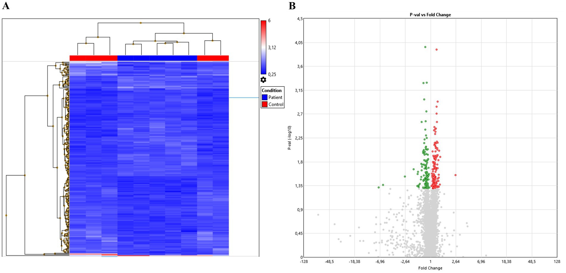
Figure 1. Overview of microarray analysis. (A) Heatmap of relative miRNAs expression. Differential expression is shown between Controls and HSCR samples, color scale represents up- (red), no threshold (gray), or downregulation (blue). (B) Volcano plot showing miRNAs distribution after ANOVA analysis.
After statistical analysis, data was sorted out based in fold changes in order to select the miRNAs that were most dysregulated. Results showed hsa-miR-378 h, hsa-miR-210-5p, hsa-miR-6876-3p, hsa-miR-634 and hsa-miR-6883-3p as the most upregulated miRNAs; while hsa-miR-4443, hsa-miR-22-3p, hsa-miR-4732-5p, hsa-miR-3187-5p, and hsa-miR-371b-5p were the most downregulated miRNAs in urine of HSCR patients compared to urine of healthy controls (p-value ≤0.05) (see Table 1).
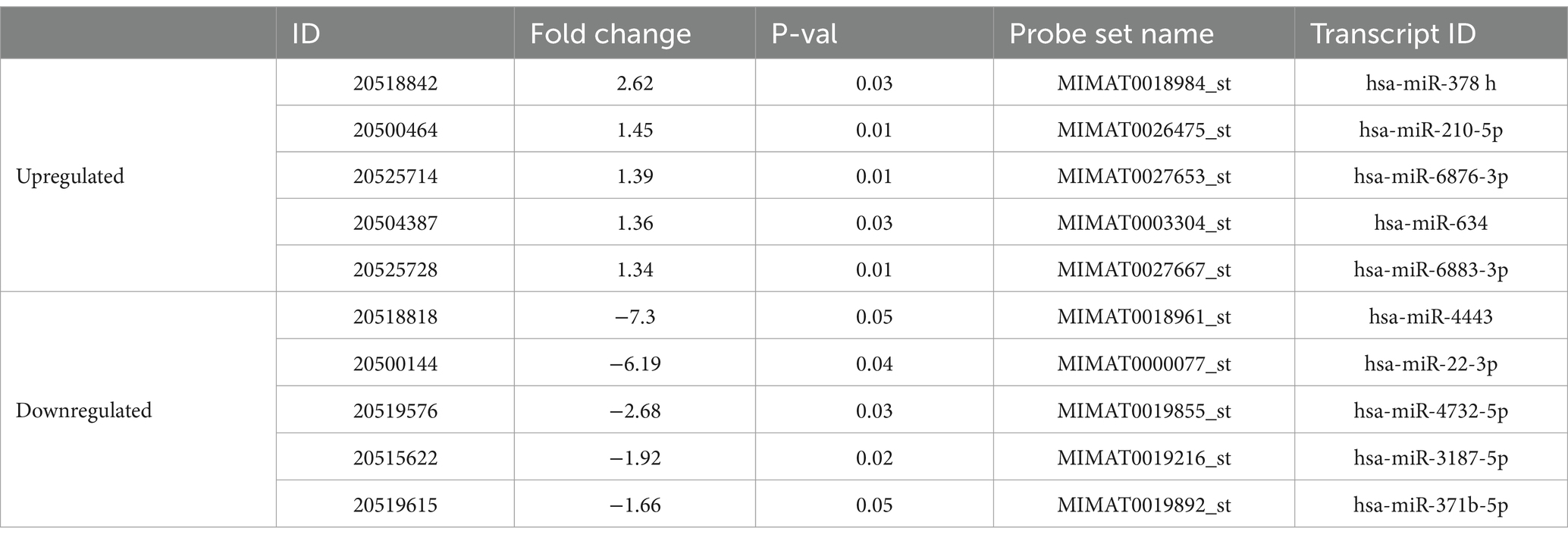
Table 1. List of the 10 selected most dysregulated miRNAs identified in HSCR urine.
With the aim of verifying the potential role of those miRNAs in HSCR development, we performed a target analysis exploration with the help of miRWalk (Sticht et al., 2018) and miRDB (Chen and Wang, 2020) databases.
Initially, we investigated the top 10 targeted genes in both databases with the highest score (see Table 2). Despite the high diversity of genes targeted by the selected miRNAs, it was possible to already find genes like ALKBH5, which was related to enteric neuronal crest migration in HSCR (Wang et al., 2021), or genes linked to neuronal migration, like DAB2IP (Lee et al., 2012) or NF1 (Sanchez-Ortiz et al., 2014), as well as to neuronal motility, like GMR5 (Turunen et al., 2018), or to other neuronal processes such as BTG2 (el-Ghissassi et al., 2002) or KIRREL3 (Hisaoka et al., 2021).
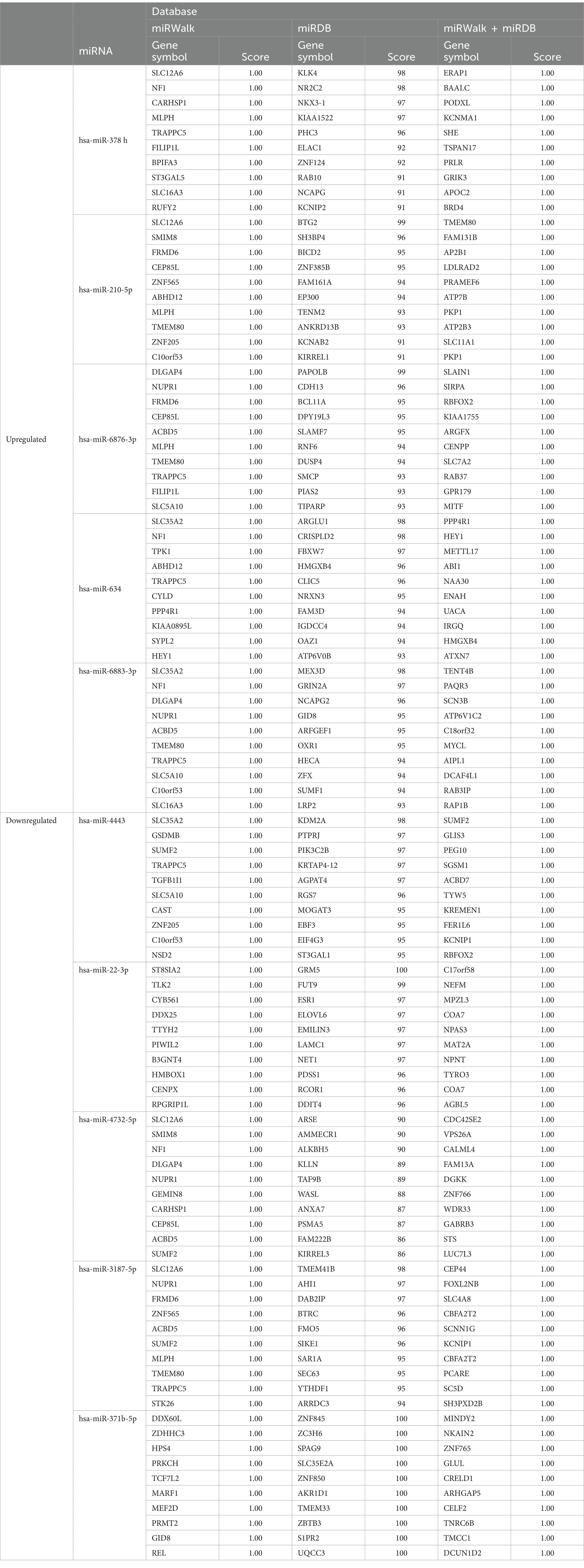
Table 2. Target database analysis of the selected dysregulated miRNAS in HSCR urine.
Further analysis in the same databases revealed that most of the dysregulated miRNAs in HSCR urine identified in the microarray regulate genes strongly associated to HSCR (see Table 3). For instance, genes like EDNRB (Puffenberger et al., 1994), RET (Luo et al., 1993; Emison et al., 2005), BDNF (Schriemer et al., 2016), GDNF (Parisi and Kapur, 2000), SOX10 (Prasad et al., 2011) etc. are targets of many of our detected miRNAs. In addition, a wide spectrum of different genes related to HSCR (Parisi and Kapur, 2000; Heuckeroth and Schäfer, 2016; Luzón-Toro et al., 2020) such as EDN3, ECE-1, ERBB3 (Gershon, 2021), NRG1 (Gui et al., 2013), NTKR3 (Sanchez-Mejias et al., 2009) or L1CAM (Jackson et al., 2009), have been also found in the databases (Table 3). Aside from those, other genes participating in signaling processes, like MAPK8 (Kuil et al., 2021), TMP3 (Camilleri et al., 2019) or PIK3C2B (Fu et al., 2020) have also recently been connected to ENS formation and HSCR.
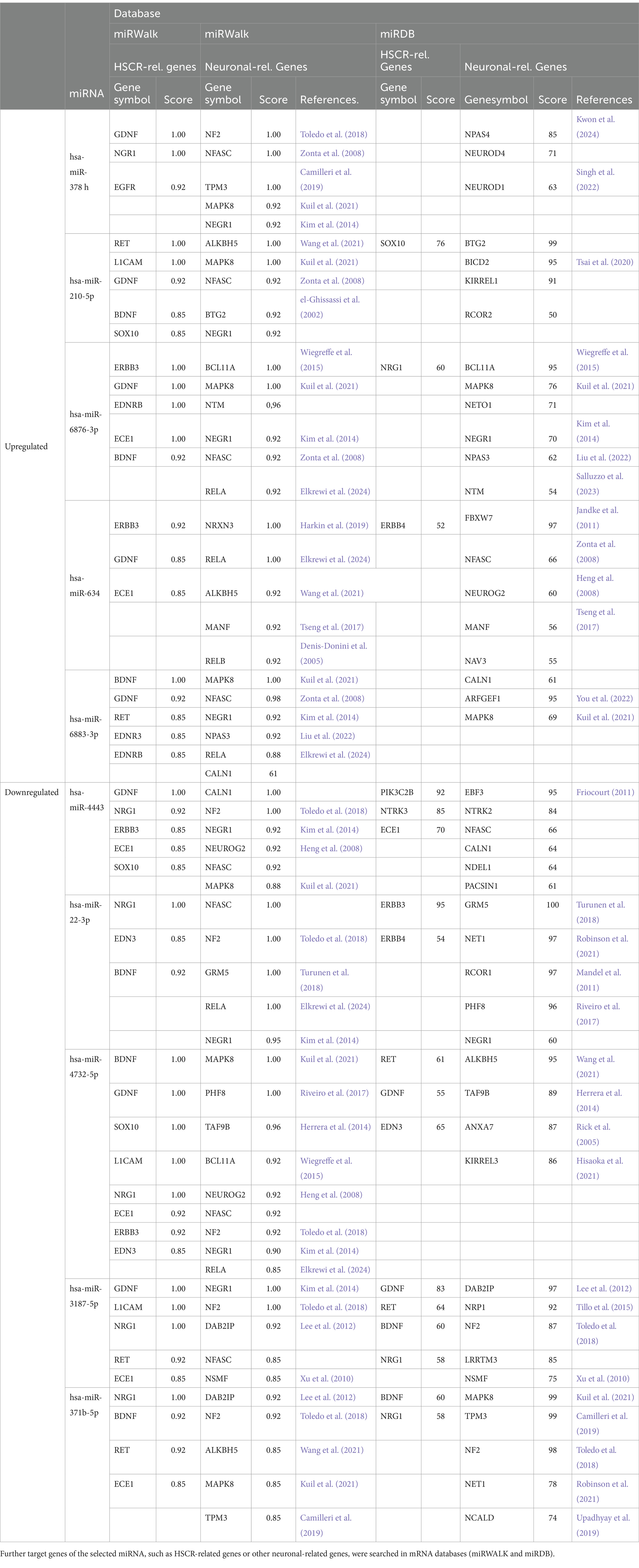
Table 3. Specific miRNA targets associated with HSCR or neuronal processes (neuronal migration, neurogenesis, etc.).
Following that, we also searched for other genes related to critical neuronal processes for a proper Enteric Nervous System (ENS) formation, like neurogenesis, neurodevelopment or neuronal migration (see Table 3). We found genes like ALKBH5 that could be critical for enteric neuronal crest migration in HSCR (Wang et al., 2021); in addition to others important for neuronal migration: as NF2 (Toledo et al., 2018), NEUROG2 (Heng et al., 2008) and NEUROD1 (Singh et al., 2022) or DAB2IP (Lee et al., 2012); as well as genes participating in neuronal proliferation NEGR1 (Kim et al., 2014), or in neuronal assembly NFASC (Zonta et al., 2008).
From all examined HSCR urinary downregulated miRNAs, miR-4732-5p was the one that was associated with the highest number of genes related to HSCR in both databases, 8 genes in miRWalk (BDNF, GDNF, SOX10, L1CAM, NRG1, ECE1, ERBB3, EDN3) together with 3 more genes in miRDB (RET, GDNF, EDN3); followed by hsa-miR-3187-5p with 5 genes in miRWalk (GDNF, L1CAM, NRG1, RET, ECE1) and 4 more in miRDB (GDNF, RET, BDNF, NRG1); and finally by hsa-miR-4443 with 5 genes in miRWalk (GDNF, NRG1, ERBB3, ECE1, SOX10) and 3 more in miRDB (PIK3C2B, NTRK3, ECE1) (Table 3).
Regarding the upregulated sequences, the miRNAs with more HSCR-associated gene targets were hsa-miR-210-5p with 5 genes in miRWalk (RET, L1CAM, GDNF, BDNF, SOX10) and 1 more in miRDB (SOX10); together with hsa-miR-6876-3p, also with 5 genes in miRWalk (ERBB3, GDNF, EDNRB, ECE1, BDNF) and one more located in miRDB (NRG1) (Table 3).
Altogether, the identified HSCR urinary-dysregulated miRNAs of interest targeted genes associated with HSCR pathology and also others related to neuronal processes that may be important also in ENS formation.
Lastly, we evaluated the expression of the above selected miRNAs (see Table 1), in the urine of the HSCR patients and controls by RT-qPCR.
Here we could verify an increased expression of hsa-miR-6883-3p and hsa-miR-6876-3p, as well as a decreased expression of hsa-miR-4732-5p (F.N Test p = 0.000015) and hsa-miR-3187-5p (F.N Test p = 0.000002) (Figure 2) in HSCR urine compared to the controls, which confirmed the previous results of those miRNAs in the microarrays.
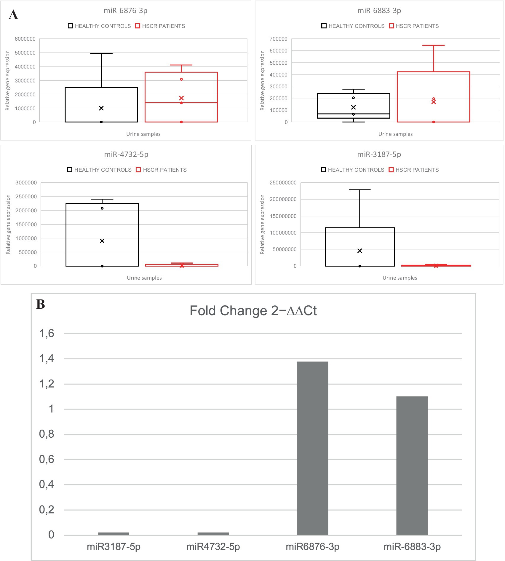
Figure 2. (A) Overview of RT-qPCR analysis of the selected miRNAs in HSCR urine samples compared to controls: miR3187-5p (F.N Test p = 0.000002), miR4732-5p (F.N Test p = 0.000015), miR6876-3p, miR-6883-3p. (B) Fold change comparison of 2 − ∆∆Ct from HSCR to control samples (HSCR/Controls).
Our study demonstrates that specific dysregulated miRNAs can be detected in the urine of HSCR patients when compared to healthy controls. In our HSCR patients’ cohort, we could identify several upregulated and downregulated miRNAs by microarray analysis that are, related to key functional aspects in ENS development. Analyzing the predicted targets of the top 5 up and downregulated miRNAs in data bases, we could verify that they do regulate genes related to HSCR pathology, and also to neuronal processes that are maybe necessary for the proper development of the Enteric Nervous System (ENS) formation, like neurogenesis, neurodevelopment or neuronal migration (Table 3).
When we compare our results with other miRNA studies in HSCR, our identified miRNAs in HSCR urine differ from those found by others in HSCR serum (Tang et al., 2014), HSCR intestine (Shen et al., 2016; Zhang et al., 2024), or in developing ENS cells (Pai et al., 2023). However, the miRNAs found by these studies are also different between each other, possibly because of differential expression between tissue and body fluids and the specific cohorts studied.
Some of the selected miRNAs found in HSCR urine regulate RELA and ERBB3 genes according to the search in miRNA databases. We recently published a downregulated expression of ERBB3 and RELA mRNAs in distal aganglionic segments of HSCR patients in a cohort of 25 patients (Elkrewi et al., 2024). ERBB3, a member of the EGF receptor tyrosine kinase family, is known to play an important role in neural crest development (Prasad et al., 2011), while RELA, the main subunit of the NF-κB pathway (Perkins, 2007), participates in embryonic neurogenesis and neural progenitor migration and differentiation (Mémet, 2006; Zhang and Hu, 2012). Therefore, it could be interesting to further validate the miRNA targets at protein level in tissue or blood HSCR samples in the future.
Regarding the potential causes of miRNA dysregulation in HSCR, miRNAs expression can be regulated at transcriptional (changes in gene expression) or post-transcriptional (changes in miRNA processing) levels (Gulyaeva and Kushlinskiy, 2016). Prospective investigation is needed to assess if already known HSCR mutations mutations, or other HSCR disease’s characteristics, i.e., alterations in intestinal tissue, are somehow responsible for these miRNA modifications.
In addition, cellular pathways (Treiber et al., 2019) and various physiological and pathological stimuli, such as hormones, stress, inflammatory cytokines, DNA methylation status, etc. have been related to affect miRNA expression (Gulyaeva and Kushlinskiy, 2016; Juźwik et al., 2019). Of these, processes like inflammation, and neurodegeneration are certainly consistent with HSCR pathology and could be considered as possible miRNA dysregulation mechanisms for future investigation.
Given that our chosen list of dysregulated miRNAs in HSCR urine regulate the expression of genes related to HSCR and to neuronal and cell migration processes (see Table 3), we think they may be good candidates for HSCR biomarkers. In particular, sequences like miR-4732-5p, hsa-miR-3187-5p and hsa-miR-6876-3p target several HSCR-related genes and were validated in our RT-qPCR analysis. As an example, high levels of hsa-miR-6876-3p, which can target GDNF, ERBB3, EDNRB and BDNF mRNAs, will lead to a lower expression of those genes and thus impair neurogenesis and neuronal migration, which finally may impact the proper gut innervation in those patients.
Importantly, the expression of specific candidate miRNAs can also be detected using nanophotonic biosensors integrated into portable lab-on-a-chip platforms, which are applicable in clinical and environmental diagnostics (Tarasov et al., 2016; Smith et al., 2017; Rebelo et al., 2019; García-Chamé et al., 2020). Thus, when at some point a defined miRNA pattern would be determined, the miRNA detection could easily be implemented in a microanalytical chip system and thereafter be used routinely to complete HSCR diagnosis, potentially helping future patients.
In summary, this work provides initial results that are promising in the search of a new complementary diagnosis method for HSCR. They show they show that indeed some miRNAs have dysregulated expression as predicted. However, further research is still necessary to validate a specific miRNA signature in a larger cohort of patients that could be used as HSCR biomarker in the future. After validation, these can be used for the creation of a non-invasive, easy to interpret and relatively simple novel diagnostic tool, complementary to histology, patient history, and pathology.
This preliminary study serves as a foundation for future investigation. A larger number of patients are required to increase the robustness of the results. Ideally, patients may be recruited from a network of hospitals at national level. Once verified, selected miRNAs must be correlated with clinical history, histology and surgery outcome. Based on that, the best miRNAs may be initially added to other diagnosis approaches until their application is fully demonstrated, and finally be implemented in a regular manner.
HSCR patients present a detectable miRNA dysregulated signature in urine that could work as an easier, non-invasive and more affordable method to complement current Hirschsprung’s disease diagnosis tests. Therefore, further research is necessary to validate this preliminary results in a larger cohort of patients.
The datasets presented in this study can be found in online repositories. The names of the repository/repositories and accession number(s) can be found at: https://www.ncbi.nlm.nih.gov/, GSE277874.
The studies involving humans were approved by the “Medizinische Ethik-Kommission II” of the Medical Faculty Mannheim, University of Heidelberg (2011-237N-MA). The studies were conducted in accordance with the local legislation and institutional requirements. Written informed consent for participation in this study was provided by the participants’ legal guardians/next of kin.
AS: Data curation, Formal analysis, Investigation, Visualization, Writing – original draft. RK: Investigation, Resources, Writing – review & editing. EE: Investigation, Writing – review & editing. CT: Data curation, Formal analysis, Writing – review & editing. JF: Resources, Writing – review & editing. AA: Resources, Writing – review & editing. RM: Resources, Writing – review & editing. NAH-C: Resources, Writing – review & editing. MB: Funding acquisition, Resources, Writing – review & editing. LW: Funding acquisition, Resources, Writing – review & editing. K-HS: Funding acquisition, Resources, Writing – review & editing. MAT-L: Conceptualization, Data curation, Project administration, Supervision, Validation, Visualization, Writing – original draft, Writing – review & editing.
The author(s) declare that financial support was received for the research, authorship, and/or publication of this article. The work of Abhijit Sreepada and Enas Zoheer Elkrewi was supported through funding from the Medical Faculty Mannheim, University of Heidelberg, under the auspices of the Master’s program “Translational Medical Research” (TMR). Note: This Master’s program has no funding registration code for their students’ practicum. For the publication fee we acknowledged financial support by Heidelberg University.
We would like to thank Camela Jost (Manager of General Core Equipment unite, Medical Faculty of Mannheim, University of Heidelberg) for her kind support and technical help.
The authors declare that the research was conducted in the absence of any commercial or financial relationships that could be construed as a potential conflict of interest.
All claims expressed in this article are solely those of the authors and do not necessarily represent those of their affiliated organizations, or those of the publisher, the editors and the reviewers. Any product that may be evaluated in this article, or claim that may be made by its manufacturer, is not guaranteed or endorsed by the publisher.
The Supplementary material for this article can be found online at: https://www.frontiersin.org/articles/10.3389/fnmol.2024.1504424/full#supplementary-material
Benz, F., Roy, S., Trautwein, C., Roderburg, C., and Luedde, T. (2016). Circulating MicroRNAs as biomarkers for Sepsis. Int. J. Mol. Sci. 17:78. doi: 10.3390/ijms17010078
Bertoli, G., Cava, C., and Castiglioni, I. (2015). MicroRNAs: new biomarkers for diagnosis, prognosis, therapy prediction and therapeutic tools for breast cancer. Theranostics 5, 1122–1143. doi: 10.7150/thno.11543
Braun, Y., Friedmacher, F., Theilen, T., Fiegel, H. C., Weber, K., Harter, P. N., et al. (2024). Diagnosis of Hirschsprung disease by analyzing acetylcholinesterase staining using artificial intelligence. J. Pediatr. Gastroenterol. Nutr. 79, 729–737. doi: 10.1002/jpn3.12339
Calkins, C. (2018). Hirschsprung disease beyond infancy. Clin. Colon Rectal Surg. 31, 051–060. doi: 10.1055/s-0037-1604034
Camilleri, M., Wieben, E., Eckert, D., Carlson, P., Hurley O’Dwyer, R., Gibbons, D., et al. (2019). Familial chronic megacolon presenting in childhood or adulthood: seeking the presumed gene association. Neurogastroenterol. Motil. 31:e13550. doi: 10.1111/nmo.13550
Chen, X., Ba, Y., Ma, L., Cai, X., Yin, Y., Wang, K., et al. (2008). Characterization of microRNAs in serum: a novel class of biomarkers for diagnosis of cancer and other diseases. Cell Res. 18, 997–1006. doi: 10.1038/cr.2008.282
Chen, Y., and Wang, X. (2020). miRDB: an online database for prediction of functional microRNA targets. Nucleic Acids Res. 48, D127–D131. doi: 10.1093/nar/gkz757
Condrat, C. E., Thompson, D. C., Barbu, M. G., Bugnar, O. L., Boboc, A., Cretoiu, D., et al. (2020). miRNAs as biomarkers in disease: latest findings regarding their role in diagnosis and prognosis. Cells 9:276. doi: 10.3390/cells9020276
de Lorijn, F., Kremer, L. C. M., Reitsma, J. B., and Benninga, M. A. (2006). Diagnostic tests in Hirschsprung disease. J. Pediatr. Gastroenterol. Nutr. 42, 496–505. doi: 10.1097/01.mpg.0000214164.90939.92
Denis-Donini, S., Caprini, A., Frassoni, C., and Grilli, M. (2005). Members of the NF-κB family expressed in zones of active neurogenesis in the postnatal and adult mouse brain. Dev. Brain Res. 154, 81–89. doi: 10.1016/j.devbrainres.2004.10.010
Diamond, I. R., Casadiego, G., Traubici, J., Langer, J. C., and Wales, P. W. (2007). The contrast enema for Hirschsprung disease: predictors of a false-positive result. J. Pediatr. Surg. 42, 792–795. doi: 10.1016/j.jpedsurg.2006.12.031
el-Ghissassi, F., Valsesia-Wittmann, S., Falette, N., Duriez, C., Walden, P. D., and Puisieux, A. (2002). BTG2TIS21/PC3 induces neuronal differentiation and prevents apoptosis of terminally differentiated PC12 cells. Oncogene 21, 6772–6778. doi: 10.1038/sj.onc.1205888
Elkrewi, E. Z., Al Abdulqader, A. A., Khasanov, R., Maas-Omlor, S., Boettcher, M., Wessel, L. M., et al. (2024). Role of inflammation and the NF-κB signaling pathway in Hirschsprung’s disease. Biomol. Ther. 14:992. doi: 10.3390/biom14080992
Emison, E. S., McCallion, A. S., Kashuk, C. S., Bush, R. T., Grice, E., Lin, S., et al. (2005). A common sex-dependent mutation in a RET enhancer underlies Hirschsprung disease risk. Nature 434, 857–863. doi: 10.1038/nature03467
Erbersdobler, A. (2024). The Pathologist’s role in the diagnosis of Hirschsprung’s disease. Eur. J. Pediatr. Surg. doi: 10.1055/s-0044-1788562
Fabris, L., Ceder, Y., Chinnaiyan, A. M., Jenster, G. W., Sorensen, K. D., Tomlins, S., et al. (2016). The potential of MicroRNAs as prostate cancer biomarkers. Eur. Urol. 70, 312–322. doi: 10.1016/j.eururo.2015.12.054
Friedmacher, F., and Puri, P. (2015). Rectal suction biopsy for the diagnosis of Hirschsprung’s disease: a systematic review of diagnostic accuracy and complications. Pediatr. Surg. Int. 31, 821–830. doi: 10.1007/s00383-015-3742-8
Friocourt, G. (2011). Identification of Arx targets unveils new candidates for controlling cortical interneuron migration and differentiation. Front. Cell. Neurosci. 5:28. doi: 10.3389/fncel.2011.00028
Fu, A. X., Lui, K. N.-C., Tang, C. S.-M., Ng, R. K., Lai, F. P.-L., Lau, S.-T., et al. (2020). Whole-genome analysis of noncoding genetic variations identifies multiscale regulatory element perturbations associated with Hirschsprung disease. Genome Res. 30, 1618–1632. doi: 10.1101/gr.264473.120
García-Chamé, M.-Á., Gutiérrez-Sanz, Ó., Ercan-Herbst, E., Haustein, N., Filipiak, M. S., Ehrnhöfer, D. E., et al. (2020). A transistor-based label-free immunosensor for rapid detection of tau protein. Biosens. Bioelectron. 159:112129. doi: 10.1016/j.bios.2020.112129
Gershon, M. D. (2021). Hirschsprung disease and more: dysregulation of ERBB2 and ERBB3. J. Clin. Invest. 131:e146389. doi: 10.1172/JCI146389
Gries, M., Christmann, A., Schulte, S., Weyland, M., Rommel, S., Martin, M., et al. (2021). Parkinson mice show functional and molecular changes in the gut long before motoric disease onset. Mol. Neurodegener. 16:34. doi: 10.1186/s13024-021-00439-2
Gui, H., Tang, W.-K., So, M.-T., Proitsi, P., Sham, P. C., Tam, P. K., et al. (2013). RET and NRG1 interplay in Hirschsprung disease. Hum. Genet. 132, 591–600. doi: 10.1007/s00439-013-1272-9
Gulyaeva, L. F., and Kushlinskiy, N. E. (2016). Regulatory mechanisms of microRNA expression. J. Transl. Med. 14:143. doi: 10.1186/s12967-016-0893-x
Harkin, L. F., Lindsay, S. J., Xu, Y., Alzu’bi, A., Ferrera, A., Gullon, E. A., et al. (2019). Corrigendum: Neurexins 1–3 each have a distinct pattern of expression in the early developing human cerebral cortex. Cereb. Cortex 29:1705. doi: 10.1093/cercor/bhz012
Heng, J. I.-T., Nguyen, L., Castro, D. S., Zimmer, C., Wildner, H., Armant, O., et al. (2008). Neurogenin 2 controls cortical neuron migration through regulation of Rnd2. Nature 455, 114–118. doi: 10.1038/nature07198
Herrera, F. J., Yamaguchi, T., Roelink, H., and Tjian, R. (2014). Core promoter factor TAF9B regulates neuronal gene expression. eLife 3:e02559. doi: 10.7554/eLife.02559
Heuckeroth, R. O. (2018). Hirschsprung disease — integrating basic science and clinical medicine to improve outcomes. Nat. Rev. Gastroenterol. Hepatol. 15, 152–167. doi: 10.1038/nrgastro.2017.149
Heuckeroth, R. O., and Schäfer, K.-H. (2016). Gene-environment interactions and the enteric nervous system: neural plasticity and Hirschsprung disease prevention. Dev. Biol. 417, 188–197. doi: 10.1016/j.ydbio.2016.03.017
Hisaoka, T., Komori, T., Fujimoto, K., Kitamura, T., and Morikawa, Y. (2021). Comprehensive expression pattern of kin of irregular chiasm-like 3 in the adult mouse brain. Biochem. Biophys. Res. Commun. 563, 66–72. doi: 10.1016/j.bbrc.2021.05.063
Hosako, H., Martin, G. S., Barrier, M., Chen, Y. A., Ivanov, I. V., and Mirkes, P. E. (2009). Gene and microRNA expression in p53 -deficient day 8.5 mouse embryos. Birth Defects Res. A Clin. Mol. Teratol. 85, 546–555. doi: 10.1002/bdra.20565
Jackson, S.-R., Guner, Y. S., Woo, R., Randolph, L. M., Ford, H., and Shin, C. E. (2009). L1CAM mutation in association with X-linked hydrocephalus and Hirschsprung’s disease. Pediatr. Surg. Int. 25, 823–825. doi: 10.1007/s00383-009-2420-0
Jandke, A., Da Costa, C., Sancho, R., Nye, E., Spencer-Dene, B., and Behrens, A. (2011). The F-box protein Fbw7 is required for cerebellar development. Dev. Biol. 358, 201–212. doi: 10.1016/j.ydbio.2011.07.030
Juźwik, C. A., Drake, S., Zhang, Y., Paradis-Isler, N., Sylvester, A., Amar-Zifkin, A., et al. (2019). microRNA dysregulation in neurodegenerative diseases: A systematic review. Prog. Neurobiol. 182:101664. doi: 10.1016/j.pneurobio.2019.101664
Kim, H., Hwang, J.-S., Lee, B., Hong, J., and Lee, S. (2014). Newly identified Cancer-associated role of human neuronal growth regulator 1 (NEGR1). J. Cancer 5, 598–608. doi: 10.7150/jca.8052
Korsager, L. E. H., Bjørn, N., Ellebæk, M. B., Christensen, L. G., and Qvist, N. (2023). Full-thickness rectal biopsy in children suspected of having Hirschsprung’s disease: the inconclusive biopsy. Children 10:1619. doi: 10.3390/children10101619
Korzeniewski, N., Tosev, G., Pahernik, S., Hadaschik, B., Hohenfellner, M., and Duensing, S. (2015). Identification of cell-free microRNAs in the urine of patients with prostate cancer. Urol. Oncol. 33, 16.e17–16.e22. doi: 10.1016/j.urolonc.2014.09.015
Kroh, E. M., Parkin, R. K., Mitchell, P. S., and Tewari, M. (2010). Analysis of circulating microRNA biomarkers in plasma and serum using quantitative reverse transcription-PCR (qRT-PCR). Methods 50, 298–301. doi: 10.1016/j.ymeth.2010.01.032
Kuil, L. E., MacKenzie, K. C., Tang, C. S., Windster, J. D., Le, T. L., Karim, A., et al. (2021). Size matters: large copy number losses in Hirschsprung disease patients reveal genes involved in enteric nervous system development. PLoS Genet. 17:e1009698. doi: 10.1371/journal.pgen.1009698
Kwon, O.-H., Choe, J., Kim, D., Kim, S., and Moon, C. (2024). Sensory stimulation-dependent Npas4 expression in the olfactory bulb during early postnatal development. Exp. Neurobiol. 33, 77–98. doi: 10.5607/en23037
Lawrie, C. H., Gal, S., Dunlop, H. M., Pushkaran, B., Liggins, A. P., Pulford, K., et al. (2008). Detection of elevated levels of tumour-associated microRNAs in serum of patients with diffuse large B-cell lymphoma. Br. J. Haematol. 141, 672–675. doi: 10.1111/j.1365-2141.2008.07077.x
Lee, G. H., Kim, S. H., Homayouni, R., and D’Arcangelo, G. (2012). Dab2ip regulates neuronal migration and neurite outgrowth in the developing neocortex. PLoS One 7:e46592. doi: 10.1371/journal.pone.0046592
Lewis, N. A., Levitt, M. A., Zallen, G. S., Zafar, M. S., Iacono, K. L., Rossman, J. E., et al. (2003). Diagnosing Hirschsprung’s disease: increasing the odds of a positive rectal biopsy result. J. Pediatr. Surg. 38, 412–416. doi: 10.1053/jpsu.2003.50070
Liu, J.-W., Li, H., and Zhang, Y. (2022). Npas3 regulates stemness maintenance of radial glial cells and neuronal migration in the developing mouse cerebral cortex. Front. Cell. Neurosci. 16:865681. doi: 10.3389/fncel.2022.865681
Liu, R., Chen, X., Du, Y., Yao, W., Shen, L., Wang, C., et al. (2012). Serum MicroRNA expression profile as a biomarker in the diagnosis and prognosis of pancreatic Cancer. Clin. Chem. 58, 610–618. doi: 10.1373/clinchem.2011.172767
Luo, Y., Ceccherini, I., Pasini, B., Matera, I., Bicocchi, M. P., Barone, V., et al. (1993). Close linkage with the RET protooncogene and boundaries of deletion mutations in autosomal dominant Hirschsprung disease. Hum. Mol. Genet. 2, 1803–1808. doi: 10.1093/hmg/2.11.1803
Luo, Y., Wang, C., Chen, X., Zhong, T., Cai, X., Chen, S., et al. (2013). Increased serum and urinary MicroRNAs in children with idiopathic nephrotic syndrome. Clin. Chem. 59, 658–666. doi: 10.1373/clinchem.2012.195297
Luzón-Toro, B., Villalba-Benito, L., Torroglosa, A., Fernández, R. M., Antiñolo, G., and Borrego, S. (2020). What is new about the genetic background of Hirschsprung disease? Clin. Genet. 97, 114–124. doi: 10.1111/cge.13615
Mall, C., Rocke, D. M., Durbin-Johnson, B., and Weiss, R. H. (2013). Stability of miRNA in human urine supports its biomarker potential. Biomark. Med 7, 623–631. doi: 10.2217/bmm.13.44
Mandel, G., Fiondella, C. G., Covey, M. V., Lu, D. D., LoTurco, J. J., and Ballas, N. (2011). Repressor element 1 silencing transcription factor (REST) controls radial migration and temporal neuronal specification during neocortical development. Proc. Natl. Acad. Sci. U.S.A. 108, 16789–16794. doi: 10.1073/pnas.1113486108
Mémet, S. (2006). NF-κB functions in the nervous system: from development to disease. Biochem. Pharmacol. 72, 1180–1195. doi: 10.1016/j.bcp.2006.09.003
Menezes, M., and Puri, P. (2006). Long-term outcome of patients with enterocolitis complicating Hirschsprung’s disease. Pediatr. Surg. Int. 22, 316–318. doi: 10.1007/s00383-006-1639-2
Mitchell, P. S., Parkin, R. K., Kroh, E. M., Fritz, B. R., Wyman, S. K., Pogosova-Agadjanyan, E. L., et al. (2008). Circulating microRNAs as stable blood-based markers for cancer detection. Proc. Natl. Acad. Sci. 105, 10513–10518. doi: 10.1073/pnas.0804549105
Muller, C., Hobeika, C., Montalva, L., Berrebi, D., and Bonnard, A. (2015). Calretinin variant in Hirschsprung disease: Pretransitional sign and surgical planning. Eur. J. Pediatr. Surg. 26, 449–453. doi: 10.1055/s-0035-1566106
Neeser, H. R., Robbiani, I., Rodewald, A.-K., Nigbur, T., di Natale, A., Moehrlen, U., et al. (2024). Enough is enough: how many rectal suction biopsies do you need to diagnose Hirschsprung’s disease? Pediatr. Surg. Int. 40:206. doi: 10.1007/s00383-024-05793-y
O’Brien, J., Hayder, H., Zayed, Y., and Peng, C. (2018). Overview of MicroRNA biogenesis, mechanisms of actions, and circulation. Front. Endocrinol. 9:402. doi: 10.3389/fendo.2018.00402
Osatakul, S., Patrapinyokul, S., and Osatakul, N. (1999). The diagnostic value of anorectal manometry as a screening test for Hirschsprung’s disease. J. Med. Assoc. Thail. 82, 1100–1105
Pai, C., Sengupta, R., and Heuckeroth, R. O. (2023). Sequencing reveals miRNAs enriched in the developing mouse enteric nervous system. Noncoding RNA 10:1. doi: 10.3390/ncrna10010001
Parisi, M. A., and Kapur, R. P. (2000). Genetics of Hirschsprung disease. Curr. Opin. Pediatr. 12, 610–617. doi: 10.1097/00008480-200012000-00017
Perkins, N. D. (2007). Integrating cell-signalling pathways with NF-κB and IKK function. Nat. Rev. Mol. Cell Biol. 8, 49–62. doi: 10.1038/nrm2083
Prasad, M. K., Reed, X., Gorkin, D. U., Cronin, J. C., McAdow, A. R., Chain, K., et al. (2011). SOX10 directly modulates ERBB3 transcription via an intronic neural crest enhancer. BMC Dev. Biol. 11:40. doi: 10.1186/1471-213X-11-40
Puffenberger, E. G., Hosoda, K., Washington, S. S., Nakao, K., deWit, D., Yanagisawa, M., et al. (1994). A missense mutation of the endothelin-B receptor gene in multigenic hirschsprung’s disease. Cell 79, 1257–1266. doi: 10.1016/0092-8674(94)90016-7
Rebelo, A. R., Liu, C., Schäfer, K.-H., Saumer, M., Yang, G., and Liu, Y. (2019). Poly(4-vinylaniline)/polyaniline bilayer-functionalized bacterial cellulose for flexible electrochemical biosensors. Langmuir 35, 10354–10366. doi: 10.1021/acs.langmuir.9b01425
Rick, M., Ramos Garrido, S. I., Herr, C., Thal, D. R., Noegel, A. A., and Clemen, C. S. (2005). Nuclear localization of Annexin A7 during murine brain development. BMC Neurosci. 6:25. doi: 10.1186/1471-2202-6-25
Riveiro, A. R., Mariani, L., Malmberg, E., Amendola, P. G., Peltonen, J., Wong, G., et al. (2017). JMJD-1.2/PHF8 controls axon guidance by regulating hedgehog-like signaling. Development. 144, 856–856. doi: 10.1242/dev.142695
Robinson, R. A., Griffiths, S. C., van de Haar, L. L., Malinauskas, T., van Battum, E. Y., Zelina, P., et al. (2021). Simultaneous binding of guidance cues NET1 and RGM blocks extracellular NEO1 signaling. Cell 184, 2103–2120.e31. doi: 10.1016/j.cell.2021.02.045
Romero, P., Burger, A., Wennberg, E., Schmitteckert, S., Holland-Cunz, S., Schwab, C., et al. (2024). Clinical relevance of pathological diagnosis of Hirschsprung’s disease with acetylcholine-esterase Histochemistry or Calretinin immunohistochemistry. Children 11:428. doi: 10.3390/children11040428
Salehi, M., and Sharifi, M. (2018). Exosomal miRNAs as novel cancer biomarkers: challenges and opportunities. J. Cell. Physiol. 233, 6370–6380. doi: 10.1002/jcp.26481
Salluzzo, M., Vianello, C., Abdullatef, S., Rimondini, R., Piccoli, G., and Carboni, L. (2023). The role of IgLON cell adhesion molecules in neurodegenerative diseases. Genes (Basel) 14:1886. doi: 10.3390/genes14101886
Sanchez-Mejias, A., Fernandez, R. M., Lopez-Alonso, M., Antinolo, G., and Borrego, S. (2009). Contribution of RET, NTRK3 and EDN3 to the expression of Hirschsprung disease in a multiplex family. J. Med. Genet. 46, 862–864. doi: 10.1136/jmg.2009.067819
Sanchez-Ortiz, E., Cho, W., Nazarenko, I., Mo, W., Chen, J., and Parada, L. F. (2014). NF1 regulation of RAS/ERK signaling is required for appropriate granule neuron progenitor expansion and migration in cerebellar development. Genes Dev. 28, 2407–2420. doi: 10.1101/gad.246603.114
Schriemer, D., Sribudiani, Y., IJpma, A., Natarajan, D., MacKenzie, K. C., Metzger, M., et al. (2016). Regulators of gene expression in enteric neural crest cells are putative Hirschsprung disease genes. Dev. Biol. 416, 255–265. doi: 10.1016/j.ydbio.2016.06.004
Shayan, K., Smith, C., and Langer, J. C. (2004). Reliability of intraoperative frozen sections in the management of Hirschsprung’s disease. J. Pediatr. Surg. 39, 1345–1348. doi: 10.1016/j.jpedsurg.2004.05.009
Shen, Z., Du, C., Zang, R., Xie, H., Lv, W., Li, H., et al. (2016). Microarray expression profiling of dysregulated long non-coding RNAs in Hirschsprung’s disease reveals their potential role in molecular diagnosis. Neurogastroenterol. Motil. 28, 266–273. doi: 10.1111/nmo.12722
Singh, A., Mahesh, A., Noack, F., Cardoso de Toledo, B., Calegari, F., and Tiwari, V. K. (2022). Tcf12 and NeuroD1 cooperatively drive neuronal migration during cortical development. Development 149:dev200250. doi: 10.1242/dev.200250
Smith, D. A., Newbury, L. J., Drago, G., Bowen, T., and Redman, J. E. (2017). Electrochemical detection of urinary microRNAs via sulfonamide-bound antisense hybridisation. Sens Actuators B Chem 253, 335–341. doi: 10.1016/j.snb.2017.06.069
Sticht, C., De La Torre, C., Parveen, A., and Gretz, N. (2018). miRWalk: an online resource for prediction of microRNA binding sites. PLoS One 13:e0206239. doi: 10.1371/journal.pone.0206239
Subramani, R., Gangwani, L., Nandy, S. B., Arumugam, A., Chattopadhyay, M., and Lakshmanaswamy, R. (2015). Emerging roles of microRNAs in pancreatic cancer diagnosis, therapy and prognosis (review). Int. J. Oncol. 47, 1203–1210. doi: 10.3892/ijo.2015.3129
Talebi, A., Kahdouei, M. T., Memar, E. H., Ashjaei, B., Raji, H., and Adel, M. G. (2024). Can the diameter of nerve fibers be effectively utilized to enhance the accuracy of determining the length of the aganglionic segment compared to intraoperative biopsy in patients with Hirschsprung’s disease? BMC. Res. Notes 17:220. doi: 10.1186/s13104-024-06873-x
Tang, W., Li, H., Tang, J., Wu, W., Qin, J., Lei, H., et al. (2014). Specific serum microRNA profile in the molecular diagnosis of Hirschsprung’s disease. J. Cell. Mol. Med. 18, 1580–1587. doi: 10.1111/jcmm.12348
Tarasov, A., Gray, D. W., Tsai, M.-Y., Shields, N., Montrose, A., Creedon, N., et al. (2016). A potentiometric biosensor for rapid on-site disease diagnostics. Biosens. Bioelectron. 79, 669–678. doi: 10.1016/j.bios.2015.12.086
Tillo, M., Erskine, L., Cariboni, A., Fantin, A., Joyce, A., Denti, L., et al. (2015). VEGF189 binds NRP1 and is sufficient for VEGF/NRP1-dependent neuronal patterning in the developing brain. Development. 142, 314–9. doi: 10.1242/dev.115998
Toledo, A., Grieger, E., Karram, K., Morrison, H., and Baader, S. L. (2018). Neurofibromatosis type 2 tumor suppressor protein is expressed in oligodendrocytes and regulates cell proliferation and process formation. PLoS One 13:e0196726. doi: 10.1371/journal.pone.0196726
Treiber, T., Treiber, N., and Meister, G. (2019). Regulation of microRNA biogenesis and its crosstalk with other cellular pathways. Nat. Rev. Mol. Cell Biol. 20, 5–20. doi: 10.1038/s41580-018-0059-1
Tsai, M.-H., Cheng, H.-Y., Nian, F.-S., Liu, C., Chao, N.-H., Chiang, K.-L., et al. (2020). Impairment in dynein-mediated nuclear translocation by BICD2 C-terminal truncation leads to neuronal migration defect and human brain malformation. Acta Neuropathol. Commun. 8:106. doi: 10.1186/s40478-020-00971-0
Tseng, K.-Y., Danilova, T., Domanskyi, A., Saarma, M., Lindahl, M., and Airavaara, M. (2017). MANF is essential for neurite extension and neuronal migration in the developing cortex. eNeuro 4:ENEURO.0214-17.2017. doi: 10.1523/ENEURO.0214-17.2017
Turunen, P. M., Louhivuori, L. M., Louhivuori, V., Kukkonen, J. P., and Åkerman, K. E. (2018). Endocannabinoid signaling in embryonic neuronal motility and cell–cell contact – role of mGluR5 and TRPC3 channels. Neuroscience 375, 135–148. doi: 10.1016/j.neuroscience.2018.02.005
Upadhyay, A., Hosseinibarkooie, S., Schneider, S., Kaczmarek, A., Torres-Benito, L., Mendoza-Ferreira, N., et al. (2019). Neurocalcin Delta knockout impairs adult neurogenesis whereas half reduction is not pathological. Front. Mol. Neurosci. 12:19. doi: 10.3389/fnmol.2019.00019
Wang, B., Fang, X., Sun, X., Du, C., Zhou, L., Lv, X., et al. (2021). m6A demethylase ALKBH5 suppresses proliferation and migration of enteric neural crest cells by regulating TAGLN in Hirschsprung’s disease. Life Sci. 278:119577. doi: 10.1016/j.lfs.2021.119577
Wang, J., Chen, J., and Sen, S. (2016). MicroRNA as biomarkers and diagnostics. J. Cell. Physiol. 231, 25–30. doi: 10.1002/jcp.25056
Weldon, B. A., Shubin, S. P., Smith, M. N., Workman, T., Artemenko, A., Griffith, W. C., et al. (2016). Urinary microRNAs as potential biomarkers of pesticide exposure. Toxicol. Appl. Pharmacol. 312, 19–25. doi: 10.1016/j.taap.2016.01.018
Wiegreffe, C., Simon, R., Peschkes, K., Kling, C., Strehle, M., Cheng, J., et al. (2015). Bcl11a (Ctip1) controls migration of cortical projection neurons through regulation of Sema3c. Neuron 87, 311–325. doi: 10.1016/j.neuron.2015.06.023
Xu, N., Bhagavath, B., Kim, H.-G., Halvorson, L., Podolsky, R. S., Chorich, L. P., et al. (2010). NELF is a nuclear protein involved in hypothalamic GnRH neuronal migration. Mol. Cell. Endocrinol. 319, 47–55. doi: 10.1016/j.mce.2009.11.016
You, Z., Yang, Z., Cao, S., Deng, S., and Chen, Y. (2022). The novel KLF4/BIG1 regulates LPS-mediated neuro-inflammation and migration in BV2 cells via PI3K/Akt/NF-kB signaling pathway. Neuroscience 488, 102–111. doi: 10.1016/j.neuroscience.2022.01.014
Zhang, B., Yang, J., Song, A., Feng, W., and Guo, Z. (2024). miR-146b-5p regulates the enteric nervous system development in Hirschsprung disease via targeting RET pathway. Biochem. Genet. doi: 10.1007/s10528-024-10852-z
Zhang, Y., and Hu, W. (2012). NFκB signaling regulates embryonic and adult neurogenesis. Front Biol (Beijing) 7, 277–291. doi: 10.1007/s11515-012-1233-z
Zhao, C., Dong, J., Jiang, T., Shi, Z., Yu, B., Zhu, Y., et al. (2011). Early second-trimester serum MiRNA profiling predicts gestational diabetes mellitus. PLoS One 6:e23925. doi: 10.1371/journal.pone.0023925
Zhou, S., Jin, J., Wang, J., Zhang, Z., Freedman, J. H., Zheng, Y., et al. (2018). miRNAS in cardiovascular diseases: potential biomarkers, therapeutic targets and challenges. Acta Pharmacol. Sin. 39, 1073–1084. doi: 10.1038/aps.2018.30
Keywords: Hirschsprung’s disease (HSCR), enteric nervous system (ENS), microRNA (miRNA), non-invasive diagnostic, urine extracellular vesicles
Citation: Sreepada A, Khasanov R, Elkrewi EZ, de la Torre C, Felcht J, Al Abdulqader AA, Martel R, Hoyos-Celis NA, Boettcher M, Wessel LM, Schäfer K-H and Tapia-Laliena MÁ (2025) Urine miRNA signature as potential non-invasive diagnostic biomarker for Hirschsprung’s disease. Front. Mol. Neurosci. 17:1504424. doi: 10.3389/fnmol.2024.1504424
Edited by:
Naho Fujiwara, Juntendo University, JapanReviewed by:
Raquel Oliveira, King’s College London, United KingdomCopyright © 2025 Sreepada, Khasanov, Elkrewi, de la Torre, Felcht, Al Abdulqader, Martel, Hoyos-Celis, Boettcher, Wessel, Schäfer and Tapia-Laliena. This is an open-access article distributed under the terms of the Creative Commons Attribution License (CC BY). The use, distribution or reproduction in other forums is permitted, provided the original author(s) and the copyright owner(s) are credited and that the original publication in this journal is cited, in accordance with accepted academic practice. No use, distribution or reproduction is permitted which does not comply with these terms.
*Correspondence: María Ángeles Tapia-Laliena, bWFyaWFhbmdlbGVzLnRhcGlhLWxhbGllbmFAbWVkbWEudW5pLWhlaWRlbGJlcmcuZGU=: Karl-Herbert Schäfer, S2FybEhlcmJlcnQuU2NoYWVmZXJAaHMta2wuZGU=
†These authors have contributed equally to this work and share first authorship
Disclaimer: All claims expressed in this article are solely those of the authors and do not necessarily represent those of their affiliated organizations, or those of the publisher, the editors and the reviewers. Any product that may be evaluated in this article or claim that may be made by its manufacturer is not guaranteed or endorsed by the publisher.
Research integrity at Frontiers
Learn more about the work of our research integrity team to safeguard the quality of each article we publish.