
95% of researchers rate our articles as excellent or good
Learn more about the work of our research integrity team to safeguard the quality of each article we publish.
Find out more
REVIEW article
Front. Mol. Neurosci. , 01 July 2024
Sec. Molecular Signalling and Pathways
Volume 17 - 2024 | https://doi.org/10.3389/fnmol.2024.1405415
This article is part of the Research Topic Protein Post-Translational Modifications in the Nervous System: from Development to Disease and Ageing View all 11 articles
 Chloé Bizingre1,2†Clara Bianchi1,2†Anne Baudry1,2Aurélie Alleaume-Butaux1,2
 Benoit Schneider1,2,3*
 Mathéa Pietri1,2
More than 650 reversible and irreversible post-translational modifications (PTMs) of proteins have been listed so far. Canonical PTMs of proteins consist of the covalent addition of functional or chemical groups on target backbone amino-acids or the cleavage of the protein itself, giving rise to modified proteins with specific properties in terms of stability, solubility, cell distribution, activity, or interactions with other biomolecules. PTMs of protein contribute to cell homeostatic processes, enabling basal cell functions, allowing the cell to respond and adapt to variations of its environment, and globally maintaining the constancy of the milieu interieur (the body’s inner environment) to sustain human health. Abnormal protein PTMs are, however, associated with several disease states, such as cancers, metabolic disorders, or neurodegenerative diseases. Abnormal PTMs alter the functional properties of the protein or even cause a loss of protein function. One example of dramatic PTMs concerns the cellular prion protein (PrPC), a GPI-anchored signaling molecule at the plasma membrane, whose irreversible post-translational conformational conversion (PTCC) into pathogenic prions (PrPSc) provokes neurodegeneration. PrPC PTCC into PrPSc is an additional type of PTM that affects the tridimensional structure and physiological function of PrPC and generates a protein conformer with neurotoxic properties. PrPC PTCC into PrPSc in neurons is the first step of a deleterious sequence of events at the root of a group of neurodegenerative disorders affecting both humans (Creutzfeldt–Jakob diseases for the most representative diseases) and animals (scrapie in sheep, bovine spongiform encephalopathy in cow, and chronic wasting disease in elk and deer). There are currently no therapies to block PrPC PTCC into PrPSc and stop neurodegeneration in prion diseases. Here, we review known PrPC PTMs that influence PrPC conversion into PrPSc. We summarized how PrPC PTCC into PrPSc impacts the PrPC interactome at the plasma membrane and the downstream intracellular controlled protein effectors, whose abnormal activation or trafficking caused by altered PTMs promotes neurodegeneration. We discussed these effectors as candidate drug targets for prion diseases and possibly other neurodegenerative diseases.
The structure, dynamics, and functionalities of proteins depend on or are influenced by post-translational modifications (PTMs), i.e., chemical reactions that occur after the synthesis of proteins. Protein PTMs can be reversible or irreversible and are, most often, driven by enzymes. Reversible PTMs of proteins are associated with the covalent addition of functional or chemical groups, such as the acetyl, phosphate, methyl, glycan, short- and long-chain acyl, or ubiquitin groups, among others, on the side-chain of key amino acids of the targeted protein (Ramazi and Zahiri, 2021). The addition of those chemical functions relies on the activity of specific enzymes (acetylases, kinases, methylases, glycanases, ubiquitinases, etc) that catalyze the transfer of the group from a specific donor to the protein. Those PTMs are reversible as they are removed by hydrolytic enzymes (deacetylases, phosphatases, demethylases, deubiquitinases, etc.) that regenerate a naked protein with functional properties distinct from those of the post-translational modified counterpart. Reversible PTMs contribute to the spatio-temporal regulation of biological processes that notably relate to cell signaling events, genome plasticity, the regulation of gene expression, or energy metabolism (Humphrey et al., 2015; Millán-Zambrano et al., 2022). By contrast, irreversible PTMs of proteins sometimes refer to glycation and deamidation but mostly relate to the proteolytic modifications of proteins. Several proteases achieve the cleavage of the concerned protein, giving rise to protein fragments with distinct biological functions (Walsh et al., 2005). For example, some enzymes (e.g., digestive enzymes) and hormones (e.g., insulin) are synthesized in an immature form, called the pro-form, and turn activated after the proteolytic removal of one or several fragments (Triebel et al., 2022). Those PTMs are irreversible, so the degradation of the modified protein is necessary to regulate the biological process in which the protein is involved. Another type of irreversible, non-classical PTMs relates to the transconformational conversion of proteins, which differs from the subtle protein structural changes associated with protein activity and regulation. The transconformational conversion of proteins consists of deep modifications of the protein folding, i.e., the rearrangement of the protein with changes in the ratio between α-helix and β-sheet elementary folding motifs (Louros et al., 2023). These PTMs that affect the global architecture of the protein cause changes in the physicochemical properties of the protein, such as the surface charges and solubility, thereby leading to protein conformers that often display biological activity distinct from the conformer they derive (Louros et al., 2023). One unfortunate famous example of such post-translational conformational conversion (PTCC) of proteins relates to prions. Prions were highly publicized in the late 20th century with the mad cow disease crisis and the emergence of the variant Creutzfeldt–Jakob disease in humans caused by the transmission of prion pathology from cows to humans through food contaminated with prions (Knight, 2017). Even if the mad cow crisis is behind us, the emergence of chronic wasting disease (CWD) that concerns elk and deer and, for the moment, is confined to North America, Japan, and Scandinavia (Tranulis and Tryland, 2023), combined with the risk of CWD transmission to humans (Hannaoui et al., 2022), necessitates improving our knowledge of the mechanisms underlying prion diseases to rationalize therapeutic strategies to combat these devastating neurodegenerative disorders.
Prion diseases are caused by an infectious and neurotoxic protein, the scrapie protein (PrPSc), which results from the post-translational conformational conversion of the normal cellular prion protein PrPC (Prusiner, 1998). While the 3D structure of PrPC contains three α-helices in the ordered globular domain of the protein, the α-helices are remodeled in favor of the formation of β-sheets in the 3D structure of PrPSc. Such changes in PrP structure confer PrPSc insoluble properties in detergents and partial resistance to proteolysis. Moreover, PrPSc β-sheets are responsible for the aggregation of PrPSc molecules and the formation of fibrillar amyloid assemblies (Cobb et al., 2007; Kraus et al., 2021). As PrPSc promotes the conversion of PrPC through direct interaction of PrPSc with PrPC, post-translational conformational changes of PrPC into PrPSc are autocatalytic, which sustains the prion concept formulated in the 80s (Prusiner, 1986). It is established that PrPC conversion into PrPSc in neurons is at the root of prion diseases (Mallucci et al., 2003). PrPC is a ubiquitous protein that is more expressed in neurons and is present at the cell surface. Acting at the plasma membrane as a neuronal receptor or co-receptor (Mouillet-Richard et al., 2000) or a scaffolding protein that governs the dynamic assembly of signaling modules (Linden et al., 2008), PrPC controls signaling effectors that all contribute to the regulation of neuronal functions (Schneider, 2011). As PrPC is subjected to several PTMs, we here review how those PTMs of PrPC impact the transconformational conversion of PrPC into PrPSc. We also summarize how PrPC PTCC into PrPSc (i) affects PrPC interactome at the plasma membrane and (ii) impacts PrPC downstream signaling effectors, whose activity or trafficking disturbed by imbalanced PTMs contribute to the progression of prion diseases.
PrPC is coded by the PRNP gene located in chromosome 20 in the human genome (chromosome 2 in mice). The translation of the protein generates a single polypeptide chain of 253 amino-acids that folds as three α-helices and two short anti-parallel β-sheets in the C-terminal PrPC domain, while the N-terminal domain remains flexible (Zahn et al., 2000) and its conformation varies depending on the ligand interacting with this region (Zahn, 2003). Several PTMs occur on PrPC consisting of the removal of the N-terminus signal sequence when the protein enters the endoplasmic reticulum, the attachment of a GlycosylPhosphatidylInositol (GPI) moiety at residue 230 of PrPC C-terminus, two N-glycosylations at Asn181 and Asn197, the formation of a disulfide bridge between Cys179 and Cys214 that ensures the stability of the C-terminal domain of PrPC, and the cleavage of the protein between amino-acids 111 and 112 by ADAM10 and TACE (aka ADAM17) α-secretases (Figure 1) (Vincent et al., 2001; Rudd et al., 2002). Such PTMs of PrPC give rise to a heterogeneous population of GPI-anchored PrPC molecules at the plasma membrane, i.e., full-length and truncated PrPC carrying no, one, or two N-glycans (Figure 1). Nevertheless, unglycosylated PrPC represents a minor proportion of PrPC molecules present in the plasma membrane. The nature of the sugars composing the glycans (Ermonval et al., 2009b), which are more or less decorated with sialic acid (Baskakov and Katorcha, 2016), expands the diversity of cell surface PrPC molecules with an apparent molecular mass ranging from 17 to 35 kDa. In cells, sialoglycosylated PrPC resides in lipid rafts of the plasma membrane (Taylor and Hooper, 2006). Sialylation of PrPC GPI anchor acts as a signal that targets the protein to the synapse in neurons (Figure 1) (Bate et al., 2016).

Figure 1. PrPC PTMs influence the post-translational conformational conversion of PrPC into PrPSc and determine features of prion diseases. Schematic representations of (i) PrPC structure showing PrPC PTMs (glycosylation, sialylation, disulfide bond, GPI anchoring, and α-cleavage) and (ii) PrPC isoforms (non, mono, or biglycosylated full-length and truncated PrPC) ranging from 17 to 35 kDa revealed by PrP immunoblotting (insert 1). The post-translational conformational conversion (PTCC) of PrPC into pathogenic prions (PrPSc) changes the 3D structure of PrPC with the suppression of the three α-helices (H1, H2, and H3) and modification of the two β-sheets (β1 and β2) in favor of a global β-sheet enrichment in PrPSc. PrPSc displays physicochemical properties distinct from PrPC, including partial resistance to proteolysis of PrPSc submitted to proteinase K (PK) digestion (insert 2). Depending on the nature of PrPC PTMs, PrPC PTMs influence, positively or negatively, PrPC PTCC into PrPSc and take part in prion strain selection, prion accumulation, fibril formation, prion neurotoxicity, and spreading of prion infectivity.
As the GPI anchor is found attached to the C-terminus of both PrPC and PrPSc (Stahl et al., 1990), the GPI anchor has been initially proposed to play an active role in the replication of PrPSc. However, PrPSc replication occurs when using recombinant PrPC purified from bacteria, i.e., devoid of GPI anchor (Colby et al., 2007). PrPC PTCC into PrPSc also occurs in mice expressing GPI anchorless-PrPC (Chesebro et al., 2005; Aguilar-Calvo et al., 2017) and in patients with Gerstmann–Sträussler–Scheinker (GSS) Syndrome who express anchorless PrPC due to Q227X stop codon mutation in PRNP gene, i.e., before the site of insertion of the GPI anchor (Shen et al., 2021). Thus, the GPI moiety does not appear to be fundamental to PrPC PTCC into PrPSc. Nevertheless, based on the observation that mice expressing GPI anchorless-PrPC and infected with prions display fibril-containing plaques larger than those in prion-infected wild-type mice, the GPI anchor is suspected to obstruct fibril assembly (Aguilar-Calvo et al., 2017).
Additional experiments combining cell-based assays and in vivo approaches also revealed that the GPI anchor is needed for the establishment and maintenance of chronic prion infection within a cell (McNally et al., 2009). Such a role of the GPI anchor in the persistence of prion infection is presumably not attributed to the targeting of PrPC to specific membrane environments compatible with prion formation. GPI-anchored PrPC would rather serve to amplify PrPSc production, thus enabling the spreading of infectivity between cells (Figure 1) according to different cell modalities (Vilette et al., 2018).
Mutagenesis experiments designed to suppress the two N-linked glycosylation sites in the C-terminal PrPC domain indicated that glycans limit the formation of fibrils and spongiosis in the brains of prion-infected mice (Sevillano et al., 2020). By stabilizing intramolecular interactions with the N-terminal domain of PrPC, the N-glycans maintain a PrPC physiological fold that resists the acquisition of a toxic conformation (Schilling et al., 2023). In addition, the sialylation of the N-linked carbohydrates, but not of the GPI anchor, was shown to create a prion replication barrier due to electrostatic repulsion forces between sialic residues that constraint PrPC structure (Katorcha et al., 2015, 2016). Thus, PrPC molecules that are poorly glycosylated with hyposialylated N-linked glycans would be more prone to convert into PrPSc (Bosques and Imperiali, 2003). Baskakov’s laboratory further showed that the sialoglycan profile of cell surface PrPC dictates the selective recruitment of PrPC molecules by pathogenic prions, leading to the emergence of a prion strain with a unique sialoglycoform signature and prion disease phenotype (Figure 1) (Makarava et al., 2020). Of note, the sialylation state of PrPSc evolves with PrPSc invasion of secondary lymphoid organs, such as the spleen, in which PrPSc is more sialylated than in the brain. The hypersialylation of N-glycans of PrPSc in secondary lymphoid organs vs. the brain reflects different equipment of sialyltransferases between organs and is proposed as a mechanism that dissimulates PrPSc from the immune survey (Srivastava et al., 2015). The selection of PrPC molecules with a definite level of sialic acid on N-glycans enters the complex process of prion strain evolution that balances the rapid conversion of PrPC into PrPSc and PrPSc protection against the immune system (Makarava and Baskakov, 2023).
ADAM10/17-mediated α-cleavage of PrPC between the amino acids 111 and 112 exerts protection against prion infection. Truncated PrPC (also called PrP C1 fragment) resists the post-translational conformational conversion induced by PrPSc and exerts a dominant negative effect on the conversion of full-length PrPC into PrPSc (Westergard et al., 2011). However, prion infection cancels PrPC α-cleavage in favor of a redox-induced β-cleavage of PrPC between residues 89/90, generating PrP C2 fragment (Chen et al., 1995). As the C2 fragment converts into PrPSc and does not inhibit the conversion of full-length PrPC into PrPSc, the PTM switch between the α- and β-cleavage of PrPC contributes to the exponential accumulation of PrPSc. ADAM10 α-secretase also displays the capacity to execute PrPC cleavage upstream of the GPI anchor, which generates a GPI-anchorless PrP called shed PrP (Kovač and Čurin Šerbec, 2018). Shed PrP floats in bodily fluids, binds PrPSc oligomers, prevents their replication, or acts as nucleation seeds that promote the deposition and neutralization of PrPSc (Mohammadi et al., 2023). Deficit in ADAM10-mediated shedding of PrPC within a prion infectious context contributes to PrPSc accumulation and progression of prion diseases (Figure 1) (Chen et al., 2014; Altmeppen et al., 2015).
While PrPC PTCC into PrPSc is at the root of all prion diseases, these neurodegenerative diseases constitute a heterogeneous group. Considering the same host, prion diseases can exhibit different phenotypes characterized by distinct clinical signs, magnetic resonance imaging (MRI) signals, disease incubation times, brain lesion profiles, and PrPSc deposit types. These phenotypes are associated with specific PrPSc biochemical properties in terms of electrophoretic profiles, degree of N-glycosylation, and resistance to proteinase K digestion (Silva et al., 2015). The biochemical and pathological characteristics of PrPSc are stable and conserved when PrPSc is successively transmitted to the same host species, leading to the concept of prion strains and their classification into several disease-associated PrPSc types (Parchi et al., 1999; Collinge and Clarke, 2007; Carta and Aguzzi, 2022). The structure analysis of ex vivo pathogenic prions at high resolution by cryogenic electron microscopy (Cryo-Em) revealed that prion strains display analogous β-arch topologies but differ in their conformation details (Kraus et al., 2021; Hoyt et al., 2022; Manka et al., 2022; Cracco et al., 2023). Conformational differences between strains would relate to constraints exerted by PrPC PTMs at the level of N-glycans and the GPI anchor (Vázquez-Fernández et al., 2016).
As mentioned above, the post-translational modifications of PrPC by N-glycosylation, sialylation, and cleavage give rise to a great diversity of PrPC molecules at the cell surface. The PTM profile of PrPC varies according to the cell context due to cell-type specific equipment in sialyltransferases, glycosidases, α-secretases, etc. Specific PTM combinations at the proximal level of PrPC in defined brain areas and peripheral tissues would thus sustain the regio-selective emergence of peculiar prion strains, their accumulation, tropism toward definite neuronal cell types, and the susceptibility of specific neuronal populations to respond to prion strain infection, ultimately leading to neurodegeneration. Supporting the idea of an intricate link between PrPC PTM profiles, regionalized PTCC of PrPC into PrPSc, and prion strain-associated neuropathological lesions, post-mortem detection of spongiform degeneration in sporadic Creutzfeldt–Jakob disease (CJD) brain using diffusion MRI showed that the location of the epicenter and the propagation profile of lesions depend on the prion strain (Pascuzzo et al., 2020). In the near future, single-cell approaches, transcriptomic and proteomic analyses, and the profiling of PrPC PTMs should permit the categorization of brain cell populations that select, replicate, and propagate specific prion strains.
In prion diseases, PrPC PTCC into PrPSc represents the first critical step in neuropathogenesis, which is, positively or negatively, influenced by a set of limited PTMs that concern the N-glycans, GPI-anchor, and cleavages of PrPC (Figure 1). In the PrPSc-induced neurodegenerative domino game, PrPC PTCC into PrPSc impacts PrPC signaling partners in the plasma membrane (i.e., PrPC interactome) and downstream PrPC-coupled neuronal signaling effectors, whose deregulation causes the death of prion-infected neurons.
Many studies have been conducted to define the interactome of PrPC, which currently includes more than 30 protein and non-protein partners. These interactors of PrPC include soluble factors (copper, STI-1, etc.), components of the extracellular matrix (laminin, vitronectin, etc.), or membrane proteins (caveolin 1, NCAM, MARCKS, neuronal receptors, β-integrins, laminin receptor, etc.) (Miranzadeh Mahabadi and Taghibiglou, 2020). The interaction of PrPC with all those partners underlies the complex role of PrPC in membrane signalosomes, i.e., signaling platforms in lipid rafts of the plasma membrane in which PrPC modulates the interactions of cell adhesion molecules with the extracellular matrix or the signaling activity of receptors (Linden et al., 2008; Schneider, 2011). This regulatory role of PrPC in the assembly, dynamics, and activity of membrane signalosomes depends on PrPC-induced reversible PTMs of PrPC interactors. Within a prion infectious context, the conversion of PrPC into PrPSc impacts the PrPC interactome and the PTMs physiologically involved in the regulation of PrPC signalosomes.
Our laboratory identified the first PrPC-associated signaling platform in neurite extensions of 1C11-derived serotonergic and noradrenergic neuronal cells (Mouillet-Richard et al., 2000). This platform results from the assembly of GPI-anchored PrPC with caveolin-1 (Cav1), a scaffolding protein partially inserted into the inner leaflet of the plasma membrane. Cav1 would interact with the GPI anchor of PrPC via palmitoyl groups present at cysteine residues in the carboxy-terminal domain of Cav1 (Dietzen et al., 1995; Arbuzova et al., 2000). The PrPC-Cav1 complex activates the Src tyrosine kinase Fyn on the cytosolic face of the plasma membrane by dephosphorylating the Fyn inhibitory site at Tyr528 (Mouillet-Richard et al., 2000), possibly via the protein tyrosine phosphatase α (PTPα) (Wang et al., 2009). Subsequent phosphorylation of Cav1 at Tyr14 by activated Fyn stabilizes the PrPC-Cav1-Fyn platform (Pantera et al., 2009; Gottlieb-Abraham et al., 2013) and initiates downstream intracellular signaling events (Schneider et al., 2003). Depending on the cell type and cell compartment, the PrPC-Cav1 complex recruits and activates Fyn but also other tyrosine kinases of the Src family, such as Lyn or Src, in neurons (Lopes et al., 2005; Toni et al., 2006; Caetano et al., 2008), astrocytes (Dias et al., 2016), or immune cells (Stuermer et al., 2004; Krebs et al., 2006).
In addition to its role in the formation and regulation of signalosomes, Cav1 is the major scaffolding protein involved in caveolae formation. Cav1 oligomerization enables the formation of the caveolar coat. The Cav1 phosphorylation or dephosphorylation at Tyr14 destabilizes or stabilizes Cav1 oligomers, respectively, sustaining the bidirectional “kiss-and-run” movement of caveolae between the plasma membrane and the cytosol (Parton et al., 1994; Parton and Simons, 2007; Zimnicka et al., 2016). The Cav1 phosphorylation also drives caveolae anchorage to actin filaments thanks to the interaction between phosphorylated Cav1 and phosphorylated filamin A (Sverdlov et al., 2009). By promoting Cav1 phosphorylation and influencing caveolae movement, PrPC would thus modulate the signaling activity of receptors present in caveolae (Luo et al., 2021).
Within a prion infectious context, co-immunoprecipitation experiments showed that PrPSc also interacts with Cav1 in brain homogenates of prion-infected hamsters (Shi et al., 2013). Our laboratory provided evidence in 1C11 neuronal cells and mouse brains infected by prions that PrPSc chronically activates Fyn kinase (Pietri et al., 2006; Pradines et al., 2013) and disrupts the “kiss-and-run” dynamics of caveolae, leading to the accumulation and freezing of Cav1-enriched vesicles underneath the plasma membrane (Figure 2) (Pietri et al., 2013). The freezing of Cav1-enriched vesicles within prion-infected neurons likely reduces the stock of Cav1 available at the plasma membrane and dampens Cav1 regulatory functions in signal transduction, lipid raft-dependent endocytosis, etc. (Cha et al., 2015). We anticipate that, in prion-infected neurons, the sequestration of signalosomes in intracellular freeze caveolae alters the physiological homeostatic activity of signalosomes at the plasma membrane.
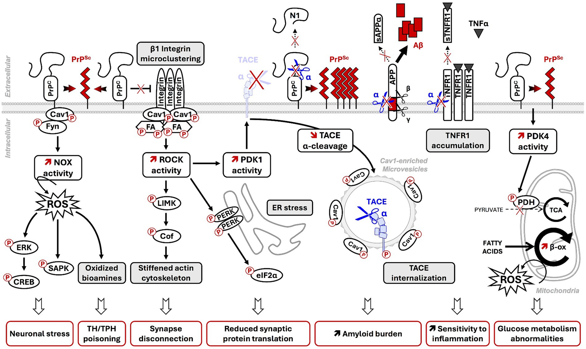
Figure 2. Contribution of PrPSc-induced PTMs dysregulation of PrPC membrane partners and coupled signaling effectors to prion neuropathogenesis. In prion diseases, post-translational conformational conversion (PTCC) of PrPC into PrPSc alters PTMs of PrPC membrane partners in PrPC signalosomes [Cav1, Fyn, components of focal adhesions (FA)] and deregulates downstream intracellular signaling effectors (NADPH oxidase-NOX, ROCK, PDK1, PDK4). The deregulation of PrPC-coupled signaling pathways on PrPC PTCC into PrPSc contributes to neurodegeneration through multiple deleterious events: onset of oxidative stress conditions, MAPK/SAPK-dependent neuronal stress, alteration of neuronal polarity and functions, amplification of PrPSc and Aβ production, increased vulnerability to TNFα inflammation by reduced shedding of plasma membrane TNFR into sTNFR, and energy metabolism abnormalities. NOX: NADPH oxidase; ROS: reactive oxygen species; ERK: extracellular-regulated kinases; SAPK: stress-associated protein kinases (p38 and JNK); ROCK: RhoA-associated coiled-coil containing kinases; PERK: PKR-related endoplasmic reticulum kinase; PDK1: 3-phosphoinositide-dependent kinase 1; TACE: TNFα converting enzyme; (s)TNFR: (soluble) TNFα receptor; N1: N-terminal fragment of α-cleaved PrPC; α/β/γ: α-, β-, and γ-secretases; sAPPα: neuroprotective α-cleaved APP fragment; Aβ: neurotoxic amyloid β-peptides; PDK4: Pyruvate Dehydrogenase Kinase 4; PDH: Pyruvate Dehydrogenase. TCA: tricyclic acid cycle or Krebs cycle; β-ox: fatty acid β-oxidation.
Interestingly, the fluidity of the plasma membrane also depends on caveolae fusion with the plasma membrane (Sohn et al., 2018; Yang et al., 2020). Whether, in prion-infected neurons, the freezing of caveolae in the cytosol caused by the excessive phosphorylation of Cav1 at Tyr14 affects the elastic properties of the plasma membrane and thus contributes to neurodegeneration deserves further investigation.
Although varying with the cell type, the molecular composition of the PrPC interactome always includes several proteins involved in cell adhesion, e.g., extracellular matrix proteins, such as laminin, vitronectin, and fibronectin, and membrane proteins, including integrins, neural cell adhesion molecule (NCAM), and myristoylated alanine-rich C-kinase substrate (MARCKS) proteins (Ghodrati et al., 2018). MARCKS are proteins inserted into the inner leaflet of the plasma membrane via a positively charged domain and a myristoyl group added to Gly2 (Arbuzova et al., 2000). These proteins likely interact with the GPI anchor of PrPC.
A global proteomic study comparing PrPC interactomes of four mouse cell lines and the mouse brain revealed a highly conserved functional relationship between PrPC, MARCKS, and NCAM1 (Mehrabian et al., 2016). PrPC directly interacts with NCAM1, promoting NCAM1 recruitment to lipid rafts and NCAM1-dependent Fyn kinase activation (Santuccione et al., 2005; Lehembre et al., 2008). Through its coupling to MARCKS, PrPC indirectly controls NCAM1 sialylation by regulating the expression of the polysialyltransferase ST8SIA2 (Boutin et al., 2009; Mehrabian et al., 2015). The PrPC-induced increase in NCAM1 polysialylation limits homophilic and heterophilic NCAM1 interactions and thereby modulates cell adhesion, a critical event for the onset and maintenance of neuronal polarity (Mehrabian et al., 2016). Any PrPSc-induced disturbance of the PrPC-NCAM1-MARCKS signalosome would affect neuronal polarity in prion diseases.
Beyond NCAM1, PrPC is also functionally involved in the regulation of other adhesion proteins, such as β1 integrins, which orchestrate the assembly and the turnover of focal adhesions (FAs). PrPC prevents β1 integrin microclustering and attenuates β1 integrin signaling in 1C11 and PC12 cells (Loubet et al., 2012). By interacting with β1 integrins, PrPC would block the structural modifications required for β1 integrin activation. Alternatively, PrPC would limit β1 integrin signaling by neutralizing some β1 integrin activators such as CD98 or thrombospondin-1 (Ghodrati et al., 2018). In prion-infected neuronal cells (Alleaume-Butaux et al., 2015), as well as in the brains of mice infected with the Chandler or Rocky Mountain Laboratory (RML) prion strains (Albert-Gasco et al., 2024), PrPSc-induced depletion of plasma membrane PrPC causes a loss of PrPC’s regulatory role toward β1 integrins. This depletion leads to β1 integrin microclustering and oversignaling (Alleaume-Butaux et al., 2015). β1 integrin oversignaling induced by PrPSc likely promotes chronic Src kinase phosphorylation at Tyr418 and activation, the subsequent phosphorylation of focal adhesion kinase (FAK) at Tyr861 by Src kinases, and the phosphorylation of paxillin at Tyr31 and Tyr118 by both Src kinases and FAK, as observed in cells depleted for PrPC (Loubet et al., 2012; Alleaume-Butaux et al., 2015; Ezpeleta et al., 2017). Such an overphosphorylated state of FA components enhances FA stability and reduces FA dynamics that disturb the adhesion properties of prion-infected neurons. This mechanism would contribute, at least in part, to the loss of neuronal polarity on prion infection (Figure 2) (Alleaume-Butaux et al., 2015).
Finally, PrPC modulates the laminin-mediated attachment of neurons to the extracellular matrix. PrPC interacts with laminin and laminin receptors (LRs) and limits laminin binding to its membrane receptor (Baloui et al., 2004). Our laboratory further showed in 1C11 neuronal cells that PrPC, through its coupling to Cav1 and Fyn, regulates the activity of tissue non-specific alkaline phosphatase (TNAP), which phosphorylates laminin and reduces laminin-dependent adhesion of cells (Ermonval et al., 2009a). As PrPSc interacts with LR in neuronal cells (Morel et al., 2005) and overactivates the PrPC-Cav1-Fyn platform, the excessive phosphorylation of laminin induced by PrPSc would also destabilize the neuronal cell-matrix interaction and neuronal polarity in prion-infected neurons.
Thus, by affecting several homeostatic PTMs in the PrPC adhesion interactome, PrPSc alters cell–cell and cell-extracellular matrix contacts required for the onset and stability of the neuronal polarity and the neuronal plasticity.
At the presynaptic and post-synaptic membrane of neurons, PrPC interacts with several ionotropic (NMDAR, AMPAR GluA1/2, KAR GluR6/7, and α7nAChR) and metabotropic (mGluR1/5) receptors (Kleene et al., 2007; Beraldo et al., 2010, 2011; Carulla et al., 2011; Watt et al., 2012; Um et al., 2013).
Several studies have highlighted that group I metabotropic glutamate receptors (mGluR1/5) act as co-receptors of PrPC. The binding of laminin or STI1 to PrPC activates PrPC, which in turn recruits and activates mGluR1/5 (Coitinho et al., 2006; Beraldo et al., 2011). mGluR1/5 are G protein-coupled receptors (GPCR) coupled to heterotrimeric Gαq/11 proteins involved in glutamate-dependent memory consolidation through neuritogenesis and neuronal plasticity events. The molecular mechanisms underlying PrPC modulation of mGluR1/5 activity remain, however, elusive. Nevertheless, the beneficial effect of mGluR1/5 inhibition in scrapie-infected mice suggests that PrPSc impacts the PrPC-mediated regulation of mGluR1/5 activity (Goniotaki et al., 2017). Since activation of mGluR1/5 involves binding of Fyn kinase to the C-terminal domain of mGluR1/5 and Fyn-dependent phosphorylation of mGluR1/5 at Tyr937 (Jin et al., 2017), chronic activation of Fyn by PrPSc may corrupt mGluR1/5 activity by imbalanced phosphorylation of mGluR1/5.
NMDAR is a calcium-permeable channel composed of two GluN1 and two GluN2 or GluN3 subunits involved in glutamate-mediated neuronal plasticity and excitotoxicity. PrPC interacts with GluN2D and GluN2B subunits (Khosravani et al., 2008; Barnes et al., 2020) and controls NMDA receptor activity through nitrosylation of GluN1 and GluN2A subunits (Gasperini et al., 2015). PrPC-bound Cu2+ acts as an electron acceptor that induces NO oxidation and subsequent S-nitrosylation of two cysteines on GluN1 and three cysteines on GluN2A, including Cys399, which mediates the predominant inhibitory effect on NMDAR activity. The reduction of NMDAR S-nitrosylation in prion-infected mice before the onset of clinical signs increases NMDAR-dependent excitation (Ratté et al., 2008). PrPSc also induces phosphorylation of the NMDAR GluN2B subunit at Tyr1472, probably via sustained activation of Fyn, and potentiates NMDAR activity in the hippocampus of a mouse model of CJD (Bertani et al., 2017). By decreasing S-nitrosylation and increasing phosphorylation of NMDAR, PrPSc enhances NMDAR activity and renders prion-infected neurons hypersensitive to NMDA-induced excitotoxicity (Meneghetti et al., 2019). Disturbance of PrPC-governed PTMs of neuronal receptors by PrPSc thus alters neurotransmission in prion diseases.
Of note, the synapse interactome of PrPC also includes synapse-associated proteins (synaptophysin and PSD-95), vesicle-associated proteins (synapsin), and ion pumps (Kv4.2 DPP6 and VGCC). Through these interactions, PrPC contributes to the assembly of functional complexes involved in neurotransmission at the pre- and post-synaptic membranes. Further investigations are needed to assess whether PrPSc-mediated disruption of those complexes or imbalanced PTMs of the above-mentioned synapse effectors would also contribute to the alteration of neurotransmission in prion diseases (Russelakis-Carneiro et al., 2004).
The use of diverse neuronal cell lines (N2a neuroblastoma cells, 1C11 neuronal stem cells and their serotonergic or noradrenergic neuronal progenies, PC12 pheochromocytoma cells, etc.) and primary cultures of neurons (cerebellar granule neurons, cortical or hippocampal neurons) showed that PrPC is involved in the regulation of a wide range of cellular functions, including cell adhesion (see Section 3.3), neuronal differentiation, synaptic plasticity, cell survival, redox equilibrium, stress protection, or energy metabolism (Schneider, 2011; Castle and Gill, 2017; Wulf et al., 2017; Schneider et al., 2021). This multifaceted role of PrPC involves its capacity to act as a receptor/co-receptor, governing a complex signaling network, and as a scaffolding protein that regulates the lipid rafts of the plasma membrane. This regulation affects the assembly and stoichiometry of interaction between partners such as integrins, laminin receptors, and mGluR, thereby controlling the activity of diverse signaling modules. Regardless of the context, PrPC is coupled to several intracellular signaling effectors, including Src kinase Fyn, NADPH oxidase, ERK1/2 MAP kinases, glycogen synthase kinase 3β (GSK3β), Protein kinase A (PKA), RhoA-associated coiled-coil containing kinases (ROCKs), 3-phosphoinositide-dependent kinase 1 (PDK1), Pyruvate Dehydrogenase Kinase 4 (PDK4), TACE α-secretase (aka ADAM17), and CREB transcription factor. These effectors all play a part in maintaining the homeostasis of neuronal functions (Chiarini et al., 2002; Hernandez-Rapp et al., 2014; Arnould et al., 2021; Schneider et al., 2021). For most of the signaling intermediates downstream of PrPC, their regulation depends on reversible and transient PTMs by phosphorylation, which sustains the fine-tuning of neuronal functions. Within a prion infectious context, the corruption of PrPC signaling in response to PrPC PTCC into PrPSc leads to imbalance PTMs of PrPC-coupled signaling effectors, thus generating aberrant and deleterious signals for prion-infected neurons (Figure 2). A global comparative phospho-proteome analysis between PrPSc-infected N2a cells and non-infected cells identified 105 proteins differentially phosphorylated with chronic and excessive phosphorylation of some effectors (e.g., cofilin) or loss of phosphorylation for some others (e.g., stathmin) (Wagner et al., 2010). We here review PTMs of some PrPC-coupled signaling effectors affected by prion infection and the consequences thereof for neurons.
One signaling pathway impacted by PrPSc in prion-infected neurons is the PrPC/Fyn/NADPH oxidase cascade. The chronic stimulation of this pathway triggers excessive production of reactive oxygen species (ROS) by NADPH oxidase (NOX) at the root of oxidative stress conditions. Overproduced ROS promote robust phosphorylation of MAPKs ERK1/2 (at Thr185/Tyr187) and additionally recruit stress-associated protein kinases (SAPKs) p38 and JNK1/2, which are activated by phosphorylations at Thr180/Tyr182 and Thr183/Tyr185, respectively. The subsequent sustained activation of MAPKs and SAPKs contributes to the death of prion-infected neurons by apoptosis (Figure 2) (Pietri et al., 2006; Pradines et al., 2013). The local rise of p38 phosphorylation at Thr180 and Tyr182 in dendritic spines of prion-infected hippocampal neurons was also shown to promote synaptic degeneration and decrement in synaptic transmission (Fang et al., 2018). Augmented phosphorylation of ERK1/2, p38, and JNK was confirmed in vivo in the brains of hamsters infected with the 263 K prion strain (Lee et al., 2005; Pamplona et al., 2008). Of note, downregulating this pathway through the use of siRNAs against Fyn or the p22phox subunit of NADPH oxidase reverts those PTMs on ERK1/2, p38, and JNK induced by prion infection, supporting the therapeutic potential of targeting Fyn or NADPH oxidase to protect neurons in prion diseases (Pradines et al., 2013).
In addition, ROS overproduced by NADPH oxidase in prion-infected serotonergic or noradrenergic neurons promote the generation of oxidized derivatives of serotonin (5-HT), such as tryptamine 4,5-dione (T-4,5-D) and 5,6-dihydroxytryptamine (5,6-DHT), or catecholamines, such as 6-hydroxydopamine and tetrahydroisoquinolines. These derivatives are considered neurotoxins (Mouillet-Richard et al., 2008). Their neurotoxic action relies on the poisoning of metabolic enzymes involved in the synthesis of 5-HT or noradrenalin (NE), that is, a set of “toxic and accidental PTMs” affecting the tryptophan hydroxylase TPH (the rate-limiting enzyme for 5-HT synthesis) and possibly the tyrosine hydroxylase TH (the rate-limiting enzyme for NE synthesis) through the covalent grafting of 5-HT/catecholamine-derived neurotoxins at catalytic Cys residues of those biosynthetic enzymes (Figure 2).
Another consequence of ERK1/2 activation is the downstream activation of the transcription factor CREB by phosphorylation at Ser133 (Figure 2) (Lee et al., 2005; Pradines et al., 2013). In prion-infected neurons, sustained CREB phosphorylation stimulates the expression of the immediate-early genes Egr-1 implicated in cell survival but lockdowns the transcription of the MMP9 encoding gene, which attenuates the metalloproteinase activity of MMP9. The decrease in MMP9 enzymatic activity leads to a reduction of β-dystroglycan cleavage at the neuronal cell surface that alters the interactions between neurons and the extracellular matrix (Pradines et al., 2013). Such CREB PTMs and subsequent modifications of gene expression in prion-infected neurons would contribute to the alterations of neuronal plasticity associated with prion diseases.
Our study documented that PrPSc abrogates the negative regulatory role exerted by PrPC on ROCK signaling due to a loss of PrPC control of β1 integrin microclustering and signaling activity (Alleaume-Butaux et al., 2015). In prion-infected 1C11 neuronal cells, N2a58 neuroblastoma cells, mouse cerebellar granule neurons, hippocampal neurons, and the brains of prion-infected mice, ROCK overactivity leads to excessive phosphorylation of LIMK1/2 at Thr505 and Thr508 and cofilin at Ser3. Stable phosphorylation of cofilin in prion-infected neurons cancels the severing activity of cofilin on the actin cytoskeleton (Figure 2) (Wagner et al., 2010; Alleaume-Butaux et al., 2015; Kim et al., 2020). The resulting PrPSc-induced stiffening of the actin cytoskeleton associated with fewer dynamics of focal adhesions (see section 3.3) disrupts neuronal polarity and provokes synaptic disconnection and dendrite/axon degeneration (Alleaume-Butaux et al., 2015; Kim et al., 2020). Such neuronal damages are counteracted by ROCK inhibition with pharmacological compounds (Alleaume-Butaux et al., 2015). Alterations in ROCK, LIMK1, and cofilin were also evidenced in the post-mortem cortex and cerebellum samples of sporadic Creutzfeldt–Jakob disease (sCJD) patients at clinical and pre-clinical stages (Zafar et al., 2018), paving the road for developing therapeutic strategies targeting ROCK to limit neurodegeneration in prion diseases.
Apart from the action of ROCK on the actin cytoskeleton, our laboratory showed that overactivated ROCK plays a role in the unfolded protein response (UPR). In 1C11-derived serotonergic neurons infected by mouse-adapted human GSS prions (Fukuoka strain), overactivated ROCK enhances the phosphorylation of PERK at Thr980 in the endoplasmic reticulum. Phosphorylated PERK, in turn, promotes the hyperphosphorylation of the translational initiation factor eiF2α at Ser51 (Schneider et al., 2021), which halts the translation of some proteins involved in the maintenance of synaptic connections (Moreno et al., 2012), likely contributing to synapse failure in prion-infected neurons (Figure 2). It remains unknown whether PERK is a direct substrate of ROCK. In any case, ROCK inhibition decreases the phosphorylation of PERK and eiF2α (Schneider et al., 2021), allowing restarting the expression of synaptic proteins and preserving neuronal transmission within a prion-infectious context.
In 2013, our laboratory provided prime evidence that the corruption of PrPC coupling to 3-phosphoinositide-dependent kinase 1 (PDK1) and downstream TACE α-secretase plays a critical role in the neuropathogenesis of prion diseases (Pietri et al., 2013). In prion-infected neurons, overactivated PDK1 promotes TACE phosphorylation at Thr735 and TACE displacement from the plasma membrane to caveolin-1-enriched microvesicles, which neutralizes TACE neuroprotective shedding activity (Figure 2). As PDK1 only admits AGC kinases as substrates (Leroux and Biondi, 2023), it is unlikely that TACE phosphorylation results from a direct action of PDK1 on TACE. The phosphorylation of TACE at Thr735 would be part of signals that impact the subcellular localization of TACE and modulate its shedding activity. Internalized, phosphorylated TACE in prion-infected neurons becomes uncoupled from three major substrates: (i) TNFα receptors (TNFR), which accumulate at the cell surface, rendering neurons hypersensitive to TNFα, (ii) PrPC, where the loss of PrPC PTM by α-cleavage between amino-acids 111/112 strongly reduces the C1 fragment of PrPC in favor of full-length PrPC (and C2 fragment), which is highly prone to converting into PrPSc, and (iii) the amyloid precursor protein (APP), where the loss of APP PTM by α-cleavage in favor of APP PTM by β- and γ-secretases leads to the accumulation of neurotoxic Aβ peptides (Figure 2) (Pietri et al., 2013; Ezpeleta et al., 2019). Importantly, inhibiting PDK1 suppresses TACE phosphorylation at Thr735. The subsequent redirection of TACE at the plasma membrane allows TACE to reintegrate cell surface signalosomes and recover its protective shedding activity. This activity includes the cleavages of TNFR, PrPC, and APP. Such restored irreversible PTMs of TNFR, PrPC, and APP protect prion-infected neurons from TNFα toxicity and limit the production of toxic amyloids (Pietri et al., 2013; Ezpeleta et al., 2019).
PDK1 activity is known to be governed by several events: (i) translocation to the plasma membrane, (ii) post-translational modifications by phosphorylation, (iii) conformational changes, and (iv) interaction with different effectors (Sacerdoti et al., 2023). We demonstrated that the overactivation of PDK1 in prion-infected neurons depends on the upstream kinase ROCK (Alleaume-Butaux et al., 2015). ROCK interacts with PDK1 and promotes phosphorylation of PDK1, a PTM that accounts for the increase in PDK1 enzymatic activity within a prion infectious context. Of note, ROCK-dependent phosphorylation of PDK1 occurs only after autophosphorylation of PDK1 at Ser241. The additional phosphorylation of PDK1 by ROCK leads to sustained PDK1 activity in prion-infected neurons. Importantly, as the pharmacological inhibition of PDK1 or ROCK reduces motor impairment, lowers brain PrPSc and Aβ levels, and prolongs the lifespan of prion-infected mice, PDK1 and ROCK are currently considered as potential therapeutic targets to combat prion diseases (Pietri et al., 2013; Alleaume-Butaux et al., 2015; Ezpeleta et al., 2019). Therefore, any strategies aiming at rescuing the α-cleavage or shedding of PrPC and APP should help to limit the production of PrPSc and Aβ and thereby mitigate prion diseases (Linsenmeier et al., 2021).
We evidenced that prion infection also cancels the regulatory role of PrPC on glucose metabolism, leading to a metabolic reprogramming of infected neurons, i.e., a conversion from glucose oxidative degradation to β-oxidation of fatty acids (Arnould et al., 2021). From a mechanistic point of view, PrPSc abrogates the negative control exerted by PrPC on the expression of Pyruvate Deshydrogenase Kinase 4 (PDK4) encoding gene, causing a high rise in PDK4 enzymatic activity. By phosphorylating the mitochondrial Pyruvate Dehydrogenase (PDH) complex, overactivated PDK4 decreases the activity of PDH that normally ensures the transfer of cytosolic pyruvate in the mitochondria and its conversion into acetylCoA for the production of energy. The consequences of such PTM of PDH and subsequently reduced activity of PDH are a slowdown of the glycolytic flux and limited oxidative degradation of glucose. To compensate for energy restriction, prion-infected neurons divert their metabolism toward fatty acids β-oxidation. Since fatty acids can act as pro-oxidant molecules, the oxidative stress resulting from the degradation of fatty acids by the β-oxidation pathway has been shown to contribute to neurodegeneration in prion diseases (Figure 2). Interestingly, pharmacological inhibition of PDK4 with dichloroacetate (DCA), a medicine approved for treating congenital lactic acidosis, restores, at least partly, PDH activity in the brains of prion-infected mice, which favors the recovery of glucose metabolism over fatty acids β-oxidation and extends the lifespan of DCA-treated prion-infected mice (Arnould et al., 2021).
PrPSc-induced dysregulation of ionotropic (NMDAR) and metabotropic receptors (mGluR) increases the intracellular Ca2+ level (Hu et al., 2022). Exposure of mouse cerebellar granule neurons to the neurotoxic PrP amyloidogenic polypeptide (PrP90-231) increases NMDAR-dependent uptake of Ca2+ (Thellung et al., 2017). In prion-infected SMB-S15 cells, mGluR oversignaling enhances the release of Ca2+ from ER in response to PLC-mediated hydrolysis of 4.5-biphosphate phosphatidylinositol and subsequent IP3 increase, combined with an increased level of IP3 receptor in the ER (Hu et al., 2022). Calmodulin (CaM), a transducer of Ca2+ signals that activates different kinases, is upregulated in the cortex of sporadic CJD (sCJD) patients and in the brains of prion-infected hamsters (Shi et al., 2015; Zhang et al., 2017). On prion infection, the upregulation of the Ca2+/calmodulin complex increases the Ca2+/calmodulin-dependent calcineurin (CaN) phosphatase activity. CaN is a type 2 phosphatase highly expressed in neurons, physiologically involved in synaptic plasticity, memory, and neuronal death. In prion-infected neurons, CaN dephosphorylates the pro-apoptotic protein Bad at Ser112, causing Bad translocation in mitochondria and the subsequent release of cytochrome c from mitochondria to the cytoplasm where it activates caspase-dependent apoptosis pathways (Agostinho et al., 2008). PrPSc-induced CaN overactivity would also alter synaptic plasticity and trigger neurodegeneration by dephosphorylating the substrate slingshot 1 (SHH1) in prion diseases. SHH1 activates cofilin and triggers the formation of cofilin-actin rods, which are involved in glutamate-mediated excitotoxicity (Bamburg et al., 2021). Pharmacological inhibition of CaN with the immunosuppressive drug FK506 limits neurodegeneration, reduces motor deficits, and increases the survival of mice infected with RML or Fukuoka-1 strains (Mukherjee et al., 2010; Nakagaki et al., 2013), introducing an additional way of therapeutic intervention for prion diseases.
The Calpain non-lysosomal cysteine proteases are other enzymes dysregulated by Ca2+ overload in prion-infected neurons (Baudry and Bi, 2024). In a sCJD mouse model, prion infection downregulates the neuroprotective Calpain-1 and upregulates the neurodegenerative Calpain-2, leading to global Calpain overactivity in the brain. Overactivated pathological Calpain- 2 enhances the cleavage of Calpain substrates such as Neurofilament Light Chain and γ-tubulin, additionally contributing to the loss of neuronal polarity (Llorens et al., 2017).
In prion diseases, the initial aberrant PTM concerns normal cellular prion protein PrPC with the post-translational conformational conversion (PTCC) of PrPC into pathogenic prions PrPSc. This dramatic change in PrPC folding is influenced by several PrPC PTMs (glycosylation, sialylation, cleavages, etc.) that oppose or favor the production of PrPSc. One consequence of the conversion of PrPC PTCC into PrPSc is the deregulation of PTMs at the proximal level of PrPC partners in PrPC signalosomes. Altered PTMs of PrPC partners in prion-infected neurons affect the homeostatic activity of plasma membrane adhesion proteins, neuronal receptors, or ion channels, likely contributing to neuronal polarity and neurotransmission defects in prion diseases. Altered PTMs in PrPC signalosomes impact downstream intracellular effectors such as Src kinases, ROCK, PDK1, PDK4, α-secretases, CREB transcription factor, and others. The imbalanced PTMs modify the biological activity or subcellular localization of these signaling effectors, thus hampering the signaling pathways they are involved in. It manifests by changes in redox equilibrium, metabolic reprogramming toward pro-oxidant fatty acids metabolism, high sensitivity to several stresses such as inflammation, and possibly autophagy derangement (López-Pérez et al., 2019), which compromise neuronal homeostasis and contribute to neurodegeneration in prion diseases. Most of the signaling effectors with disturbed PTMs listed in this review were identified with the help of prion-infected cell lines and primary neuronal cultures and confirmed in vivo in the brains of mouse models with prion-like diseases or even in the post-mortem brains of Creutzfeldt–Jakob disease patients. These signaling effectors currently represent attractive therapeutic targets to combat prion diseases and possibly other neurodegenerative diseases, such as Alzheimer’s and Parkinson’s diseases. Several studies have reported on the mechanistic convergence of these amyloid-based neurodegenerative diseases to PrPC. PrPC displays the capacity to interact with several unrelated amyloid proteins, including PrPSc, Alzheimer-linked Aβ oligomers, and Parkinson-linked pre-formed fibrils of pathological α-synuclein, and to relay their neurotoxicity (Laurén et al., 2009; Um et al., 2012; Aulić et al., 2017). Oligomers of Aβ or α-synuclein bind with a nanomolar affinity PrPC at the same epitopes, including one epitope in the hinge region of PrPC involved in PrPC PTCC into PrPSc (Chen et al., 2010; Smith et al., 2019; Corbett et al., 2020; Scialò and Legname, 2020). It would be tempting to genetically modify those PrPC epitopes with gene-editing technologies to generate PrPC molecules that are unable to bind amyloids or to be converted into PrPSc with the perspective of limiting or canceling amyloid neurotoxicity. Because PrPC is essential for the homeostasis of neurons and other cell types, such a genetic protective approach will be successful only if we are able to keep the PrPC cell functions intact, including PrPC signaling activity.
Of note, the binding to PrPC of Aβ or α-synuclein does not promote the conversion of PrPC into PrPSc, as PrPC PTCC into PrPSc is restricted to prion diseases. The binding to PrPC of Aβ/α-synuclein oligomers would deregulate PrPC signalosomes and downstream coupled signaling effectors on the removal of PrPC from signalosomes (loss-of-PrPC function). Alternatively, the binding to PrPC of Aβ/α-synuclein oligomers would freeze PrPC in signalosomes, maintaining PrPC signalosomes in an active state (gain-of-PrPC function). Whatever the scenario, the corruption of PrPC signalosomes by PrPSc, Aβ, or α-synuclein leads to post-translation modifications and deregulation of the same signaling effectors, such as ROCK, PDK1, and others (Pietri et al., 2013; Ferreira et al., 2017; Koch et al., 2018; Abd-Elrahman et al., 2020). In conclusion, common PTM patterns appear to contribute to neurodegeneration in prion diseases, Alzheimer’s diseases, and Parkinson’s diseases. However, the pending question remains as to which other specific PTM patterns can be specifically associated with each of these amyloid-based neurodegenerative diseases, as these diseases display different clinical manifestations.
ChB: Writing – original draft, Writing – review & editing. ClB: Writing – original draft, Writing – review & editing. AB: Writing – original draft, Writing – review & editing. AA-B: Writing – original draft, Writing – review & editing. BS: Writing – original draft, Writing – review & editing. MP: Writing – original draft, Writing – review & editing.
The author(s) declare financial support was received for the research, authorship, and/or publication of this article. This study was supported by the INSERM. ChB was supported by the Fondation pour la Recherche Medicale (FRM).
The authors declare that the research was conducted in the absence of any commercial or financial relationships that could be construed as a potential conflict of interest.
The author(s) declared that they were an editorial board member of Frontiers, at the time of submission. This had no impact on the peer review process and the final decision.
All claims expressed in this article are solely those of the authors and do not necessarily represent those of their affiliated organizations, or those of the publisher, the editors and the reviewers. Any product that may be evaluated in this article, or claim that may be made by its manufacturer, is not guaranteed or endorsed by the publisher.
Abd-Elrahman, K. S., Albaker, A., de Souza, J. M., Ribeiro, F. M., Schlossmacher, M. G., Tiberi, M., et al. (2020). Aβ oligomers induce pathophysiological mGluR5 signaling in Alzheimer’s disease model mice in a sex-selective manner. Sci. Signal. 13:eabd2494. doi: 10.1126/scisignal.abd2494
Agostinho, P., Lopes, J. P., Velez, Z., and Oliveira, C. R. (2008). Overactivation of calcineurin induced by amyloid-beta and prion proteins. Neurochem. Int. 52, 1226–1233. doi: 10.1016/j.neuint.2008.01.005
Aguilar-Calvo, P., Xiao, X., Bett, C., Eraña, H., Soldau, K., Castilla, J., et al. (2017). Post-translational modifications in PrP expand the conformational diversity of prions in vivo. Sci. Rep. 7:43295. doi: 10.1038/srep43295
Albert-Gasco, H., Smith, H. L., Alvarez-Castelao, B., Swinden, D., Halliday, M., Janaki-Raman, S., et al. (2024). Trazodone rescues dysregulated synaptic and mitochondrial nascent proteomes in prion neurodegeneration. Brain 147, 649–664. doi: 10.1093/brain/awad313
Alleaume-Butaux, A., Nicot, S., Pietri, M., Baudry, A., Dakowski, C., Tixador, P., et al. (2015). Double-edge sword of sustained ROCK activation in prion diseases through Neuritogenesis defects and prion accumulation. PLoS Pathog. 11:e1005073. doi: 10.1371/journal.ppat.1005073
Altmeppen, H. C., Prox, J., Krasemann, S., Puig, B., Kruszewski, K., Dohler, F., et al. (2015). The sheddase ADAM10 is a potent modulator of prion disease. eLife 4:e04260. doi: 10.7554/eLife.04260
Arbuzova, A., Wang, L., Wang, J., Hangyás-Mihályné, G., Murray, D., Honig, B., et al. (2000). Membrane binding of peptides containing both basic and aromatic residues. Experimental studies with peptides corresponding to the scaffolding region of Caveolin and the effector region of MARCKS. Biochemistry 39, 10330–10339. doi: 10.1021/bi001039j
Arnould, H., Baudouin, V., Baudry, A., Ribeiro, L. W., Ardila-Osorio, H., Pietri, M., et al. (2021). Loss of prion protein control of glucose metabolism promotes neurodegeneration in model of prion diseases. PLoS Pathog. 17:e1009991. doi: 10.1371/journal.ppat.1009991
Aulić, S., Masperone, L., Narkiewicz, J., Isopi, E., Bistaffa, E., Ambrosetti, E., et al. (2017). α-Synuclein amyloids hijack prion protein to gain cell entry, facilitate cell-to-cell spreading and block prion replication. Sci. Rep. 7:10050. doi: 10.1038/s41598-017-10236-x
Baloui, H., Von Boxberg, Y., Vinh, J., Weiss, S., Rossier, J., Nothias, F., et al. (2004). Cellular prion protein/laminin receptor: distribution in adult central nervous system and characterization of an isoform associated with a subtype of cortical neurons. Eur. J. Neurosci. 20, 2605–2616. doi: 10.1111/j.1460-9568.2004.03728.x
Bamburg, J. R., Minamide, L. S., Wiggan, O., Tahtamouni, L. H., and Kuhn, T. B. (2021). Cofilin and actin dynamics: multiple modes of regulation and their impacts in neuronal development and degeneration. Cells 10:2726. doi: 10.3390/cells10102726
Barnes, J. R., Mukherjee, B., Rogers, B. C., Nafar, F., Gosse, M., and Parsons, M. P. (2020). The relationship between glutamate dynamics and activity-dependent synaptic plasticity. J. Neurosci. 40, 2793–2807. doi: 10.1523/JNEUROSCI.1655-19.2020
Baskakov, I. V., and Katorcha, E. (2016). Multifaceted role of sialylation in prion diseases. Front. Neurosci. 10:358. doi: 10.3389/fnins.2016.00358
Bate, C., Nolan, W., McHale-Owen, H., and Williams, A. (2016). Sialic acid within the Glycosylphosphatidylinositol anchor targets the cellular prion protein to synapses *. J. Biol. Chem. 291, 17093–17101. doi: 10.1074/jbc.M116.731117
Baudry, M., and Bi, X. (2024). Revisiting the calpain hypothesis of learning and memory 40 years later. Front. Mol. Neurosci. 17:1337850. doi: 10.3389/fnmol.2024.1337850
Beraldo, F. H., Arantes, C. P., Santos, T. G., Machado, C. F., Roffe, M., Hajj, G. N., et al. (2011). Metabotropic glutamate receptors transduce signals for neurite outgrowth after binding of the prion protein to laminin γ1 chain. FASEB. J. Off. Publ. Fed. Am. Soc. Exp. Biol. 25, 265–279. doi: 10.1096/fj.10-161653
Beraldo, F. H., Arantes, C. P., Santos, T. G., Queiroz, N. G. T., Young, K., Rylett, R. J., et al. (2010). Role of α7 nicotinic acetylcholine receptor in calcium signaling induced by prion protein interaction with stress-inducible protein 1. J. Biol. Chem. 285, 36542–36550. doi: 10.1074/jbc.M110.157263
Bertani, I., Iori, V., Trusel, M., Maroso, M., Foray, C., Mantovani, S., et al. (2017). Inhibition of IL-1β signaling normalizes NMDA-dependent neurotransmission and reduces seizure susceptibility in a mouse model of Creutzfeldt–Jakob disease. J. Neurosci. 37, 10278–10289. doi: 10.1523/JNEUROSCI.1301-17.2017
Bosques, C. J., and Imperiali, B. (2003). The interplay of glycosylation and disulfide formation influences fibrillization in a prion protein fragment. Proc. Natl. Acad. Sci. USA 100, 7593–7598. doi: 10.1073/pnas.1232504100
Boutin, C., Schmitz, B., Cremer, H., and Diestel, S. (2009). NCAM expression induces neurogenesis in vivo. Eur. J. Neurosci. 30, 1209–1218. doi: 10.1111/j.1460-9568.2009.06928.x
Caetano, F. A., Lopes, M. H., Hajj, G. N. M., Machado, C. F., Arantes, C. P., Magalhães, A. C., et al. (2008). Endocytosis of prion protein is required for ERK1/2 signaling induced by stress-inducible protein 1. J. Neurosci. 28, 6691–6702. doi: 10.1523/JNEUROSCI.1701-08.2008
Carta, M., and Aguzzi, A. (2022). Molecular foundations of prion strain diversity. Curr. Opin. Neurobiol. 72, 22–31. doi: 10.1016/j.conb.2021.07.010
Carulla, P., Bribián, A., Rangel, A., Gavín, R., Ferrer, I., Caelles, C., et al. (2011). Neuroprotective role of PrPC against kainate-induced epileptic seizures and cell death depends on the modulation of JNK3 activation by GluR6/7-PSD-95 binding. Mol. Biol. Cell 22, 3041–3054. doi: 10.1091/mbc.E11-04-0321
Castle, A. R., and Gill, A. C. (2017). Physiological functions of the cellular prion protein. Front. Mol. Biosci. 4:19. doi: 10.3389/fmolb.2017.00019
Cha, S.-H., Choi, Y. R., Heo, C.-H., Kang, S.-J., Joe, E.-H., Jou, I., et al. (2015). Loss of parkin promotes lipid rafts-dependent endocytosis through accumulating caveolin-1: implications for Parkinson’s disease. Mol. Neurodegener. 10:63. doi: 10.1186/s13024-015-0060-5
Chen, C., Lv, Y., Zhang, B.-Y., Zhang, J., Shi, Q., Wang, J., et al. (2014). Apparent reduction of ADAM10 in scrapie-infected cultured cells and in the brains of scrapie-infected rodents. Mol. Neurobiol. 50, 875–887. doi: 10.1007/s12035-014-8708-7
Chen, S. G., Teplow, D. B., Parchi, P., Teller, J. K., Gambetti, P., and Autilio-Gambetti, L. (1995). Truncated forms of the human prion protein in Normal brain and in prion diseases. J. Biol. Chem. 270, 19173–19180. doi: 10.1074/jbc.270.32.19173
Chen, S., Yadav, S. P., and Surewicz, W. K. (2010). Interaction between human prion protein and amyloid-β (Aβ) oligomers role of n-terminal residues. J. Biol. Chem. 285, 26377–26383. doi: 10.1074/jbc.M110.145516
Chesebro, B., Trifilo, M., Race, R., Meade-White, K., Teng, C., LaCasse, R., et al. (2005). Anchorless prion protein results in infectious amyloid disease without clinical scrapie. Science 308, 1435–1439. doi: 10.1126/science.1110837
Chiarini, L. B., Freitas, A. R. O., Zanata, S. M., Brentani, R. R., Martins, V. R., and Linden, R. (2002). Cellular prion protein transduces neuroprotective signals. EMBO J. 21, 3317–3326. doi: 10.1093/emboj/cdf324
Cobb, N. J., Sönnichsen, F. D., McHaourab, H., and Surewicz, W. K. (2007). Molecular architecture of human prion protein amyloid: a parallel, in-register beta-structure. Proc. Natl. Acad. Sci. USA 104, 18946–18951. doi: 10.1073/pnas.0706522104
Coitinho, A. S., Freitas, A. R. O., Lopes, M. H., Hajj, G. N. M., Roesler, R., Walz, R., et al. (2006). The interaction between prion protein and laminin modulates memory consolidation. Eur. J. Neurosci. 24, 3255–3264. doi: 10.1111/j.1460-9568.2006.05156.x
Colby, D. W., Zhang, Q., Wang, S., Groth, D., Legname, G., Riesner, D., et al. (2007). Prion detection by an amyloid seeding assay. Proc. Natl. Acad. Sci. USA 104, 20914–20919. doi: 10.1073/pnas.0710152105
Collinge, J., and Clarke, A. R. (2007). A general model of prion strains and their pathogenicity. Science 318, 930–936. doi: 10.1126/science.1138718
Corbett, G. T., Wang, Z., Hong, W., Colom-Cadena, M., Rose, J., Liao, M., et al. (2020). PrP is a central player in toxicity mediated by soluble aggregates of neurodegeneration-causing proteins. Acta Neuropathol. (Berl.) 139, 503–526. doi: 10.1007/s00401-019-02114-9
Cracco, L., Puoti, G., Cornacchia, A., Glisic, K., Lee, S.-K., Wang, Z., et al. (2023). Novel histotypes of sporadic Creutzfeldt-Jakob disease linked to 129MV genotype. Acta Neuropathol. Commun. 11:141. doi: 10.1186/s40478-023-01631-9
Dias, M. V. S., Teixeira, B. L., Rodrigues, B. R., Sinigaglia-Coimbra, R., Porto-Carreiro, I., Roffé, M., et al. (2016). PRNP/prion protein regulates the secretion of exosomes modulating CAV1/caveolin-1-suppressed autophagy. Autophagy 12, 2113–2128. doi: 10.1080/15548627.2016.1226735
Dietzen, D. J., Hastings, W. R., and Lublin, D. M. (1995). Caveolin is Palmitoylated on multiple cysteine residues: palmitoylation is not necessary for localization of caveolin to caveolae (∗). J. Biol. Chem. 270, 6838–6842. doi: 10.1074/jbc.270.12.6838
Ermonval, M., Baudry, A., Baychelier, F., Pradines, E., Pietri, M., Oda, K., et al. (2009a). The cellular prion protein interacts with the tissue non-specific alkaline phosphatase in membrane microdomains of Bioaminergic neuronal cells. PLoS One 4:e6497. doi: 10.1371/journal.pone.0006497
Ermonval, M., Petit, D., Le Duc, A., Kellermann, O., and Gallet, P.-F. (2009b). Glycosylation-related genes are variably expressed depending on the differentiation state of a bioaminergic neuronal cell line: implication for the cellular prion protein. Glycoconj. J. 26, 477–493. doi: 10.1007/s10719-008-9198-5
Ezpeleta, J., Baudouin, V., Arellano-Anaya, Z. E., Boudet-Devaud, F., Pietri, M., Baudry, A., et al. (2019). Production of seedable amyloid-β peptides in model of prion diseases upon PrPSc-induced PDK1 overactivation. Nat. Commun. 10:3442. doi: 10.1038/s41467-019-11333-3
Ezpeleta, J., Boudet-Devaud, F., Pietri, M., Baudry, A., Baudouin, V., Alleaume-Butaux, A., et al. (2017). Protective role of cellular prion protein against TNFα-mediated inflammation through TACE α-secretase. Sci. Rep. 7:7671. doi: 10.1038/s41598-017-08110-x
Fang, C., Wu, B., Le, N. T. T., Imberdis, T., Mercer, R. C. C., and Harris, D. A. (2018). Prions activate a p38 MAPK synaptotoxic signaling pathway. PLoS Pathog. 14:e1007283. doi: 10.1371/journal.ppat.1007283
Ferreira, D. G., Temido-Ferreira, M., Vicente Miranda, H., Batalha, V. L., Coelho, J. E., Szegö, É. M., et al. (2017). α-Synuclein interacts with PrPC to induce cognitive impairment through mGluR5 and NMDAR2B. Nat. Neurosci. 20, 1569–1579. doi: 10.1038/nn.4648
Gasperini, L., Meneghetti, E., Pastore, B., Benetti, F., and Legname, G. (2015). Prion protein and copper cooperatively protect neurons by modulating NMDA receptor through S-nitrosylation. Antioxid. Redox Signal. 22, 772–784. doi: 10.1089/ars.2014.6032
Ghodrati, F., Mehrabian, M., Williams, D., Halgas, O., Bourkas, M. E. C., Watts, J. C., et al. (2018). The prion protein is embedded in a molecular environment that modulates transforming growth factor β and integrin signaling. Sci. Rep. 8:8654. doi: 10.1038/s41598-018-26685-x
Goniotaki, D., Lakkaraju, A. K. K., Shrivastava, A. N., Bakirci, P., Sorce, S., Senatore, A., et al. (2017). Inhibition of group-I metabotropic glutamate receptors protects against prion toxicity. PLoS Pathog. 13:e1006733. doi: 10.1371/journal.ppat.1006733
Gottlieb-Abraham, E., Shvartsman, D. E., Donaldson, J. C., Ehrlich, M., Gutman, O., Martin, G. S., et al. (2013). Src-mediated caveolin-1 phosphorylation affects the targeting of active Src to specific membrane sites. Mol. Biol. Cell 24, 3881–3895. doi: 10.1091/mbc.e13-03-0163
Hannaoui, S., Zemlyankina, I., Chang, S. C., Arifin, M. I., Béringue, V., McKenzie, D., et al. (2022). Transmission of cervid prions to humanized mice demonstrates the zoonotic potential of CWD. Acta Neuropathol. (Berl.) 144, 767–784. doi: 10.1007/s00401-022-02482-9
Hernandez-Rapp, J., Martin-Lannerée, S., Hirsch, T. Z., Pradines, E., Alleaume-Butaux, A., Schneider, B., et al. (2014). A PrPC-caveolin-Lyn complex negatively controls neuronal GSK3β and serotonin 1B receptor. Sci. Rep. 4:4881. doi: 10.1038/srep04881
Hoyt, F., Standke, H. G., Artikis, E., Schwartz, C. L., Hansen, B., Li, K., et al. (2022). Cryo-EM structure of anchorless RML prion reveals variations in shared motifs between distinct strains. Nat. Commun. 13:4005. doi: 10.1038/s41467-022-30458-6
Hu, C., Chen, C., Xia, Y., Chen, J., Yang, W., Wang, L., et al. (2022). Different aberrant changes of mGluR5 and its downstream signaling pathways in the scrapie-infected cell line and the brains of scrapie-infected experimental rodents. Front. Cell Dev. Biol. 10:844378. doi: 10.3389/fcell.2022.844378
Humphrey, S. J., James, D. E., and Mann, M. (2015). Protein phosphorylation: a major switch mechanism for metabolic regulation. Trends Endocrinol. Metab. 26, 676–687. doi: 10.1016/j.tem.2015.09.013
Jin, D.-Z., Mao, L.-M., and Wang, J. Q. (2017). An essential role of Fyn in the modulation of metabotropic glutamate receptor 1 in neurons. eNeuro 4, ENEURO.0096–ENEU17.2017. doi: 10.1523/ENEURO.0096-17.2017
Katorcha, E., Makarava, N., Savtchenko, R., and Baskakov, I. V. (2015). Sialylation of the prion protein glycans controls prion replication rate and glycoform ratio. Sci. Rep. 5:16912. doi: 10.1038/srep16912
Katorcha, E., Srivastava, S., Klimova, N., and Baskakov, I. V. (2016). Sialylation of Glycosylphosphatidylinositol (GPI) anchors of mammalian prions is regulated in a host-, tissue-, and cell-specific manner *. J. Biol. Chem. 291, 17009–17019. doi: 10.1074/jbc.M116.732040
Khosravani, H., Zhang, Y., Tsutsui, S., Hameed, S., Altier, C., Hamid, J., et al. (2008). Prion protein attenuates excitotoxicity by inhibiting NMDA receptors. J. Cell Biol. 181, 551–565. doi: 10.1083/jcb.200711002
Kim, H.-J., Kim, M.-J., Mostafa, M. N., Park, J.-H., Choi, H.-S., Kim, Y.-S., et al. (2020). RhoA/ROCK regulates prion pathogenesis by controlling Connexin 43 activity. Int. J. Mol. Sci. 21:1255. doi: 10.3390/ijms21041255
Kleene, R., Loers, G., Langer, J., Frobert, Y., Buck, F., and Schachner, M. (2007). Prion protein regulates glutamate-dependent lactate transport of astrocytes. J. Neurosci. 27, 12331–12340. doi: 10.1523/JNEUROSCI.1358-07.2007
Knight, R. (2017). “Chapter thirteen - infectious and sporadic prion diseases,” in Progress in Molecular Biology and Translational Science, eds. G. Legname and S. Vanni (Academic Press), 293–318.
Koch, J. C., Tatenhorst, L., Roser, A.-E., Saal, K.-A., Tönges, L., and Lingor, P. (2018). ROCK inhibition in models of neurodegeneration and its potential for clinical translation. Pharmacol. Ther. 189, 1–21. doi: 10.1016/j.pharmthera.2018.03.008
Kovač, V., and Čurin Šerbec, V. (2018). Prion proteins without the glycophosphatidylinositol anchor: potential biomarkers in neurodegenerative diseases. Biomark. Insights 13:1177271918756648. doi: 10.1177/1177271918756648
Kraus, A., Hoyt, F., Schwartz, C. L., Hansen, B., Artikis, E., Hughson, A. G., et al. (2021). High-resolution structure and strain comparison of infectious mammalian prions. Mol. Cell 81, 4540–4551.e6. doi: 10.1016/j.molcel.2021.08.011
Krebs, B., Dorner-Ciossek, C., Schmalzbauer, R., Vassallo, N., Herms, J., and Kretzschmar, H. A. (2006). Prion protein induced signaling cascades in monocytes. Biochem. Biophys. Res. Commun. 340, 13–22. doi: 10.1016/j.bbrc.2005.11.158
Laurén, J., Gimbel, D. A., Nygaard, H. B., Gilbert, J. W., and Strittmatter, S. M. (2009). Cellular prion protein mediates impairment of synaptic plasticity by amyloid-β oligomers. Nature 457, 1128–1132. doi: 10.1038/nature07761
Lee, H.-P., Jun, Y.-C., Choi, J.-K., Kim, J.-I., Carp, R. I., and Kim, Y.-S. (2005). Activation of mitogen-activated protein kinases in hamster brains infected with 263K scrapie agent. J. Neurochem. 95, 584–593. doi: 10.1111/j.1471-4159.2005.03429.x
Lehembre, F., Yilmaz, M., Wicki, A., Schomber, T., Strittmatter, K., Ziegler, D., et al. (2008). NCAM-induced focal adhesion assembly: a functional switch upon loss of E-cadherin. EMBO J. 27, 2603–2615. doi: 10.1038/emboj.2008.178
Leroux, A. E., and Biondi, R. M. (2023). The choreography of protein kinase PDK1 and its diverse substrate dance partners. Biochem. J. 480, 1503–1532. doi: 10.1042/BCJ20220396
Linden, R., Martins, V. R., Prado, M. A. M., Cammarota, M., Izquierdo, I., and Brentani, R. R. (2008). Physiology of the prion protein. Physiol. Rev. 88, 673–728. doi: 10.1152/physrev.00007.2007
Linsenmeier, L., Mohammadi, B., Shafiq, M., Frontzek, K., Bär, J., Shrivastava, A. N., et al. (2021). Ligands binding to the prion protein induce its proteolytic release with therapeutic potential in neurodegenerative proteinopathies. Sci. Adv. 7:eabj1826. doi: 10.1126/sciadv.abj1826
Llorens, F., Thüne, K., Sikorska, B., Schmitz, M., Tahir, W., Fernández-Borges, N., et al. (2017). Altered Ca2+ homeostasis induces Calpain-Cathepsin axis activation in sporadic Creutzfeldt-Jakob disease. Acta Neuropathol. Commun. 5:35. doi: 10.1186/s40478-017-0431-y
Lopes, M. H., Hajj, G. N. M., Muras, A. G., Mancini, G. L., Castro, R. M. P. S., Ribeiro, K. C. B., et al. (2005). Interaction of cellular prion and stress-inducible protein 1 promotes Neuritogenesis and neuroprotection by distinct signaling pathways. J. Neurosci. 25, 11330–11339. doi: 10.1523/JNEUROSCI.2313-05.2005
López-Pérez, Ó., Otero, A., Filali, H., Sanz-Rubio, D., Toivonen, J. M., Zaragoza, P., et al. (2019). Dysregulation of autophagy in the central nervous system of sheep naturally infected with classical scrapie. Sci. Rep. 9:1911. doi: 10.1038/s41598-019-38500-2
Loubet, D., Dakowski, C., Pietri, M., Pradines, E., Bernard, S., Callebert, J., et al. (2012). Neuritogenesis: the prion protein controls β1 integrin signaling activity. FASEB J. 26, 678–690. doi: 10.1096/fj.11-185579
Louros, N., Schymkowitz, J., and Rousseau, F. (2023). Mechanisms and pathology of protein misfolding and aggregation. Nat. Rev. Mol. Cell Biol. 24, 912–933. doi: 10.1038/s41580-023-00647-2
Luo, S., Yang, M., Zhao, H., Han, Y., Jiang, N., Yang, J., et al. (2021). Caveolin-1 regulates cellular metabolism: a potential therapeutic target in kidney disease. Front. Pharmacol. 12:768100. doi: 10.3389/fphar.2021.768100
Makarava, N., and Baskakov, I. V. (2023). Role of sialylation of N-linked glycans in prion pathogenesis. Cell Tissue Res. 392, 201–214. doi: 10.1007/s00441-022-03584-2
Makarava, N., Chang, J. C.-Y., Molesworth, K., and Baskakov, I. V. (2020). Posttranslational modifications define course of prion strain adaptation and disease phenotype. J. Clin. Invest. 130, 4382–4395. doi: 10.1172/JCI138677
Mallucci, G., Dickinson, A., Linehan, J., Klöhn, P.-C., Brandner, S., and Collinge, J. (2003). Depleting neuronal PrP in prion infection prevents disease and reverses Spongiosis. Science 302, 871–874. doi: 10.1126/science.1090187
Manka, S. W., Zhang, W., Wenborn, A., Betts, J., Joiner, S., Saibil, H. R., et al. (2022). 2.7 Å cryo-EM structure of ex vivo RML prion fibrils. Nat. Commun. 13:4004. doi: 10.1038/s41467-022-30457-7
McNally, K. L., Ward, A. E., and Priola, S. A. (2009). Cells expressing anchorless prion protein are resistant to scrapie infection. J. Virol. 83, 4469–4475. doi: 10.1128/jvi.02412-08
Mehrabian, M., Brethour, D., Wang, H., Xi, Z., Rogaeva, E., and Schmitt-Ulms, G. (2015). The prion protein controls Polysialylation of neural cell adhesion molecule 1 during cellular morphogenesis. PLoS One 10:e0133741. doi: 10.1371/journal.pone.0133741
Mehrabian, M., Hildebrandt, H., and Schmitt-Ulms, G. (2016). NCAM1 Polysialylation: the prion Protein’s elusive reason for being? ASN Neuro 8:175909141667907. doi: 10.1177/1759091416679074
Meneghetti, E., Gasperini, L., Virgilio, T., Moda, F., Tagliavini, F., Benetti, F., et al. (2019). Prions strongly reduce NMDA receptor S-Nitrosylation levels at pre-symptomatic and terminal stages of prion diseases. Mol. Neurobiol. 56, 6035–6045. doi: 10.1007/s12035-019-1505-6
Millán-Zambrano, G., Burton, A., Bannister, A. J., and Schneider, R. (2022). Histone post-translational modifications — cause and consequence of genome function. Nat. Rev. Genet. 23, 563–580. doi: 10.1038/s41576-022-00468-7
Miranzadeh Mahabadi, H., and Taghibiglou, C. (2020). Cellular prion protein (PrPc): putative interacting partners and consequences of the interaction. Int. J. Mol. Sci. 21:7058. doi: 10.3390/ijms21197058
Mohammadi, B., Song, F., Matamoros-Angles, A., Shafiq, M., Damme, M., Puig, B., et al. (2023). Anchorless risk or released benefit? An updated view on the ADAM10-mediated shedding of the prion protein. Cell Tissue Res. 392, 215–234. doi: 10.1007/s00441-022-03582-4
Morel, E., Andrieu, T., Casagrande, F., Gauczynski, S., Weiss, S., Grassi, J., et al. (2005). Bovine prion is endocytosed by human enterocytes via the 37 kDa/67 kDa laminin receptor. Am. J. Pathol. 167, 1033–1042. doi: 10.1016/S0002-9440(10)61192-3
Moreno, J. A., Radford, H., Peretti, D., Steinert, J. R., Verity, N., Martin, M. G., et al. (2012). Sustained translational repression by eIF2α-P mediates prion neurodegeneration. Nature 485, 507–511. doi: 10.1038/nature11058
Mouillet-Richard, S., Ermonval, M., Chebassier, C., Laplanche, J. L., Lehmann, S., Launay, J. M., et al. (2000). Signal transduction through prion protein. Science 289, 1925–1928. doi: 10.1126/science.289.5486.1925
Mouillet-Richard, S., Nishida, N., Pradines, E., Laude, H., Schneider, B., Féraudet, C., et al. (2008). Prions impair bioaminergic functions through serotonin- or catecholamine-derived neurotoxins in neuronal cells. J. Biol. Chem. 283, 23782–23790. doi: 10.1074/jbc.M802433200
Mukherjee, A., Morales-Scheihing, D., Gonzalez-Romero, D., Green, K., Taglialatela, G., and Soto, C. (2010). Calcineurin inhibition at the clinical phase of prion disease reduces neurodegeneration, improves behavioral alterations and increases animal survival. PLoS Pathog. 6:e1001138. doi: 10.1371/journal.ppat.1001138
Nakagaki, T., Satoh, K., Ishibashi, D., Fuse, T., Sano, K., Kamatari, Y. O., et al. (2013). FK506 reduces abnormal prion protein through the activation of autolysosomal degradation and prolongs survival in prion-infected mice. Autophagy 9, 1386–1394. doi: 10.4161/auto.25381
Pamplona, R., Naudí, A., Gavín, R., Pastrana, M. A., Sajnani, G., Ilieva, E. V., et al. (2008). Increased oxidation, glycoxidation, and lipoxidation of brain proteins in prion disease. Free Radic. Biol. Med. 45, 1159–1166. doi: 10.1016/j.freeradbiomed.2008.07.009
Pantera, B., Bini, C., Cirri, P., Paoli, P., Camici, G., Manao, G., et al. (2009). PrPc activation induces neurite outgrowth and differentiation in PC12 cells: role for caveolin-1 in the signal transduction pathway. J. Neurochem. 110, 194–207. doi: 10.1111/j.1471-4159.2009.06123.x
Parchi, P., Giese, A., Capellari, S., Brown, P., Schulz-Schaeffer, W., Windl, O., et al. (1999). Classification of sporadic Creutzfeldt-Jakob disease based on molecular and phenotypic analysis of 300 subjects. Ann. Neurol. 46, 224–233. doi: 10.1002/1531-8249(199908)46:2<224::AID-ANA12>3.0.CO;2-W
Parton, R. G., Joggerst, B., and Simons, K. (1994). Regulated internalization of caveolae. J. Cell Biol. 127, 1199–1215. doi: 10.1083/jcb.127.5.1199
Parton, R. G., and Simons, K. (2007). The multiple faces of caveolae. Nat. Rev. Mol. Cell Biol. 8, 185–194. doi: 10.1038/nrm2122
Pascuzzo, R., Oxtoby, N. P., Young, A. L., Blevins, J., Castelli, G., Garbarino, S., et al. (2020). Prion propagation estimated from brain diffusion MRI is subtype dependent in sporadic Creutzfeldt-Jakob disease. Acta Neuropathol. (Berl.) 140, 169–181. doi: 10.1007/s00401-020-02168-0
Pietri, M., Caprini, A., Mouillet-Richard, S., Pradines, E., Ermonval, M., Grassi, J., et al. (2006). Overstimulation of PrPC signaling pathways by prion peptide 106-126 causes oxidative injury of Bioaminergic neuronal cells *. J. Biol. Chem. 281, 28470–28479. doi: 10.1074/jbc.M602774200
Pietri, M., Dakowski, C., Hannaoui, S., Alleaume-Butaux, A., Hernandez-Rapp, J., Ragagnin, A., et al. (2013). PDK1 decreases TACE-mediated α-secretase activity and promotes disease progression in prion and Alzheimer’s diseases. Nat. Med. 19, 1124–1131. doi: 10.1038/nm.3302
Pradines, E., Hernandez-Rapp, J., Villa-Diaz, A., Dakowski, C., Ardila-Osorio, H., Haik, S., et al. (2013). Pathogenic prions deviate PrPC signaling in neuronal cells and impair A-beta clearance. Cell Death Dis. 4:e456. doi: 10.1038/cddis.2012.195
Prusiner, S. B. (1986). Prions are novel infectious pathogens causing scrapie and Creutzfeldt-Jakob disease. BioEssays news rev. Mol. Cell. Dev. Biol. 5, 281–286. doi: 10.1002/bies.950050612
Prusiner, S. B. (1998). Prions. Proc. Natl. Acad. Sci. USA 95, 13363–13383. doi: 10.1073/pnas.95.23.13363
Ramazi, S., and Zahiri, J. (2021). Post-translational modifications in proteins: resources, tools and prediction methods. Database 2021:baab012. doi: 10.1093/database/baab012
Ratté, S., Prescott, S. A., Collinge, J., and Jefferys, J. G. R. (2008). Hippocampal bursts caused by changes in NMDA receptor-dependent excitation in a mouse model of variant CJD. Neurobiol. Dis. 32, 96–104. doi: 10.1016/j.nbd.2008.06.007
Rudd, P. M., Merry, A. H., Wormald, M. R., and Dwek, R. A. (2002). Glycosylation and prion protein. Curr. Opin. Struct. Biol. 12, 578–586. doi: 10.1016/s0959-440x(02)00377-9
Russelakis-Carneiro, M., Hetz, C., Maundrell, K., and Soto, C. (2004). Prion replication alters the distribution of Synaptophysin and Caveolin 1 in neuronal lipid rafts. Am. J. Pathol. 165, 1839–1848. doi: 10.1016/S0002-9440(10)63439-6
Sacerdoti, M., Gross, L. Z. F., Riley, A. M., Zehnder, K., Ghode, A., Klinke, S., et al. (2023). Modulation of the substrate specificity of the kinase PDK1 by distinct conformations of the full-length protein. Sci. Signal. 16:eadd3184. doi: 10.1126/scisignal.add3184
Santuccione, A., Sytnyk, V., Leshchyns’ka, I., and Schachner, M. (2005). Prion protein recruits its neuronal receptor NCAM to lipid rafts to activate p59fyn and to enhance neurite outgrowth. J. Cell Biol. 169, 341–354. doi: 10.1083/jcb.200409127
Schilling, K. M., Jorwal, P., Ubilla-Rodriguez, N. C., Assafa, T. E., Gatdula, J. R. P., Vultaggio, J. S., et al. (2023). N-glycosylation is a potent regulator of prion protein neurotoxicity. J. Biol. Chem. 299:105101. doi: 10.1016/j.jbc.2023.105101
Schneider, B. (2011). Understanding the neurospecificity of prion protein signaling. Front. Biosci. 16, 169–186. doi: 10.2741/3682
Schneider, B., Baudry, A., Pietri, M., Alleaume-Butaux, A., Bizingre, C., Nioche, P., et al. (2021). The cellular prion protein-ROCK connection: contribution to neuronal homeostasis and neurodegenerative diseases. Front. Cell. Neurosci. 15:660683. doi: 10.3389/fncel.2021.660683
Schneider, B., Mutel, V., Pietri, M., Ermonval, M., Mouillet-Richard, S., and Kellermann, O. (2003). NADPH oxidase and extracellular regulated kinases 1/2 are targets of prion protein signaling in neuronal and nonneuronal cells. Proc. Natl. Acad. Sci. USA 100, 13326–13331. doi: 10.1073/pnas.2235648100
Scialò, C., and Legname, G. (2020). The role of the cellular prion protein in the uptake and toxic signaling of pathological neurodegenerative aggregates. Prog. Mol. Biol. Transl. Sci. 175, 297–323. doi: 10.1016/bs.pmbts.2020.08.008
Sevillano, A. M., Aguilar-Calvo, P., Kurt, T. D., Lawrence, J. A., Soldau, K., Nam, T. H., et al. (2020). Prion protein glycans reduce intracerebral fibril formation and spongiosis in prion disease. J. Clin. Invest. 130, 1350–1362. doi: 10.1172/JCI131564
Shen, P., Dang, J., Wang, Z., Zhang, W., Yuan, J., Lang, Y., et al. (2021). Characterization of anchorless human PrP with Q227X stop mutation linked to Gerstmann-Sträussler-Scheinker syndrome in vivo and in vitro. Mol. Neurobiol. 58, 21–33. doi: 10.1007/s12035-020-02098-8
Shi, Q., Chen, L.-N., Zhang, B.-Y., Xiao, K., Zhou, W., Chen, C., et al. (2015). Proteomics analyses for the global proteins in the brain tissues of different human prion diseases. Mol. Cell. Proteomics MCP 14, 854–869. doi: 10.1074/mcp.M114.038018
Shi, Q., Jing, Y.-Y., Wang, S.-B., Chen, C., Sun, H., Xu, Y., et al. (2013). PrP octarepeats region determined the interaction with caveolin-1 and phosphorylation of caveolin-1 and Fyn. Med. Microbiol. Immunol. 202, 215–227. doi: 10.1007/s00430-012-0284-8
Silva, C. J., Vázquez-Fernández, E., Onisko, B., and Requena, J. R. (2015). Proteinase K and the structure of PrPSc: the good, the bad and the ugly. Virus Res. 207, 120–126. doi: 10.1016/j.virusres.2015.03.008
Smith, L. M., Kostylev, M. A., Lee, S., and Strittmatter, S. M. (2019). Systematic and standardized comparison of reported amyloid-β receptors for sufficiency, affinity, and Alzheimer’s disease relevance. J. Biol. Chem. 294, 6042–6053. doi: 10.1074/jbc.RA118.006252
Sohn, J., Lin, H., Fritch, M. R., and Tuan, R. S. (2018). Influence of cholesterol/caveolin-1/caveolae homeostasis on membrane properties and substrate adhesion characteristics of adult human mesenchymal stem cells. Stem Cell Res Ther 9:86. doi: 10.1186/s13287-018-0830-4
Srivastava, S., Makarava, N., Katorcha, E., Savtchenko, R., Brossmer, R., and Baskakov, I. V. (2015). Post-conversion sialylation of prions in lymphoid tissues. Proc. Natl. Acad. Sci. 112, E6654–E6662. doi: 10.1073/pnas.1517993112
Stahl, N., Borchelt, D. R., and Prusiner, S. B. (1990). Differential release of cellular and scrapie prion proteins from cellular membranes by phosphatidylinositol-specific phospholipase C. Biochemistry 29, 5405–5412. doi: 10.1021/bi00474a028
Stuermer, C. A. O., Langhorst, M. F., Wiechers, M. F., Legler, D. F., Hanwehr, S. H.Von, Guse, A. H., et al. (2004). PrPc capping in T cells promotes its association with the lipid raft proteins reggie-1 and reggie-2 and leads to signal transduction. FASEB J. 18, 1731–1733. doi: 10.1096/fj.04-2150fje
Sverdlov, M., Shinin, V., Place, A. T., Castellon, M., and Minshall, R. D. (2009). Filamin a regulates Caveolae internalization and trafficking in endothelial cells. Mol. Biol. Cell 20, 4531–4540. doi: 10.1091/mbc.e08-10-0997
Taylor, D. R., and Hooper, N. M. (2006). The prion protein and lipid rafts (review). Mol. Membr. Biol. 23, 89–99. doi: 10.1080/09687860500449994
Thellung, S., Gatta, E., Pellistri, F., Villa, V., Corsaro, A., Nizzari, M., et al. (2017). Different molecular mechanisms mediate direct or glia-dependent prion protein fragment 90–231 neurotoxic effects in cerebellar granule neurons. Neurotox. Res. 32, 381–397. doi: 10.1007/s12640-017-9749-2
Toni, M., Spisni, E., Griffoni, C., Santi, S., Riccio, M., Lenaz, P., et al. (2006). Cellular prion protein and Caveolin-1 interaction in a neuronal cell line precedes Fyn/Erk 1/2 signal transduction. Biomed. Res. Int. 2006:e69469. doi: 10.1155/JBB/2006/69469
Tranulis, M. A., and Tryland, M. (2023). The zoonotic potential of chronic wasting disease—a review. Food Secur. 12:824. doi: 10.3390/foods12040824
Triebel, J., Robles, J. P., Zamora, M., Clapp, C., and Bertsch, T. (2022). New horizons in specific hormone proteolysis. Trends Endocrinol. Metab. TEM 33, 371–377. doi: 10.1016/j.tem.2022.03.004
Um, J. W., Kaufman, A. C., Kostylev, M., Heiss, J. K., Stagi, M., Takahashi, H., et al. (2013). Metabotropic glutamate receptor 5 is a coreceptor for Alzheimer aβ oligomer bound to cellular prion protein. Neuron 79, 887–902. doi: 10.1016/j.neuron.2013.06.036
Um, J. W., Nygaard, H. B., Heiss, J. K., Kostylev, M. A., Stagi, M., Vortmeyer, A., et al. (2012). Alzheimer amyloid-β oligomer bound to postsynaptic prion protein activates Fyn to impair neurons. Nat. Neurosci. 15, 1227–1235. doi: 10.1038/nn.3178
Vázquez-Fernández, E., Vos, M. R., Afanasyev, P., Cebey, L., Sevillano, A. M., Vidal, E., et al. (2016). The structural architecture of an infectious mammalian prion using Electron Cryomicroscopy. PLoS Pathog. 12:e1005835. doi: 10.1371/journal.ppat.1005835
Vilette, D., Courte, J., Peyrin, J. M., Coudert, L., Schaeffer, L., Andréoletti, O., et al. (2018). Cellular mechanisms responsible for cell-to-cell spreading of prions. Cell. Mol. Life Sci. 75, 2557–2574. doi: 10.1007/s00018-018-2823-y
Vincent, B., Paitel, E., Saftig, P., Frobert, Y., Hartmann, D., Strooper, B. D., et al. (2001). The Disintegrins ADAM10 and TACE contribute to the constitutive and Phorbol Ester-regulated Normal cleavage of the cellular prion protein *. J. Biol. Chem. 276, 37743–37746. doi: 10.1074/jbc.M105677200
Wagner, W., Ajuh, P., Löwer, J., and Wessler, S. (2010). Quantitative phosphoproteomic analysis of prion-infected neuronal cells. Cell Commun. Signal 8:28. doi: 10.1186/1478-811X-8-28
Walsh, C. T., Garneau-Tsodikova, S., and Gatto, G. J. (2005). Protein posttranslational modifications: the chemistry of proteome diversifications. Angew. Chem. Int. Ed. Engl. 44, 7342–7372. doi: 10.1002/anie.200501023
Wang, P.-S., Wang, J., Xiao, Z.-C., and Pallen, C. J. (2009). Protein-tyrosine phosphatase α acts as an upstream regulator of Fyn signaling to promote oligodendrocyte differentiation and myelination. J. Biol. Chem. 284, 33692–33702. doi: 10.1074/jbc.M109.061770
Watt, N. T., Taylor, D. R., Kerrigan, T. L., Griffiths, H. H., Rushworth, J. V., Whitehouse, I. J., et al. (2012). Prion protein facilitates uptake of zinc into neuronal cells. Nat. Commun. 3:1134. doi: 10.1038/ncomms2135
Westergard, L., Turnbaugh, J. A., and Harris, D. A. (2011). A naturally occurring C-terminal fragment of the prion protein (PrP) delays disease and acts as a dominant-negative inhibitor of PrPSc formation. J. Biol. Chem. 286, 44234–44242. doi: 10.1074/jbc.M111.286195
Wulf, M.-A., Senatore, A., and Aguzzi, A. (2017). The biological function of the cellular prion protein: an update. BMC Biol. 15:34. doi: 10.1186/s12915-017-0375-5
Yang, W., Geng, C., Yang, Z., Xu, B., Shi, W., Yang, Y., et al. (2020). Deciphering the roles of caveolin in neurodegenerative diseases: the good, the bad and the importance of context. Ageing Res. Rev. 62:101116. doi: 10.1016/j.arr.2020.101116
Zafar, S., Younas, N., Sheikh, N., Tahir, W., Shafiq, M., Schmitz, M., et al. (2018). Cytoskeleton-associated risk modifiers involved in early and rapid progression of sporadic Creutzfeldt-Jakob disease. Mol. Neurobiol. 55, 4009–4029. doi: 10.1007/s12035-017-0589-0
Zahn, R. (2003). The Octapeptide repeats in mammalian prion protein constitute a pH-dependent folding and aggregation site. J. Mol. Biol. 334, 477–488. doi: 10.1016/j.jmb.2003.09.048
Zahn, R., Liu, A., Lührs, T., Riek, R., von Schroetter, C., López García, F., et al. (2000). NMR solution structure of the human prion protein. Proc. Natl. Acad. Sci. USA 97, 145–150. doi: 10.1073/pnas.97.1.145
Zhang, R.-Q., Chen, C., Xiao, L.-J., Sun, J., Ma, Y., Yang, X.-D., et al. (2017). Aberrant alterations of the expressions and S-nitrosylation of calmodulin and the downstream factors in the brains of the rodents during scrapie infection. Prion 11, 352–367. doi: 10.1080/19336896.2017.1367082
Keywords: neurodegenerative diseases, signaling, sialylation, phosphorylation, α-Secretases, ROCK, PDK1 (PDPK1), PDK4
Citation: Bizingre C, Bianchi C, Baudry A, Alleaume-Butaux A, Schneider B and Pietri M (2024) Post-translational modifications in prion diseases. Front. Mol. Neurosci. 17:1405415. doi: 10.3389/fnmol.2024.1405415
Edited by:
Jean-Marc Taymans, Institut National de la Santé et de la Recherche Médicale (INSERM), FranceReviewed by:
Jana Alonso, Spanish National Research Council (CSIC), SpainCopyright © 2024 Bizingre, Bianchi, Baudry, Alleaume-Butaux, Schneider and Pietri. This is an open-access article distributed under the terms of the Creative Commons Attribution License (CC BY). The use, distribution or reproduction in other forums is permitted, provided the original author(s) and the copyright owner(s) are credited and that the original publication in this journal is cited, in accordance with accepted academic practice. No use, distribution or reproduction is permitted which does not comply with these terms.
*Correspondence: Benoit Schneider, QmVub2l0LnNjaG5laWRlckBwb2x5dGVjaG5pcXVlLmVkdQ==
†These authors have contributed equally to this work and share first authorship
Disclaimer: All claims expressed in this article are solely those of the authors and do not necessarily represent those of their affiliated organizations, or those of the publisher, the editors and the reviewers. Any product that may be evaluated in this article or claim that may be made by its manufacturer is not guaranteed or endorsed by the publisher.
Research integrity at Frontiers
Learn more about the work of our research integrity team to safeguard the quality of each article we publish.