
94% of researchers rate our articles as excellent or good
Learn more about the work of our research integrity team to safeguard the quality of each article we publish.
Find out more
CORRECTION article
Front. Neurosci. , 09 July 2021
Sec. Brain Imaging Methods
Volume 15 - 2021 | https://doi.org/10.3389/fnins.2021.723831
 Klara Holikova1
 Hanne Laakso2Raimo Salo2
 Artem Shatillo3Antti Nurmi3
 Martin Bares4,5Jiri Vanicek1
 Shalom Michaeli6
 Silvia Mangia6
 Alejandra Sierra2
 Olli Gröhn2*
A Corrigendum on
RAFF-4, Magnetization Transfer and Diffusion Tensor MRI of Lysophosphatidylcholine Induced Demyelination and Remyelination in Rats
by Holikova, K., Laakso, H., Salo, R., Shatillo, A., Nurmi, A., Bares, M., et al. (2021). Front. Neurosci. 15:625167. doi: 10.3389/fnins.2021.625167
In the original article, there was a mistake in Figures 2 and 4 as published. In both figures, radial diffusivity (RD) maps were accidently changed to incorrect ones in the final phase of Figure production. The corrected Figures 2 and 4 below. All quantitative analysis and data interpretation was performed using correct RD maps.
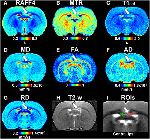
Figure 2. Quantitative MRI maps in the demyelination phase, on day 3: RAFF4 (A), magnetization transfer ratio, MTR (B), T1sat (C), mean diffusivity, MD (D), fractional anisotropy, FA (E), axial diffusivity, AD (F), radial diffusivity, RD (G), T2w image with lesion (H) and representative example of ROIs for analyzing the lesion on a grayscale RAFF4 map (I). White arrow points to the lesion in the corpus callosum.
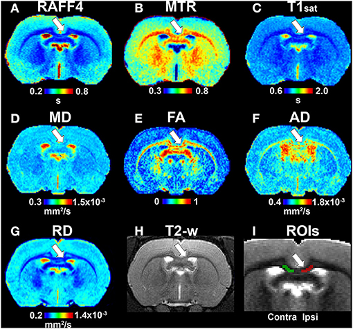
Figure 4. Quantitative MRI maps in the remyelination phase, on day 38. Relaxation time constant map of RAFF4 (A), magnetization transfer ratio, MTR (B), T1sat (C), mean diffusivity, MD (D), fractional anisotropy, FA (E), axial diffusivity, AD (F), radial diffusivity, RD (G), T2w image with the lesion (H) and a representative example of ROIs for analyzing lesion on a grayscale RAFF4 map (I). White arrow points to the lesion in the corpus callosum.
The authors apologize for this error and state that this does not change the scientific conclusions of the article in any way. The original article has been updated.
Keywords: myelin, demyelination, remyelination, MRI, diffusion, rotating frame relaxation
Citation: Holikova K, Laakso H, Salo R, Shatillo A, Nurmi A, Bares M, Vanicek J, Michaeli S, Mangia S, Sierra A and Gröhn O (2021) Corrigendum: RAFF-4, Magnetization Transfer and Diffusion Tensor MRI of Lysophosphatidylcholine Induced Demyelination and Remyelination in Rats. Front. Neurosci. 15:723831. doi: 10.3389/fnins.2021.723831
Received: 11 June 2021; Accepted: 21 June 2021;
Published: 09 July 2021.
Edited and reviewed by: Yu-Chien Wu, Indiana University Bloomington, United States
Copyright © 2021 Holikova, Laakso, Salo, Shatillo, Nurmi, Bares, Vanicek, Michaeli, Mangia, Sierra and Gröhn. This is an open-access article distributed under the terms of the Creative Commons Attribution License (CC BY). The use, distribution or reproduction in other forums is permitted, provided the original author(s) and the copyright owner(s) are credited and that the original publication in this journal is cited, in accordance with accepted academic practice. No use, distribution or reproduction is permitted which does not comply with these terms.
*Correspondence: Olli Gröhn, b2xsaS5ncm9obkB1ZWYuZmk=
Disclaimer: All claims expressed in this article are solely those of the authors and do not necessarily represent those of their affiliated organizations, or those of the publisher, the editors and the reviewers. Any product that may be evaluated in this article or claim that may be made by its manufacturer is not guaranteed or endorsed by the publisher.
Research integrity at Frontiers
Learn more about the work of our research integrity team to safeguard the quality of each article we publish.