- IRCCS SYNLAB SDN S.p.A., Naples, Italy
Neurofeedback (NF) is a biofeedback technique that teaches individuals self-control of brain functions by measuring brain activations and providing an online feedback signal to modify emotional, cognitive, and behavioral functions. NF approaches typically rely on a single modality, such as electroencephalography (EEG-NF) or a brain imaging technique, such as functional magnetic resonance imaging (fMRI-NF). The introduction of simultaneous EEG-fMRI tools has opened up the possibility of combining the high temporal resolution of EEG with the high spatial resolution of fMRI, thereby increasing the accuracy of NF. However, only a few studies have actively combined both techniques. In this study, we conducted a systematic review of EEG-fMRI-NF studies (N = 17) to identify the potential and effectiveness of this non-invasive treatment for neurological conditions. The systematic review revealed a lack of homogeneity among the studies, including sample sizes, acquisition methods in terms of simultaneity of the two procedures (unimodal EEG-NF and fMRI-NF), therapeutic targets field, and the number of sessions. Indeed, because most studies are based on a single session of NF, it is difficult to draw any conclusions regarding the therapeutic efficacy of NF. Therefore, further research is needed to fully understand non-clinical and clinical potential of EEG-fMRI-NF.
1. Introduction
In the late 1970s, Sterman showed evidence of an electroencephalogram (EEG) response to cold pressor stimulation, thus highlighting a feedback function in regulating electrical cerebral reactivity EEG rhythms (Sterman et al., 1966). Such an EEG response derives from the intrinsic electrical activity generated by the excitation and inhibition of pyramidal neurons (Lorente De Nó, 1947). Physiological processes, such as sleep state or progression to sleep, may produce changes in EEG frequency patterns, as well as motor and cognitive behaviors, leading, for example, to what is known as “alpha desynchronization” (Klimesch et al., 1998). Based on Pavlov's experiments, Sterman et al. (1966) used operant conditioning in opposition to classical conditioning (Staddon and Cerutti, 2003) to voluntarily modulate EEG activity in response to a sensory stimulus event or cognitive task. The resulting EEG's change in response to an external event, namely EEG feedback, could be an increase or a decrease in the activity, and it is respectively called “event-related desynchronization” or “event-related synchronization”. The first rhythmic EEG pattern identified was the sensorimotor rhythm (SMR), a particular brain rhythm in the range of 12–15 Hz.
Initially, EEG-biofeedback studies were done on experimental animals, then, this research was translated to humans, aiming at regulating SMR in uncontrolled epilepsy to reduce motor seizure rates (Sterman, 2000). In Attention Deficit Disorder and Hyperactivity (ADHD), EEG-biofeedback may improve the clinical pattern, reducing hyperactivity and enhancing attention (Lubar and Shouse, 1976). In anxiety, protocols involving the upregulation of the theta/alpha ratio were used to enhance the state of relaxation (Budzynski, 1999). Throughout the years, EEG-biofeedback was branded as EEG-neurofeedback (EEG-NF), and different protocols and approaches have been validated, such as SMR and SMR ratio, alpha/theta ratio, frontal alpha training, etc. (Gruzelier, 2014). These pioneering studies highlight the opportunity to volitionally control human electrocortical activity, modifying cognitive and motor functions in health and disease. Groppe et al. described the associations between the typical EEG rhythms and their functions, i.e., alpha rhythm is mainly related to attention and cognitive functions at all ages, theta rhythm to working memory load, and low beta to inhibition of sensorimotor cortex (Groppe et al., 2013).
NF has emerged as an alternative non-pharmacological treatment for several neurological and behavioral disorders. With the EEG digitalization in the 1990s and the advent of brain-computer interface (BCI), easier access to electrophysiological information was enabled by decoding brain activation patterns generated by NF tasks, thus increasing the use of the EEG-NF in neurology and psychiatry. The EEG is non-invasive, inexpensive, portable, and benefits from a high temporal resolution. However, EEG is very sensitive to noises, such as environmental components, or movements. Also, it lacks specificity because of its low spatial resolution (order of the centimeters) (Iannaccone et al., 2015). The introduction of BCI was fostered by its application to functional magnetic resonance imaging (fMRI): the fMRI-NF was introduced for the first time by Weiskopf et al. (2004).
In fMRI-NF, blood oxygenation level-dependent activity (BOLD), which is related to the vascular response to neural activity, is directly fed back to participants in the magnetic scanner. Participants are trained to voluntarily modulate this activity, increasing or decreasing it. Such training targets specific regions of interest (ROI) and modulates motor behavior, attention, working memory, and emotional and cognitive processes. For instance, the upregulation of BOLD activity of the primary motor region and amygdala is associated with improved motor performance and emotional functions (anxiety, stress, depression, etc.), respectively (Zotev et al., 2011).
Albeit fMRI brings significant added value to NF protocols, along with BCIs, many researchers still debate the real usefulness of fMRI-NF in clinical contexts. A concern is the amount of physiological noise and artifacts in the magnetic scanner, whilst a key advantage is that fMRI-NF provides feedback about the whole brain's ongoing neural activation, allowing for a spatial resolution in the range of millimeters. The BOLD correlation between two or more neural networks is called “functional connectivity”, NF can regulate the connectivity, and it may lead to long-term effects. The neural mechanisms engaged by neurofeedback and how they affect learning still need elucidation (Sulzer et al., 2013). These mechanisms involve wide and complex brain networks, including basal ganglia, temporoparietal areas, anterior cingulate cortex (ACC), and the anterior insula. These brain regions appear to be activated during the NF training regardless of the ROI targeted, hence questioning if their activation reflects the actual learning processes (Emmert et al., 2016).
Recently associations between electrophysiological and hemodynamic signatures of fMRI demonstrated their usefulness for diagnostic purposes (Marchitelli et al., 2016; Mele et al., 2019), overcoming the lack of specificity of the EEG, hence opening new insights in the field of neuromodulation. This correlation between the EEG and BOLD activity encouraged simultaneous EEG-fMRI-NF, providing a novel approach in neuroscience. Studying brain plasticity and reorganization through the continuous self-regulation training of specific brain areas allows for restoring lost neurological functions (Dewiputri and Auer, 2013). The self-regulation training process during EEG-fMRI-NF requires participants to modulate two different types of signals, driving back two feedbacks. How the feedback is represented may vary, as a function of stimulation type (e.g., verbal, visual, auditory, olfactory, or a combination of these, for instance, a size/height of a bar or changes in tones). In the literature, the application of either EEG-NF or fMRI-NF as a therapeutic tool in clinical populations suffering from neurological and psychiatric disorders is documented (Lofthouse et al., 2012; Arns et al., 2017).
EEG-fMRI-NF has also been applied to healthy subjects to boost and improve their cognitive and behavioral functioning (Gruzelier, 2014). However, the assessment of such a novel and promising procedure remains quite unexplored in the literature (but see Zotev et al., 2014), and to the best of our knowledge, there is no systematic review assessing the use of simultaneous EEG-fMRI-NF. Therefore, this study aims to summarize scientific evidence about the simultaneous EEG-fMRI-NF, highlighting experimental protocols, fields of application, and clinical effectiveness.
2. Material and methods
2.1. Search strategy and study selection
We searched PubMed, Scopus, and Embase according to the recommendation of the preferred reporting items for systematic reviews and meta-analysis (PRISMA) guidelines. We used the following keywords combined with the “AND” logic operator: EEG, fMRI and neurofeedback. Only English-language articles were included in the search. A total of 98 articles were found until May 2022 (first record: 2014). No time-frame window was set due to the shortfall of published research. When selecting literature, inclusion criteria were the simultaneity of the EEG and fMRI and the presence of NF training. The exclusion criteria were using one single modality with NF training (only EEG-NF or fMRI-NF), duplicate studies, the absence of NF training, and the use of NF without real-time EEG or fMRI, pre-prints, symposium presentations and book issues. The resulting articles were selected or rejected based on the criteria described in Figure 1.
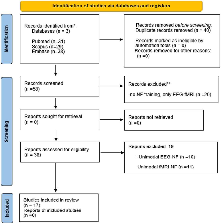
Figure 1. PRISMA study selection diagram. The diagram moves through the four stages of PRISMA study selection: identification screening, screening, eligibility and included. From an original count of 98 studies, 17 were finally selected for this review. NF, neurofeedback; EEG, electroencephalograph; fMRI, functional magnetic resonance imaging.
2.2. Data collection process and extraction
Papers' titles were evaluated by two-independent operators to assess the eligibility of the selected papers; a third evaluator was included in cases of discordant assessment. When the inclusion or exclusion criteria were not met, also the abstract was included in the assessment. Finally, full-text screening was performed on the remaining papers to identify those that were not matched the eligibility requirements. In the tables, the studies are listed in chronological order. Table 1 includes studies that use bimodal EEG-fMRI-NF; Table 2 includes studies with unimodal fMRI or unimodal EEG NF with respectively passive simultaneous EEG and fMRI. The characteristics of the included studies were extracted according to the first author, aim of the study, sample, methods, protocol, results, and conclusion.
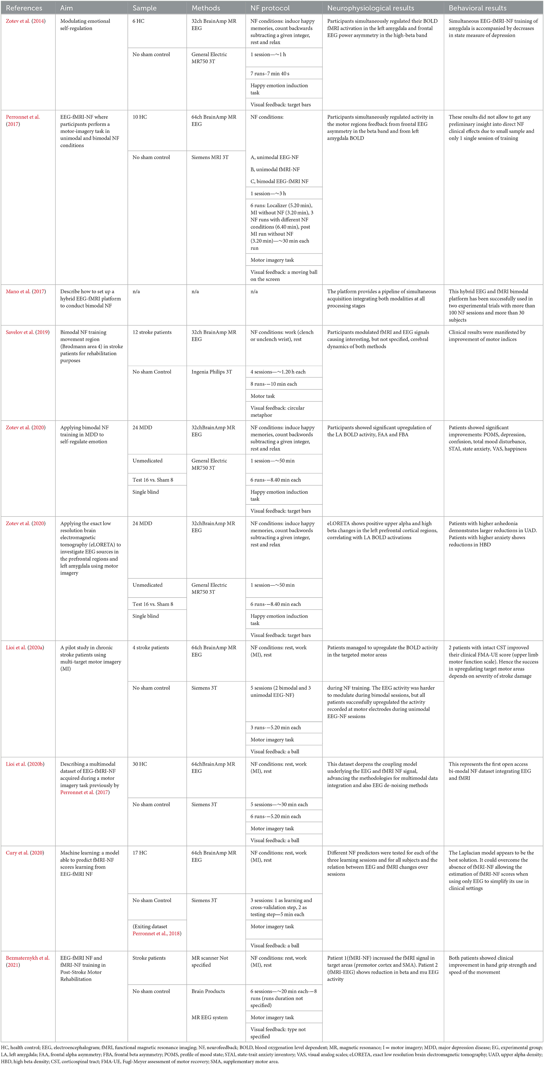
Table 1. Ten EEG-fMRI-NF studies included in the review, organized by: authors, aim of the study, sample, methods, NF protocol, neurophysiological results and behavioral results.
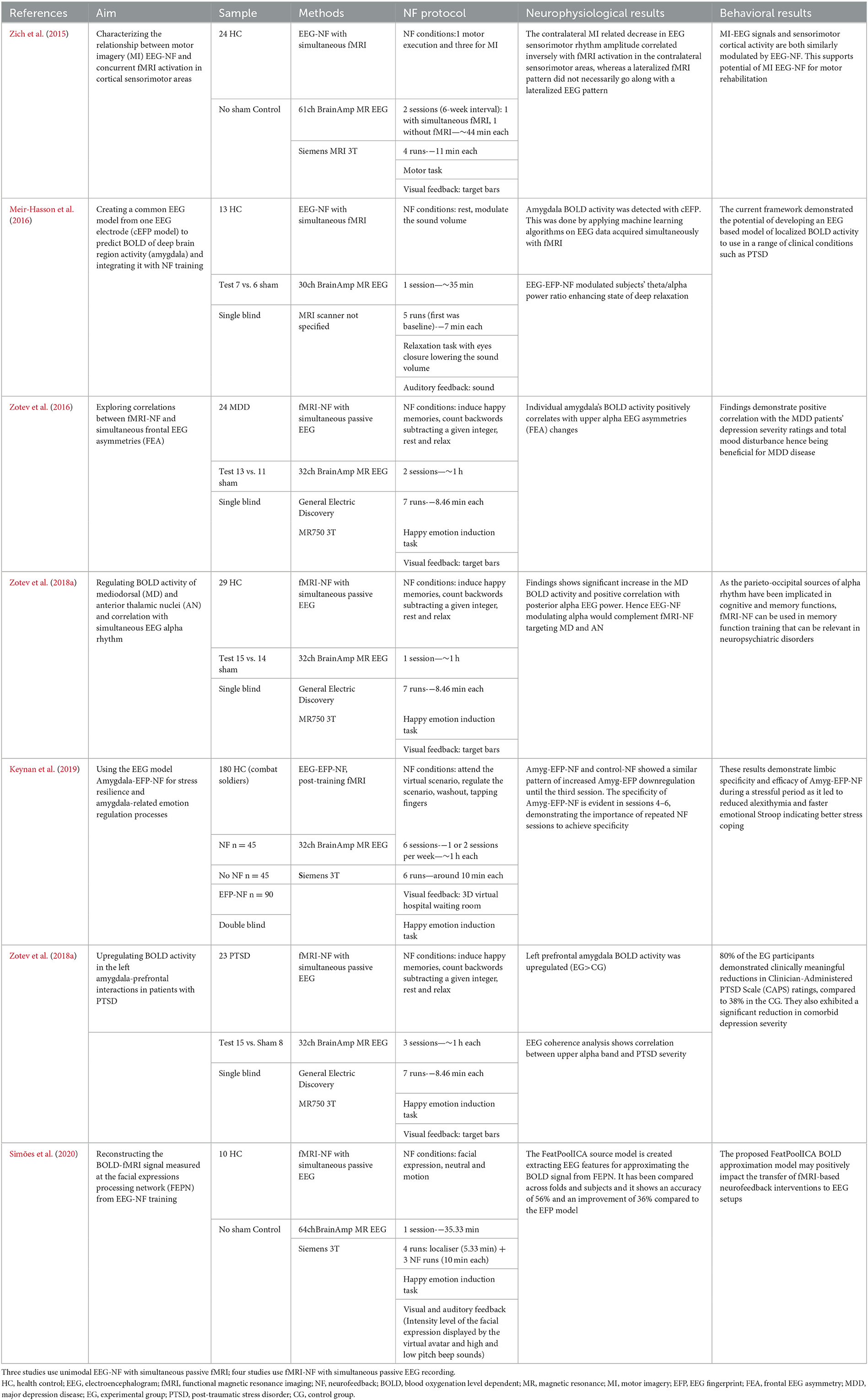
Table 2. Seven unimodal studies (EEG-NF with simultaneous fMRI-NF or vice versa) included in the review organized by: authors, aim of the study, sample, methods, NF protocol, neurophysiological results, and behavioral results.
3. Results
3.1. Study selection
A total of 98 articles were retrieved from the PubMed (N = 31), Scopus (N = 29), and Embase (N = 38) databases. After duplicate removal (N = 40), the remaining 58 papers were screened based on titles and abstracts. Of these 58 articles, 20 records were excluded because they did not involve neurofeedback training but only simultaneous EEG-fMRI technique. The remaining 38 articles were then read in full and assessed for eligibility, and 19 were then excluded: 10 because they focused on unimodal EEG-NF, 11 because they evaluated unimodal fMRI-NF. Ultimately, 18 records were included for qualitative analysis in this review. The PRISMA flow diagram describes the process of choice of the studies (Figure 1), and their characteristics are summarized in Tables 1, 2, for EEG-fMRI-NF studies with bimodal or unimodal feedback, respectively (Figure 2). Ten studies investigated the use of simultaneous acquired EEG and fMRI in a bimodal neurofeedback acquisition (Table 1). Simultaneous bimodal EEG-fMRI studies estimated the real-time NF from both signals. This means that EEG was recorded in the MRI scanner with an MR-compatible EEG cap in one or more sessions during fMRI acquisition. Participants were required to execute a task and modulate BOLD and EEG activities. Following a real-time quality pre-processing of BOLD and EEG, the NF was exported to a custom-made visualization software and shown to subjects using either a video or a display screen. Seven studies used the NF training only through one single modality (or fMRI-NF or EEG-NF), but both modality measures are simultaneously acquired for offline analysis (Table 2). Acquiring EEG synchronously with fMRI-NF allowed the detection of cerebral electrical activity features that are related to activations of BOLD activity. NF training may use EEG-correlates of fMRI responses instead of the expensive and complex MRI techniques, thus enabling the NF application in any clinical setting using the more affordable EEG (Formaggio et al., 2010).
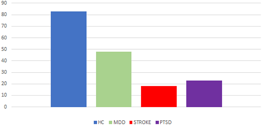
Figure 2. Proportion of EEG-fMRI-NF studies. Number of studies in relation to neuropsychological diseases (MDD, stroke, PTSD) and healthy control subjects.
3.2. Multimodal EEG-fMRI-NF on healthy participants
Four of the bimodal EEG-fMRI-NF studies involved healthy participants without employing a control group (CG) or a control condition (Zotev et al., 2014; Perronnet et al., 2017; Cury et al., 2020; Lioi et al., 2020b) (Figures 3, 4). The first-ever approach of multimodal EEG-fMRI-NF was introduced by Zotev et al. The complex implementation of the bimodal real-time system included a General Electric Discovery MR750 whole-body 3 T MRI scanner and an MR-compatible EEG from Brain Products. Specifically, the system used a real-time fMRI feature reading software (AFNI) and a real-time BrainVision Recview EEG software. Real-time export of both signals was sent through a TCP/IP socket. Control and communication programs were scripted in Phyton and ran on a Linux computer. The fMRI BOLD was updated every TR from a selected ROI, whilst the EEG is updated faster from individual or group of channels (F3 and F4 electrodes in this paper), and real-time FFT spectrum analysis was computed for specific frequency bands. The multimodal NF graphical user interface (mGUI) integrated both EEG and fMRI signal, converting them into a graphical representation shown to the subject, which could be colored bars, text, or dynamic images. The aim of this study was to train the left amygdala [i.e., a sphere of 7 mm radius centered at (−21, −5, −16) in the Talairach space] and frontal EEG power asymmetries in the beta frequency band emotional through emotion self-regulation in healthy controls (HC) (Zotev et al., 2014). The results confirmed the feasibility of this multimodal NF technique which can then be implemented in therapeutic treatments for major depressive disorders. Perronnet et al. (2017) made the first comparison of unimodal fMRI-NF and EEG-NF during a motor imagery task versus bimodal EEG-fMRI-NF. The main aim was to determine the additional value of bimodal NF, hence the simultaneity of this hybrid system. The simultaneous system used MR-compatible EEG BrainVision and 3T MR. Feedback and visual instructions were transmitted using the Nordic Neurolab hardware and presented to the participant via an LCD screen and a rear-facing mirror fixed on the coil. This multimodal system is the same one used by Mano et al. (2017). Eighteen scalp channels on the primary motor area were used for EEG analysis, and the following Brodmann areas were used for fMRI: 44, 40, 47, 2 and 42. Post-hoc fMRI analysis showed that motor activations in the primary motor cortex (M1) were substantially greater during EEG-fMRI-NF than during EEG-NF. These areas are known to be involved in sensorimotor processes and visuospatial reasoning (e.g., Federico et al., 2022; Osiurak et al., 2022). They also found similar results of the EEG and fMRI of the emotional task study by Zotev et al. (2014). Perronnet et al. in this study also proposed a novel feedback metaphor for the bimodal NF that merges both EEG and fMRI signals into one single two-dimensional (2D) feedback. Another study focused on the possibility of computing and detecting fMRI-NF or EEG-fMRI-NF scores in motor imagery tasks, from EEG signals only (Cury et al., 2020). They compared different NF predictors, computed through a mathematical model, aiming to extract NF scores from the EEG-fMRI-NF data recorded from a pre-existing database previously published by Perronnet et al. (2017). The major goal of this study was to develop a system that can use only EEG to predict an NF score comparable to the NF score that might be obtained with a concurrent NF-EEG-fMRI session. The strategy was based on machine learning. To calculate and synchronize NF-EEG and NF-fMRI scores in real-time during a training phase, EEG and fMRI were both gathered at the same time. The resulting scores were then combined. Lioi et al. (2020b) validated a multimodal brain imaging data structure (BIDS) dataset of EEG-fMRI-NF during an imagery motor task to improve the learning of fMRI-informed EEG and the coupling mechanisms underlying both NF modalities. Such results emphasize how brain areas related to motor and visuospatial functions (e.g., Federico et al., 2021, 2022; Osiurak et al., 2022) may benefit significantly from using EEG-fMRI-NF. The proposed structure included in the format info relates to acquisition procedures, analyses, de-noising operators, and NF scores. Moreover, Mano et al. (2017) aimed to set up a hybrid EEG and fMRI platform for bimodal NF experiments, outlining all the steps required to build a multimodal NF system. It guides researchers in choosing software, hardware, experimental protocols, neurofeedback presentation and calculation.
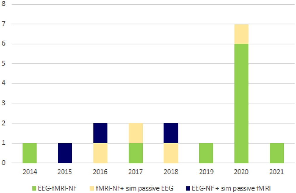
Figure 3. Distribution over the years of EEG-fMRI-NF studies. EEG-fMRI-NF research began surging in 2014; primary research continues to rise. This graph presents the composition of the publications found in our literature search. NF, neurofeedback; EEG, electroencephalograph; fMRI, functional magnetic resonance imaging.
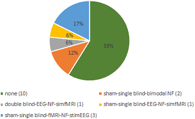
Figure 4. Distribution of controls employed in the review's studies. Sham neurofeedback is used to describe non-contingent feedback method. We consider controls absent if all participants of the studies received genuine feedback. With single blind we intend that participants of control groups were not aware of that. In the double-blind study both participants and experimenters were blind to NF group allocation. NF, neurofeedback; EEG, electroencephalograph; fMRI, functional magnetic resonance imaging.
3.3. Multimodal EEG-fMRI-NF on clinical population
Two bimodal EEG-fMRI-NF studies enrolled unmedicated patients affected by major depressive disorder (MDD) (Zotev et al., 2020; Zotev and Bodurka, 2020), both including a CG (Figures 3, 4). Zotev et al. (2020) evaluated EEG power asymmetries in frontal alpha and beta bands (FAA, FBA) during self-emotion regulation through autobiographical memories. Changes in EEG activities correlated with neural activations, and activations in prefrontal regions were associated with motivation and happiness (Davidson, 1995). Similarly, left amygdala BOLD activations were associated with FAA/FBA (Zotev et al., 2016). Therefore, simultaneous NF modulation of BOLD and FAA/FBA in MDD patients showed significant clinical improvements. The same research group (Zotev and Bodurka, 2020) investigated the effects of bimodal NF with EEG source activations through exact low-resolution brain electromagnetic tomography (eLORETA). The eLORETA values were computed using MNI coordinates of the EEG electrodes arranged according to the international 10–20 system. FFT power spectrum and consequent eLORETA were calculated for each given condition (happy memories, count and rest) in individual alpha frequency bands and for each run. Similar associations were found in EEG source density over prefrontal regions. MDD patients showed a large reduction of the upper alpha current source density in the left prefrontal region, associated with approach motivation. Concurrently, a significant reduction of high beta current density was observed in the right prefrontal region, indicating a reduction in avoidance-motivation related to anxiety (Zotev and Bodurka, 2020). Three studies enrolled stroke patients (Savelov et al., 2019; Lioi et al., 2020a; Bezmaternykh et al., 2021). Bezmaternykh et al. used a hybrid EEG-fMRI system and the OpenNFT suite for real-time estimation of the feedback. The ROIs chosen for fMRI were in the premotor and supplementary motor regions; EEG target was alpha and beta band in C3 and C4 electrodes (Bezmaternykh et al., 2021). Lioi et al. used the integrated EEG-fMRI-NF platform introduced by Mano et al. (2017), computing NF signal with Matlab. EEG 8–30 Hz frequency range was considered, and primary and supplementary motor areas were the fMRI ROIs (Lioi et al., 2020a). Savelov et al. referred to the hybrid platform (Mano et al., 2017), targeting BA 4 and certain EEG frequency bands which are not specified. In all cases, the clinical benefit derived from EEG-fMRI-NF training has been demonstrated, particularly regarding symptom improvement and gain of function. Simultaneous EEG-fMRI-NF training during motor imagery tasks of the affected limbs demonstrated clinical efficacy for motor improvement. In detail, NF treatment elicited activation in the ipsilesional supplementary motor area (SMA) and primary motor cortex (M1), according to the severity of the brain injury.
3.4. Unimodal fMRI-NF with passive simultaneous EEG
Four studies employed the fMRI-NF with simultaneous passive EEG recording, aiming to evaluate the electrophysiological activity deriving from the EEG signal that correlates with the hemodynamic active changes during a NF task. In two of these four studies, participants were HC (Zotev et al., 2018a; Simões et al., 2020), and the other two studies included groups of patients with MDD and post-traumatic stress disorder (PTSD) (Zotev et al., 2016, 2018b). Only three studies employed a control group (Zotev et al., 2016, 2018a,b), and apart from the PTSD paper, the other two studies show a statistical significance between EG and CG. Simões et al. (2020) investigated the possibility of reconstructing fMRI BOLD activity in the facial expressions processing network (FEPN) from simultaneous EEG recording in HC. The authors used the MR-compatible EEG NeuroScan SynAmps2 system, and the MaglinkTM software, and seven EEG source features were extracted on the electrodes in proximity to the FEPN in theta, low beta, high beta, gamma, and broadband). The extraction process of the EEG features was done through the independent component analysis, thus creating the FeatPoolICA-source model to predict the BOLD fMRI signal in FEPN. Studying the correlations between fMRI-NF BOLD activations and passive simultaneous EEG recording gave the possibility of shedding light on the neural networks underpinning cortical activations. Positive posterior alpha EEG power correlations with BOLD fluctuations was found in the mediodorsal (MD) and anterior (AN) thalamic nuclei, during a happy emotion induction task. These nuclei are involved in many functions, such as memory, emotion, motivation, learning, and decision-making (Zotev et al., 2018a). Zotev et al. (2016, 2018b) investigated correlations between FAA changes and the amygdala BOLD laterality in MDD patients upon emotion training (Zotev et al., 2016). FAA and alpha EEG coherence had a positive correlation with the left amygdala in PTSD patients (Zotev et al., 2018b), therefore showing significant reductions in the total mood disturbance. In all these three studies Zotev et al. used an EEG-fMRI system configuration where EEG was simultaneous but passive, i.e., no EEG information was used for real-time experiments. The target fMRI ROIs were defined as 14-mm diameter spheres in the Talairach space, and EEG electrodes used were F3, F4, F7, and F8. fMRI-NF was implemented using the custom real-time fMRI system utilizing the real-time functionality of AFNI as described previously (Zotev et al., 2011).
3.5. Unimodal EEG-NF with simultaneous passive fMRI
In three papers, fMRI was used as a passive simultaneous recording during EEG-NF training in HC. Two of them (Meir-Hasson et al., 2016; Keynan et al., 2019) applied algorithms to simultaneous EEG-fMRI-NF data to create an EEG prediction model called the electrical fingerprint (EFP), useful to probe the BOLD in the amygdala, thus relieving the need of a prior fMRI. They both enrolled HC with a CG, although Meir-Hasson et al. (2016) employed a relaxation task in HC, while Keynan et al. (2019) used an emotion-inducing (happiness) task in combat soldiers, which led to better stress coping. This framework showed that by using a single electrode in a single area, it was possible to create an EEG-EFP-NF model to predict BOLD activity. The effects of the feedback generated by this model were evaluated, and participants were able to modulate the activities resulting in a reduction of anxiety. The approach suggested in the paper by Meir-Hasson et al. used a regression model based on a representation of the EEG signal from a single channel in terms of time, frequency, and delay. The BOLD signal in the regions of interest was well estimated by the results, although the use of fMRI neurofeedback in this study simply supported the paradigm. The technique was intended to target the amygdala during NF-EEG sessions. Zich et al. (2015) instead applied EEG-NF with simultaneous passive fMRI during motor imagery tasks. The aim of the study was to investigate the concurrent BOLD correlating changes in the sensory-motor cortices upon EEG-NF training during a motor imagery task. Movement imagination results are like movement execution ones, and it showed that a complex relationship exists between sensorimotor cortex BOLD activations and scalp EEG signal, namely the SMR. These motor imagery-related findings support the usefulness of EEG-NF for motor recovery.
4. Discussion
The combination of new neuroimaging and electrophysiological techniques has encouraged an emerging field of research, namely the simultaneous application of EEG and fMRI in neurofeedback (EEG-fMRI-NF). This novel hybrid NF system takes advantage of the high spatial resolution of fMRI and the high temporal resolution of the EEG. Whilst unimodal NF is known to be a promising non-invasive therapeutic tool in neurological/psychiatric disorders, the multimodal EEG-fMRI-NF approach has been poorly explored so far. Therefore, there are still doubts about the clinical significance of these complementary measures on cognitive and behavioral performances. The present systematic review evaluated 17 EEG-fMRI NF studies in the literature according to their methodological characteristics as well as their clinical and neurophysiological outcomes. Such studies were heterogeneous in their design, methodology and application. Nine of these studies involved healthy participants (HC; Zotev et al., 2014, 2018a; Zich et al., 2015; Meir-Hasson et al., 2016; Perronnet et al., 2017; Keynan et al., 2019; Cury et al., 2020; Lioi et al., 2020b; Simões et al., 2020), while seven studies enrolled patients (Zotev et al., 2016, 2018b, 2020; Savelov et al., 2019; Lioi et al., 2020a; Zotev and Bodurka, 2020; Bezmaternykh et al., 2021).
Studies involving patients included small sample sizes and quite a compact clinical field of application, principally psychiatric. Therefore, such studies may appear not very well suited for detecting mild or moderate effects. Hence, future research should require larger samples to study NF effects in clinical populations. Eight papers explored the amygdala through emotional imagery (usually retrieving happy autobiographical memories), thus proving the potential use of EEG-fMRI-NF in MDD and PTSD (Zotev et al., 2014, 2016, 2018a,b, 2020; Keynan et al., 2019; Simões et al., 2020; Zotev and Bodurka, 2020). Interestingly, some studies in the past two decades used the SMR-NF protocol to control and reduce seizure threshold in epileptic patients, hence reducing sensorimotor system excitability (Bernstein et al., 2021; Loriette et al., 2021). Critically, SMR-NF protocol has poorly been used with the advanced procedure as simultaneous EEG-fMRI-NF, resulting in little research in areas such as epilepsy, thus suggesting a need for further studies of EEG-fMRI-NF in a wider clinical population type.
Six studies employed a motor imagery-motor task to explore the motor function, which has been linked to the activation of the primary motor cortex and supplementary motor area (Zich et al., 2015; Perronnet et al., 2017; Cury et al., 2020; Lioi et al., 2020a,b; Bezmaternykh et al., 2021; Federico et al., 2021, 2022; Osiurak et al., 2022). It should be noted that motor function was investigated in stroke patients only in three studies, with small sample sizes (Savelov et al., 2019; Lioi et al., 2020a; Bezmaternykh et al., 2021). Also, the clinical criterions used for these patients were unclear (e.g., Lioi et al., 2020a) as patients were presented with different clinical features, such as the severity of the stroke and its symptoms, and the level of damage to the cortico-spinal tract. In addition, it was not specified how long after the stroke onset the NF training was performed, and, finally, it has not been proven whether the benefits of NF training were long-lasting. On these bases, further studies should consider longer follow-up periods and document NF clinical effects.
According to our data, for the majority of studies, a key methodological concern is the number of NF sessions. It is common to implement a fixed number of sessions based on the effect sizes of similar protocols or studies. However, a training goal could also be established regarding a particular performance pattern, such as the reduction of certain symptoms as measured by the results of clinical questionnaires. In such cases, the number of sessions cannot be determined before the intervention, as it might be required to monitor subjects' learning curves to individually adapt the number of training sessions. Hence, practical factors and learner-specific traits should be considered while choosing the NF protocol. However, neuroplasticity may contribute to NF training outcomes. Congruently, there is a wide intra- and inter-variability among subjects depending on the type of disease and subjects' premorbid characteristics, such as cognitive reserve. With the type of disease, it might be important to consider whether there is structural and organic damage causing the impairment, such as strokes studies (Savelov et al., 2019; Lioi et al., 2020a), or if there is only a functional impairment with no structural changes, such as MDD studies (Zotev et al., 2016, 2020; Zotev and Bodurka, 2020). This strictly influences the target areas to choose and hence which neural connections to train during NF sessions. Hence, it becomes difficult to choose a fixed number of sessions. Up to now, little is known about neurofeedback sessions within a certain time interval being more helpful for learning to self-regulate brain activity, and less is known, even regarding the length of an effective gap between training sessions. Further studies should evaluate structural and functional changes after NF training, aiming to demonstrate the clinical efficacy and effects based on the number of sessions. Such future studies might repeat fMRI, and EEG following the training, and introduce a questionnaire to assess premorbid subjects' characteristics (e.g., cognitive reserve, which may moderate the relationship between brain pathology and clinical outcomes; Rosenich et al., 2020).
In our data, CG and/or a sham condition are only rarely represented (i.e., 41% of the studies do not employ a CG) (Zotev et al., 2014; Zich et al., 2015; Perronnet et al., 2017; Savelov et al., 2019; Lioi et al., 2020a,b; Bezmaternykh et al., 2021), without following consensus on the reporting and experimental design of clinical and cognitive-behavioral neurofeedback studies (CRED-NF checklist; Ros et al., 2020). In a few studies, single-blinded CG showed clinical and NF change improvements that were not statistically different from EG (Zotev et al., 2016, 2020). The reason why this happened could be associated with a placebo effect deriving from a beneficial psychological impact of the regular interaction with the clinical team during the study. However, it is to be noted that the CRED-NF checklist was just recently issued (2020). Hence, some authors were unable to use them as a reference for their investigations.
Recent unimodal fMRI-NF studies, which used the high spatial resolution of fMRI, found that learned control of localized BOLD activity in the amygdala corresponds with better emotion regulation in healthy individuals and may reduce the symptoms of stress and mood disorders (Keynan et al., 2019). However, the MRI approach's scalability (accessibility and cost-effectiveness), particularly in clinical applications, is significantly hampered by cost. A few intrinsic limitations make the MRI not always feasible, such as its costs, noises, contraindications, and patient compliance (i.e., claustrophobia, metal implant, etc.). The combination of EEG and fMRI enables regulating of discrete and defined brain areas, including the amygdala or thalamic nuclei. It also enables the analysis of associated EEG activity. The use of simultaneous EEG-fMRI recording in the context of real-time clinical neurofeedback is a recent application that was first introduced, and its feasibility was demonstrated by Zotev et al. (2014), Mano et al. (2017), and Perronnet et al. (2017). The creation of a new class of data known as EEG-fMRI-NF data, like the dataset provided by Perronnet et al. (2017), is made possible by recent technologies (Mano et al., 2017) that synchronize both EEG and fMRI signals for real-time neurofeedback. Perronnet's research also demonstrated that employing both neurofeedback modalities simultaneously in EEG-fMRI-NF sessions enhances the quality of the sessions. Hence, the opportunity to recreate an EEG-fMRI-NF session in real-time when using EEG only would reduce the use of fMRI in neurofeedback.
To get beyond MRI limitations, such as its costs and often reduced feasibility, mainly in clinical settings, some studies focused on the possibility of predicting and detecting fMRI-NF values through complex machine learning processes. Indeed, to export fMRI BOLD information, different methods have been exploited, which simulate and predict BOLD in a specific region of interest by learning from an EEG signal recorded simultaneously inside the fMRI scanner. Hence, specific EEG prediction models have been proposed in some studies, considering EEG recording linked to simultaneous fMRI changes (Meir-Hasson et al., 2016; Keynan et al., 2019; Simões et al., 2020). In addition, NF sessions might be used as learning sessions to improve the prediction of NF-fMRI scores. To tailor the model more effectively to the subject or patient, each new bi-modal neurofeedback session might be added to the subject-specific model. Future studies should investigate such an aspect. Another promising approach is called EEG finger-print (EFP), a time-frequency representation of the EEG that corresponds to the BOLD activity in a specific location. Additionally, finding correlations between frontal EEG frequencies asymmetries and amygdala BOLD activity, implies that EEG-NF designed to boost FAA might be complementary with fMRI-NF that is amygdala-based (Meir-Hasson et al., 2016; Keynan et al., 2019; Simões et al., 2020). The amygdala's role in associative learning, decision-making, memory, and attention is widely known. Thus, more brain disorders such as schizophrenia, frontotemporal dementia, and attention deficit hyperactive disorder might be further investigated with the multimodal approach EEG-fMRI-NF, as done with unimodal one. Similar functions are solved also by other subcortical brain structures, such as the thalamus nuclei, which are involved in memory and cognitive roles. For instance, Bernstein et al. (2021) demonstrated the impairment of thalamus nuclei in the early stages of MCI and Alzheimer's disease, as recently shown in the clinical neurological literature (Ilardi et al., 2022). Zotev et al. (2018a) employed fMRI-NF with simultaneous passive EEG to modulate EEG and BOLD activity in these brain areas, resulting in a promising use of NF training in prodromal stages of neurodegenerative disease aiming to improve neuroplasticity.
Multimodal EEG-fMRI-NF has limitations in setting, as providing feedback with both modalities requires complex hardware and software installation and longer time preparation for the patient. Both raw data from EEG and fMRI need to go through some pre-processing steps before being shown to participants, to reduce the signal-to-noise ratio and guarantee accurate and precise feedback (e.g., remotion of gradient artifact, ballistocardiogram artifact, motion correction, features extraction) (Loriette et al., 2021). The simultaneity of the two modalities increases the cognitive load of the subjects when aiming to modulate both measures, also according to the feedback metaphor's choice (1D or 2D). Only Perronnet et al. (2017) evaluated different feedback metaphor interfaces. 1D merges EEG and fMRI information in one gauge as a single regulation task, like the thermometer, whilst the 2D feedback is a plot that sees the two modalities in two different dimensions. This approach shows a successful regulation of EEG and fMRI simultaneously with both strategies. The difference stands in the complexity of the neural networks recruited with the 1D or the 2D feedback. 1D results are easier to control hence it relieves the cognitive load of the subject, 2D instead has a complex representation, and it invites participants to seek out and investigate more specialized brain patterns to regulate both measurements increasing the cognitive load (Gaume et al., 2016). Certain studies revealed that NF efficacy on the learning curve depends more on EEG than fMRI modulation. This may be because EEG measure is more challenging to control and modulate, necessitating greater effort at fMRI expenses. Given the difference in modulating the two measures, the choice of the NF metaphor's interface depends on the clinical population included in the studies. However, it is only partially understood which substantial theoretical aspects may underpin the EEG-fMRI-NF learning processes. Therefore, a larger investigation and use of these technologies is required to improve the clinical use of such novel hybrid systems.
5. Conclusion
This review presents a compelling argument for using simultaneous EEG-fMRI-NF protocols. By combining EEG and fMRI, EEG-fMRI-NF may enhance brain rehabilitation techniques, enabling the volitional regulation of two complementary bio-signals, namely, the electric brain activity (EEG) and BOLD (fMRI). This systematic review shows potential therapeutic effects of EEG-fMRI coupling in NF training on certain brain regions, despite methodological issues, small sample sizes, and intrinsic heterogeneity limiting the generalizability of findings. However, given most studies use a single session of NF with either EEG, fMRI, or their combination, it is difficult to draw conclusions about the clinical efficacy of NF. Therefore, future studies are needed to fully understand both clinical and non-clinical potentialities of EEG-fMRI-NF.
Data availability statement
The original contributions presented in the study are included in the article/supplementary material, further inquiries can be directed to the corresponding author.
Author contributions
GF, CC, and GC contributed to conception and design of the work. GC performed the literature research and draft the manuscript. GF and GM reviewed and selected the articles to include in the systematic review. CRI, MM, ADC, and VA revised the drafts. MS performed manuscript supervision and project administration. All authors have read and agreed to the published version of the manuscript.
Funding
This work was supported by a Ricerca Corrente Grant from the Italian Ministry of Health (IRCCS SYNLAB SDN).
Conflict of interest
GC, GF, GM, ADC, MM, CRI, VA, MS, and CC were employed by IRCCS SYNLAB SDN S.p.A.
Publisher's note
All claims expressed in this article are solely those of the authors and do not necessarily represent those of their affiliated organizations, or those of the publisher, the editors and the reviewers. Any product that may be evaluated in this article, or claim that may be made by its manufacturer, is not guaranteed or endorsed by the publisher.
References
Arns, M., Batail, J. M., Bioulac, S., Congedo, M., Daudet, C., Drapier, D., et al. (2017). Neurofeedback: One of today's techniques in psychiatry? Encephale 43, 135–145. doi: 10.1016/j.encep.2016.11.003
Bernstein, A. S., Rapcsak, S. Z., Hornberger, M., and Saranathan, M. (2021). Structural changes in thalamic nuclei across prodromal and clinical Alzheimer's disease. J. Alzheimer's Disease 82, 361–371. doi: 10.3233/JAD-201583
Bezmaternykh, D. D., Kalgin, K.V, Maximova, P. E., Mel'nikov, M. Y., Petrovskii, E. D., et al. (2021). Application of fMRI and simultaneous fMRI-EEG neurofeedback in post-stroke motor rehabilitation. Bull. Exp. Biol. Med. 171, 379–383. doi: 10.1007/s10517-021-05232-1
Budzynski, T. H. (1999). “From EEG to neurofeedback,” in Introduction to Quantitative EEG Neurofeedback, 1st Edn, eds J. R. Invans and A. Arbarbanel (San Diego, CA: Academic Press Inc), 65–79. doi: 10.1016/B978-012243790-8/50004-3
Cury, C., Maurel, P., Gribonval, R., and Barillot, C. (2020). A sparse EEG-informed fMRI model for hybrid EEG-fMRI neurofeedback prediction. Front. Neurosci. 13, 1451. doi: 10.3389/fnins.2019.01451
Davidson, R. (1995). “Cerebral asymmetry, emotion, and affective style,” in Brain Asymmetry, eds R. Davidson and K. Hugdahl (Cambridge, MA: MIT Press), 362–387.
Dewiputri, W. I., and Auer, T. (2013). Functional magnetic resonance imaging (FMRI) neurofeedback: implementations and applications. Malays. J. Med. Sci. 20, 5–15.
Emmert, K., Kopel, R., Sulzer, J., Brühl, A. B., Berman, B. D., Linden, D. E. J., et al. (2016). Meta-analysis of real-time fMRI neurofeedback studies using individual participant data: How is brain regulation mediated? Neuroimage 124, 806–812. doi: 10.1016/j.neuroimage.2015.09.042
Federico, G., Osiurak, F., Reynaud, E., and Brandimonte, M. A. (2021). Semantic congruency effects of prime words on tool visual exploration. Brain Cogn. 152, 105758. doi: 10.1016/j.bandc.2021.105758
Federico, G., Reynaud, E., Navarro, J., Lesourd, M., Gaujoux, V., Lamberton, F., et al. (2022). The cortical thickness of the area PF of the left inferior parietal cortex mediates technical-reasoning skills. Sci. Rep. 12, 11840. doi: 10.1038/s41598-022-15587-8
Formaggio, E., Storti, S. F., Cerini, R., Fiaschi, A., and Manganotti, P. (2010). Brain oscillatory activity during motor imagery in EEG-fMRI coregistration. Magn. Reson. Imaging 28, 1403–1412. doi: 10.1016/j.mri.2010.06.030
Gaume, A., Vialatte, A., Mora-Sánchez, A., Ramdani, C., and Vialatte, F. B. (2016). A psychoengineering paradigm for the neurocognitive mechanisms of biofeedback and neurofeedback. Neurosci. Biobehav. Rev. 68, 891–910. doi: 10.1016/j.neubiorev.2016.06.012
Groppe, D. M., Bickel, S., Keller, C. J., Jain, S. K., Hwang, S. T., Harden, C., et al. (2013). Dominant frequencies of resting human brain activity as measured by the electrocorticogram. Neuroimage 79, 223–233. doi: 10.1016/j.neuroimage.2013.04.044
Gruzelier, J. H. (2014). EEG-neurofeedback for optimising performance. I: a review of cognitive and affective outcome in healthy participants. Neurosci. Biobehav. Rev. 44, 124–141. doi: 10.1016/j.neubiorev.2013.09.015
Iannaccone, R., Hauser, T. U., Staempfli, P., Walitza, S., Brandeis, D., and Brem, S. (2015). Conflict monitoring and error processing: new insights from simultaneous EEG-fMRI. Neuroimage 105, 395–407. doi: 10.1016/j.neuroimage.2014.10.028
Ilardi, C. R., Iavarone, A., La Marra, M., Iachini, T., and Chieffi, S. (2022). Hand movements in mild cognitive impairment: clinical implications and insights for future research. J. Integr. Neurosci. 21, 67. doi: 10.31083/j.jin2102067
Keynan, J. N., Cohen, A., Jackont, G., Green, N., Goldway, N., Davidov, A., et al. (2019). Electrical fingerprint of the amygdala guides neurofeedback training for stress resilience. Nat. Hum. Behav. 3, 63–73. doi: 10.1038/s41562-018-0484-3
Klimesch, W., Doppelmayr, M., Russegger, H., Pachinger, T., and Schwaiger, J. (1998). Induced alpha band power changes in the human EEG and attention. Neurosci. Lett. 244, 73–76. doi: 10.1016/S0304-3940(98)00122-0
Lioi, G., Butet, S., Fleury, M., Bannier, E., Lécuyer, A., Bonan, I., et al. (2020a). A multi-target motor imagery training using bimodal EEG-fMRI neurofeedback: a pilot study in chronic stroke patients. Front. Hum. Neurosci. 14, 37. doi: 10.3389/fnhum.2020.00037
Lioi, G., Cury, C., Perronnet, L., Mano, M., Bannier, E., Lécuyer, A., et al. (2020b). Simultaneous EEG-fMRI during a neurofeedback task, a brain imaging dataset for multimodal data integration. Sci. Data. 7, 173. doi: 10.1038/s41597-020-0498-3
Lofthouse, N., Arnold, L. E., Hersch, S., Hurt, E., and DeBeus, R. (2012). A review of neurofeedback treatment for pediatric ADHD. J. Atten. Disord. 16, 351–372. doi: 10.1177/1087054711427530
Lorente De Nó, R. (1947). Action potential of the motoneurons of the hypoglossus nucleus. J. Cell. Comp. Physiol. 29, 207–287. doi: 10.1002/jcp.1030290303
Loriette, C., Ziane, C., and ben Hamed, S. (2021). Neurofeedback for cognitive enhancement and intervention and brain plasticity. Rev. Neurol. 177, 1133–1144. doi: 10.1016/j.neurol.2021.08.004
Lubar, J. F., and Shouse, M. N. (1976). EEG and behavioral changes in a hyperkinetic child concurrent with training of the sensorimotor rhythm (SMR). Biofeedback Self Regul. 1, 293–306. doi: 10.1007/BF01001170
Mano, M., Lécuyer, A., Bannier, E., Perronnet, L., Noorzadeh, S., and Barillot, C. (2017). How to build a hybrid neurofeedback platform combining EEG and fMRI. Front. Neurosci. 11, 140. doi: 10.3389/fnins.2017.00140
Marchitelli, R., Minati, L., Marizzoni, M., Bosch, B., Bartrés-Faz, D., Müller, B. W., et al. (2016). Test-retest reliability of the default mode network in a multi-centric fMRI study of healthy elderly: effects of data-driven physiological noise correction techniques. Hum. Brain Mapp. 37, 2114–2132. doi: 10.1002/hbm.23157
Meir-Hasson, Y., Keynan, J. N., Kinreich, S., Jackont, G., Cohen, A., Podlipsky-Klovatch, I., et al. (2016). One-class FMRI-inspired EEG model for self-regulation training. PLoS ONE 11, e0154968. doi: 10.1371/journal.pone.0154968
Mele, G., Cavaliere, C., Alfano, V., Orsini, M., Salvatore, M., and Aiello, M. (2019). Simultaneous EEG-fMRI for functional neurological assessment. Front. Neurol. 10, 848. doi: 10.3389/fneur.2019.00848
Osiurak, F., Claidière, N., Bluet, A., Brogniart, J., Lasserre, S., Bonhoure, T., et al. (2022). Technical reasoning bolsters cumulative technological culture through convergent transformations. Sci. Adv. 8, eabl7446. doi: 10.1126/sciadv.abl7446
Perronnet, L., Lécuyer, A., Mano, M., Bannier, E., Lotte, F., Clerc, M., et al. (2017). Unimodal versus bimodal EEG-fMRI neurofeedback of a motor imagery task. Front. Hum. Neurosci. 11, 193. doi: 10.3389/fnhum.2017.00193
Perronnet, L., Lécuyer, A., Mano, M., Fleury, M., Lioi, G., Cury, C., et al. (2018). Learning 2-in-1: towards integrated EEG-fMRI-neurofeedback. BioRxiv [Preprint].
Ros, T., Enriquez-Geppert, S., Zotev, V., Young, K. D., Wood, G., Whitfield-Gabrieli, S., et al. (2020). Consensus on the reporting and experimental design of clinical and cognitive-behavioural neurofeedback studies (CRED-nf checklist). Brain 143, 1674–1685. doi: 10.1093/brain/awaa009
Rosenich, E., Hordacre, B., Paquet, C., Koblar, S. A., and Hillier, S. L. (2020). Cognitive reserve as an emerging concept in stroke recovery. Neurorehabil. Neural Repair 34, 187–199. doi: 10.1177/1545968320907071
Savelov, A. A., Shtark, M. B., Mel'nikov, M. E., Kozlova, L. I., Bezmaternykh, D. D., Verevkin, E. G., et al. (2019). Prospects of synchronous fMRI-EEG recording as the basis for neurofeedback (exemplified on patient with stroke sequelae). Bull. Exp. Biol. Med. 166, 390–393. doi: 10.1007/s10517-019-04357-8
Simões, M., Abreu, R., Direito, B., Sayal, A., Castelhano, J., Carvalho, P., et al. (2020). How much of the BOLD-fMRI signal can be approximated from simultaneous EEG data: relevance for the transfer and dissemination of neurofeedback interventions. J. Neural Eng. 17, 046007. doi: 10.1088/1741-2552/ab9a98
Staddon, J. E. R., and Cerutti, D. T. (2003). Operant conditioning. Annu. Rev. Psychol. 54, 115–144. doi: 10.1146/annurev.psych.54.101601.145124
Sterman, M. B. (2000). Basic concepts and clinical findings in the treatment of seizure disorders with EEG operant conditioning. Clin. Electroencephalogr. 31, 45–55. doi: 10.1177/155005940003100111
Sterman, M. B., Clemens, T. L., and Wenger, M. A. (1966). Autonomic and clinical correlates of differential electroencephalogram response to cold pressor stimulation. Psychophysiology 2, 327–337. doi: 10.1111/j.1469-8986.1966.tb02662.x
Sulzer, J., Haller, S., Scharnowski, F., Weiskopf, N., Birbaumer, N., Blefari, M. L., et al. (2013). Real-time fMRI neurofeedback: progress and challenges. Neuroimage. 76, 386–399. doi: 10.1016/j.neuroimage.2013.03.033
Weiskopf, N., Scharnowski, F., Veit, R., Goebel, R., Birbaumer, N., and Mathiak, K. (2004). Self-regulation of local brain activity using real-time functional magnetic resonance imaging (fMRI). J. Physiol. Paris 98, 357–373. doi: 10.1016/j.jphysparis.2005.09.019
Zich, C., Debener, S., Kranczioch, C., Bleichner, M. G., Gutberlet, I., and de Vos, M. (2015). Real-time EEG feedback during simultaneous EEG-fMRI identifies the cortical signature of motor imagery. Neuroimage 114, 438–447. doi: 10.1016/j.neuroimage.2015.04.020
Zotev, V., and Bodurka, J. (2020). Effects of simultaneous real-time fMRI and EEG neurofeedback in major depressive disorder evaluated with brain electromagnetic tomography. Neuroimage Clin. 28. doi: 10.1016/j.nicl.2020.102459
Zotev, V., Krueger, F., Phillips, R., Alvarez, R. P., Simmons, W. K., Bellgowan, P., et al. (2011). Self-regulation of amygdala activation using real-time fMRI neurofeedback. PLoS ONE 6, e24522. doi: 10.1371/journal.pone.0024522
Zotev, V., Mayeli, A., Misaki, M., and Bodurka, J. (2020). Emotion self-regulation training in major depressive disorder using simultaneous real-time fMRI and EEG neurofeedback. Neuroimage Clin. 27, 102331. doi: 10.1016/j.nicl.2020.102331
Zotev, V., Misaki, M., Phillips, R., Wong, C. K., and Bodurka, J. (2018a). Real-time fMRI neurofeedback of the mediodorsal and anterior thalamus enhances correlation between thalamic BOLD activity and alpha EEG rhythm. Hum. Brain Mapp. 39, 1024–1042. doi: 10.1002/hbm.23902
Zotev, V., Phillips, R., Misaki, M., Wong, C. K., Wurfel, B. E., Krueger, F., et al. (2018b). Real-time fMRI neurofeedback training of the amygdala activity with simultaneous EEG in veterans with combat-related PTSD. Neuroimage Clin. 19, 106–121. doi: 10.1016/j.nicl.2018.04.010
Zotev, V., Phillips, R., Yuan, H., Misaki, M., and Bodurka, J. (2014). Self-regulation of human brain activity using simultaneous real-time fMRI and EEG neurofeedback. Neuroimage. 85, 985–995. doi: 10.1016/j.neuroimage.2013.04.126
Keywords: EEG-fMRI, neurofeedback, simultaneous, biofeedback, clinical neuroscience
Citation: Ciccarelli G, Federico G, Mele G, Di Cecca A, Migliaccio M, Ilardi CR, Alfano V, Salvatore M and Cavaliere C (2023) Simultaneous real-time EEG-fMRI neurofeedback: A systematic review. Front. Hum. Neurosci. 17:1123014. doi: 10.3389/fnhum.2023.1123014
Received: 13 December 2022; Accepted: 15 March 2023;
Published: 31 March 2023.
Edited by:
Hamid Soltanian-Zadeh, University of Tehran, IranReviewed by:
Estate M. Sokhadze, University of South Carolina, United StatesOlga R. Dobrushina, Reichman University, Israel
Copyright © 2023 Ciccarelli, Federico, Mele, Di Cecca, Migliaccio, Ilardi, Alfano, Salvatore and Cavaliere. This is an open-access article distributed under the terms of the Creative Commons Attribution License (CC BY). The use, distribution or reproduction in other forums is permitted, provided the original author(s) and the copyright owner(s) are credited and that the original publication in this journal is cited, in accordance with accepted academic practice. No use, distribution or reproduction is permitted which does not comply with these terms.
*Correspondence: Giovanni Federico, cmVzZWFyY2hAZ2lvdmFubmlmZWRlcmljby5uZXQ=
 Giuseppina Ciccarelli
 Giovanni Federico
 Giulia Mele
 Angelica Di Cecca
 Vincenzo Alfano
 Carlo Cavaliere