94% of researchers rate our articles as excellent or good
Learn more about the work of our research integrity team to safeguard the quality of each article we publish.
Find out more
CORRECTION article
Front. Hum. Neurosci., 26 November 2021
Sec. Brain Imaging and Stimulation
Volume 15 - 2021 | https://doi.org/10.3389/fnhum.2021.780797
This article is part of the Research TopicAdvanced Neuroimaging Methods in Brain DisordersView all 20 articles
This article is a correction to:
Repeatability and Reproducibility of in-vivo Brain Temperature Measurements
 Ayushe A. Sharma1,2,3*
 Rodolphe Nenert3,4
 Christina Mueller1
 Andrew A. Maudsley5
 Jarred W. Younger1
 Jerzy P. Szaflarski2,3,4,6
A Corrigendum on
Repeatability and Reproducibility of in-vivo Brain Temperature Measurements
by Sharma, A. A., Nenert, R., Mueller, C., Maudsley, A. A., Younger, J. W., and Szaflarski, J. P. (2020). Front. Hum. Neurosci. 14:598435. doi: 10.3389/fnhum.2020.598435
In the original article, there was a mistake in Figure 1 as published.
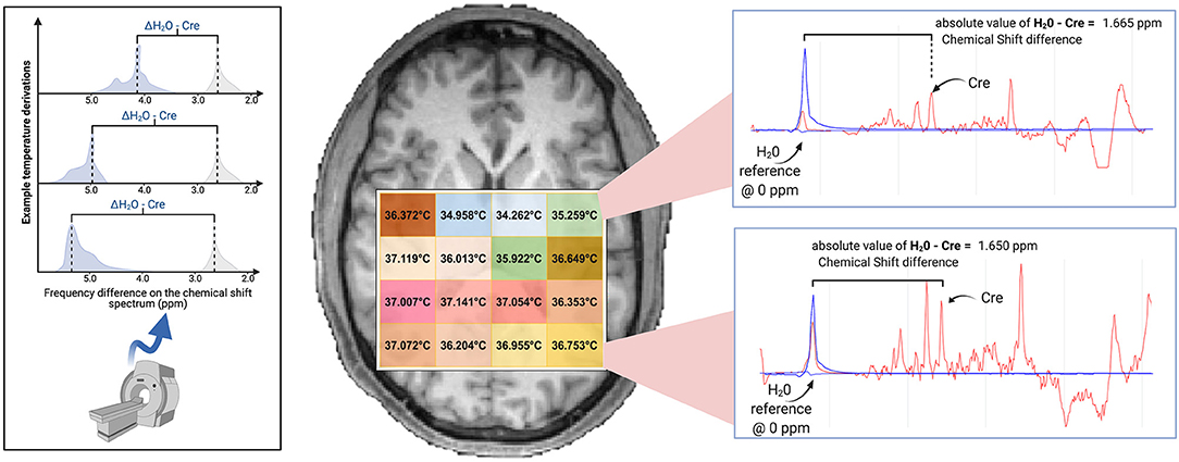
The original figure was adapted from Dehkharghani et al. (2015), but this adaptation was not appropriately described and referenced in the manuscript. We apologize for this oversight. The figure has been revised: the adapted portion has been replaced with a new graphic and the caption now appropriately indicates that a portion was adapted from a previously published article. The corrected Figure 1 appears below.

Figure 1. Brain temperature can be non-invasively derived from volumetric magnetic resonance spectroscopic imaging (MRSI) data by calculating the frequency difference between the temperature-sensitive water peak and one or more metabolite peaks that are temperature-insensitive (left*). When using creatine as the reference, voxel-level brain temperature can be calculated according to the following equation: TCRE = −102.61(ΔH20−CRE) + 206.1°C, ΔH20−CRE = chemical shift difference between the creatine and water resonances. Example TCRE calculations are provided for a participant's single tissue slice (right). Representative spectra illustrate ΔH20−CRE derivations, with plots depicting a water-suppressed metabolite spectrum (red line), with an overlay that indicates the location of the reference water signal (blue line). Spectral plots were created within the Metabolite Imaging and Data Analysis System (MIDAS) software package, and the figure was created using BioRender. *Adapted from Dehkharghani et al. (2015).
The authors apologize for this error and state that this does not change the scientific conclusions of the article in any way. The original article has been updated.
All claims expressed in this article are solely those of the authors and do not necessarily represent those of their affiliated organizations, or those of the publisher, the editors and the reviewers. Any product that may be evaluated in this article, or claim that may be made by its manufacturer, is not guaranteed or endorsed by the publisher.
Dehkharghani, S., Mao, H., Howell, L., Zhang, X., Pate, K. S., Magrath, P. R., et al. (2015). Proton resonance frequency chemical shift thermometry: experimental design and validation toward high-resolution noninvasive temperature monitoring and in vivo experience in a non-human primate model of acute ischemic stroke. Am. J. Neuroradiol. 36, 1128–1135. doi: 10.3174/ajnr.A4241
Keywords: MRS, brain temperature, MR thermometry, neuroinflammation, neuroimaging
Citation: Sharma AA, Nenert R, Mueller C, Maudsley AA, Younger JW and Szaflarski JP (2021) Corrigendum: Repeatability and Reproducibility of in-vivo Brain Temperature Measurements. Front. Hum. Neurosci. 15:780797. doi: 10.3389/fnhum.2021.780797
Received: 21 September 2021; Accepted: 04 November 2021;
Published: 26 November 2021.
Edited and reviewed by: Dajiang Zhu, University of Texas at Arlington, United States
Copyright © 2021 Sharma, Nenert, Mueller, Maudsley, Younger and Szaflarski. This is an open-access article distributed under the terms of the Creative Commons Attribution License (CC BY). The use, distribution or reproduction in other forums is permitted, provided the original author(s) and the copyright owner(s) are credited and that the original publication in this journal is cited, in accordance with accepted academic practice. No use, distribution or reproduction is permitted which does not comply with these terms.
*Correspondence: Ayushe A. Sharma, c2hhcm1hODdAdWFiLmVkdQ==
Disclaimer: All claims expressed in this article are solely those of the authors and do not necessarily represent those of their affiliated organizations, or those of the publisher, the editors and the reviewers. Any product that may be evaluated in this article or claim that may be made by its manufacturer is not guaranteed or endorsed by the publisher.
Research integrity at Frontiers

Learn more about the work of our research integrity team to safeguard the quality of each article we publish.