- 1Department of Neurosurgery, People’s Hospital of Xinjiang Uygur Autonomous Region, Urumchi, China
- 2Xinjiang Second Medical College, Karamay, China
Background: Normal pressure hydrocephalus (NPH) has drawn an increasing amount of attention over the last 20 years. At present, there is a shortage of intuitive analysis on the trends in development, key contributors, and research hotspots topics in the NPH field. This study aims to analyze the evolution of NPH research, evaluate publications both qualitatively and quantitatively, and summarize the current research hotspots.
Methods: A bibliometric analysis was conducted on data retrieved from the Web of Science Core Collection (WoSCC) database between 2003 and 2023. Quantitative assessments were conducted using bibliometric analysis tools such as VOSviewer and CiteSpace software.
Results: A total of 2,248 articles published between 2003 and 2023 were retrieved. During this period, the number of publications steadily increased. The United States was the largest contributor. The University of Gothenburg led among institutions conducting relevant research. Eide P. K. was the most prolific author. The Journal of Neurosurgery is the leading journal on NPH. According to the analysis of the co-occurrence of keywords and co-cited references, the primary research directions identified were pathophysiology, precise diagnosis, and individualized treatment. Recent research hotspots have mainly focused on epidemiology, the glymphatic system, and CSF biomarkers.
Conclusion: The comprehensive bibliometric analysis of NPH offers insights into the main research directions, highlights key countries, contributors, and journals, and identifies significant research hotspots. This information serves as a valuable reference for scholars to further study NPH.
Introduction
Normal pressure hydrocephalus (NPH) is a significant condition that impacts the physical and mental health of the elderly, first reported by Adams et al. (1) in 1965. It is characterized by normal cerebrospinal fluid (CSF) pressure, ventricular enlargement, and a classic triad of symptoms: gait and balance disorders, cognitive impairment, and urinary incontinence (2, 3). NPH, a specific type of communicating hydrocephalus, is classified into primary or idiopathic (iNPH) and secondary (sNPH) (4). Secondary NPH can occur at any age and is typically associated with specific causes such as subarachnoid hemorrhage, cerebral hemorrhage, encephalitis, craniocerebral trauma, etc. It is generally easier to diagnose due to its clear causal relationship, with symptoms manifesting after the primary disease. However, iNPH is more prevalent among the elderly. The nonspecific nature of its triad of symptoms makes differential diagnosis challenging, as these symptoms can overlap with those of other age-related conditions. Additionally, imaging findings of iNPH may be confounded by brain atrophy, leading to potential misdiagnosis as neurodegenerative diseases such as Alzheimer’s disease (AD) (5). Therefore, distinguishing iNPH from other neurological disorders remains a diagnostic challenge (6). In Norway, epidemiological studies on NPH reported an annual incidence of 5.5 per 100,000 population and a prevalence exceeding 21.9 per 100,000 population (7). However, there are reasons to suspect that these rates may be underestimated. First, the primary challenge lies in differentiating NPH from similar diseases. Second, there is evidence indicating a notable age-related increase in NPH incidence (8). Given the global rise in life expectancy, particularly over the past two decades, it is anticipated that the number of elderly patients with NPH will increase (9, 10). In recent years, the emergence of NPH as a significant public health challenge has driven a surge in research interest, reflected by a notable increase in the number of research and review articles on this topic. However, comprehensive reviews of NPH are currently unavailable. There are some reviews articles on NPH in the past, including epidemiological studies of NPH (11, 12), surgical treatment for NPH (13, 14), and pathogenesis and pathophysiology of NPH (15, 16). While these studies offer preliminary insight into the field of NPH, a comprehensive scientometric analysis of NPH is not available in literature.
Bibliometric analysis is an emerging tool that, unlike traditional literature reviews, focuses on quantifying literature, publications, authors, institutions, funding, and keywords (17). It utilizes statistical methods and visualization to explore the structure and trends of a subject or domain, enabling rapid analysis of publication patterns, characteristics, and interrelationships (18). Despite methodological limitations, bibliometrics is a valuable tool for evaluating scientific research within a specific subject or field. Therefore, bibliometric analysis has been extensively utilized in medical fields including neurology (19), neurosurgery (20), and oncology (21).
Therefore, we aimed to propose a scientometric approach for NPH and provide a comprehensive overview of the advancements in NPH research. Our objective was to explore the trending research fields in NPH and identify current hotspots. Additionally, we conducted detailed discussions on significant subtopics identified through bibliometric analysis. This study aims to assist both novice researchers and specialists in understanding the breadth of research topics, identifying new areas of interest, and shaping future research directions in the field of NPH.
Methods
Data source and retrieval strategy
We utilized the Web of Science Core Collection (WoSCC) to extract relevant literature on NPH published from January 2003 to December 2023. The data source edition was limited to the Web of Science Citation Index Expanded (SCI-Expanded) to ensure the high quality and authority of the included studies. All literature searches were performed on a single day (March 21, 2024) to minimize the impact of database updates on the number of retrieved documents. The search strategy was Topic = (“Normal Pressure Hydrocephalus” or “NPH”). We limited the literature types to articles and reviews, and restricted the language to English.
Data extraction and collection
The literature was independently searched and screened by two researchers (TC and XH), and the results of both searches were then compared. Discrepancies in opinion were resolved by discussion with the third researcher (JW) and the optimal outcome chosen.
Based on the search strategies and restrictions outlined above, a total of 2,248 studies were found. The eligible publications were saved as plain text files and exported. Subsequently, the bibliographic records of these articles were imported into statistical software for analysis. Additionally, other information, such as the total number of citations, average citations per item (ACI), H-index, and Journal Impact Factor (IF), was obtained using the “Create Citation Report” function and Journal Citation Reports (JCR). Figure 1 illustrates the steps of the literature search and selection process.
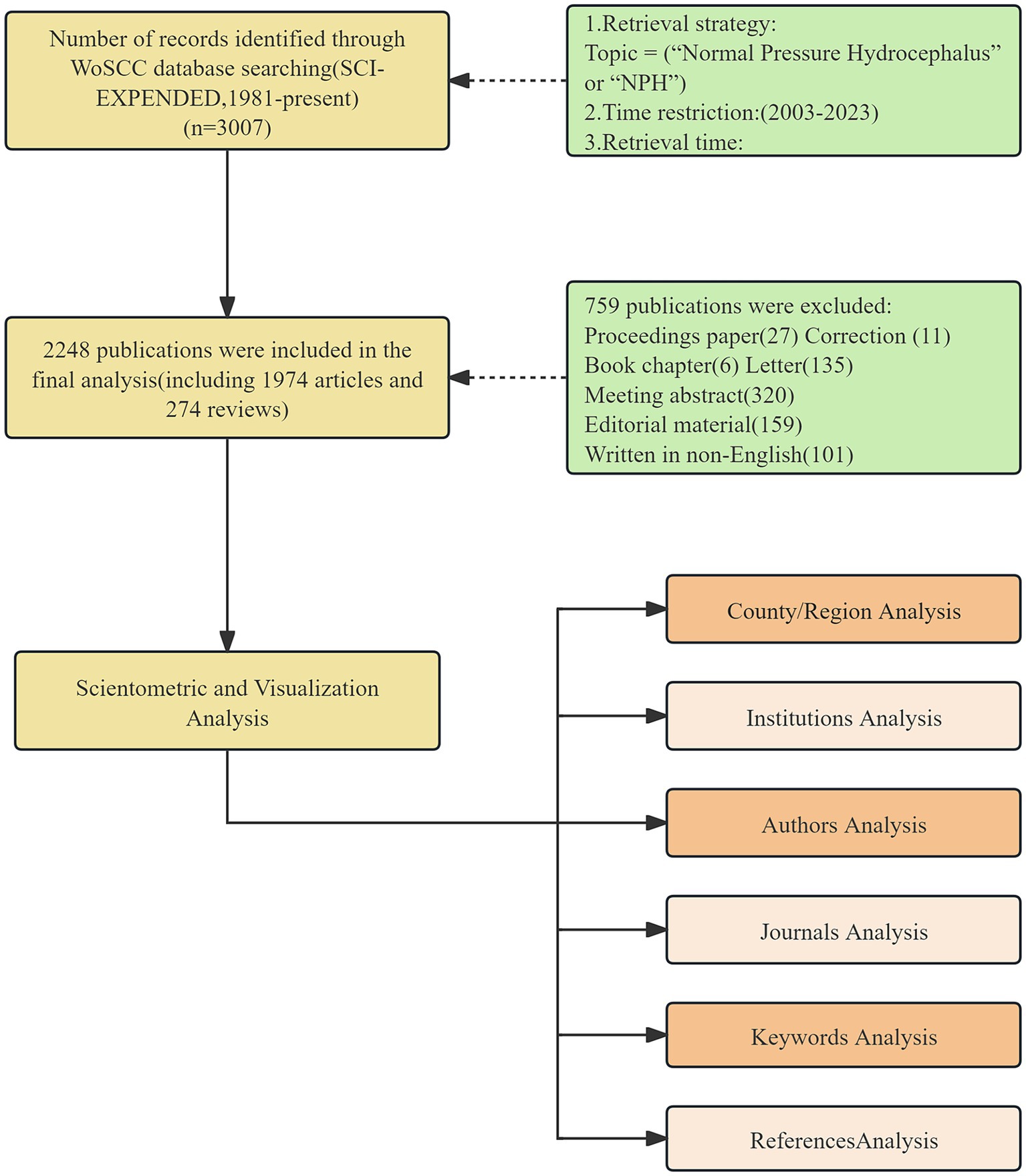
Figure 1. The flow chart of the included publications and methods used in the bibliometric analysis.
Data analysis and network mapping
We employed Microsoft Excel 2019 to analyze the annual number of published documents and citations. We utilized VOSviewer 1.6.18, developed by Leiden University in the Netherlands (22), to analyze the distribution and collaboration of countries or regions, institutions, authors and keywords. We employed CiteSpace 6.3, developed by Chen et al. (23), to calculate keywords burst, dual graph overlay of journals, and reference co-citation analysis. The visual graphs were generated using the aforementioned software tools, and the layout was refined using Pajek and SCImago Graphica for drawing visual maps and analyzing the research status, hotspots, and trends in NPH. In visualization maps, each node is depicted as a labeled circle. The size of each node is proportional to its frequency in the co-occurrence analysis. The color of each circle corresponds to the cluster to which it belongs. The thickness of the lines between nodes represents the strength of the connection and relevance between them.
Research ethics
The data were obtained from public databases and did not involve human or animal participants. Therefore, there were no ethical considerations associated with the use of this data. No Ethics Committee approval was necessary.
Results
Scientific output
Using the described search strategy and selection process, a total of 2,248 related documents were identified, comprising 1,974 original articles and 274 reviews published between 2003 and 2023. The total number of citations for all publications was 53,710, with an average of approximately 23.89 citations per document. The H-index of the literature was determined to be 94.
The changing trends in annual publications and citations of NPH research are depicted in Figure 2. Our analysis identified a significant increasing trend in the annual number of scientific publications over the past 20 years, with an R2 value of 0.7694. The number of publications increased from 39 in 2003 to 196 in 2022, representing a 402.56% increase. Regarding the citation number, the chart illustrates a similar increasing trend as observed in the annual publication number, with an R2 value of 0.99. Based on these results, it is evident that the interest in NPH has significantly increased in recent years, as indicated by the substantial growth in annual publication volume and citations.

Figure 2. Distribution of the annual published documents and citations on HPH research from 2003 to 2023.
Analysis of countries and regions
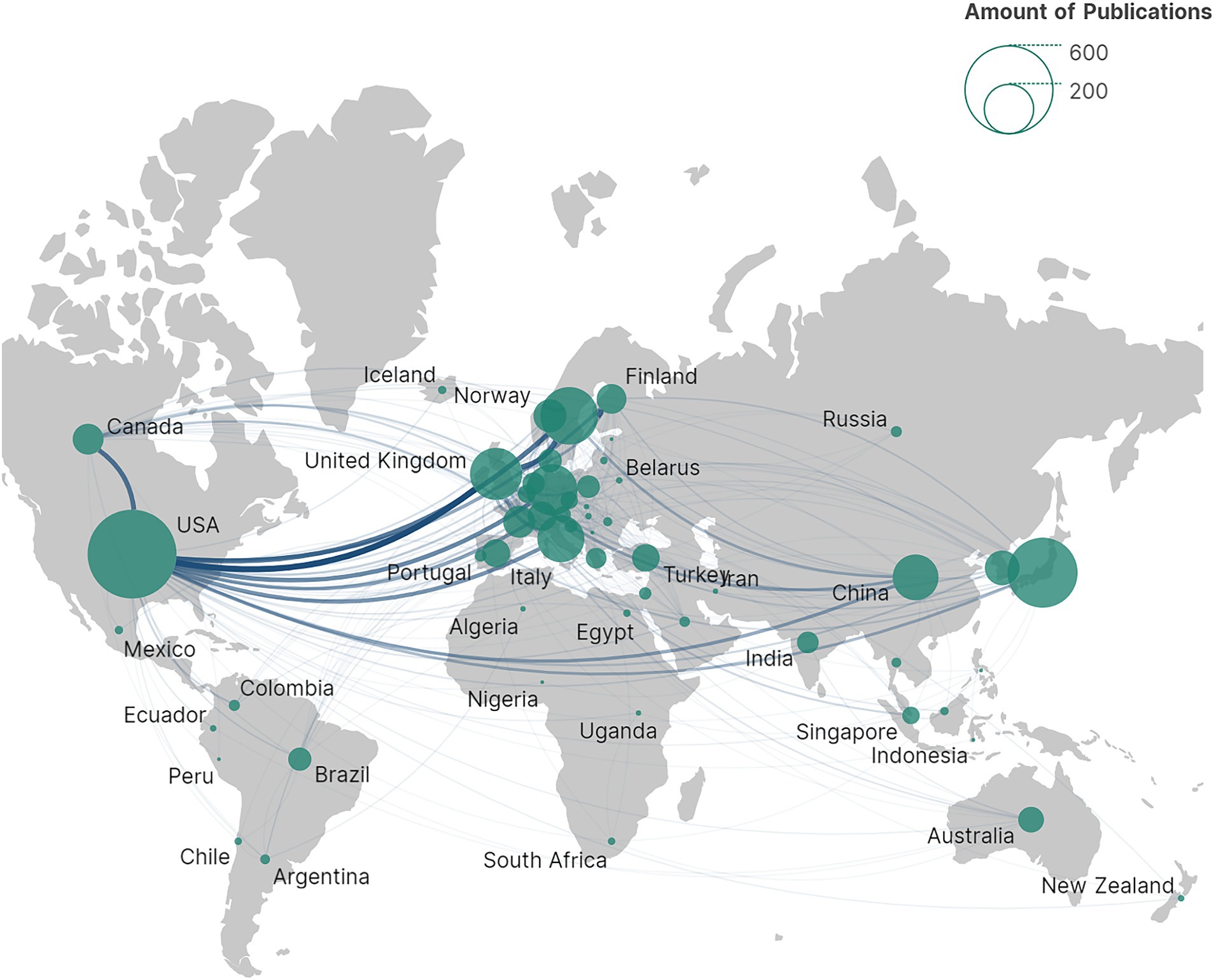
NPH is a global health challenge, with contributions from a total of 61 countries/regions to NPH research, as depicted in Figure 3. The data illustrates the worldwide distribution of publications, highlighting a concentration of publications in economically developed countries in Europe and the United States.
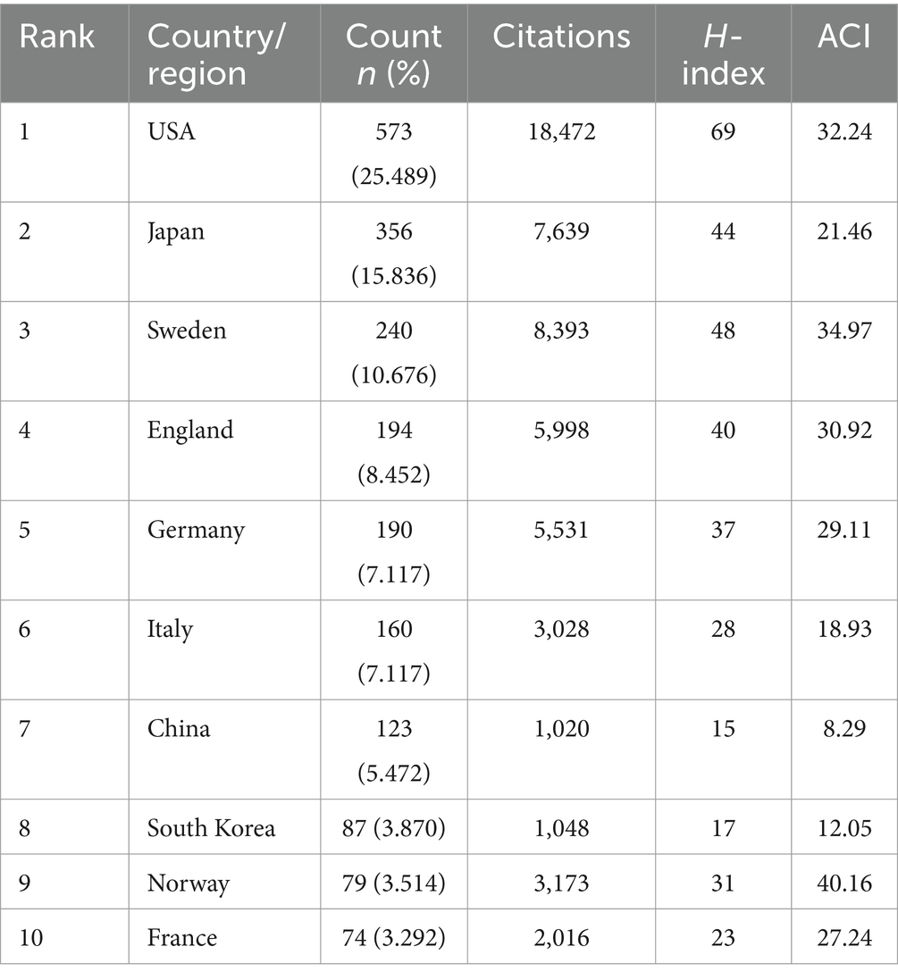
Table 1 presents the top 10 most productive countries. Regarding national research strength, the USA led with 573 publications and 18,472 citations, followed by Japan with 356 publications and 7,639 citations, and Sweden with 240 publications and 8,393 citations. The H-index is a dominant metric used to quantify the productivity and impact of authors, countries, or institutions. In terms of the H-index, the United States maintained the top position with an H-index of 69, followed by Sweden and Japan, both with H-indices above 40 (Table 1). Our study found that the United States led in both the total number of publications and H-index. Therefore, in terms of both research quantity and quality, the United States dominated in this research field. Additionally, it is noteworthy that although some countries had fewer publications, they obtained a high average number of citations per item (ACI). For example, Norway ranked highest in terms of ACI (average citations per item) at 40.16, which could be attributed to the publication of highly influential studies.
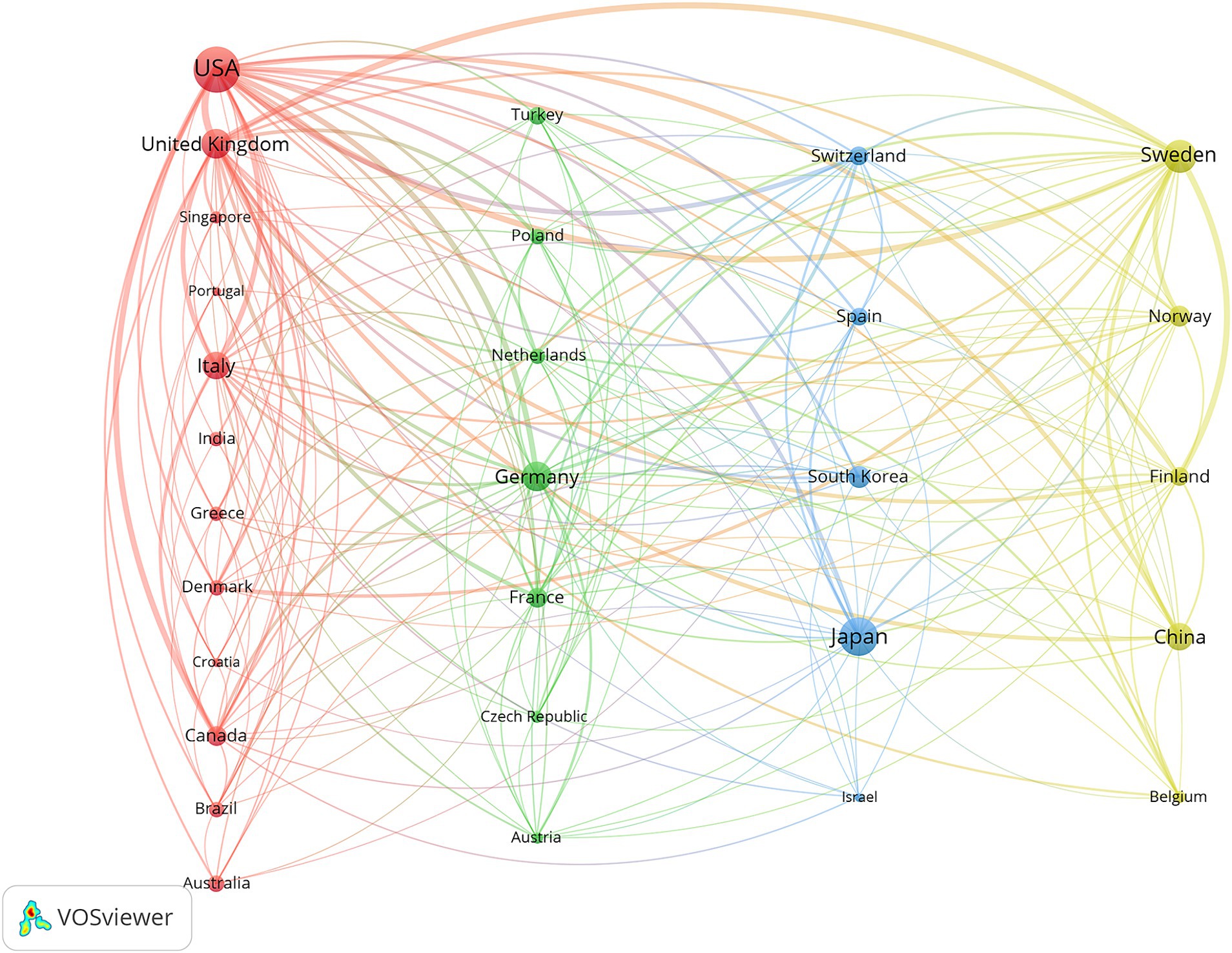
VOSviewer was used for co-authorship analysis of the countries/regions to reveal international collaborations in NPH field. Figure 4 displays international cooperation among relevant countries/regions and only countries/regions with more than 10 papers were included. Of the 29 countries/regions that met this threshold. The 29 countries/regions were divided into 4 clusters represented by different colors. The largest cluster (in red), consisting of 12 countries, centered on the United States, United Kingdom and Italy. The USA had the most significant number of cooperating partners (n = 21). For NPH research, there is close cooperation between countries, highlighting NPH as a global challenge.
VOSviewer was utilized to conduct co-authorship analysis of countries/regions, revealing international collaborations in the field of NPH. Figure 4 illustrates international cooperation among relevant countries/regions, including only those with more than 10 papers. Among the 29 countries/regions that met this threshold, these countries/regions were divided into 4 clusters represented by different colors. The largest cluster (highlighted in red) consisted of 12 countries, centered around the United States, United Kingdom, and Italy. The USA had the highest number of cooperating partners (n = 21). In NPH research, close cooperation between countries highlights NPH as a global challenge.
Analysis of institutions
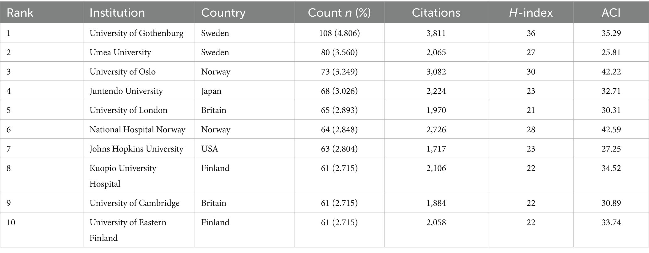
In the analysis of institutions, a total of 2,132 institutions contributed to the published research in the field. The top 10 most prolific institutions are listed in Table 2. Among the top 10 institutions, eight were from Europe, one was from the United States, and one was Asian. Among the most productive institutions, the University of Gothenburg had 108 publications and 3,811 citations, followed by Umea University with 80 publications and 2,065 citations, and the University of Oslo with 73 publications and 3,082 citations. As shown in Table 2, the University of Gothenburg had the highest H-index value (n = 36), followed by the University of Oslo (n = 30) and the National Hospital Norway (n = 28). Regarding ACI, the top three institutions with the largest ACI were the National Hospital Norway (n = 42.59 times), followed by the University of Oslo (n = 42.22 times), both of which are from Norway.
The University of Gothenburg’s most frequent collaborator is Sahlgrenska University Hospital, with 51 joint publications. The university’s most cited article, “Prevalence of Idiopathic Normal-Pressure Hydrocephalus,” was published in 2014. This study investigates the prevalence of normal pressure hydrocephalus among adults aged 70 years and older, providing robust epidemiological data on this condition in an aging population. The most prolific author affiliated with the University of Gothenburg in this field is M. Tullberg, who has authored 42 articles on normal pressure hydrocephalus, ranking 9th among the top 10 authors in terms of total publications.
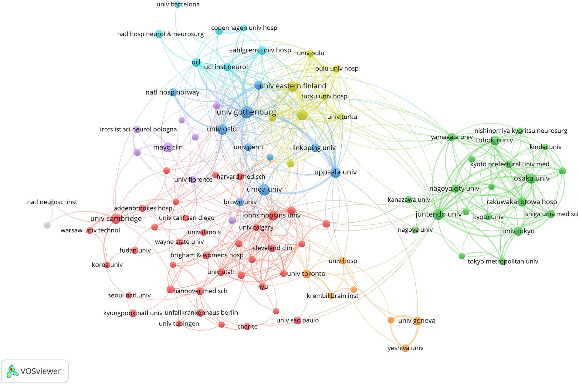
VOSviewer was used to create the network visualization map depicting institutions’ cooperation (Figure 5). With a minimum threshold of 10 articles published by institutions, 100 institutions met the criteria. There were seven clusters of co-authorship represented by different colors. The largest cluster, highlighted in red, consisted of 37 institutions centered around the University of Cambridge, Johns Hopkins University, and the Cleveland Clinic. The map shows that there is a certain amount of collaboration and exchange between research institutions around the world, but mostly restricted either to a country or a particular region.
Analysis of authors
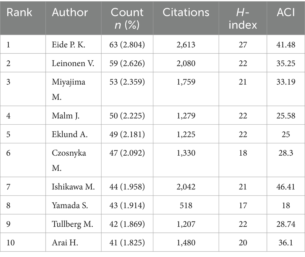
In total, 8,363 authors are involved in research related to NPH, and Table 3 lists the top 10 authors. In terms of publications, Eide P. K. has published 63 articles and had 2,613 citations, followed by Leinonen V. with 59 publications and 2,080 citations, and Miyajima M. with 53 publications and 1,759 citations. The author with the highest ACI was Ishikawa M. (n = 46.41), followed by Eide P. K. (n = 41.48) and Arai H. (n = 36.1).
In our analysis of Eide P. K.’s articles, we discovered that his research on normal pressure hydrocephalus (NPH) is comprehensive, covering epidemiology, pathogenesis, and surgical treatment, with a particular focus on the role of the glymphatic system in the pathogenesis of NPH. Notably, a 2017 paper he co-authored, titled “Glymphatic MRI in Idiopathic Normal Pressure Hydrocephalus,” has been cited 400 times. This landmark study was the first to evaluate glymphatic system function in NPH patients using magnetic resonance imaging.
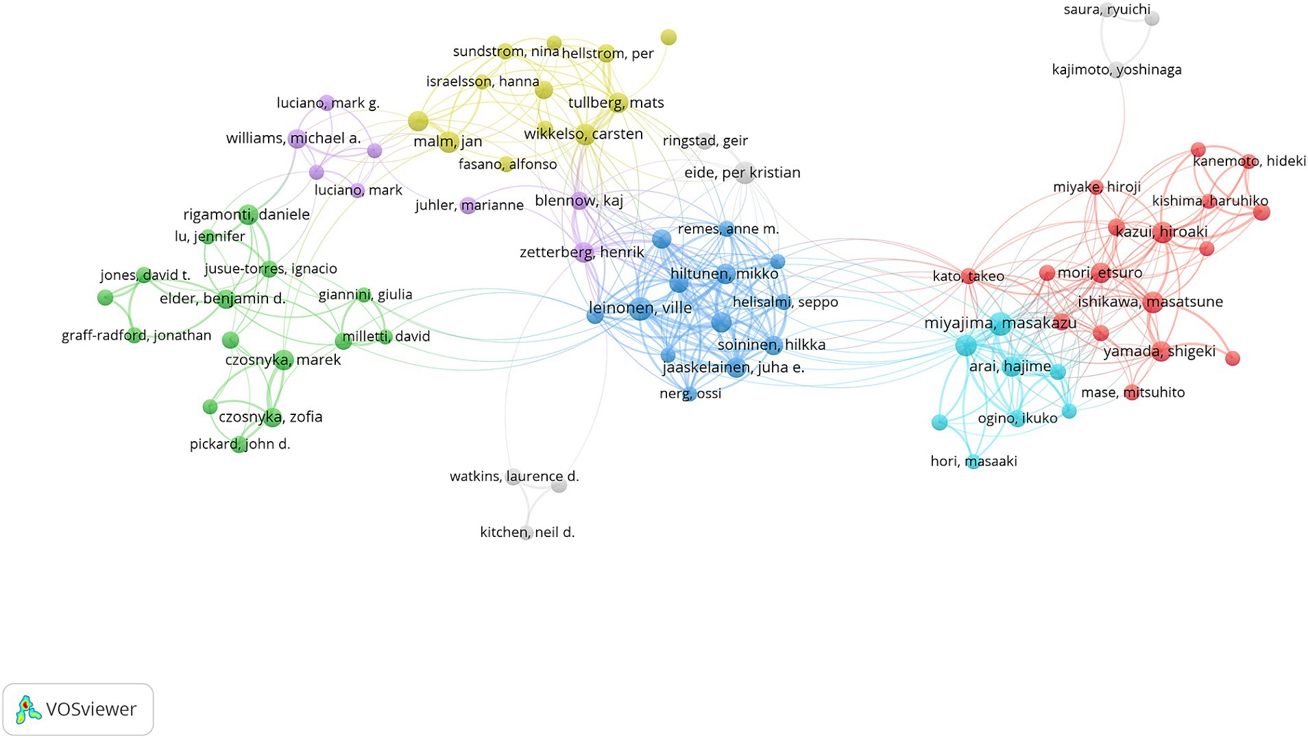
Closely following is Leinonen V., who, along with his team, focused their research on biomarkers for the diagnosis and prognosis of NPH. Their representative article, “Cerebrospinal Fluid Biomarker and Brain Biopsy Findings in Idiopathic Normal Pressure Hydrocephalus,” is a notable contribution to the field. Although Eide P. K. and Leinonen V. have both published extensively on NPH, they have only collaborated twice. This suggests that most studies in this field are conducted independently rather than through the collaborative efforts of the authors listed in Table 3.
We used VOSviewer to perform co-authorship analysis, including only authors with a minimum of 10 publications (Figure 6). Co-authorship networks involved 93 authors and were divided into six color-coded clusters. The largest cluster, represented in red and consisting of 17 authors, stood out prominently. The majority of cluster members are native authors; therefore, international research teams should improve their communication.
Analysis of journals
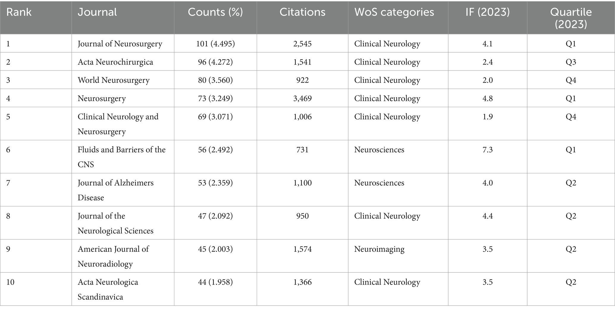
Relevant articles were published in 510 journals. The Journal of Neurosurgery (101 publications, 4.495% of total) ranks first, followed by Acta Neurochirurgica (96 publications, 4.272%) and World Neurosurgery (80 publications, 3.560%). All of the top 10 journals belong to the field of neurology. With the exception of Clinical Neurology and Neurosurgery, these journals all have a 2023 impact factor greater than 2. Fluids and Barriers of the CNS has the highest impact factor (n = 7.3).
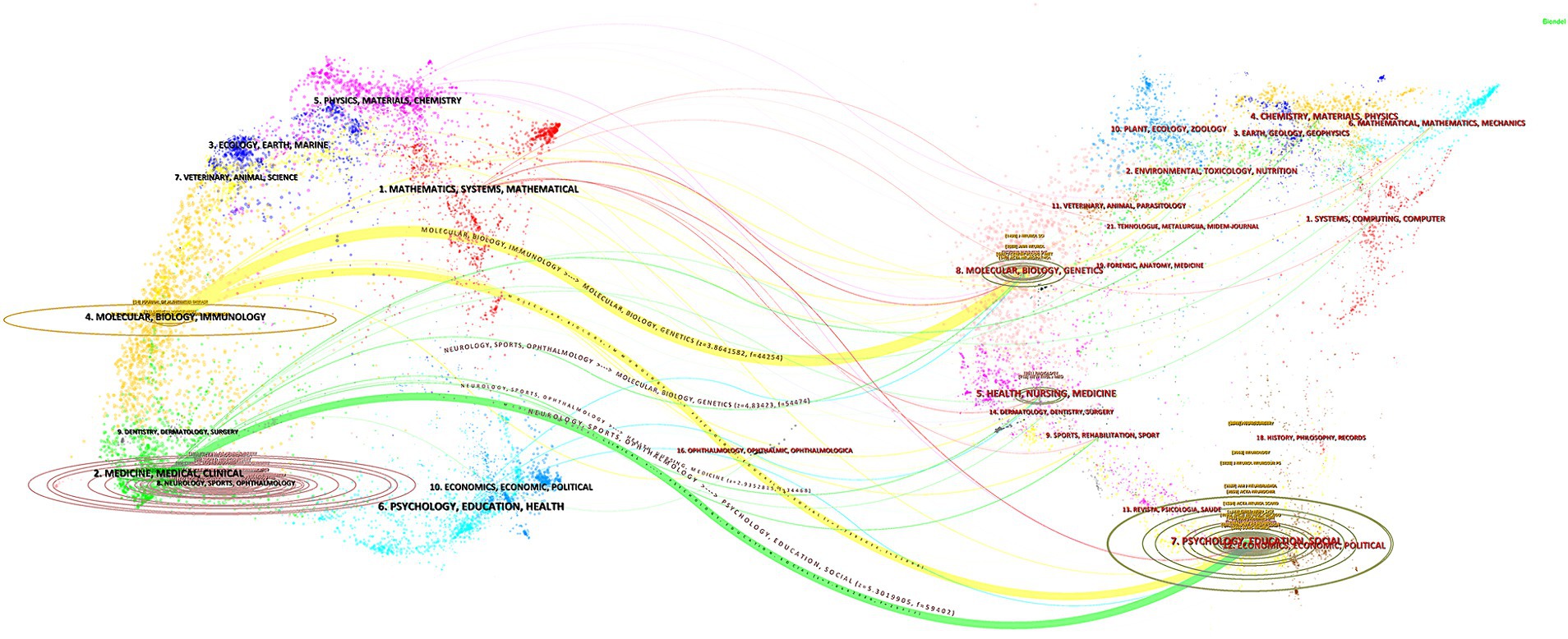
Dual-map overlays allowed us to accomplish several innovative visual analytic tasks that were previously not possible to perform intuitively. By following the citation arcs from the origin branch to the concentrated landing zones, it became straightforward to determine whether a set of publications incorporated prior work from multiple disciplines (24). We conducted an analysis of published and co-cited journals using a journal dual-map overlay, depicted in Figure 7. The citing journals are represented on the left, and the cited journals are represented on the right. Citation relationships are depicted as colored lines from left to right. We identified two distinct citation paths represented by orange and green lines. The green path is an independent development pattern, the citing journals mainly belong to Molecular Biology/Clinics, and cited journals mainly belong to Psychology/Education/Social. Whereas, the orange pathway is a confluent pattern of development, indicating that the two crosscutting areas evolved into a common research theme, the citing journals mainly belong to Molecular Biology/Immunology, and cited journals mainly belong to Molecular Biology/Clinics and Psychology/Education/Social.
Analysis of co-occurring keywords and burst terms
Indexing keywords facilitates understanding the main content of a paper, so to a certain extent, keyword analysis illustrates the hotspots and focus of this research area. Before analyzing, we remove to the search terms, and we merged some terms with the same meaning, (e.g., “csf” and “cerebrospinal fluid,” “Alzheimer’s disease” and “Alzheimer disease,” “biomarker” and “biomarkers”). In this study, a comprehensive analysis was conducted on 6,334 extracted keywords, and the top 10 frequently occurring keywords are “Cerebrospinal fluid,” “diagnosis,” “management,” “Alzheimer’s-disease,” “dementia,” “ventriculoperitoneal shunt,” “magnetic resonance imaging,” “brain,” “disease,” “shunt surgery.”
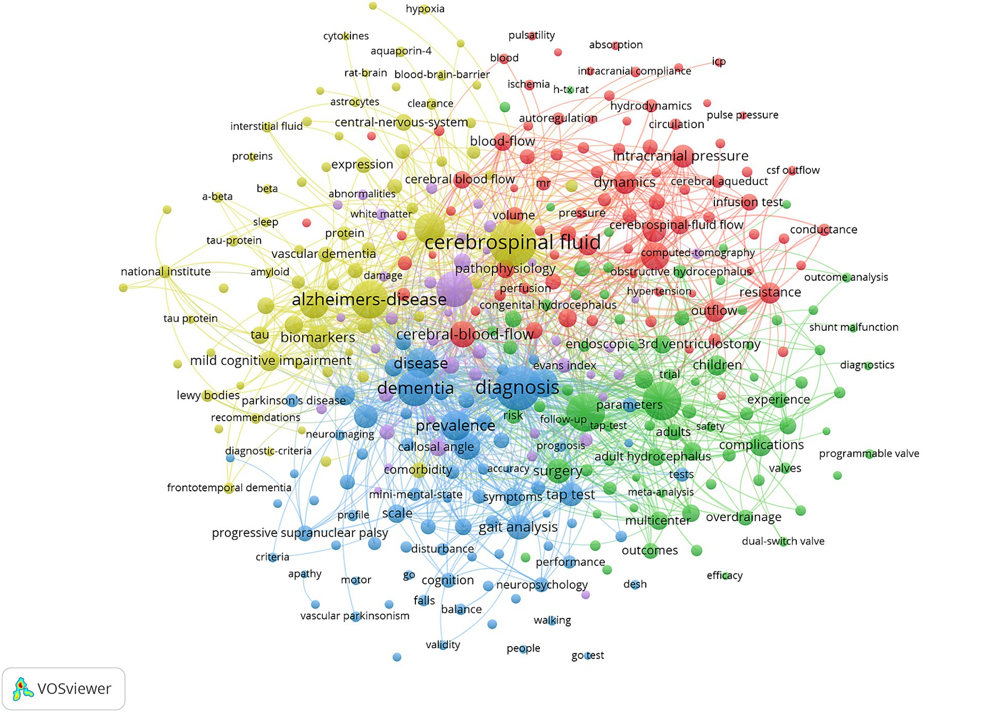
In the keyword co-occurrence network (Figure 8), the keywords were assigned to 5 clusters according to the color: cluster 1 (red) included keywords related to pathogenesis and pathophysiology, such as “intracranial pressure,” “blood flow,” “dynamics,” etc. Cluster 2 (green) included keywords related to treatment, such as “ventriculoperitoneal shunt,” “endoscopic 3rd ventriculostomy,” and “shunt.” Cluster 3 (blue) included keywords related to diagnosis, such as “callosal angle,” “dementia.” Cluster 4 (yellow) included keywords related to differential diagnosis such as “Alzheimer disease,” “biomarkers,” and “proteins.”
Burstness highlights the period when the occurrence or use of specific nodes is significantly higher compared to other nodes. Specifically, when there is a sudden spike in publications on a particular subject or area of interest, a keyword burst can reveal new developments related to that field. Furthermore, burstness indicates nodes that have garnered significant attention in a short period, showing substantial changes in frequency over a brief timeframe. We used CiteSpace software to extract citation bursts for all keywords, specifically focusing on the top 20 keywords (Figure 9). The red timeline in Figure 9 illustrates the duration of the outbreak. Based on the development trends shown in the Figure 9, the study period can be divided into three phases: 2003–2009, 2010–2018, and 2019–2023. The strongest bursts in 2003–2009 included the following keywords: “predictive value,” “cerebral blood flow,” “resistance,” “CSF,” “communicating hydrocephalus,” “shunt operation,” obstructive hydrocephalus.” Studies in this period largely focused on the pathogenesis and differential diagnosis of normal pressure hydrocephalus. Regarding pathogenesis, researchers concentrated on cerebrospinal fluid circulation disorders and intracerebral microcirculation and vascular dysfunction. The period from 2010 to 2018 recorded the keywords with the strongest citation bursts: “MRI” and “diffusion tensor imaging.” During this time, research primarily began utilizing imaging technologies to explore the pathogenesis of normal pressure hydrocephalus (NPH) and identify potential imaging biomarkers. The period from 2016 to 2021 shows five keywords that have recently received significant attention: “callosal angle,” “scale,” “shunt surgery,” “glymphatic system,” and “guidelines.” The most notable keyword, “guidelines,” attained the highest burst strength during this period. This suggests that researchers worldwide are increasingly focusing on accurate diagnosis and treatment of NPH, and are eager to establish standardized guidelines.
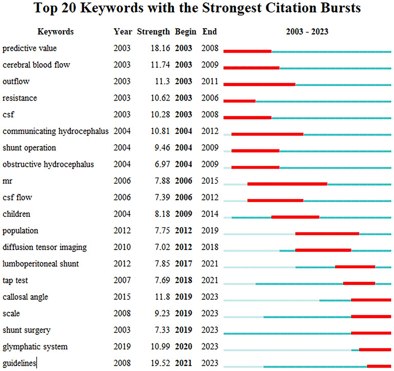
Figure 9. The top keywords with the strongest citation bursts. The long blue line depicts the timeline (2003–2023), and the short red line indicates the burst period of certain keyword.
Analysis of co-cited references
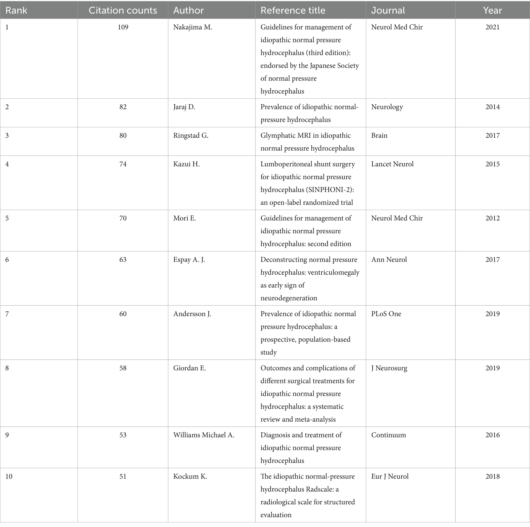
A co-citation relationship is established when two studies are concurrently cited by a third study (24). Co-citation analysis further illustrates the frequency with which articles are cited together. This analysis of co-cited works helps constitutes the theoretical basis and knowledge framework of a scientific research topic. We identified the top 10 co-cited references in Table 4. The most frequently cited reference is the guidelines for management of INPH (third edition), and the second version of the guide is also cited at fifth. The second article primarily addresses the prevalence of iNPH, which is related to the topic of the seventh article. The third article discusses the role of the glymphatic system in the pathogenesis of NPH.
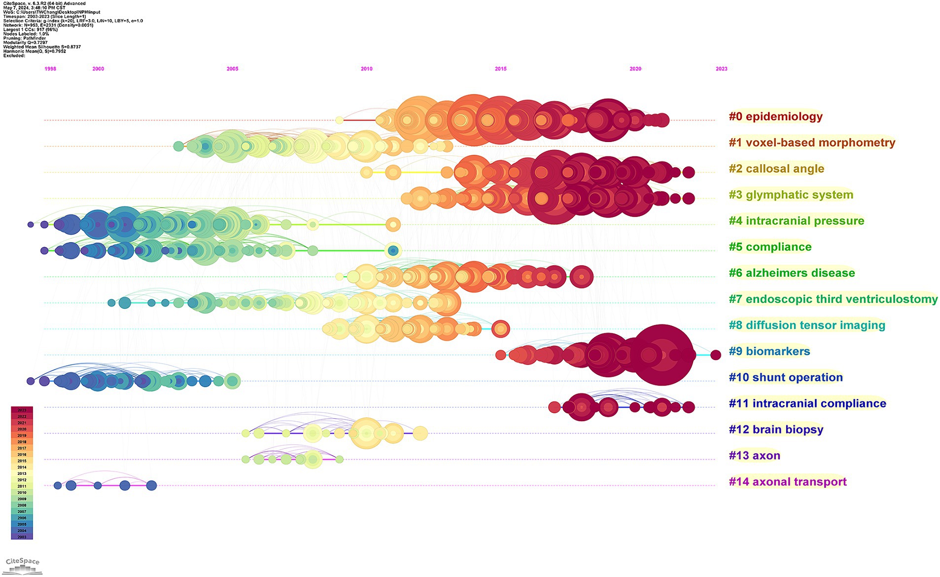
The co-citation network can be segmented into distinct clusters using CiteSpace. Papers within the same cluster are closely related based on their co-citation patterns. Each cluster is defined by terms extracted from the keywords field of the referenced documents in that cluster. We can find the top 14 clusters, which are #0 epidemiology, #1 voxel-based morphometry, #2 shunt operation, #3 intracranial compliance #4 brain biopsy, #5 axon, #6 axonal transport, #7 callosal angle, #8 glymphatic system, #9 intracranial pressure, #10 compliance, #11 Alzheimer’s disease, #12 endoscopic third ventriculostomy, #13 diffusion tensor imaging, and #14 biomarkers (Figure 10). Eight of these clusters (#3, #4, #5, #4, #6, #8, #6, and #10) are about the pathogenesis of NPH, and three of these clusters (#1, #7, and #13) focus on imaging study of NPH. Whereas the other two (#2 and #12) focus on surgical treatment of NPH.
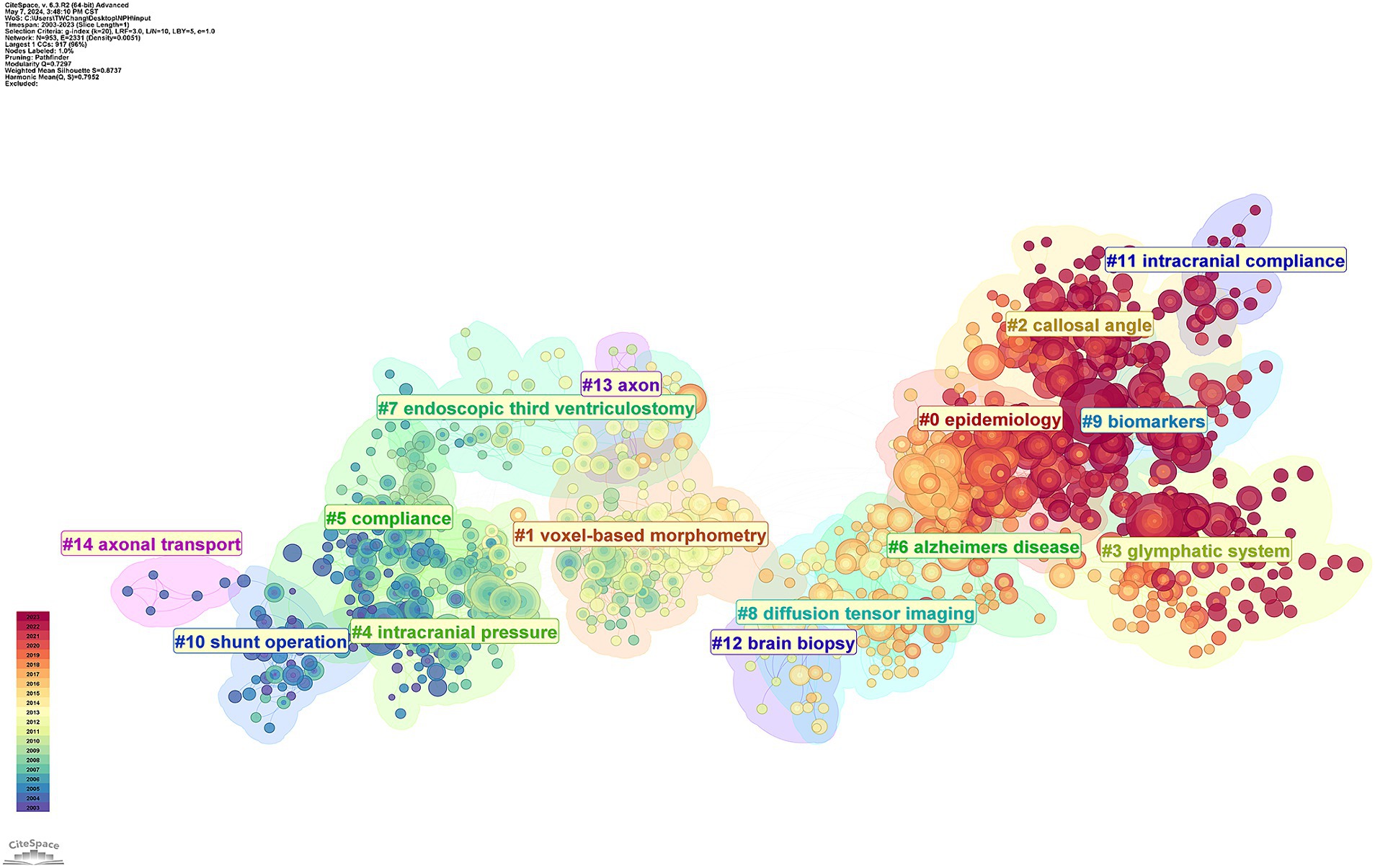
Figure 10. CiteSpace visualization clusters of the co-cited references. Terms from the title field of the citing papers within each cluster are used as the definition of that cluster.
The cluster map can be transformed into a timeline diagram to analyze research evolution and development within the listed clusters over time. Early studies primarily focused on the surgical treatment and pathogenesis of normal pressure hydrocephalus (Figure 11). At the same time, we can also observe that the research on the pathogenesis continues to the present, and the main concern is the role of the intracranial lymphatic system in the pathogenesis. Another current area of interest is to identify biomarkers that can accurately diagnose and predict the prognosis of normal pressure hydrocephalus.
Discussion
Global trends in NPH research
Research on normal pressure hydrocephalus (NPH) has entered a phase of rapid development. Research on NPH has shown consistent growth over the past two decades, as shown in Figure 2. NPH has emerged as a compelling topic across multiple disciplines and is anticipated to continue gaining momentum in the future. This trend can be attributed to the development of the economy and society, as developed countries continue to experience population aging. As the population ages, the incidence of normal pressure hydrocephalus continues to rise. Consequently, an increasing number of researchers are focusing on this field.
Research initiatives in the field of NPH have been initiated in several countries, with the United States leading the way. A total of 1,573 publications originated from the USA, representing approximately a quarter of all publications. Figure 4 illustrates extensive intercountry cooperation among countries/regions. The USA emerged as the most productive country and the hub of international cooperation. Therefore, the USA led the forefront of scientific and academic research.
Among the top 10 most productive institutes, eight were European institutions, one was Asian, and one was American. European institutions led in terms of quantity of NPH research. Figure 6 illustrates that there is a certain amount of collaboration and exchange between research institutions around the world, but mostly restricted either to a country or a particular region.
Additionally, the top three most prolific authors in this field are Eide P. K., Leinonen V., and Miyajima M. They are leaders in the field and are likely to continue shaping its development. These top scholars are ideally suited for collaboration and communication.
Within this domain, there are specific journals that deserve researchers’ attention. The journals listed in Table 5, such as Journal of Neurosurgery, Acta Neurochirurgica, and World Neurosurgery, are likely core journals in this field, making them recommended venues for submitting relevant papers. Researchers should also prioritize keeping up with the latest articles published in these journals.
Hot spots and future research direction
Through keyword burst detection and cited references clustering analysis, research hotspots and current research directions may be discovered. Thus, combined with keywords and references that continue to show high bursts, we analyzed current research hotspots and predicted future research trends.
Epidemiology
The variations in diagnostic criteria, survey populations, age groups, gender distributions, and racial compositions employed across different studies pose challenges when comparing the epidemiological findings of iNPH. In a prevalence study conducted in Norway in 2008, using the diagnostic criteria outlined in the 2005 international guidelines, the reported prevalence rates were 21.9 per 100,000 individuals for “probably iNPH” and 29 per 100,000 individuals for “possible iNPH” within hospital settings.
A Swedish study revealed that among the general population, the prevalence rate of “probably iNPH” was 3.7%, with rates varying between 0.2% for individuals aged 70–79 years and 5.9% for those aged above 80, without significant gender disparity observed. However, an epidemiological survey in Sweden in 2019, using Japanese iNPH diagnostic criteria, indicated a lower prevalence rate of “probably iNPH,” at approximately 1.5% (25, 26). Another survey in Japan during 2008, employing Japanese diagnostic criteria, demonstrated a prevalence rate of “MRI-supported possible iNPH” at around 2.9%. A recent MRI-based study (27) focusing primarily on 70-year-olds in Sweden, using international guideline diagnostic criteria, reported a higher prevalence rate of approximately 1.5% for “possible iNPH,” significantly greater than the rate reported in 2019.
Future research efforts should focus on developing more authoritative diagnostic guidelines for NPH. Researchers can then conduct large-scale population studies involving multiple countries and research centers, based on these standardized guidelines. Such comprehensive studies will provide more accurate epidemiological data on the prevalence and incidence of NPH, which will be invaluable for clinical practitioners.
Glymphatic system
The glymphatic system is a brain fluid transport system composed of several key components. Current research indicates that the glymphatic system includes cerebrospinal fluid (CSF), perivascular spaces (PVS), aquaporin-4 (AQP4) channels located on astrocytic end feet, and interstitial fluid (ISF) (28). AQP4 plays a critical role in mediating the exchange of CSF and ISF within this system, facilitating the clearance of metabolic byproducts and toxic proteins in the central nervous system. This mechanism is vital for maintaining brain homeostasis and supporting overall brain health.
Cerebrospinal fluid (CSF) in the subarachnoid space flows into perivascular spaces surrounding arteries, and water within the CSF passes through aquaporin-4 (AQP4) channels located on astrocytic end feet into the brain parenchyma for cerebrospinal fluid-interstitial fluid (CSF-ISF) exchange. After this exchange, the interstitial fluid (ISF) containing significant metabolic waste enters AQP4 channels, flows into perivascular spaces around veins, and then gradually returns to the subarachnoid space before entering venous circulation, thereby constituting the glymphatic system (29).
PVS and AQP4 are two crucial structures involved in the glymphatic system with implications for various central nervous system diseases (30). PVS represent microscopic tissue spaces surrounding arteries and veins within the brain; their loose fibrous matrix provides low-resistance pathways for efficient CSF-ISF flow essential for intracranial fluid transport. Astrocytes possess orthogonal arrangements of AQP4 on their end feet which facilitate efficient water transportation. When AQP4 is polarly distributed on the outer wall of the PVS end feet, it facilitates the rapid exchange of CSF with ISF in the brain tissue, promoting the clearance of brain metabolic waste. Furthermore, the polar distribution of AQP4 is synchronized with the strength of the astrocytic lymphatic system. Therefore, structural damage to the PVS and abnormal polar distribution of AQP4 may be an important cause of impaired CSF-ISF exchange, metabolic waste deposition, and ultimately the development or exacerbation of neurological diseases.
Loss of arterial compliance can lead to glymphatic system damage in iNPH (16). In patients diagnosed with iNPH, the incidence of systemic atherosclerotic diseases, including primary hypertension, dyslipidemia, coronary artery disease, and peripheral arterial disease, is significantly higher (31). As the vessels become increasingly rigid, the weakening of vascular pulsatility and the diminished driving force for glymphatic fluid flow lead to the accumulation of waste in the brain interstitium, increasing the resistance to outflow along the lymphatic pathways, which is part of the reason why CSF retrograde into the ventricular system. The restoration of cerebrospinal fluid dynamics after shunting may promote glymphatic fluid flow and improve cognitive function.
The physiological function of aquaporins is to facilitate the transport of water across cell membranes. Reduced expression of AQP-4 has been confirmed as another potential mechanism for lymphatic vessel damage in iNPH patients (32). In Alzheimer’s disease, decreased expression of AQP-4 leads to impaired clearance of misfolded proteins, resulting in neurotoxicity and cognitive decline. Glymphatic system damage in iNPH may cause accumulation of metabolic waste, leading to neurotoxicity and cognitive impairment.
Impairment to the glymphatic system disrupts brain fluid dynamics and hinders the clearance of metabolic waste, critically contributing to the pathogenesis of NPH. Given these established mechanisms and targets, future research should prioritize investigating methods to ameliorate NPH symptoms and prognosis through modulation of glymphatic system function.
Biomarkers
Cerebrospinal fluid (CSF) biomarker testing can yield valuable biological information. There has been increasing interest in identifying appropriate biomarkers for diagnosing, differentiating diagnoses, and potentially predicting outcomes using CSF.
Total tau protein (t-tau) (33) is generally considered a non-specific marker of neuronal/axonal degeneration, while phospho-tau (p-tau) (34) reflects tau hyperphosphorylation as well as tangle formation. Aβ42 is indicative of the presence of amyloid pathology (35). Kapaki et al. (36) analyzing CSF biomarkers in iNPH, which showed that t-tau was significantly increased in iNPH, as compared with the control group, whereas Aβ42 was decreased. Ray et al. (37) reported different results that showed a significant decrease in CSF Aβ42 concentration in NPH patients as compared with the control group, but no significant difference in t-tau or p-tau between these two groups. Taghdiri et al. (38) showed that lower CSF Aβ42 and p-tau concentrations were observed in patients with iNPH compared to health controls.
The researchers proposed using cerebrospinal fluid biomarkers to distinguish diseases similar to iNPH, such as the most common Alzheimer’s disease. Taghdiri et al. (38) suggested that the concentration of Aβ42 in cerebrospinal fluid of patients with iNPH was higher than that of patients with AD, and the concentration of t-tau and p-tau was lower than that of patients with AD. Mazzeo et al. (39) agreed that Aβ42/Aβ40, p-tau, and t-tau were significantly lower in iNPH than AD, but found no significant difference in Aβ42 concentrations.
Meanwhile, researchers have started to investigate the potential correlation between biomarker concentrations and shunt responsiveness. Thavarajasingam et al. (40) compared two groups of patients with and without improvement in symptoms after shunt surgery, and found significantly higher levels of t-tau and p-tau in the cerebrospinal fluid of iNPH patients who did not experience symptom improvement post-shunting, as compared to those who did. However, Tullberg et al. (41) argued that there were no significant differences in cerebrospinal fluid biomarkers between patients who showed postoperative improvement and those who did not.
In addition to the above well-studied cerebrospinal fluid markers, researchers are looking for novel cerebrospinal fluid biomarkers that can diagnose and predict prognosis. Multiple studies have shown that neurofilament light chain (NFL) (42) is associated with axonal injury or degeneration. Furthermore, the concentration of NFL in the cerebrospinal fluid of iNPH patients is higher than that of healthy controls. However, Jeppsson et al. (43) reported opposite results, stating that there was no difference in NFL concentration between iNPH patients and healthy controls. There are also studies suggesting that glial fibrillary acidic protein (GFAP) could serve as a potential biomarker (44).
Some researchers have also investigated cerebrospinal fluid biomarkers for other neuronal injuries, including lipocalin-type prostaglandin D synthase (PGDS) (45), myelin basic protein (MBP) (46), protein tyrosine phosphatase receptor type Q (PTPRQ) (47), and various other molecules. However, these studies have produced contradictory results thus far.
Currently, there is substantial heterogeneity among studies investigating cerebrospinal fluid biomarkers. Various research cohorts have employed diverse inclusion and exclusion criteria, and their laboratory measurement techniques are inconsistently applied. Consequently, many studies have yielded inconsistent or conflicting findings. Further large-scale cohort studies are necessary to collectively establish a unified biomarker profile for normal pressure hydrocephalus in cerebrospinal fluid. This would enable clinicians to accurately diagnose the syndrome and tailor personalized treatment plans for each patient’s benefit.
Interestingly, the majority of researchers concentrate on biomarkers found in cerebrospinal fluid. However, we can leverage methodologies employed in the study of other neurodegenerative diseases, like Alzheimer’s disease, to investigate novel biomarkers. Xu et al. (48) explored immune cell infiltration and the expression patterns of genes related to immune function in Alzheimer’s disease patients. Employing machine learning algorithms, they identified five genes (PFKFB4, PDK3, KIAA0319L, CEBPD, and PHC2T) associated with immune cells and functions in AD, validating their accurate diagnostic potential. This implies potential for discovering or validating related genetic biomarkers for diagnosing NPH. In recent decades, the ongoing advancement of neuroimaging technologies, such as resting-state and task-based functional MRI, alongside electroencephalography, has facilitated the development of biomarkers for diagnosing cognitive and motor disorders.
Yin et al. (49) compared Alzheimer’s disease patients with normal controls and observed significant alterations in both the function and structure of the dorsal attention network among AD patients. Additionally, cognitive performance showed a close correlation with these observed alterations. Our comprehension of neurobrain network changes in NPH remains incomplete. Future research could utilize these advanced neuroimaging technologies to investigate potential neuroimaging biomarkers, which could offer substantial research insights.
Limitations
This study is based on bibliometric and bibliographic visualization analyses of the literature, which can assist researchers in better understanding the developmental trends and academic frontiers of the field. However, this study has some limitations. Firstly, only English-language articles and reviews from SCI Expanded-indexed journals were included. Secondly, some details may be omitted due to the inability of VOSviewer and CiteSpace to analyze the full text of publications. Lastly, some newly published excellent articles may be excluded due to time lag.
Conclusion
Bibliometric analysis revealed that current research on NHP is developing rapidly. The United States has contributed many outstanding scientific research achievements and breakthroughs in this field and ranked first among high output countries. Bibliometric analysis indicates that research on NHP is experiencing rapid growth. The USA has made significant contributions to scientific research and advancements in this area, ranking at the top among high output countries.
The university of Gothenburg has made novel progress and published most studies in this field. Better understanding the pathophysiological mechanisms of normal pressure hydrocephalus and identifying more accurate biomarkers in cerebrospinal fluid are hot topics for future research. The University of Gothenburg has achieved significant advancements and produced most researches in this area. Exploring the pathophysiological mechanisms of NPH and discovering more precise biomarkers in cerebrospinal fluid are important areas for future investigation.
Author contributions
TC: Writing – original draft, Software, Visualization, Data curation, Formal analysis, Project administration. XH: Data curation, Software, Visualization, Writing – original draft. XZ: Software, Visualization, Writing – original draft. JL: Validation, Visualization, Writing – original draft. WB: Software, Visualization, Writing – original draft. JW: Conceptualization, Funding acquisition, Supervision, Writing – review & editing.
Funding
The author(s) declare that financial support was received for the research, authorship, and/or publication of this article. This study has received funding by National Natural Science Foundation of China (82060316).
Conflict of interest
The authors declare that the research was conducted in the absence of any commercial or financial relationships that could be construed as a potential conflict of interest.
Publisher’s note
All claims expressed in this article are solely those of the authors and do not necessarily represent those of their affiliated organizations, or those of the publisher, the editors and the reviewers. Any product that may be evaluated in this article, or claim that may be made by its manufacturer, is not guaranteed or endorsed by the publisher.
References
1. Tan, C, Wang, X, Wang, Y, Wang, C, Tang, Z, Zhang, Z, et al. The pathogenesis based on the glymphatic system, diagnosis, and treatment of idiopathic normal pressure hydrocephalus. Clin Interv Aging. (2021) 16:139–53. doi: 10.2147/cia.S290709
2. Hakim, S, and Adams, RD. The special clinical problem of symptomatic hydrocephalus with normal cerebrospinal fluid pressure. Observations on cerebrospinal fluid hydrodynamics. J Neurol Sci. (1965) 2:307–27. doi: 10.1016/0022-510x(65)90016-x
3. Adams, RD, Fisher, CM, Hakim, S, Ojemann, RG, and Sweet, WH. Symptomatic occult hydrocephalus with “normal” cerebrospinal-fluid pressure. A treatable syndrome. N Engl J Med. (1965) 273:117–26. doi: 10.1056/nejm196507152730301
4. Meier, U, and Mutze, S. Correlation between decreased ventricular size and positive clinical outcome following shunt placement in patients with normal-pressure hydrocephalus. J Neurosurg. (2004) 100:1036–40. doi: 10.3171/jns.2004.100.6.1036
5. Yin, R, Wen, J, and Wei, J. Progression in neuroimaging of normal pressure hydrocephalus. Front Neurol. (2021) 12:700269. doi: 10.3389/fneur.2021.700269
6. Fasano, A, Espay, AJ, Tang-Wai, DF, Wikkelsö, C, and Krauss, JK. Gaps, controversies, and proposed roadmap for research in normal pressure hydrocephalus. Mov Disord. (2020) 35:1945–54. doi: 10.1002/mds.28251
7. Brean, A, and Eide, PK. Prevalence of probable idiopathic normal pressure hydrocephalus in a Norwegian population. Acta Neurol Scand. (2008) 118:48–53. doi: 10.1111/j.1600-0404.2007.00982.x
8. Rabiei, K, Jaraj, D, Marlow, T, Jensen, C, Skoog, I, and Wikkelsø, C. Prevalence and symptoms of intracranial arachnoid cysts: a population-based study. J Neurol. (2016) 263:689–94. doi: 10.1007/s00415-016-8035-1
9. Skalický, P, Mládek, A, Vlasák, A, De Lacy, P, Beneš, V, and Bradáč, O. Normal pressure hydrocephalus-an overview of pathophysiological mechanisms and diagnostic procedures. Neurosurg Rev. (2020) 43:1451–64. doi: 10.1007/s10143-019-01201-5
10. Hebb, AO, and Cusimano, MD. Idiopathic normal pressure hydrocephalus: a systematic review of diagnosis and outcome. Neurosurgery. (2001) 49:1166–86. doi: 10.1097/00006123-200111000-00028
11. Passos-Neto, CEB, Lopes, CCB, Teixeira, MS, Studart Neto, A, and Spera, RR. Normal pressure hydrocephalus: an update. Arq Neuropsiquiatr. (2022) 80:42–52. doi: 10.1590/0004-282x-anp-2022-s118
12. Martín-Láez, R, Caballero-Arzapalo, H, López-Menéndez, L, Arango-Lasprilla, JC, and Vázquez-Barquero, A. Epidemiology of idiopathic normal pressure hydrocephalus: a systematic review of the literature. World Neurosurg. (2015) 84:2002–9. doi: 10.1016/j.wneu.2015.07.005
13. Thavarajasingam, SG, El-Khatib, M, Rea, M, Russo, S, Lemcke, J, Al-Nusair, L, et al. Clinical predictors of shunt response in the diagnosis and treatment of idiopathic normal pressure hydrocephalus: a systematic review and meta-analysis. Acta Neurochir. (2021) 163:2641–72. doi: 10.1007/s00701-021-04922-z
14. Thavarajasingam, SG, El-Khatib, M, Vemulapalli, K, Iradukunda, HAS, Sajeenth Vishnu, K, Borchert, R, et al. Radiological predictors of shunt response in the diagnosis and treatment of idiopathic normal pressure hydrocephalus: a systematic review and meta-analysis. Acta Neurochir. (2023) 165:369–419. doi: 10.1007/s00701-022-05402-8
15. Bräutigam, K, Vakis, A, and Tsitsipanis, C. Pathogenesis of idiopathic normal pressure hydrocephalus: a review of knowledge. J Clin Neurosci. (2019) 61:10–3. doi: 10.1016/j.jocn.2018.10.147
16. Wang, Z, Zhang, Y, Hu, F, Ding, J, and Wang, X. Pathogenesis and pathophysiology of idiopathic normal pressure hydrocephalus. CNS Neurosci Ther. (2020) 26:1230–40. doi: 10.1111/cns.13526
17. Ninkov, A, Frank, JR, and Maggio, LA. Bibliometrics: methods for studying academic publishing. Perspect Med Educ. (2022) 11:173–6. doi: 10.1007/s40037-021-00695-4
18. Gong, C, Zhong, W, Zhu, C, Chen, B, and Guo, J. Research trends and hotspots of neuromodulation in neuropathic pain: a bibliometric analysis. World Neurosurg. (2023) 180:155–162.e2. doi: 10.1016/j.wneu.2023.06.090
19. Liu, Z, Cheng, Y, Hai, Y, Chen, Y, and Liu, T. Developments in congenital scoliosis and related research from 1992 to 2021: a thirty-year bibliometric analysis. World Neurosurg. (2022) 164:e24–44. doi: 10.1016/j.wneu.2022.02.117
20. Levy, AS, Bhatia, S, Merenzon, MA, Andryski, AL, Rivera, CA, Daggubati, L, et al. Exploring the landscape of machine learning applications in neurosurgery: a bibliometric analysis and narrative review of trends and future directions. World Neurosurg. (2023) 181:108–15. doi: 10.1016/j.wneu.2023.10.042
21. Pei, Z, Chen, S, Ding, L, Liu, J, Cui, X, Li, F, et al. Current perspectives and trend of nanomedicine in cancer: a review and bibliometric analysis. J Control Release. (2022) 352:211–41. doi: 10.1016/j.jconrel.2022.10.023
22. van Eck, NJ, and Waltman, L. Software survey: VOSviewer, a computer program for bibliometric mapping. Scientometrics. (2010) 84:523–38. doi: 10.1007/s11192-009-0146-3
23. Synnestvedt, MB, Chen, C, and Holmes, JH. CiteSpace II: visualization and knowledge discovery in bibliographic databases. AMIA Annu Symp Proc. (2005) 2005:724–8.
24. Effah, NAA, Wang, Q, Owusu, GMY, Otchere, OAS, and Owusu, B. Contributions toward sustainable development: a bibliometric analysis of sustainability reporting research. Environ Sci Pollut Res Int. (2023) 30:104–26. doi: 10.1007/s11356-022-24010-8
25. Andersson, J, Rosell, M, Kockum, K, Lilja-Lund, O, Söderström, L, and Laurell, K. Prevalence of idiopathic normal pressure hydrocephalus: a prospective, population-based study. PLoS One. (2019) 14:e0217705. doi: 10.1371/journal.pone.0217705
26. Pyykkö, OT, Nerg, O, Niskasaari, HM, Niskasaari, T, Koivisto, AM, Hiltunen, M, et al. Incidence, comorbidities, and mortality in idiopathic normal pressure hydrocephalus. World Neurosurg. (2018) 112:e624–31. doi: 10.1016/j.wneu.2018.01.107
27. Constantinescu, C, Wikkelsø, C, Westman, E, Ziegelitz, D, Jaraj, D, Rydén, L, et al. Prevalence of possible idiopathic normal pressure hydrocephalus in Sweden: a population-based MRI study in 791 70-year-old participants. Neurology. (2024) 102:e208037. doi: 10.1212/wnl.0000000000208037
28. Hablitz, LM, and Nedergaard, M. The glymphatic system: a novel component of fundamental neurobiology. J Neurosci. (2021) 41:7698–711. doi: 10.1523/jneurosci.0619-21.2021
29. Plog, BA, and Nedergaard, M. The glymphatic system in central nervous system health and disease: past, present, and future. Annu Rev Pathol. (2018) 13:379–94. doi: 10.1146/annurev-pathol-051217-111018
30. Yankova, G, Bogomyakova, O, and Tulupov, A. The glymphatic system and meningeal lymphatics of the brain: new understanding of brain clearance. Rev Neurosci. (2021) 32:693–705. doi: 10.1515/revneuro-2020-0106
31. Reeves, BC, Karimy, JK, Kundishora, AJ, Mestre, H, Cerci, HM, Matouk, C, et al. Glymphatic system impairment in Alzheimer’s disease and idiopathic normal pressure hydrocephalus. Trends Mol Med. (2020) 26:285–95. doi: 10.1016/j.molmed.2019.11.008
32. Peng, S, Liu, J, Liang, C, Yang, L, and Wang, G. Aquaporin-4 in glymphatic system, and its implication for central nervous system disorders. Neurobiol Dis. (2023) 179:106035. doi: 10.1016/j.nbd.2023.106035
33. Chen, Z, Liu, C, Zhang, J, Relkin, N, Xing, Y, and Li, Y. Cerebrospinal fluid Aβ42, t-tau, and p-tau levels in the differential diagnosis of idiopathic normal-pressure hydrocephalus: a systematic review and meta-analysis. Fluids Barriers CNS. (2017) 14:13. doi: 10.1186/s12987-017-0062-5
34. Migliorati, K, Panciani, PP, Pertichetti, M, Borroni, B, Archetti, S, Rozzini, L, et al. P-tau as prognostic marker in long term follow up for patients with shunted iNPH. Neurol Res. (2021) 43:78–85. doi: 10.1080/01616412.2020.1831300
35. Manniche, C, Hejl, AM, Hasselbalch, SG, and Simonsen, AH. Cerebrospinal fluid biomarkers in idiopathic normal pressure hydrocephalus versus Alzheimer’s disease and subcortical ischemic vascular disease: a systematic review. J Alzheimers Dis. (2019) 68:267–79. doi: 10.3233/jad-180816
36. Kapaki, EN, Paraskevas, GP, Tzerakis, NG, Sfagos, C, Seretis, A, Kararizou, E, et al. Cerebrospinal fluid tau, phospho-tau181 and beta-amyloid1-42 in idiopathic normal pressure hydrocephalus: a discrimination from Alzheimer’s disease. Eur J Neurol. (2007) 14:168–73. doi: 10.1111/j.1468-1331.2006.01593.x
37. Ray, B, Reyes, PF, and Lahiri, DK. Biochemical studies in normal pressure hydrocephalus (NPH) patients: change in CSF levels of amyloid precursor protein (APP), amyloid-beta (Aβ) peptide and phospho-tau. J Psychiatr Res. (2011) 45:539–47. doi: 10.1016/j.jpsychires.2010.07.011
38. Taghdiri, F, Gumus, M, Algarni, M, Fasano, A, Tang-Wai, D, and Tartaglia, MC. Association between cerebrospinal fluid biomarkers and age-related brain changes in patients with normal pressure hydrocephalus. Sci Rep. (2020) 10:9106. doi: 10.1038/s41598-020-66154-y
39. Mazzeo, S, Emiliani, F, Bagnoli, S, Padiglioni, S, Del Re, LM, Giacomucci, G, et al. Alzheimer’s disease CSF biomarker profiles in idiopathic normal pressure hydrocephalus. J Pers Med. (2022) 12:935. doi: 10.3390/jpm12060935
40. Thavarajasingam, SG, El-Khatib, M, Vemulapalli, KV, Iradukunda, HAS, Laleye, J, Russo, S, et al. Cerebrospinal fluid and venous biomarkers of shunt-responsive idiopathic normal pressure hydrocephalus: a systematic review and meta-analysis. Acta Neurochir. (2022) 164:1719–46. doi: 10.1007/s00701-022-05154-5
41. Tullberg, M, Månsson, JE, Fredman, P, Lekman, A, Blennow, K, Ekman, R, et al. CSF sulfatide distinguishes between normal pressure hydrocephalus and subcortical arteriosclerotic encephalopathy. J Neurol Neurosurg Psychiatry. (2000) 69:74–81. doi: 10.1136/jnnp.69.1.74
42. Gaetani, L, Blennow, K, Calabresi, P, Di Filippo, M, Parnetti, L, and Zetterberg, H. Neurofilament light chain as a biomarker in neurological disorders. J Neurol Neurosurg Psychiatry. (2019) 90:870–81. doi: 10.1136/jnnp-2018-320106
43. Jeppsson, A, Höltta, M, Zetterberg, H, Blennow, K, Wikkelsø, C, and Tullberg, M. Amyloid mis-metabolism in idiopathic normal pressure hydrocephalus. Fluids Barriers CNS. (2016) 13:13. doi: 10.1186/s12987-016-0037-y
44. Eng, LF, Ghirnikar, RS, and Lee, YL. Glial fibrillary acidic protein: GFAP-thirty-one years (1969–2000). Neurochem Res. (2000) 25:1439–51. doi: 10.1023/a:1007677003387
45. Nishida, N, Nagata, N, Shimoji, K, Jingami, N, Uemura, K, Ozaki, A, et al. Lipocalin-type prostaglandin D synthase: a glymphopathy marker in idiopathic hydrocephalus. Front Aging Neurosci. (2024) 16:1364325. doi: 10.3389/fnagi.2024.1364325
46. Jeppsson, A, Bjerke, M, Hellström, P, Blennow, K, Zetterberg, H, Kettunen, P, et al. Shared CSF biomarker profile in idiopathic normal pressure hydrocephalus and subcortical small vessel disease. Front Neurol. (2022) 13:839307. doi: 10.3389/fneur.2022.839307
47. Nakajima, M, Rauramaa, T, Mäkinen, PM, Hiltunen, M, Herukka, SK, Kokki, M, et al. Protein tyrosine phosphatase receptor type Q in cerebrospinal fluid reflects ependymal cell dysfunction and is a potential biomarker for adult chronic hydrocephalus. Eur J Neurol. (2021) 28:389–400. doi: 10.1111/ene.14575
48. Xu, J, Gou, S, Huang, X, Zhang, J, Zhou, X, Gong, X, et al. Uncovering the impact of aggrephagy in the development of Alzheimer’s disease: insights into diagnostic and therapeutic approaches from machine learning analysis. Curr Alzheimer Res. (2023) 20:618–35. doi: 10.2174/0115672050280894231214063023
Keywords: normal pressure hydrocephalus, CiteSpace, VOSviewer, visual analysis, bibliometric
Citation: Chang T, Huang X, Zhang X, Li J, Bai W and Wang J (2024) A bibliometric analysis and visualization of normal pressure hydrocephalus. Front. Neurol. 15:1442493. doi: 10.3389/fneur.2024.1442493
Edited by:
Nobuyuki Kobayashi, Jikei University School of Medicine, JapanReviewed by:
Jieying Zhang, First Teaching Hospital of Tianjin University of Traditional Chinese Medicine, ChinaDongyan Nan, Sungkyunkwan University, Republic of Korea
Copyright © 2024 Chang, Huang, Zhang, Li, Bai and Wang. This is an open-access article distributed under the terms of the Creative Commons Attribution License (CC BY). The use, distribution or reproduction in other forums is permitted, provided the original author(s) and the copyright owner(s) are credited and that the original publication in this journal is cited, in accordance with accepted academic practice. No use, distribution or reproduction is permitted which does not comply with these terms.
*Correspondence: Jichao Wang, eGpzandrQHNpbmEuY29t
 Tengwu Chang
 Xiaoyuan Huang
 Xu Zhang
 JinYong Li
 Wenju Bai
 Jichao Wang1*