 Lei Sun
 Weihong Lin
 Hongmei Meng
 Wuqiong Zhang
- Department of Neurology, The First Hospital of Jilin University, Changchun, China
Background: Leukodystrophies are hereditary white matter diseases characterized by genetic polymorphisms and considerable phenotypic variability. They can be classified into myelin and non-myelin malformations. These diseases are rare, affecting 1 out of 250,000–500,000 individuals and can manifest at any age. A subtype of leukodystrophy, associated with missense mutations in the RNA polymerase subunit III (POLR3A) gene, is inherited in an autosomal recessive manner.
Case report: We report and analyse a case of a 34-year-old female who presented with ataxia. Magnetic Resonance Imaging (MRI) of the brain revealed demyelinating lesions in the white matter. Genetic testing identified the c.4044C > G and c.1186-2A > G variants in the POLR3A gene. The patient was diagnosed with hypomyelinating leukodystrophy type 7 and received neurotrophic and symptomatic supportive therapy. However, after 1 month of follow-up, there was no improvement in her symptoms.
Conclusion: POLR3A-induced leukodystrophy is relatively rare and not well understood, making it challenging to diagnose and easy to overlook. The prognosis for this disease is generally poor, significantly impacting the quality of life of affected individuals. Currently, no cure is available for this condition, and treatment is limited to managing symptoms. Further research into new treatment methods for POLR3A-induced leukodystrophy is imperative to improve the quality of life and potentially extend the life expectancy of patients.
1. Introduction
Leukodystrophy is a group of rare hereditary diseases with multiple inheritance patterns, including X-linked, autosomal dominant, and autosomal recessive inheritance patterns, and may also be caused by pathogenic variants of hundreds of genes encoding proteins involved in RNA translation, such as RNA polymerase three subunit A (POLR3A), POLR3B, POLR1C, and POLR3K (1). It is characterized by white matter abnormalities in the central nervous system (CNS) and often leads to progressive neurodegeneration and premature death (2).
Depending on whether white matter abnormalities observed on brain magnetic resonance imaging (MRI) result from insufficient initial myelin deposition or alterations in myelin balance, leukodystrophy can be classified as either poorly myelinated or non-myelinated (3). Hypomyelinating leukodystrophy type 7 (HLD 7) is an autosomal recessive oligodendrocyte-associated myelin disorder associated with several nucleotide mutations in the POLR3A gene (4). Because such diseases are rare, we report a case of leukodystrophy caused by a missense mutation in POLR3A and review the relevant literature to integrate diagnostic experience and provide ideas for the diagnosis of such diseases in the future.
2. Case report
A 34-year-old Han female patient with ataxia was admitted to our department. The patient had a slight limp without obvious inducement 7 or 8 years previously. Approximately 1 year prior to the presentation, she had given birth to a boy and subsequently developed progressive walking instability. Five months prior to the evaluation, she started experiencing static tremors in her hands and head, difficulty speaking, occasional numbness in the limbs, and a cough after drinking water. The tremor was significantly aggravated during movement. The patient also reported headaches, dizziness, and nausea. One year prior, she lacked appetite and had lost more than 10 pounds. The patient had a history of cephalosporin allergy and had undergone an appendectomy 20 years prior. She had a history of tobacco and alcohol use for 10 years and had quit smoking 6 months ago. Her parents were still alive, and she was the only child. She also reported that she had no relatives with similar symptoms. She had 27 teeth, a smaller tooth size, and normal sex hormone levels. And her menstruation was normal. At the time of presentation, she had dysarthria and upbeating nystagmus, with right gaze-evoked nystagmus. Her limb reflexes were absent, and her bilateral finger-nose test was unstable. She demonstrated instability on her calcaneus tibial test, and had a positive Romberg sign and bilateral Babinski and Chaddock's signs.
The total score of the scale for the assessment and rating of ataxia was 24, and the single-item scores were gait (score 5), stance (4), sitting (2), speech (2), finger chase (2), nose–finger test (2), fast alternating hand movement (3), and heel–shin slide (4). The patient's Mini-Mental State Examination score was within the normal range, and she failed to complete the Montreal Cognitive Scale due to tremors of the hands and head. The results for tumor markers, ceruloplasmin, immune-related autoantibodies such as the five items of thyroid function (thyroid stimulating hormone, free T3, free T4, thyroglobulin antibody and thyroid peroxidase antibody), three items of rheumatism (rheumatoid factors, anti-streptolysin O and high-sensitivity C-reactive protein), five items of immunity (immunoglobulin G, immunoglobulin A, immunoglobulin M, complement C3 and complement C4), antinuclear antibody, anticardiolipin antibody, and screening and confirmation of anti-neutrophil cytoplasmic antibody were not significantly abnormal. Lumbar puncture results for the patient showed no abnormalities. Brain MRI revealed speckled and small patchy abnormal signals bilaterally in the corona radiata, the paraventricular and frontal lobes, and the left temporal lobe. The lesions appeared hypointense on T1 images and hyperintense on T2 and FLAIR images. There were no obvious abnormal signals on diffusion-weighted images (DWI) and apparent diffusion coefficient maps (Figures 1A–E). The radiological diagnosis was multiple white matter demyelinating lesions. Color Doppler ultrasonography of the gynecological, abdominal, cardiac, carotid, and intracranial arteries revealed no obvious abnormalities.
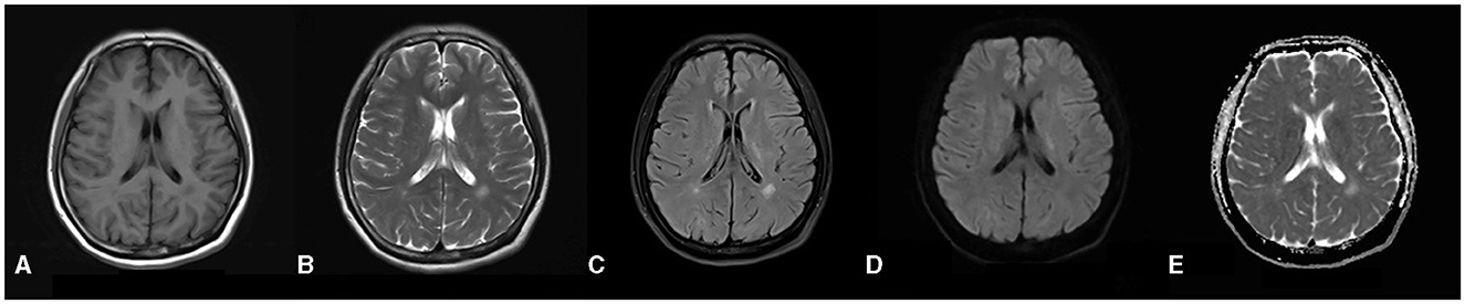
Figure 1. (A–E) Brain MRI reveals a distribution of abnormal signals localized around the ventricles, as well as within the frontal and temporal lobes. In the T1-weighted images, there is a depiction of slightly hypo-intense signals (A), while the T2-weighted images illustrate hyper-intense signals (B). The dark-field images similarly exhibit hyper-intense signals (C). Furthermore, both diffusion-weighted images and apparent diffusion coefficient (5) images do not indicate any restrictions in diffusion (D, E).
The detection of hereditary disease genes revealed mutations, namely c.4044C > G and c.1186-2A > G, in POLR3A (Figure 2). According to the American College of Medical Genetics and Genomics (ACMG) guidelines, the genetic variant c.4044C > G is classified as PM2; however, its clinical significance remains unclear. The genetic variant c.1186-2A > G, classified as PVS1 and PM2, has the potential to influence RNA splicing, subsequently impacting protein function and causing disease. Respecting the patient's opinion, we did not perform genetic testing for her parents or son. Based on the results of physical examination, laboratory tests, imaging tests and gene reports, her condition was diagnosed as POLR3A-related leukodystrophy (HLD7).
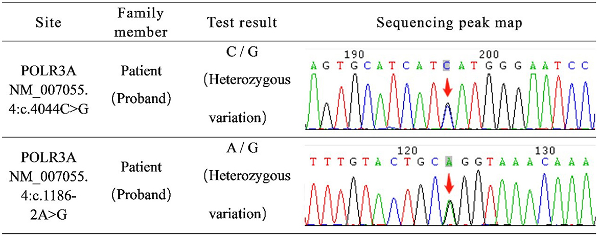
Figure 2. Genetic disease gene detection report: c.4044C > G and c.1186-2A > G of POLR3A gene were mutated.
We administered oral vitamin B12 and injectable acetylglutamine to nourish the nerves. No other drugs were used during the hospital stay. However, the symptoms did not show significant improvement during the 1 month of follow-up and appeared aggravated. The patient required assistance to walk and was unable to hold objects in her hands. After discharge, the patient was treated with vitamin B12, vitamin E, coenzyme Q, and traditional Chinese medicine, with Ganoderma lucidum, ginseng, and Poria Cocos as the main ingredients. As of recent follow-up (1 year after she was discharged), she is able to walk slowly alone and climb stairs, and the degree of head and hand tremors has reduced significantly. She is also able to hold objects and cook independently. However, her speech is slower and more indistinct than before.
3. Discussion
Leukodystrophies are a group of rare and refractory genetic diseases characterized by white matter abnormalities in the CNS, with a reported incidence of 1 in 7,500 live births (6). Onset may occur at any age, from prenatal life to senescence. Studies have shown that autosomal recessive mutations in POLR3A account for 3% of the unclassified autosomal recessive inherited and sporadic cases (7); therefore, early diagnosis and classification are essential. The clinical features of leukodystrophy caused by POLR3A mutations include neurological, dental, ophthalmic, and endocrine changes, with at least one of these criteria noted in each patient. However, the endocrine symptoms and developmental abnormalities associated with this disease have not been thoroughly studied (3). Neurological signs and symptoms include cognitive impairment and cerebellar symptoms such as ataxia and nystagmus. More than 80% of patients have cerebellar atrophy, while only a few have extrapyramidal signs such as dystonia and tremors (8). At the time of onset, the only manifestation may be simple cognitive impairment, as motor symptoms may not be present. This disease often exhibits non-neural manifestations, including dental problems such as tooth loss, endocrine problems such as hypogonadism reduction, and ophthalmic problems such as optic atrophy and myopia (9). Brain MRI often shows diffuse hypomyelination in the whole-brain white matter, which can accumulate in the internal capsule, external capsule, deep white matter, and corpus callosum. Subcortical U fibers are often not involved, and cerebellar atrophy and superior cerebellar peduncle hyperintensity are occasionally seen (10). In studies published in the last 5 years (8, 11–26), we collected data from cases of myelin dystrophic leukodystrophy caused by POLR3A mutation and summarized the relevant characteristics in Table 1.
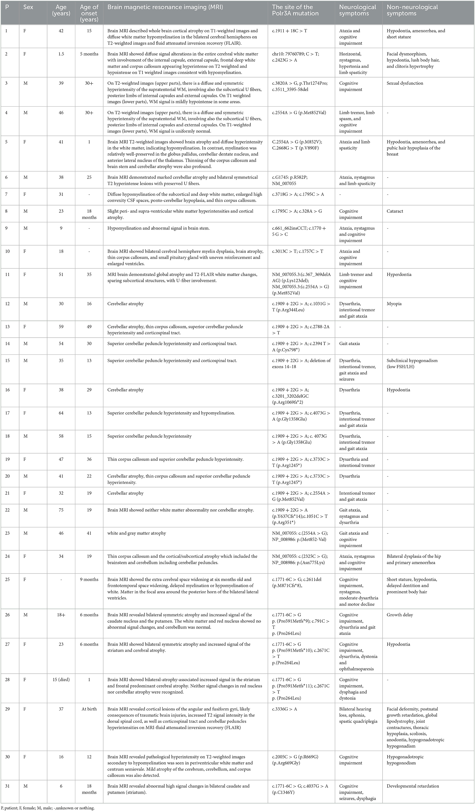
Table 1. The table shows the cases of leukodystrophy caused by POLR3A mutation published in recent years, and summarizes the relevant characteristics.
The two pathogenic genes that encode the largest subunit of human RNA polymerase III (Pol III) are POLR3A and POLR3B. Mutations in these two genes can result in four overlapping phenotypes of hypomyelinating leukodystrophy, which can be divided into the following syndromes according to clinical features: hypomyelination, hypodontia, and hypogonadotropic hypogonadism (4H syndrome); ataxia, delayed dentition, and hypomyelination (ADDH); tremor-ataxia with central hypomyelination (TACH); leukodystrophy with oligodontia (LO); and hypomyelination with cerebellar atrophy and hypoplasia of the corpus callosum (HCAHC) (27). It has been shown that in almost all patients with POLR3A and POLR3B related leukodystrophy, myelination of the optic radiation is always accompanied by hypomyelination and that the ventrolateral thalamus and dentate nucleus have relatively low signal intensity on T2. Supratentorial atrophy is common in adult patients and rare in children younger than 10 years (28). Reports also state that different genes cause some differences in brain MRI. Patients with POLR3B gene mutations have smaller cerebellums, larger fissures, thinner cortices, and decreased basal white matter, suggesting cerebellar atrophy. MR images of patients with POLR3A mutations show significantly lighter changes in the cerebellar hemispheres and cerebellar vermis, indicating only a slight myelin sheath reduction in the centrum semiovale and cerebellar white matter (29). The symptoms and prognoses of the two patients were also different. Patients with POLR3A mutations progress faster and have a shorter life expectancy than those with POLR3B mutations. It is worth noting that in patients with the POLR3A mutation, the disease onset is slightly later than in patients with other mutations. Compared with patients with POLR3B mutations, most patients with POLR3A mutations achieve independent walking (28).
In this case, the patient had the non-neurological symptoms of tooth loss. Based on the results of genetic testing, the patient was diagnosed with leukodystrophy type 7 and further classified as having LO syndrome based on the MRI report and the presence of cerebellar ataxia and nystagmus. For this patient, we used aceglutamide which is the acetyl compound of glutamine, which passes the blood-cerebrospinal fluid barrier and decomposes into glutamic acid and gamma-aminobutyric acid (GABA). Glutamate is involved in information transmission in the central nervous system. Gamma-aminobutyric acid can antagonize the excitability of glutamate, improve nerve cell metabolism, reduce oxygen consumption and reduce the effect of blood ammonia, and improve brain function (30). However, this drug was not effective in this patient. To date, no effective treatment has been found for this disease; however, symptomatic treatment can still be useful in improving muscle tone disorders and swallowing dysfunction, thereby further improving the quality of life of patients. For the treatment of dystonia, the first-line drugs we can use are anticholinergic drugs, such as trihexyphenidyl; the second-line drugs are reversible dopamine depleting agents, such as tetrabenazine; other drugs such as baclofen, benzodiazepines, levodopa, botulinum toxin, etc. For refractory patients, we can try deep brain stimulation (31). It has been reported that a patient with POLR3A associated leukodystrophy and Parkinson's disease had complete remission of bradykinesia and left leg tremor after deep brain stimulation. However, there is no systematic data to confirm the effectiveness of deep brain stimulation in patients with POLR3A associated leukodystrophy (18). Notably, ibuprofen may be used for the treatment of HLD 7; however, its efficacy and mechanism require further exploration (4).
4. Conclusion
As leukodystrophy caused by the POLR3A mutation is relatively rare, reports in this area are minimal. Although MRI features are helpful for diagnosis in patients lacking basic non-neurological features, the disease remains difficult to diagnose because it is not well understood. Therefore, we report and analyse a case of leukodystrophy caused by a missense mutation in POLR3A. Leukodystrophy caused by the POLR3A mutation often has a poor prognosis, affecting the life span of patients; thus far, there is no cure for the disease except for some symptomatic treatments, such as reducing muscle tension. We still need to further explore and study new treatment methods to improve the quality of life of patients while increasing their life expectancy, which is an important work in the future.
Data availability statement
The original contributions presented in the study are included in the article/supplementary material, further inquiries can be directed to the corresponding author.
Ethics statement
The studies involving humans were approved by Clinical Research Management Committee of the First Hospital of Jilin University. The studies were conducted in accordance with the local legislation and institutional requirements. The authors declare that all the medical records in this study are from previous diagnoses and treatments. The subject's privacy and personally identifiable information are protected. Written informed consent was obtained from the individual for the publication of any potentially identifiable images or data included in this article.
Author contributions
LS: Investigation, Writing–original draft. SH: Funding acquisition, Writing—review & editing. WL: Conceptualization, Writing—review & editing. HM: Writing—review & editing. WZ: Writing—review & editing.
Funding
The author(s) declare financial support was received for the research, authorship, and/or publication of this article. The study was supported by grants by the National Natural Science Foundation of China (No. 81801145).
Conflict of interest
The authors declare that the research was conducted in the absence of any commercial or financial relationships that could be construed as a potential conflict of interest.
Publisher's note
All claims expressed in this article are solely those of the authors and do not necessarily represent those of their affiliated organizations, or those of the publisher, the editors and the reviewers. Any product that may be evaluated in this article, or claim that may be made by its manufacturer, is not guaranteed or endorsed by the publisher.
References
1. Kohler W, Curiel J, Vanderver A. Adulthood leukodystrophies. Nat Rev Neurol. (2018) 14:94–105. doi: 10.1038/nrneurol.2017.175
2. van der Knaap MS, Bugiani M. Leukodystrophies: a proposed classification system based on pathological changes and pathogenetic mechanisms. Acta Neuropathol. (2017) 134:351–82. doi: 10.1007/s00401-017-1739-1
3. Pelletier F, Perrier S, Cayami FK, Mirchi A, Saikali S, Tran LT, et al. Endocrine and growth abnormalities in 4H leukodystrophy caused by variants in POLR3A, POLR3B, and POLR1C. J Clin Endocrinol Metab. (2021) 106:e660–74. doi: 10.1210/clinem/dgaa700
4. Sawaguchi S, Tago K, Oizumi H, Ohbuchi K, Yamamoto M, Mizoguchi K, et al. Hypomyelinating Leukodystrophy 7 (HLD7)-associated mutation of POLR3A is related to defective oligodendroglial cell differentiation, which is ameliorated by ibuprofen. Neurol Int. (2021) 14:11–33. doi: 10.3390/neurolint14010002
5. Pio Jude T, Panchadcharam E, Masilamani K. Computing zagreb and randic indices of PEG-cored dendrimers used for drug and gene delivery. J Chemist. (2020). doi: 10.1155/2020/2821510
6. Bonkowsky JL, Nelson C, Kingston JL, Filloux FM, Mundorff MB, Srivastava R. The burden of inherited leukodystrophies in children. Neurology. (2010) 75:718–25. doi: 10.1212/WNL.0b013e3181eee46b
7. Minnerop M, Kurzwelly D, Wagner H, Soehn AS, Reichbauer J, Tao F, et al. Hypomorphic mutations in POLR3A are a frequent cause of sporadic and recessive spastic ataxia. Brain. (2017) 140:1561–78. doi: 10.1093/brain/awx095
8. Campopiano R, Ferese R, Zampatti S, Giardina E, Biagioni F, Colonnese C, et al. A novel POLR3A genotype leads to leukodystrophy type-7 in two siblings with unusually late age of onset. BMC Neurol. (2020) 20:258. doi: 10.1186/s12883-020-01835-9
9. Choquet K, Forget D, Meloche E, Dicaire M-J, Bernard G, Vanderver A, et al. Leukodystrophy-associated mutations down-regulate the RNA polymerase III transcript and important regulatory RNA. J Biol Chem. (2019) 294:7445–59. doi: 10.1074/jbc.RA118.006271
10. Cayami FK, Bugiani M, Pouwels PJW, Bernard G, van der Knaap MS, Wolf NI. 4H leukodystrophy: lessons from 3T imaging. Neuropediatrics. (2018) 49:112–7. doi: 10.1055/s-0037-1608780
11. Tewari VV, Mehta R, Sreedhar CM, Tewari K, Mohammad A, Gupta N, et al. A novel homozygous mutation in POLR3A gene causing 4H syndrome: a case report. BMC Pediatr. (2018) 18:126. doi: 10.1186/s12887-018-1108-9
12. Wu S, Bai Z, Dong X, Yang D, Chen H, Hua J, et al. Novel mutations of the POLR3A gene caused POLR3-related leukodystrophy in a Chinese family: a case report. BMC Pediatr. (2019) 19:289. doi: 10.1186/s12887-019-1656-7
13. Yang YM, Zhao ZM, Jia YL, Jia YJ, Han N, Wang JH. A 42-year-old woman with 4H leukodystrophy caused by a homozygous mutation in POLR3A gene. Chin Med J (Engl). (2019) 132:1879–80. doi: 10.1097/CM9.0000000000000328
14. Hiraide T, Kubota K, Kono Y, Watanabe S, Matsubayashi T, Nakashima M, et al. POLR3A variants in striatal involvement without diffuse hypomyelination. Brain Dev. (2020) 42:363–8. doi: 10.1016/j.braindev.2019.12.012
15. Uygun Ö, Gündüz T, Eraksoy M, Kürtüncü M. Adult-onset 4H leukodystrophy: a case presentation and review of the literature. Acta Neurol Belg. (2020) 120:1461–2. doi: 10.1007/s13760-020-01297-3
16. Di Bella D, Magri S, Benzoni C, Farina L, Maccagnano C, Sarto E, et al. Hypomyelinating leukodystrophies in adults: clinical and genetic features. Eur J Neurol. (2021) 28:934–44. doi: 10.1111/ene.14646
17. Furukawa S, Kunii M, Doi H, Kondo N, Ogura A, Hirabuki K, et al. Case report: severe osteoporosis and preventive therapy in RNA polymerase III-related leukodystrophy. Front Neurol. (2021) 12:622355. doi: 10.3389/fneur.2021.622355
18. Kyle K, Mason X, Bordelon Y, Pouratian N, Bronstein J. Adult onset POLR3A leukodystrophy presenting with parkinsonism treated with pallidal deep brain stimulation. Parkinsonism Relat Disord. (2021) 85:23–5. doi: 10.1016/j.parkreldis.2021.02.018
19. Yan H, Ji H, Kubisiak T, Wu Y, Xiao J, Gu Q, et al. Genetic analysis of 20 patients with hypomyelinating leukodystrophy by trio-based whole-exome sequencing. J Hum Genet. (2021) 66:761–8. doi: 10.1038/s10038-020-00896-5
20. Di Donato I, Gallo A, Ricca I, Fini N, Silvestri G, Gurrieri F, et al. POLR3A variants in hereditary spastic paraparesis and ataxia: clinical, genetic, and neuroradiological findings in a cohort of Italian patients. Neurol Sci. (2022) 43:1071–7. doi: 10.1007/s10072-021-05462-1
21. Lessel D, Rading K, Campbell SE, Thiele H, Altmüller J, Gordon LB, et al. A novel homozygous synonymous variant further expands the phenotypic spectrum of POLR3A-related pathologies. Am J Med Genet A. (2022) 188:216–23. doi: 10.1002/ajmg.a.62525
22. Musumeci A, Calì F, Scuderi C, Vinci M, Vitello GA, Musumeci SA, et al. Identification of a novel missense mutation of POLR3A gene in a cohort of sicilian patients with leukodystrophy. Biomedicines. (2022) 10:2276. doi: 10.3390/biomedicines10092276
23. Nikkhah A, Rezakhani S. Developmental regression and movement disorder as a phenotypic variant of POLR3A mutation-case report. Clin Case Rep. (2022) 10:e6556. doi: 10.1002/ccr3.6556
24. Sytsma TM, Chen DH, Rolf B, Dorschner M, Jayadev S, Keene CD, et al. Spinal cord-predominant neuropathology in an adult-onset case of POLR3A-related spastic ataxia. Neuropathology. (2022) 42:58–65. doi: 10.1111/neup.12775
25. Buyukyilmaz G, Erozan Cavdarli B, Toksoy Adiguzel K, Adiguzel M, Kasapkara CS, Gurbuz F, et al. The first case of 4H syndrome with type 1 diabetes mellitus. J Clin Res Pediatr Endocrinol. (2023). doi: 10.4274/jcrpe.galenos.2023.2023-1-15
26. Yang H, Wu Z, Li X, Huang Y, Li J, He F, et al. A novel variant of the POLR3A gene in a Chinese patient with POLR3-related leukodystrophy. Neurol Sci. (2023) 44:3363–8. doi: 10.1007/s10072-023-06767-z
27. Thomas A, Thomas AK. POLR3-related leukodystrophy. J Clin Imaging Sci. (2019) 9:45. doi: 10.25259/JCIS_116_2019
28. Wolf NI, Vanderver A, van Spaendonk RML, Schiffmann R, Brais B, Bugiani M, et al. Clinical spectrum of 4H leukodystrophy caused by POLR3A and POLR3B mutations. Neurology. (2014) 83:1898–905. doi: 10.1212/WNL.0000000000001002
29. Vrij-van den Bos S, Hol JA, La Piana R, Harting I, Vanderver A, Barkhof F, et al. 4H leukodystrophy: a brain magnetic resonance imaging scoring system. Neuropediatrics. (2017) 48:152–60. doi: 10.1055/s-0037-1599141
30. Zhang R, Yang N, Ji C, Zheng J, Liang Z, Hou CY, et al. Neuroprotective effects of Aceglutamide on motor function in a rat model of cerebral ischemia and reperfusion. Restor Neurol Neurosci. (2015) 33:741–59. doi: 10.3233/RNN-150509
Keywords: POLR3A, hypomyelinating leukodystrophy-7, 4H syndrome, diagnosis, demyelinating diseases
Citation: Sun L, Lin W, Meng H, Zhang W and Hou S (2023) A Chinese patient with POLR3A-related leukodystrophy: a case report and literature review. Front. Neurol. 14:1269237. doi: 10.3389/fneur.2023.1269237
Received: 29 July 2023; Accepted: 10 October 2023;
Published: 27 October 2023.
Edited by:
Bruce Miller, University of California, San Francisco, United StatesReviewed by:
Xenos Mason, University of Southern California, United StatesHaojun Yang, Central South University, China
Copyright © 2023 Sun, Lin, Meng, Zhang and Hou. This is an open-access article distributed under the terms of the Creative Commons Attribution License (CC BY). The use, distribution or reproduction in other forums is permitted, provided the original author(s) and the copyright owner(s) are credited and that the original publication in this journal is cited, in accordance with accepted academic practice. No use, distribution or reproduction is permitted which does not comply with these terms.
*Correspondence: Shuai Hou, aG91c2h1YWlAamx1LmVkdS5jbg==