- 1Brain Center, Zhejiang Hospital, Hangzhou, China
- 2The Second Clinical Medical College, Zhejiang Chinese Medical University, Hangzhou, China
Background: In-stent restenosis (ISR) is an adverse and notable event in the treatment of intracranial atherosclerotic stenosis (ICAS) with percutaneous transluminal angioplasty and stenting (PTAS). The incidence and contributing factors have not been fully defined. This study was performed to evaluate factors associated with ISR after PTAS.
Data source: We identified studies on ISR after PTAS from an electronic search of articles in PubMed, Ovid MEDLINE, and the Cochrane Central Database (dated up to July 2022).
Results: A total of 19 studies, including 452 cases of ISR after 2,047 PTAS, were included in the meta-analysis. The pooled incidence rate of in-stent restenosis was 22.08%. ISR was more likely to occur in patients with coronary artery disease (OR = 1.686; 95% CI: 1.242–2.288; p = 0.0008), dissection (OR = 6.293; 95% CI: 3.883–10.197; p < 0.0001), and higher residual stenosis (WMD = 3.227; 95% CI: 0.142–6.311; p = 0.0404). Patients treated with Wingspan stents had a significantly higher ISR rate than those treated with Enterprise stents (29.78% vs. 14.83%; p < 0.0001).
Conclusions: The present study provides the current estimates of the robust effects of some risk factors for in-stent restenosis in intracranial atherosclerotic stenosis. The Enterprise stent had advantages compared with the Wingspan stent for ISR. The significant risk factors for ISR were coronary artery disease, dissection, and high residual stenosis. Local anesthesia was a suspected factor associated with ISR.
Introduction
Intracranial atherosclerotic stenosis (ICAS) leads to a dramatic decline in cerebral perfusion and is the main cause of approximately 8%-10% of all ischemic strokes (1, 2). Current treatments for ICAS include medical and endovascular therapies, but rarely surgical therapy. Percutaneous transluminal angioplasty and stenting (PTAS) is considered a minimally invasive approach to reduce stroke recurrence in patients with symptomatic ICAS and has shown potential efficacy and acceptable periprocedural morbidity in initial studies (3–6). Stents commonly used in PTAS include self-expanding stents (SES) and balloon-expandable stents, each with its own advantages and disadvantages. Balloon-expandable stents have relatively rapid one-step exchange systems that do not need more complex exchange length guidewires than self-expanding stents (7, 8). In addition, with balloon-mounted stents (BMS), the lesion does not need to be navigated more than once, which may reduce the risk of embolic stroke and hemorrhagic complications (9–11). In-stent restenosis (ISR) is an adverse and notable event in PTAS, especially with balloon-mounted bare-metal stents, and has been shown to be reduced with drug-eluting stents (12, 13). It is significantly associated with long-term stroke recurrence in stent-treated patients. The incidence of ISR varies from 5% to 30% in present studies, and systematic research on risk factors for ISR is still lacking (14–17). To investigate the risk factors related to in-stent restenosis, we performed this meta-analysis.
Materials and methods
Search strategy
This study searched the following electronic databases for potentially relevant studies published up to July 2022: PubMed, Ovid MEDLINE, and the Cochrane Central Database. The keywords and medical subject headings (MeSH) used in the searches were “Arterial Disease, Intracranial” OR “Intracranial Arterial Disease” OR “Intracranial Arterial Disorders” OR “Arterial Disorder, Intracranial” OR “Arterial Disorders, Intracranial” OR “Intracranial Arterial Disorder” OR “Arterial Diseases, Intracranial” OR “Brain Diseases, Arterial” OR “Arterial Brain Disease” OR “Arterial Diseases, Brain” OR “Arterial Disease, Brain” OR “Brain Arterial Disease” OR “Brain Arterial Diseases” OR “Brain Disorders, Arterial” OR “Arterial Brain Disorder” OR “Arterial Brain Disorders” OR “Brain Disorder, Arterial” OR “Arterial Brain Diseases” AND “risk factor” OR “risk factors” AND “restenosis”. These words were combined using the Boolean operators OR and. The articles were limited to English as the only language of publication. In addition, the references listed in the identified articles were manually read to identify any additional eligible articles; a research assistant obtained and reviewed all potentially relevant articles.
Two authors independently analyzed the titles and abstracts of the identified articles. Inclusion criteria were as follows: (1) Full-length, peer-reviewed publications on stents for symptomatic ICAS in which the onset of restenosis was related to specific variables such as patient characteristics, stent technique, and other factors; (2) Cohort studies and single-arm studies were defined based on the study protocol; (3) Adequate data were presented to enable the computation of odds ratios (ORs) or weighted mean differences (WMDs) with 95% confidence intervals (CIs); (4) The median follow-up was at least 6 months; and (5) There were at least 10 patients per treatment group. Studies with any of the following characteristics were excluded: (1) Commentaries, reviews, protocols, letters, editorials, animal studies, or case reports; (2) Studies investigating the treatment strategy for complex cerebral artery stenosis; (3) Studies with imaging evaluation or treatment of ISR; and (4) Patients were treated without stent deployment. Disagreements in the evaluation of study inclusion were resolved by consensus between the two authors.
Data extraction and quality assessment
Data were extracted from all eligible studies by the two authors with a structured data extraction form. The following characteristics were extracted from each study: name of the first author, year of publication, country, risk factors for ISR, number of patients in the ISR and control groups, and the number of patients with each potential risk factor for ISR. Any disagreement was resolved by discussion, and consensus was reached on all data. The definition of ISR was an angiographically verified >50% stenosis after stent deployment. The quality of the included cohort studies was assessed in conformity with the Newcastle–Ottawa Scale (NOS) (18), which is recommended by the Cochrane Collaboration as a bias assessment tool in observational studies. Single-arm studies were assessed using the Methodological Index for Nonrandomized Studies (MINORS) (19). Studies with a MINORS score of >10 or a NOS score of >5 were considered high-quality studies.
Meta-analyses
This study complied with the PRISMA (Preferred Reporting Items for Systematic Reviews and Meta-Analyses) guidelines. Q-test statistics were used to qualitatively test for heterogeneity between studies, with significance set at p < 0.10 (20), and then tested by I2 statistics, with I2 >50% regarded as quantitative inconsistency. In the case of significant heterogeneity (p < 0.10 or I2 >50%), a random effects model was used to calculate pooled ORs or WMDs; otherwise, a fixed model was utilized (21). A forest plot was used to graphically summarize the meta-analysis of significant risk factors. All analyses were performed with the “meta” and “metafor” packages of the R statistical and computing software, version 4.1.2 (http://www.r-project.org/).
The possibility of publication bias was assessed by the Egger test and by framing a funnel plot of the effect size of each study relative to the standard error (Supplementary Figures 2, 3). A sensitivity analysis was performed to investigate the potential sources of heterogeneity (Supplementary Figure 4). Data on comparable factors, such as BMS and SES or local and general anesthesia, were extracted from studies with comparable results for pooled analysis; otherwise, subgroup analyses were performed. The following factors were analyzed with subgroup analyses: study type of the included studies, anesthesia type, dual antiplatelet time, and stent.
Results
Literature search and basic features of the included studies
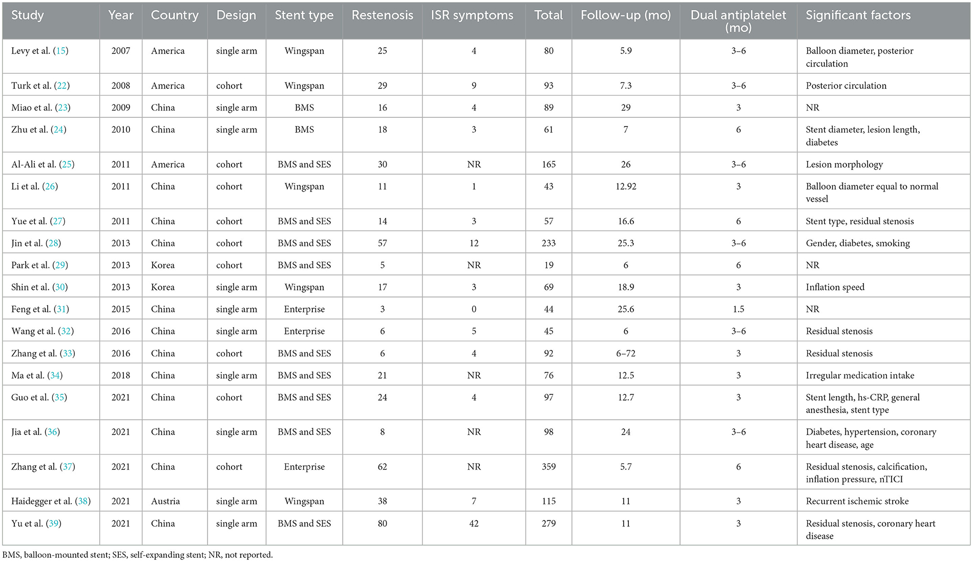
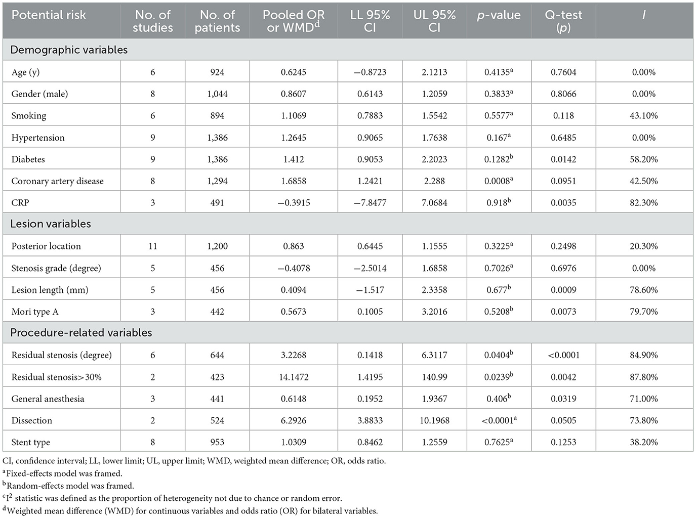
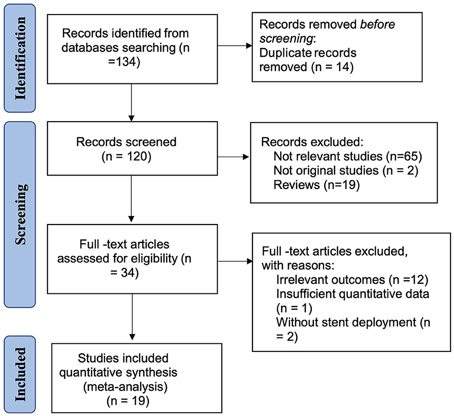
A total of 136 references were initially evaluated; 19 studies were confirmed as eligible. These consisted of 9 cohort studies and 10 single arm studies, which included 452 cases of ISR after 2,047 PTAS, giving a cumulative incidence of ISR after PTAS of 22.08% (Supplementary Figure 1). In 17 studies, in-stent restenosis was defined as >50% stenosis at the time of angiographic follow-up, in one study as >70% stenosis, and as >20% increase in stenosis comparing to the residual post-procedural stenosis in the last study. The median follow-up for these studies was 12.6 months. All subjects were treated with aspirin and clopidogrel before the procedure and stayed on dual antiplatelet therapy for at least 3 months, or 6 months if necessary. The basic features of the included studies and participants are summarized in Table 1. The analysis of 17 potential risk factors for ISR was extracted from the included studies and is presented in Table 2. The PRISMA flow diagram of this analysis is presented in Figure 1.

Table 1. The detailed information on the basic characteristics of the 23 included studies and participants.
Methodological quality assessment
The outcome of the quality assessment of the included studies was as follows: within the single arm studies, four studies received a score of 14, four studies received a score of 12, and two studies received a score of 10; within the cohort studies, five studies received a score of 8, three studies received a score of 7, one study received a score of 6, and one study received a score of 5. Detailed information on the quality assessment is shown in Supplementary Tables 1, 2.
Pooled analyses of risk factors
A meta-analysis of individual relative results indicated that various risk factors were associated with the development of ISR after PTAS (Table 2). The comprehensive OR ranged from 0.567 to 14.147. Significant heterogeneity between studies was observed for diabetes, lesion length, Mori type, residual stenosis > 30%, and CRP.
Regarding procedure-related variables, ISR was more likely to occur in patients with coronary artery disease (OR = 1.686; 95% CI: 1.242–2.288; p = 0.0008), dissection (OR = 6.293; 95% CI: 3.883–10.197; p < 0.0001), residual stenosis > 30% (OR = 14.147; 95% CI: 1.419–140.99; p = 0.0239) and higher residual stenosis (WMD = 3.227; 95% CI: 0.142–6.311; p = 0.0404) (Supplementary Figures 5–8).
Overall, ISR was not associated with gender, age, smoking, hypertension, diabetes, posterior location, degree of stenosis, lesion length, or stent type, which was divided into the self-expanding stent and the balloon-mounted stent (all p >0.05). All analysis outcomes are shown in Table 2.
Subgroup analyses and heterogeneity
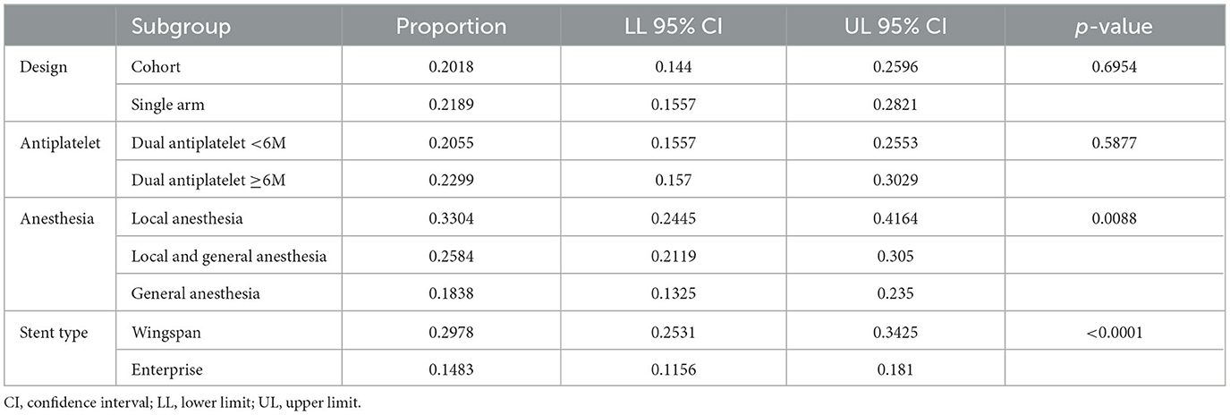
Subgroup analyses were performed to further investigate the risk factors for ISR. Different choices of anesthesia resulted in significantly different in-stent restenosis rates, with local anesthesia having the highest rate of ISR (18.4% vs. 25.8% vs. 33.0%; p = 0.0088) (Table 3). Patients treated with Wingspan stents were more prone to ISR than those treated with Enterprise stents (29.78% vs. 14.83%; p < 0.0001) (Supplementary Figures 9, 10). Study type and DAPT duration were not found to be associated with ISR (Table 3).
Significant heterogeneity between effect estimates was found for the following variables: diabetes, CRP, lesion length, Mori type A, residual stenosis, residual stenosis >30%, general anesthesia, and dissection. Mild heterogeneity between effect estimates was observed for age, gender, hypertension, smoking, coronary artery disease, posterior location, degree of stenosis, and stent type.
Discussion
ISR is an important post-procedural complication of PTAS. The present meta-analysis revealed that 22.18% of symptomatic ICAS patients may suffer from ISR after stent deployment. Risk factors for the development of ISR were identified as the Wingspan stent, coronary artery disease, dissection, and high residual stenosis. In addition, patients who received local anesthesia were more likely to develop ISR. ISR was not associated with gender, age, smoking, other morbidities, or other lesion characteristics.
Patients who were implanted with a Wingspan stent were more likely to develop in-stent restenosis than those treated with Enterprise. The ISR rate for the Enterprise procedure was found to be 14.83% compared to 29.78% for Wingspan. The Enterprise stent is a self-expanding, closed-cell stent that was originally designed for coiling assistance of wide neck intracranial aneurysms (40). This stent has been shown to perform better than the Wingspan stent in complex intracranial atherosclerotic stenoses due to its high flexibility, special carrier system structure, decreased radial force, and capability to reduce the risk of damage to the arteries and prevent elastic recoil and in-stent restenosis (31, 41). In addition, Zsolt et al. reported satisfactory ISR rates with the Enterprise stent (24.7% restenosis at 6 months follow-up) compared to the Wingspan stent (42). On the other hand, Xu et al. found that the ISR rate was significantly higher in the balloon-mounted stent group than in the Wingspan stent group (35), which was not supported by this meta-analysis. The present study revealed that patients treated with BMS had a similar ISR rate than those implanted with Wingspan. New neuro-interventional devices have been developed in recent years. For example, the drug-eluting stent was shown to have an ISR rate of 9.5% in a recent clinical trial (43), which is significantly lower than that of the present stent. Therefore, endovascular treatment of intracranial atherosclerotic stenosis will become increasingly accurate and effective with technological development.
This study found that ISR was significantly more common in patients with coronary artery disease (CAD). In fact, CAD shares the same pathology as ICAS, which is atherosclerosis of the vascular walls. The instability or rupture of atherosclerotic plaques can lead to both cardiovascular and cerebrovascular events (44, 45). Essentially, atherosclerosis appears to be an inflammatory disease, and inflammation plays an important role in the progression of atherosclerosis (46). The severity of atherosclerosis may partly reflect the level of inflammatory activity in the systemic vessel walls. ICAS patients with CAD may have more severe atherosclerosis in the systemic vessels, and the plaques may be prone to instability and rupture, which means a more vibrant systemic inflammatory response in the vessel walls and a greater opportunity for ISR. CRP is a representative inflammatory biomarker mainly produced by hepatocytes and has been suggested to be a strong predictor of intracranial ISR in two Chinese studies (35, 39). High CRP levels are a predictor of asymmetric progression of stenotic tissue because of the differential distribution of shear stress and the effect on neointimal tissue shape mediated by the inflammatory process (47). However, the association between CRP levels and ISR was not detected in Melanie et al. (38), which may be explained by the relatively old study population, as CRP increases with age and comorbidities (48). Therefore, the value of CRP as a predictive marker for ISR after stenting may be limited.
Dissection was another significant risk factor for ISR. Previous studies evaluated the association between dissection and ISR (25, 37), and concluded that dissection was associated with ISR. The presence of dissection during intervention indicates damage to the endarterium of the lesioned vessel, which may induce intimal hyperplasia and the inflammatory cascade. The exact pathophysiological mechanism requires further investigation. Therefore, a small balloon should be utilized for predilation, and the one utilized for dilatation should be selected with 80% of the diameter of the adjacent normal artery to avoid dissection of the atherosclerotic plaque (37).
High residual stenosis is also one of the risk factors for ISR. Yue et al. (49) and Zhang et al. (37) revealed that patients with residual stenosis > 30% were more likely to develop ISR than those with residual stenosis < 30%. In addition, eight other studies defined residual stenosis as a continuous variable, which summarized that patients in the restenosis group had a significantly higher stenosis rate immediately after the procedure. However, there were few detailed illustrations to explain the relationship between residual stenosis and ISR. Yue et al. suggested that higher residual stenosis might induce atherosclerotic plaques to protrude into the remodeled vessel. Some studies found the situation to be theoretically true. In our group, we thought it might be related to hemodynamics. For example, different residual stenosis rates resulted in different blood flow velocities and turbulence on either side of the stenosis.
The subgroup analysis other than the pooled analysis showed that ISR occurred more frequently in patients who underwent surgery with local anesthesia. Xu Guo et al. recently determined that local anesthesia was significantly associated with ISR (35), but Zhu et al. and Ying et al. did not reach this conclusion (24, 39). The management of anesthesia in the endovascular treatment of non-acute stroke patients with ICAS has been little discussed in the recent literature. Local anesthesia is easy to achieve during the surgical procedure, with the advantages of lower cost, less time consumption, and earlier detection of patient deterioration (24); however, it is hard for patients to keep still during the entire procedure. On the other hand, general anesthesia could minimize patient activity during surgery and allow for substantial submaximal inflation to be performed to reduce complications of technical surgery, such as iatrogenic perforation or dissection.
This study showed no association between ISR and age. Turk et al. (22) reported that ISR was more common in younger patients, with a cutoff age of 55 years. The authors hypothesized that the lesions in younger patients displayed more inflammatory arteriopathy than those with primary atherosclerosis. This study identified age as a continuous variable to be analyzed, and a negative correlation was found between the ISR and age. Different types of stents were also found not to be associated with ISR. Further studies are needed to identify whether younger patients or lesions with self-expanding stents have higher restenosis rates and physiopathological mechanisms.
The present study had several limitations that need to be discussed. First, only study-level data rather than raw data were extracted from the published literature, and the sample size in 80% of the series was < 100 patients. The target population of the studies varied with the inclusion criteria, resulting in limited generalization of population features such as distribution of lesion location, preprocedural stenosis grade, and proportion of stent type. Second, all included studies were nonrandomized observational studies, and specific biases were unavoidable. Third, the variables extracted from the studies were limited for the meta-analysis design. The complicated lesion morphology, balloon diameter of 80% of the normal vessel, stent length, inflation speed, irregular medication intake, calcifications, inflation pressure, and ulcerations, which may lead to ISR, were rarely mentioned in previous studies. This study also has its strengths, such as the comprehensive literature search, the careful evaluation of methodological quality, and the assessment of heterogeneity. In this respect, the level of evidence in this study was higher than that of some of the individual studies (50). The present results will give neurointerventionists suggestions on how to prevent ISR after PTAS and highlight the need for further studies on ISR after PTAS.
Conclusions
The present study provides the current estimates of the robust effects of some risk factors for in-stent restenosis in intracranial atherosclerotic stenosis. The Enterprise stent had advantages compared to the Wingspan stent for ISR. The significant risk factors for ISR were coronary artery disease, dissection, and high residual stenosis. Local anesthesia was a suspected factor associated with ISR. Further studies should be conducted on patients undergoing PTAS for different inciting conditions to elucidate the underlying mechanism of ISR.
Data availability statement
The original contributions presented in the study are included in the article/Supplementary material, further inquiries can be directed to the corresponding author.
Author contributions
NW wrote the manuscript. NW and YG performed the statistical analyses. NW, YL, and LF gathered the data and responsible for the integrity of the extracted data. SW, JW, and MW designed and coordinated the study. DL contributed to the analysis and interpretation of the data. All authors contributed to the article and approved the submitted version.
Funding
The work was supported by the Medical Health Science and Technology Key Project of the Zhejiang Provincial Health Commission (WKJ-ZJ-2014) and the Key Research and Development Project of the Zhejiang Provincial Department of Science and Technology (2021C03105).
Conflict of interest
The authors declare that the research was conducted in the absence of any commercial or financial relationships that could be construed as a potential conflict of interest.
Publisher's note
All claims expressed in this article are solely those of the authors and do not necessarily represent those of their affiliated organizations, or those of the publisher, the editors and the reviewers. Any product that may be evaluated in this article, or claim that may be made by its manufacturer, is not guaranteed or endorsed by the publisher.
Supplementary material
The Supplementary Material for this article can be found online at: https://www.frontiersin.org/articles/10.3389/fneur.2023.1170110/full#supplementary-material
Supplementary Figures 1–4. The cumulative incidence of ISR after PTAS along with a funnel plot, Egger test, and sensitivity analysis of the extracted data.
Supplementary Figures 5–8. Possible risk factors for ISR.
Supplementary Figures 9–10. The result of the subgroup analysis.
Supplementary Tables 1, 2. Detailed quality assessment information was provided.
References
1. Sacco RL, Kargman DE, Gu Q, Zamanillo MC. Race ethnicity and determinants of intracranial atherosclerotic cerebral infarction - the northern manhattan stroke study. Stroke. (1995) 26:14–20. doi: 10.1161/01.STR.26.1.14
2. Wityk RJ, Lehman D, Klag M, Coresh J, Ahn H, Litt B. Race and sex differences in the distribution of cerebral atherosclerosis. Stroke. (1996) 27:1974–80. doi: 10.1161/01.STR.27.11.1974
3. Aoki S, Shirouzu I, Sasaki Y, Okubo T, Hayashi N, Machinde T, et al. Enhancement of the intracranial arterial-wall at mr-imaging - relationship to cerebral atherosclerosis. Radiology. (1995) 194:477–81. doi: 10.1148/radiology.194.2.7824729
4. Derdeyn CP, Powers WJ, Grubb RL. Hemodynamic effects of middle cerebral artery stenosis and occlusion. Am J Neuroradiol. (1998) 19:1463–9.
5. Feldmann E, Wilterdink JL, Kosinski A, Lynn M, Chimowitz MI, Sarafin J, et al. The stroke outcomes and neuroimaging of intracranial atherosclerosis (SONIA) trial. Neurology. (2007) 68:2099–106. doi: 10.1212/01.wnl.0000261488.05906.c1
6. Zacharatos H, Hassan AE, Qureshi A. Intravascular ultrasound: principles and cerebrovascular applications. Am J Neuroradiol. (2010) 31:586–97. doi: 10.3174/ajnr.A1810
7. Jiang WJ, Xu XT, Du B, Dong KH, Jin M, Wang QH, et al. Comparison of elective stenting of severe vs moderate intracranial atherosclerotic stenosis. Neurology. (2007) 68:420–6. doi: 10.1212/01.wnl.0000252939.60764.8e
8. Kurre W, Brassel F, Brüning R, Buhk J, Eckert B, Horner S, et al. Complication rates using balloon-expandable and self-expanding stents for the treatment of intracranial atherosclerotic stenoses analysis of the INTRASTENT multicentric registry. Neuroradiology. (2012) 54:43–50. doi: 10.1007/s00234-010-0826-y
9. Chimowitz MI, Lynn MJ, Derdeyn CP, Turan TN, Fiorella D, Lane BF, et al. Stenting vs. aggressive medical therapy for intracranial arterial stenosis. New England Journal of Medicine. (2011) 365:993–1003. doi: 10.1056/NEJMoa1105335
10. Derdeyn CP, Fiorella D, Lynn MJ, Rumboldt Z, Cloft HJ, Gibson D, et al. Mechanisms of stroke after intracranial angioplasty and stenting in the SAMMPRIS trial. Neurosurgery. (2013) 72:777–95. doi: 10.1227/NEU.0b013e318286fdc8
11. Marks MP. Is there a future for endovascular treatment of intracranial atherosclerotic disease after stenting and aggressive medical management for preventing recurrent stroke and intracranial stenosis (SAMMPRIS)? Stroke. (2012) 43:580–4. doi: 10.1161/STROKEAHA.111.645507
12. Gupta R, Al-Ali F, Thomas AJ, Horowitz M, Barrow T, Vora NA, et al. Safety, feasibility, and short-term follow-up of drug-eluting stent placement in the intracranial and extracranial circulation. Stroke. (2006) 37:2562–6. doi: 10.1161/01.STR.0000242481.38262.7b
13. Natarajan SK, Ogilvy CS, Hopkins LN, Siddiqui AH, Levy EI. Initial experience with an everolimus-eluting, second-generation drug-eluting stent for treatment of intracranial atherosclerosis. J Neurointerv Surg. (2010) 2:104–9. doi: 10.1136/jnis.2009.001875
14. Alurkar A, Karanam LS, Oak S, Nayak S, Sorte S. Role of balloon-expandable stents in intracranial atherosclerotic disease in a series of 182 patients. Stroke. (2013) 44:2000. doi: 10.1161/STROKEAHA.113.001446
15. Levy EI, Turk AS, Albuquerque FC, Niemann DB, Aagaard-Kienitz B, Pride L, et al. Wingspan in-stent restenosis and thrombosis: incidence, clinical presentation, and management. Neurosurgery. (2007). 61:644–50. doi: 10.1227/01.NEU.0000290914.24976.83
16. Li J, Zhao Z-W, Gao G-D, Deng J-P, Yu J, Gao L, et al. Wingspan stent for high-grade symptomatic vertebrobasilar artery atherosclerotic stenosis. Cardiovasc Intervent Radiol. (2012) 35:268–78. doi: 10.1007/s00270-011-0163-5
17. Zhang L, Huang Q, Zhang Y, Liu J, Hong B, Xu Y, et al. Wingspan stents for the treatment of symptomatic atherosclerotic stenosis in small intracranial vessels: safety and efficacy evaluation. Am J Neuroradiol. (2012) 33:343–7. doi: 10.3174/ajnr.A2772
18. Stang A. Critical evaluation of the Newcastle–Ottawa scale for the assessment of the quality of nonrandomized studies in meta-analyses. Eur J Epidemiol. (2010) 25:603–5. doi: 10.1007/s10654-010-9491-z
19. Slim K, Nini E, Forestier D, Kwiatkowski F, Panis Y, Chipponi J. Methodological index for non-randomized studies (MINORS): development and validation of a new instrument. ANZ J Surg. (2003) 73:712–6. doi: 10.1046/j.1445-2197.2003.02748.x
20. Lau J, Ioannidis JP, Schmid CH. Quantitative synthesis in systematic reviews. Ann Intern Med. (1997) 127:820–826. doi: 10.7326/0003-4819-127-9-199711010-00008
21. Wei J, Yang T-B, Luo W, Qin J-B, Kong F-J. Complications following dorsal vs. volar plate fixation of distal radius fracture: a meta-analysis. J Int Med Res. (2013) 41:265–75. doi: 10.1177/0300060513476438
22. Turk AS, Levy EI, Albuquerque FC, Jr GLP, Woo H, Welch BG, et al. Influence of patient age and stenosis location on Wingspan in-stent restenosis. Am J Neuroradiol. (2008) 29:23–7. doi: 10.3174/ajnr.A0869
23. Miao ZR, Feng L, Li S, Zhu F, Ji X, Jiao L, et al. Treatment of symptomatic middle cerebral artery stenosis with balloon-mounted stents: long-term follow-up at a single center. Neurosurgery. (2009) 64:79–84. doi: 10.1227/01.NEU.0000335648.31874.37
24. Zhu SG, Zhang RL, Liu WH, Yin Q, Zhou ZM, Zhu WS, et al. Predictive factors for in-stent restenosis after balloon-mounted stent placement for symptomatic intracranial atherosclerosis. Eur J Vasc Endovasc Surg. (2010) 40:499–506. doi: 10.1016/j.ejvs.2010.05.007
25. Al-Ali F, Cree T, Hall S, Louis S, Major K, Smoker S, et al. Predictors of unfavorable outcome in intracranial angioplasty and stenting in a single-center comparison: results from the borgess medical center-intracranial revascularization registry. AJNR Am J Neuroradiol. (2011) 32:1221–6. doi: 10.3174/ajnr.A2530
26. Li J, Zhao Z-W, Gao G-D, Cheng J-M. Wingspan stenting with modified predilation for symptomatic middle cerebral artery stenosis. Catheter Cardiovasc Interv. (2011) 78:286–93. doi: 10.1002/ccd.22755
27. Yue X, Yin Q, Xi G, Zhu W, Xu G, Zhang R, et al. Comparison of BMSs with SES for symptomatic intracranial disease of the middle cerebral artery stenosis. Cardiovasc Intervent Radiol. (2011) 34:54–60. doi: 10.1007/s00270-010-9885-z
28. Jin M, Fu X, Wei Y, Du B, Xu X-T, Jiang W-J. Higher risk of recurrent ischemic events in patients with intracranial in-stent restenosis. Stroke. (2013) 44:2990–4. doi: 10.1161/STROKEAHA.113.001824
29. Park S, Kim J-H, Kwak JK, Baek HJ, Kim BH, Lee D-G, et al. Intracranial stenting for severe symptomatic stenosis: self-expandable vs. balloon-expandable stents. Interv Neuroradiol. (2013) 19:276–82. doi: 10.1177/159101991301900303
30. Shin YS, Kim BM, Suh SH, Jeon P, Kim DJ, Kim DI, et al. Wingspan stenting for intracranial atherosclerotic stenosis: clinical outcomes and risk factors for in-stent restenosis. Neurosurgery. (2013) 72:596–604. doi: 10.1227/NEU.0b013e3182846e09
31. Feng Z, Duan G, Zhang P, Chen L, Xu Y, Hong B, et al. Enterprise stent for the treatment of symptomatic intracranial atherosclerotic stenosis: an initial experience of 44 patients. BMC Neurol. (2015) 15:187. doi: 10.1186/s12883-015-0443-9
32. Wang X, Wang Z, Wang C, Ji Y, Ding X, Zang Y. Application of the enterprise stent in atherosclerotic intracranial arterial stenosis: a series of 60 cases. Turk Neurosurg. (2016) 26:69–76. doi: 10.5137/1019-5149.JTN.13350-14.1
33. Zhang F, Liu L. Complication of stenting in intracranial arterial stenosis. Arch Iranian Med. (2016) 19:317–22.
34. Ma N, Zhang Y, Shuai J, Jiang C, Zhu Q, Chen K, et al. Stenting for symptomatic intracranial arterial stenosis in China: 1-year outcome of a multicentre registry study. Stroke Vasc Neurol. (2018) 3:176–84. doi: 10.1136/svn-2017-000137
35. Guo X, Ma N, Gao F, Mo D, Luo G, Miao Z. Long-term risk factors for intracranial in-stent restenosis from a multicenter trial of stenting for symptomatic intracranial artery stenosis registry in China. Front Neurol. (2020) 11:601199. doi: 10.3389/fneur.2020.601199
36. Jia Q, Yan S. The short- and long-term efficacy of intravascular stenting in the treatment of intracranial artery stenosis. Am J Transl Res. (2021) 13:7115–23.
37. Zhang K, Li T-X, Wang Z-L, Gao B-L, Gu J-J, Gao H-L, et al. Factors affecting in-stent restenosis after angioplasty with the Enterprise stent for intracranial atherosclerotic diseases. Sci Rep. (2021) 11:10479. doi: 10.1038/s41598-021-89670-x
38. Haidegger M, Kneihsl M, Niederkorn K, Deutschmann H, Mangge H, Vetta C, et al. Blood biomarkers of progressive atherosclerosis and restenosis after stenting of symptomatic intracranial artery stenosis. Sci Rep. (2021) 11:15599. doi: 10.1038/s41598-021-95135-y
39. Yu Y, Yan L, Lou Y, Cui R, Kang K, Jiang L, et al. Multiple predictors of in-stent restenosis after stent implantation in symptomatic intracranial atherosclerotic stenosis. J Neurosurg. (2021) 136:1716–25. doi: 10.3171/2021.6.JNS211201
40. Higashida RT, Halbach VV, Dowd CF, Juravsky L, Meagher S. Initial clinical experience with a new self-expanding nitinol stent for the treatment of intracranial cerebral aneurysms: the Cordis Enterprise stent. AJNR Am J Neuroradiol. (2005) 26:1751–6.
41. Krischek O, Miloslavski E, Fischer S, Srivastava S, Hankes H. A comparison of functional and physical properties of self-expanding intracranial stents [Neuroform3, Wingspan, Solitaire, Leo+, Enterprise]. Minim Invasive Neurosurg. (2011) 54:21–8. doi: 10.1055/s-0031-1271681
42. Vajda Z, Schmid E, Güthe T, Klötzsch C, Lindner A, Niehaus L. The modified Bose method for the endovascular treatment of intracranial atherosclerotic arterial stenoses using the Enterprise stent. Neurosurgery. (2012) 70:91–101. doi: 10.1227/NEU.0b013e31822dff0f
43. Jia B, Zhang X, Ma N, Mo D, Gao F, Sun X, et al. Comparison of drug-eluting stent with bare-metal stent in patients with symptomatic high-grade intracranial atherosclerotic stenosis: a randomized clinical trial. JAMA Neurol. (2022) 79:176–184. doi: 10.1001/jamaneurol.2021.4804
44. Bhatt DL, Steg PG, Ohman EM, Hirsch AT, Ikeda Y, Mas J-L, et al. International prevalence, recognition, and treatment of cardiovascular risk factors in outpatients with atherothrombosis. JAMA. (2006) 295:180–189. doi: 10.1001/jama.295.2.180
45. Falk E. Pathogenesis of atherosclerosis. J Am Coll Cardiol. (2006) 47:C7–12. doi: 10.1016/j.jacc.2005.09.068
46. Ross R. Atherosclerosis–an inflammatory disease. N Engl J Med. (1999) 340:115–26. doi: 10.1056/NEJM199901143400207
47. Wentzel JJ, Krams R, Schuurbiers JC, Oomen JA, Kloet J, van Der Giessen WJ, et al. Relationship between neointimal thickness and shear stress after Wallstent implantation in human coronary arteries. Circulation. (2001) 103:1740–5. doi: 10.1161/01.CIR.103.13.1740
48. Friedman EM, Christ SL, Mroczek DK. Inflammation partially mediates the association of multimorbidity and functional limitations in a national sample of middle-aged and older adults: the MIDUS study. J Aging Health. (2015) 27:843–63. doi: 10.1177/0898264315569453
49. Yue X, Xi G, Lu T, Xu G, Liu W, Zhang R, et al. Influence of residual stenosis on clinical outcome and restenosis after middle cerebral artery stenting. Cardiovasc Intervent Radiol. (2011) 34:744–50. doi: 10.1007/s00270-010-9989-5
Keywords: intracranial atherosclerotic stenosis, percutaneous transluminal angioplasty and stenting, in-stent restenosis, risk factors, meta-analysis
Citation: Wang N, Lu Y, Feng L, Lin D, Gao Y, Wu J, Wang M and Wan S (2023) Identifying risk factors for in-stent restenosis in symptomatic intracranial atherosclerotic stenosis: a systematic review and meta-analysis. Front. Neurol. 14:1170110. doi: 10.3389/fneur.2023.1170110
Received: 20 February 2023; Accepted: 26 June 2023;
Published: 14 July 2023.
Edited by:
Kristian Barlinn, University Hospital Carl Gustav Carus, GermanyReviewed by:
Huaizhang Shi, First Affiliated Hospital of Harbin Medical University, ChinaHongkai Wang, Northwestern University, United States
Copyright © 2023 Wang, Lu, Feng, Lin, Gao, Wu, Wang and Wan. This is an open-access article distributed under the terms of the Creative Commons Attribution License (CC BY). The use, distribution or reproduction in other forums is permitted, provided the original author(s) and the copyright owner(s) are credited and that the original publication in this journal is cited, in accordance with accepted academic practice. No use, distribution or reproduction is permitted which does not comply with these terms.
*Correspondence: Shu Wan, d2Fuc2h1QHpqdS5lZHUuY24=
†These authors have contributed equally to this work and share first authorship
 Ning Wang
 Yuning Lu
 Lei Feng
 Dongdong Lin
 Yuhai Gao
 Jiong Wu
 Ming Wang
 Shu Wan

