
94% of researchers rate our articles as excellent or good
Learn more about the work of our research integrity team to safeguard the quality of each article we publish.
Find out more
ORIGINAL RESEARCH article
Front. Behav. Neurosci. , 04 January 2022
Sec. Emotion Regulation and Processing
Volume 15 - 2021 | https://doi.org/10.3389/fnbeh.2021.783056
This article is part of the Research Topic Resilience to Stress-Related Mood Disorders: Involvement of Oxidative Stress View all 6 articles
 Wen-Jing Cheng1,2†
 Peng Li3†
 Wen-Ya Huang1,4Yang Huang1Wen-Jie Chen1Yi-Ping Chen5Jun-Liang Shen1Jian-Kun Chen1Na-Sha Long1
 Xian-Jun Meng1,2*
Oxidative stress is closely related to the occurrence of depression. Acupuncture has been proved to be an effective method for treating depression. In order to explore the mechanism of the antidepressant effect of acupuncture, this study performed acupuncture prevention on chronic unpredictable mild stress (CUMS) depression model rats, and observed the effect of acupuncture on hippocampal oxidative stress and Nrf2 signaling pathway. Male SD rats were randomly divided into control group, CUMS group, acupuncture group, and fluoxetine group (n = 10/group). Fluoxetine, a common antidepressant, was used as a positive control drug in this study. In the fluoxetine group, rats were given fluoxetine (2.1 mg/kg) intragastrically once a day for 28 days. The acupoints of Shangxing (GV23) and Fengfu (GV16) were applied in acupuncture group, once every other day for 14 times in total. Behavioral tests and biological detections were used to evaluate the effects of the interventions and the changes of factors related to oxidative stress, Nrf2 pathway, and neuronal apoptosis. The results showed that both acupuncture and fluoxetine could increase sugar preference rate in SPT and decrease immobility time in FST in depression model rats. It also significantly decreased oxidative stress products such as ROS and H2O2, and elevated the protein and mRNA expressions of Nrf2 and HO-1. From Nissl’s staining, there were more abundant nerve cells in two intervention groups compared with CUMS group. Plus, acupuncture down-regulated the expression levels of Bax and caspase-3 and up-regulated the expression of Bcl-2. Our findings indicate that acupuncture improved depression-like behaviors of CUMS rats. And CUMS-induced depression-like behaviors in rats were related to oxidative stress and neuronal apoptosis in hippocampus. Acupuncture showed antidepressant effects in reducing oxidative stress products via regulating the Nrf2/HO-1 signaling pathway so that prevented neuronal apoptosis.
Depression is a mental illness associated with depressed mood, loss of interest, and energy fatigue (Zhou et al., 2019). Not only it causes serious damage to the patients but also imposes a huge economic burden on society due to its high morbidity rate and recurrence rate (Jun et al., 2014; Can et al., 2018). However, antidepressants were commonly used in clinic now, such as tricyclic antidepressants and serotonin reuptake inhibitors, which have a series of side effects and even no effect (Li K. et al., 2020). Therefore, the exploration of new treatments for depression is needed.
Studies have reported that the occurrence of depression is closely relevant to oxidative stress (Dantzer et al., 2008; Reiter et al., 2020), which subsequently leads to neuronal apoptosis (Khan et al., 2019). Such responses are, in a way, due to environmental stimuli which then change neuroprogression resulting in the evolution of depression-like behaviors (Berk et al., 2013). Some clinical reports have stated that oxidative stress is one of the main causes of the pathogenesis of depression (Ng et al., 2008), as indicated by elevated levels of oxidative stress markers found in the blood of patients with depression (Lindqvist et al., 2017). In addition, antidepressants are effective in reducing oxidative damage in depressive patients (Ng et al., 2008; Maes et al., 2011). What’s more, antioxidant subjects such as polyphenols exhibited antidepressant activity by modulating oxidative stress state in the brain of depression model animals (Chhillar and Dhingra, 2013).
Nuclear factor erythroid 2-like (Nrf2) is an intracellular transcription regulator, and Heme oxygenase-1 (HO-1) is one of its most important downstream regulatory products. The cascade reaction between the two is critical in the brain antioxidant system. Furthermore, the antioxidant function of the brain is linked to the pathophysiology of depression (Black et al., 2015). In a previous study, carvacrol ameliorated LPS-induced depression and anxiety-like behavior by regulating the neuroprotective properties of endogenous antioxidant protein Nrf2 (Naeem et al., 2021). Hence, Nrf2 might be considered as a potential pharmacological target for studying the mechanism of depression.
Acupuncture, as a traditional Chinese medical therapy, is often used as a complementary and alternative treatment for depression (Smith et al., 2018). Based on mountains of ancient books, we found that in Sun Simiao’s Thousand-Golden-Prescriptions (Bei Ji Qian Jin Yao Fang), thirteen ghost acupoints were often used to treat mental diseases, including depression (Si-yu et al., 2020). Although acupuncture has a certain effect in ameliorating depression, its molecular mechanism is not yet clear. For instance, previous researches found the reduction of the severity of depression in patients treated with acupuncture (Smith et al., 2018). Certainly, acupuncture is considered as an alternative treatment in primary care to significantly reduce depressive symptoms in the short to medium term (MacPherson et al., 2013). In our previous study, acupuncture improved the concentration of 5-HT in the hippocampus of CUMS-induced depressed model rats (Li et al., 2021). The hippocampus, as a key brain region belonging to the limbic system, is responsible for physical functions such as memory formation, cognitive ability, and emotion regulation (Slavich and Irwin, 2014). These observations underscore that it is indispensable to assess the rational utility of acupuncture in depression and to investigate the mechanisms involved. However, it is still unknown whether acupuncture could relieve neural injury via decreasing oxidative stress leading to apoptotic responses correlated with depressive behavior. Herein, we designed this experiment to explore whether acupuncture could suppress oxidative stress and alleviate neuronal apoptosis in the hippocampus of rats. Interestingly, our results suggest acupuncture is a potential preventive antidepressant and emphasize that oxidative stress and neuronal apoptosis are likely to be targets for the study of depression.
Male Sprague-Dawley (SD) rats of specific-pathogen-free (SPF) class weighting 130–150 g, were obtained from Shanghai Slake Laboratory Animal Co., Ltd. [Animal Certificate No. SCXK (Shanghai) 2017-0005]. Animals were kept in a room where the temperature (22 ± 2°C) and humidity (50 ± 10%) were controlled. Before the treatment, rats were adapted to the environment for 1 week. This experiment was conducted in accordance with ARRIVE’s guidelines. In addition, all operations on rats were confirmed with ethical regulations and the ethics committee of animal care of the Xiamen University, Xiamen, China (License No. XMULAC202110062).
The rat model of depression was established by chronic unpredictable mild stress (CUMS) as described previously with a slight modification (Wang et al., 2017). In this study, 40 baseline-screened rats were randomly divided into 4 groups: control group (con, n = 10), CUMS group (cums, n = 10), acupuncture group (acu, n = 10), fluoxetine group (flu, n = 10). In addition to the control group, the other three groups were isolated in individual cages and received different stressors every day during the 28 days. Each stressor could not occur continuously. Our stressors included water deprivation (24 h), food deprivation (24 h), restraint stress (6 h), wet bedding (24 h), smell stimulation, day and night reversal (24 h), strobe stimulation (6 h), and crowding (6 h).
In this experiment, acupuncture and fluoxetine should be performed 1 h before the CUMS protocol. According to the atlas in acupoints of experimental animals, two points were chosen (Fang, 1993; Figure 1): Shangxing (GV23) and Fengfu (GV16). GV23 is located in the middle of coronal line of rat anterior skull. GV16 locates at occipital posterior occipital ridge atlas joint. Both points were inserted as deep as 5–6 mm with sterilizing disposable stainless steel needles (0.25 mm diameter, HanYi, Changchun, China) for 20 min. An experimental rat head cap (no. ZL201922421814.3) was used to calm down the rats and thus ensure needles staying. Rats underwent acupuncture treatment every other day for 4 weeks. As a positive control, fluoxetine (2.1 mg/kg, 0.21 mg/ml, PHR1394-1G, Sigma-Aldrich) was diluted in double-distilled water and administered to rats daily for 4 weeks (Singh et al., 2004).
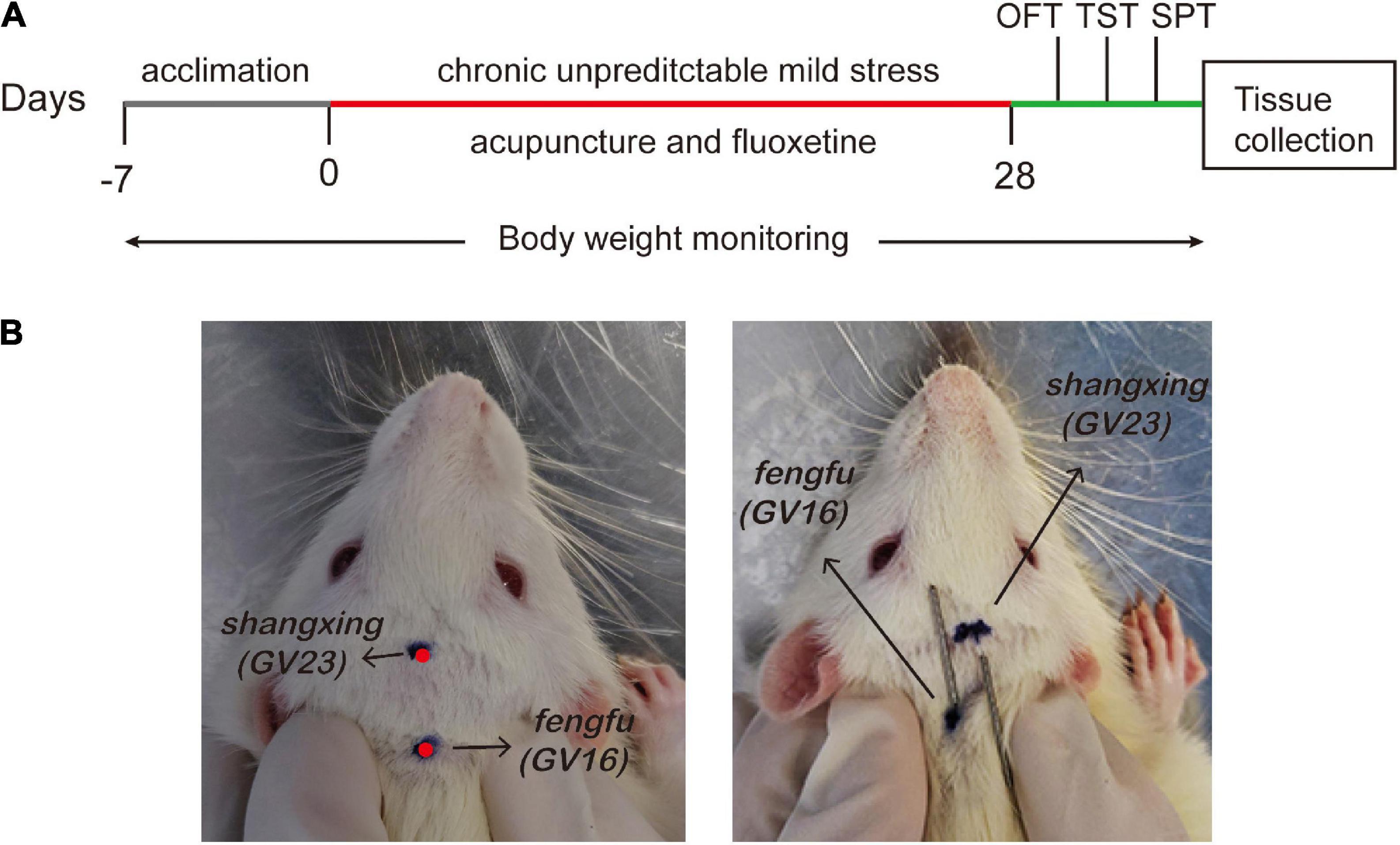
Figure 1. Schematic representation of experimental protocol. (A) Flow chart of experiment. (B) The location of acupoints (Shangxing GV23 and Fengfu GV16) in the rat.
The body weight of each rat was measured at 9 am on day 0, 7, 14, 21, and 28 of the experiment, and the changes in body weight of each group were observed.
According to previously described method (Luo et al., 2020), we performed open field test to determine locomotor activity, exploratory behavior, and depression in rodents. All rats were placed in an apparatus consisting of a dark rectangular case with a square floor (100 cm × 100 cm × 40 cm). The floor of the arena was divided into 4 × 4 equal squares. Then, each rat was placed individually in the center of the open-field apparatus and was allowed to freely explore the zone for 5 min. Finally, the total distance traveled (m) was recorded and analyzed by a behavioral recorder (Smart3.0). At the same time, the crossing numbers (defined as two forelimbs and one hindlimb in a square) were also recorded. After each trial, rat’s feces in the box were cleaned up and the open-field was thoroughly cleaned with 75% alcohol and dried.
Sucrose preference test is used to evaluate anhedonia in rodents, which is a core symptom of depression (Forbes et al., 1996). The test was carried out following the previously described protocols with minor modifications (Yao et al., 2021). Before the formal test, rats were received two bottles of 1% sucrose solution for 24 h. Then change one bottle of sugar water to pure water for 24 h, when the position of the two bottles were exchanged every 12 h. Next, rats subjected to water deprivation and fasting for 12 h. Subsequently, each rat placed in a single cage, respectively, acquired one bottle of 1% sucrose solution and a bottle of pure water, of which weight calculated. After 12 h, the weight of sugar water and pure water were weighed again. Finally, the sucrose preference rate was calculated as follows: sucrose preference rate (%) = sucrose intake/(water intake + sucrose intake) × 100. The test was conducted at the end of the period of 4-week stress.
Forced swimming test is often used to assess depression-like behavior in animals (Porsolt, 1993). After the sucrose preference test, the forced swimming test modified as described earlier was performed (Li S. Y. et al., 2020). All rats were subjected to forced swimming for 6 min in a transparent cylindrical tube (diameter 0.2 m, height 0.45 m, water depth 0.3 m, water temperature 22 ± 2°C) and then recorded its the time of immobility in water via a behavioral recorder (Smart3.0). To avoid the recording error of the instrument, the tap water in the cylinder should be changed before each rat was detected.
Rats in each group were anesthetized with 0.3% pentobarbital (1 ml/100 g). Then hippocampus tissues were taken down, added with PBS homogenate, and centrifuged at 4°C for 20 min (12,000 RPM/min). Next, the supernatant was collected for detection. According to the instructions, the expression levels of ROS, MDA, SOD and GSH-Px in the supernatant were detected by ELISA kit (E-BC-K102, Elabscience). The absorbance was measured at 450 and 550 nm, and the content of each tissue index was calculated according to the standard curve.
The hippocampal tissue was ground and added to the lysate. After centrifugation, the protein concentration was determined by the BCA Protein Assay Kit (QB214754, Thermo Scientific), and then separated on the SDS-PAGE gel, and the separated protein was transferred to the PVDF membrane (K5NA8023B, Amersham). PVDF was immersed in a blocking solution containing 5% skimmed milk powder, incubated for 1 h at room temperature, and incubated with the primary antibody overnight at 4°C (anti-Nrf2, Proteintech, 16396, 1:1,000 dilution; HO-1, Proteintech, 27282, 1:1,000 dilution). The next day, the corresponding secondary antibody was added and incubated at 4°C for 1 h. After washing the membrane, the ECL developed color, exposed and developed in a dark room, and used ImageJ software for statistical analysis.
Extract total RNA from hippocampus with 1 ml Trizol, the determined RNA concentration and purity. The Nrf2 and HO-1 primer sequences (Table 1) were identified from the consensus coding sequence (CCDS) database of the national center for biotechnology information (NCBI). Fluorescence quantitative PCR kit (RN03, Aidley Biotechnology Co., Ltd) and RT-PCR instrument were used to detect the mRNA expressions of each gene.
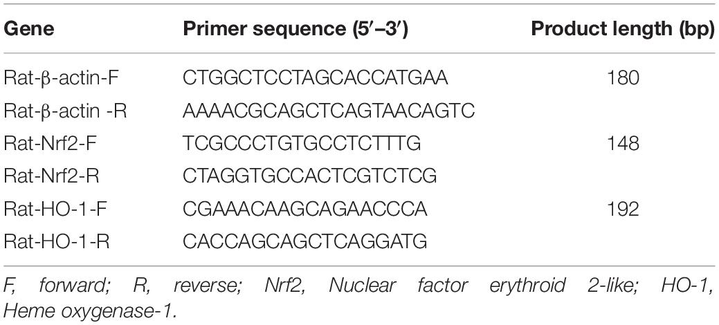
Table 1. Primer sequence.
Nissl’s staining is commonly used to observe the morphology and survival of hippocampal neurons in rats, as reported previously (Chang et al., 2019). Briefly, the brain was fixed by pouring into 4% paraformaldehyde via the vascular system through the rat’s heart. After the sample was embedded in paraffin and cut into 5 μm thick sections. Next Nissl Stain (toluidine blue method) was immersed in a temperature box at 55°C for 30 min. Finally, the hippocampal tissue lesion was observed by a fluorescence microscope (Leica DM4B, Germany) at a magnification of 400×.
Hippocampal tissue was paraffin-embedded, cut into 5 μm, and dehydrated by gradient ethanol. Then 3% H2O2 was used for incubation at room temperature for 30 min, and endogenous peroxidase was inactivated. 10% sheep serum was added for incubation at room temperature for 1 h, Next primary antibody anti-Bax (50599-2, Proteintech), anti-Bcl-2 (26593-1, Proteintech), and anti-caspase-3 (Ab184787, Abcam) were, respectively, added for incubation for 1 h, and secondary antibody was added for incubation for 1 h. After PBS rinsing, each tissue was added with 50 μl DAB chromogenic, re-dyed with hematoxylin, dehydrated, xylene permeated, and sealed. The positive expression of the protein was observed under a 100-fold and 400-fold microscope.
All data were input into the computer and processed with GraphPad Prism 8.0.2 Windows software. Normality and homogeneity of variance were tested first. For a pairwise comparison between the groups that met the normal conditions, one-way ANOVA was used. If the variance was not homogeneous, the Brown-Forsythe ANOVA test was selected and the statistical data were expressed as mean ± standard deviation (x ± s). The non-parametric rank-sum test was used when the normality was not satisfied. All results with a difference of P < 0.05 were statistically significant.
As shown in Figure 2, compared with control group, weight gain [F(3,30.82) = 62.55; P < 0.001], sucrose intake [F(3,24.99) = 6.162; P < 0.05] and total distance [F(3,26.77) = 3.827; P < 0.05] decreased, and immobility time [F(3,7.952) = 15.67; P < 0.05] increased in the model group, all of which were statistically significant. However, acupuncture significantly reduced CUMS-induced immobility time in FST, increased the distance traveled in OFT, and the sucrose preference rate in SPT (P < 0.05, P < 0.05, P < 0.05). These indicate that acupuncture can well alleviate depression-like behaviors caused by CUMS.
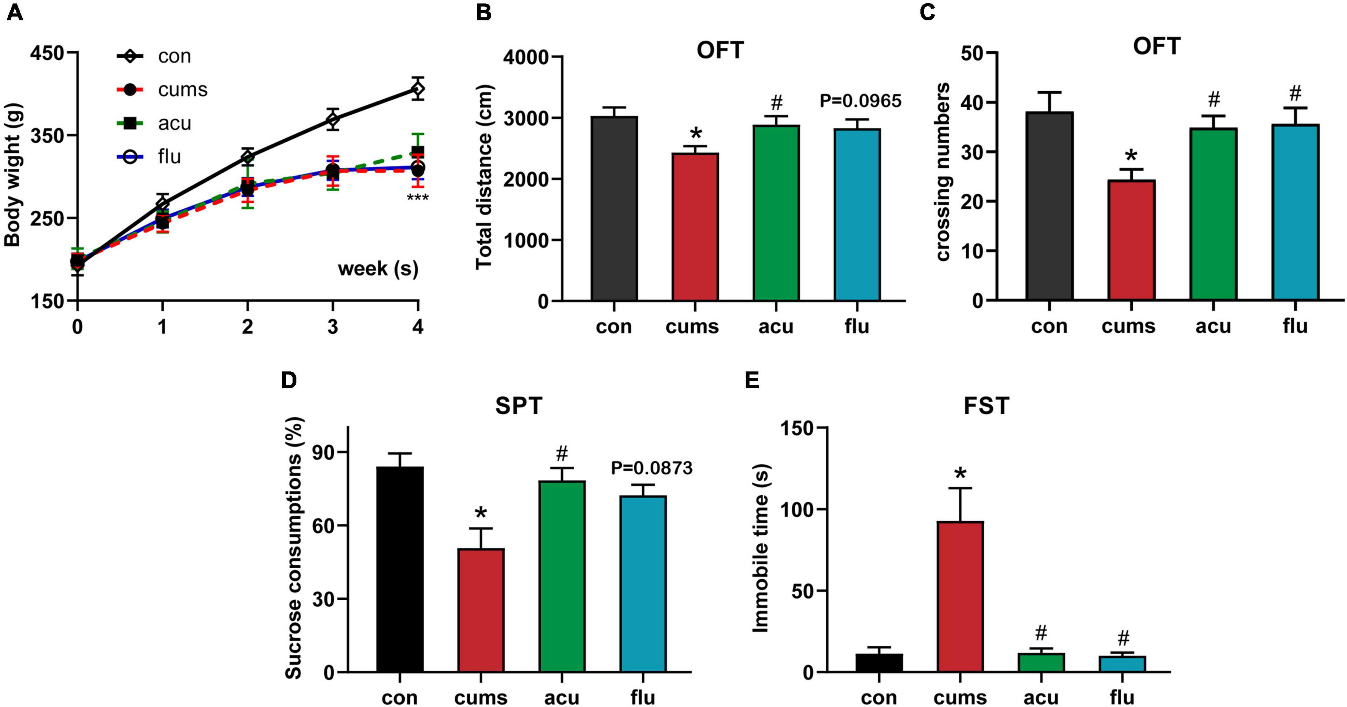
Figure 2. Acupuncture ameliorates depressive-behavior rats exposed to CUMS. (A) Acupuncture prevented the decreased the weight gain in CUMS rats. (B,C) Acupuncture or fluoxetine increases in total distance and crossing numbers of CUMS-exposed rats in the open field test. (D) Acupuncture prevented the decreased consumption of sucrose solution in CUMS rats. (E) Acupuncture or fluoxetine reversed the increases in immobility times of CUMS-exposed rats in the forced swim test. All values are presented as means ± SEM (n = 8), *P < 0.05, ***P < 0.001 compared to the control group, #P < 0.05 compared to the CUMS group.
As shown in Figure 3, the activities of antioxidant enzymes, such as SOD and GSH-Px, were significantly decreased in the depression group compared with the non-stress control group (P < 0.01, P < 0.05). And compared with the unstressed control group, the levels of malondialdehyde (MDA) (P < 0.01) and hydrogen peroxide (H2O2) [F(3,16.15) = 9.419; P < 0.001] of oxidative stress products in the hippocampus were significantly increased after 4 weeks of exposure to CUMS. In addition, excessive production of reactive oxygen species (ROS), a sign of oxidative stress, under environmental stress can cause severe damage to the brain (Salim, 2017). The results showed that compared with the unstressed control group, ROS level in the hippocampus was significantly increased after 4 weeks of exposure to CUMS [F(3,10.85) = 6.038; P < 0.01]. In contrast, acupuncture could reduce the excessive accumulation of ROS in the hippocampus of rats (P < 0.001). From the above observation, we found that acupuncture prevented the above changes in oxidative stress caused by exposure to CUMS.
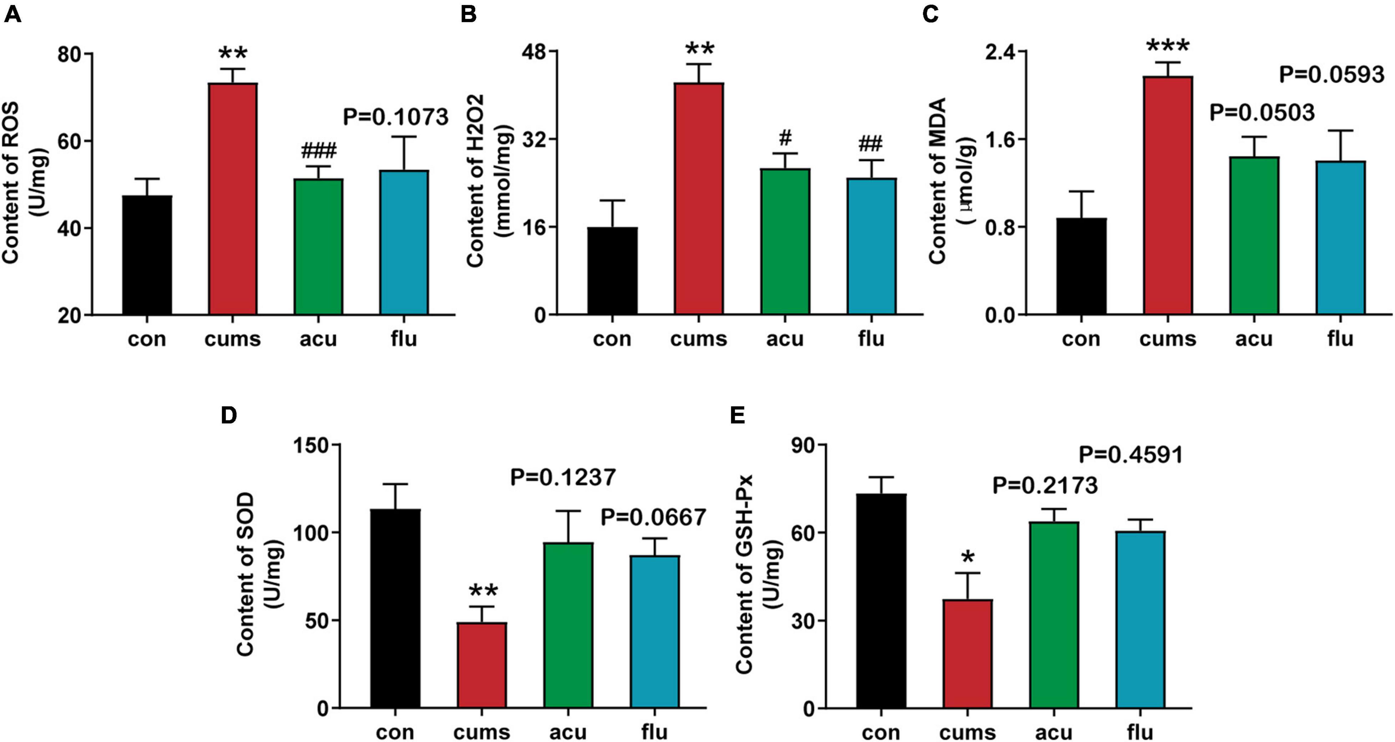
Figure 3. Acupuncture relieves the oxidative stress response in hippocampus of CUMS rats. (A) Acupuncture alleviated the increased levels of ROS within the hippocampal region caused by CUMS exposure. (B) Acupuncture and fluoxetine reduce oxidative stress product H2O2 in hippocampus of rats with depression. (C–E) Hippocampus level of MDA, SOD, GSH-Px. All values are presented as means ± SEM (n = 6), *P < 0.05, **P < 0.01, ***P < 0.001 compared to the control group. #P < 0.05, ##P < 0.01, ###P < 0.001 compared to the CUMS group.
The analysis revealed that the expression of HO-1 was significantly decreased as compared with the non-stressed control group after 4 weeks of CUMS exposure [F(3,7.884) = 19.67; P < 0.01]. Consistently, results from the RT-PCR analysis showed mRNA expressions of HO-1 significantly reduced as compared with control group [F(3,5.59) = 12.29; P < 0.01]. Interestingly, fluoxetine pretreatment elevated the protein expressions of HO-1 (P < 0.01). At the same time, the expressions of Nrf2 and HO-1 were increased via acupuncturing, although there was no statistical difference (P = 0.4158, P = 0.2137). It implied that acupuncture may improve the depression-like behaviors of rats by regulating the Nrf2 signal (Figure 4).
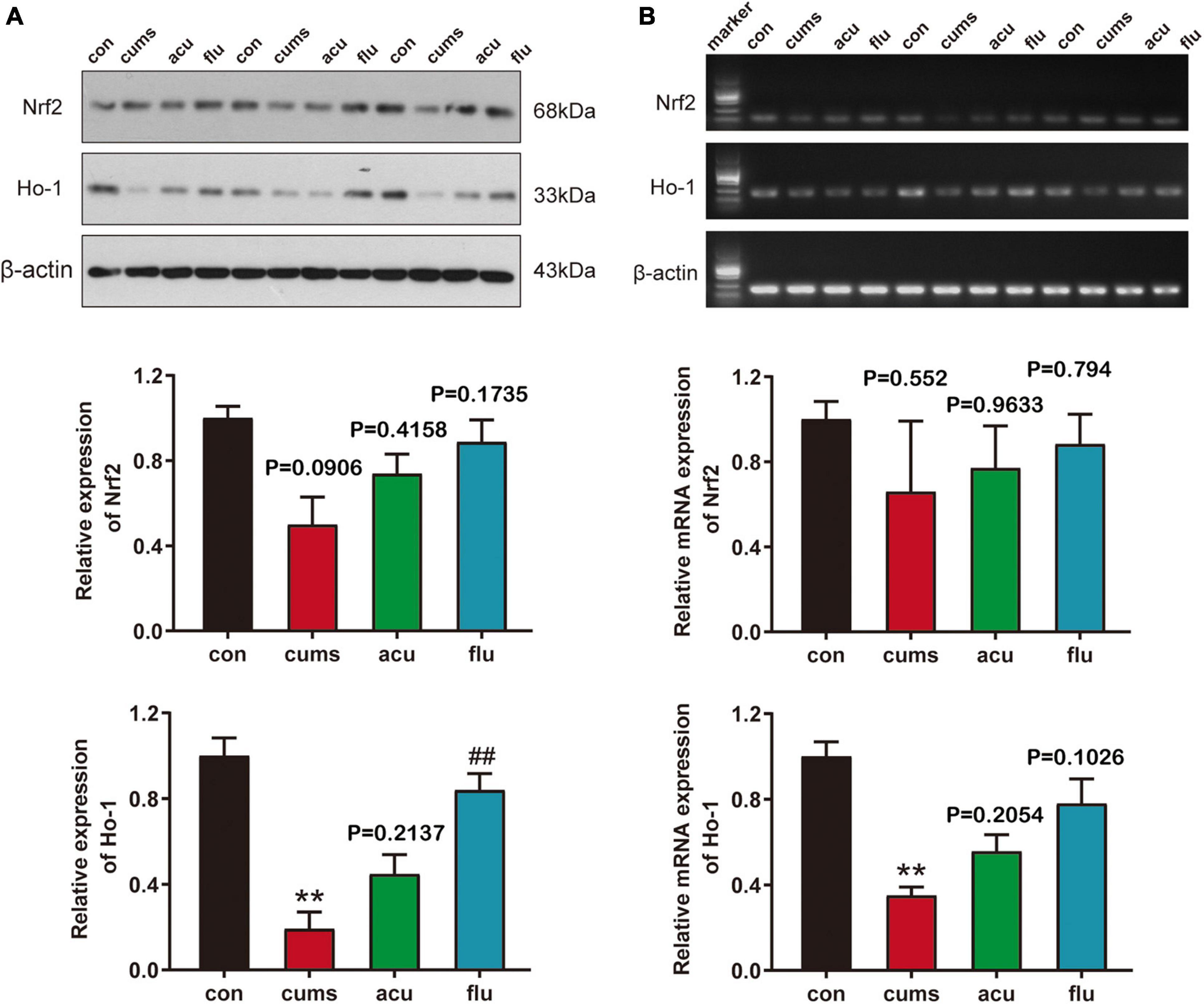
Figure 4. Acupuncture regulated the expression of Nrf2 signaling pathway in the CUMS-induce depressed rats of hippocampus. (A) The expression of Nrf2 and HO-1 protein in each group was detected by Western Blot. (B) The expression of Nrf2 and HO-1 mRNA in each group was detected by RT-PCR. All values are presented as means ± SEM (n = 3). con, control group; cums, CUMS group; acu, acupuncture group; flu, fluoxetine group, **P < 0.01 compared to the control group, ##P < 0.01 compared to the CUMS group.
The morphological and quantitative changes of hippocampal neurons were observed by Nissl staining (Figure 5). It could be found that the nerve cells of rats in control group were with high density, full cell body, and in order arrangement. At the same time, in the model group hippocampal neurons of rats were with large intercellular space and the cell layer was attenuated. The relative expression of pyramidal neurons was decreased after 4 weeks of CUMS exposure [F(3,5.008) = 11.29; P < 0.05]. Compared with the CUMS-induced group, acupuncture groups had increased numbers of normal Nissl bodies (P < 0.05).
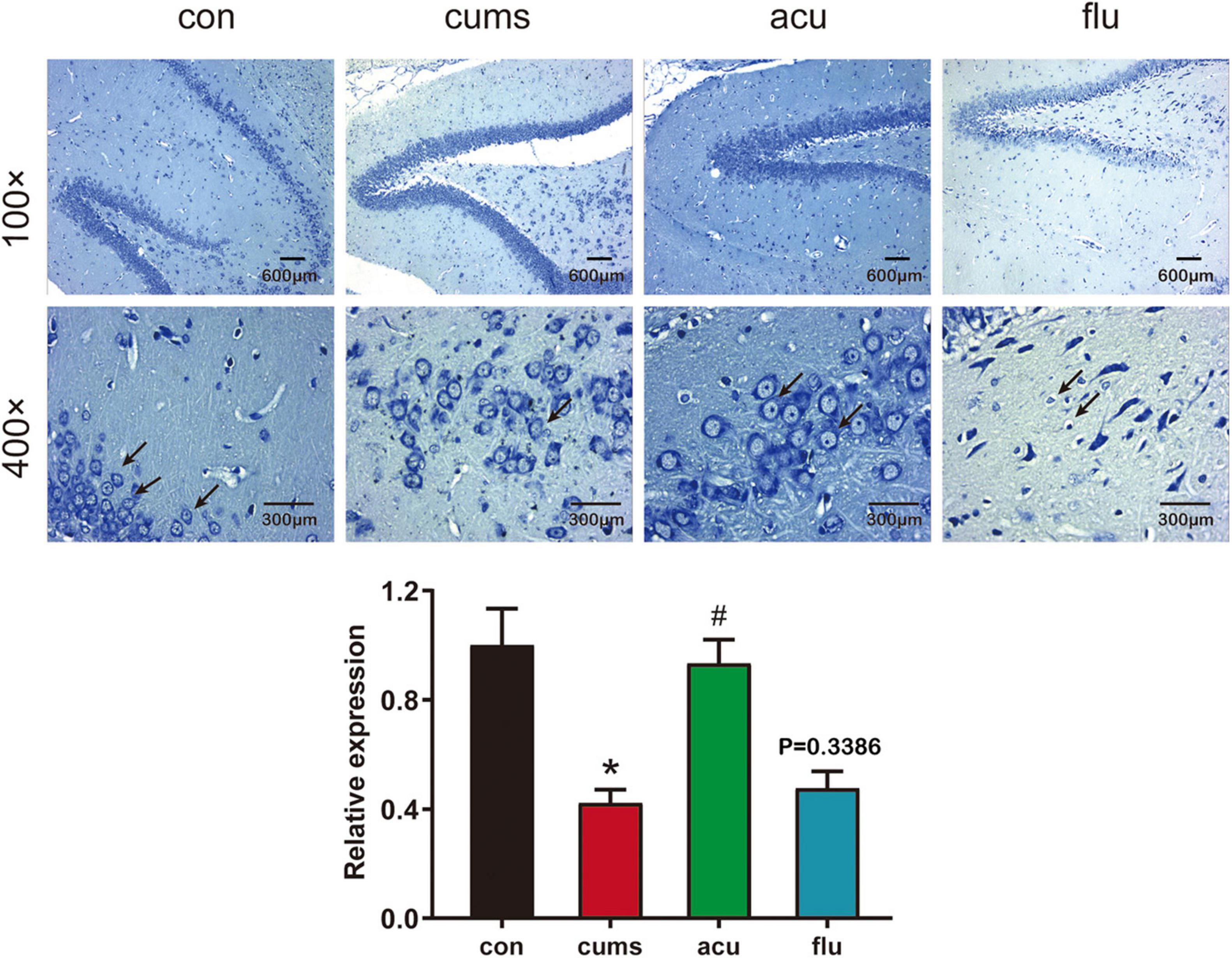
Figure 5. Acupuncture alleviated hippocampal neural injury in the CUMS-induced depressed rats. All values are presented as means ± SEM (n = 3). *P < 0.05 compared to the control group, #P < 0.05 compared to the CUMS group.
The relative expression of Bax [F(3,6.716) = 26.01; P < 0.01] and caspase-3 (P < 0.01) were significantly increased in the hippocampus of CUMS-induce rats as compared with control group. On the contrary, compared with control group, the relative expression of Bcl-2 in the hippocampus of rats induced by CUMS was significantly decreased [F(3,7.618) = 10.99; P < 0.05]. However, the changes of apoptosis-related factors in the hippocampus of CUMS-induced rats of were significantly regulated in response to acupuncture pretreatment [P < 0.05, P < 0.05, P < 0.01]. These results suggest that the neuroprotective effect of acupuncture on apoptosis may help to restore the depression-like behavior of CUMS rats (Figure 6).
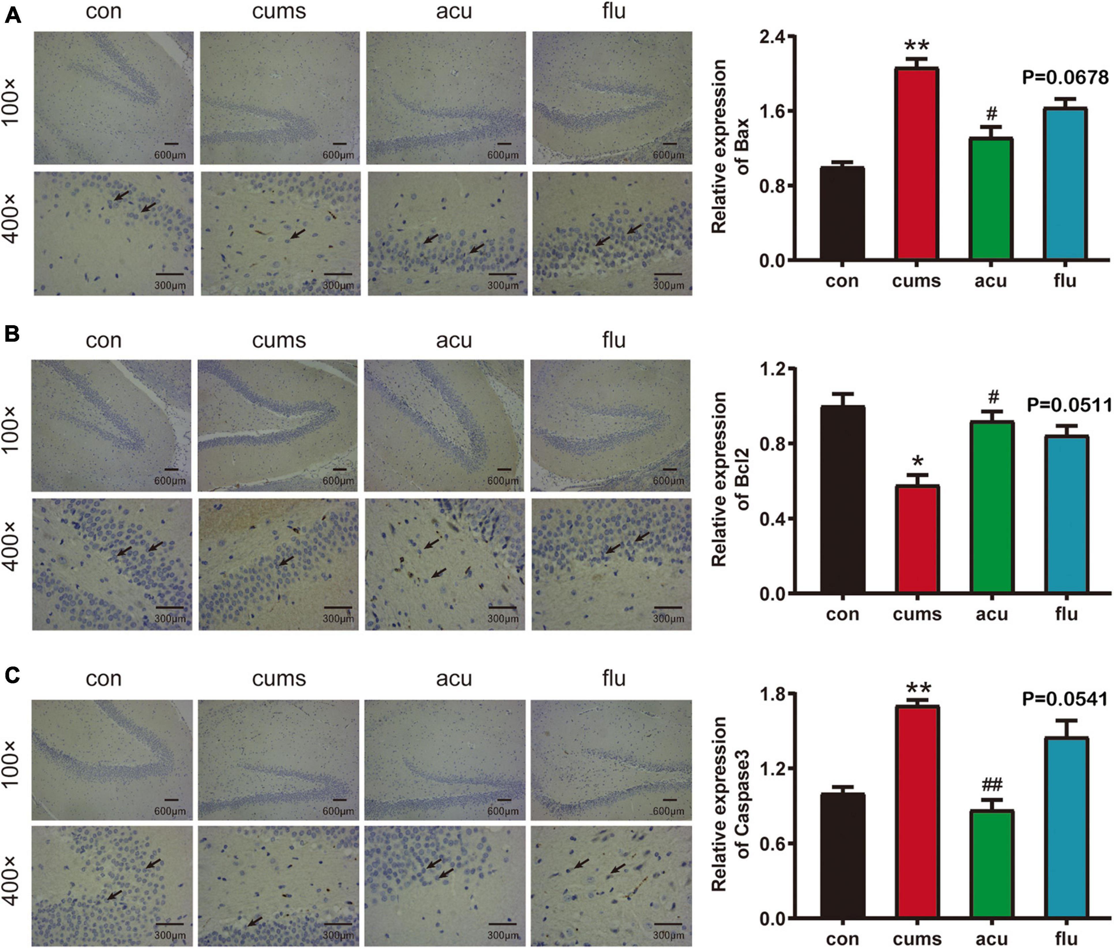
Figure 6. Acupuncture decreases the Hippocampal nerve apoptosis in the CUMS-induced depressed rats. (A–C) Are the relative expressions of Bax, Bcl-2 and caspase-3 in the hippocampus of rats in each group. All values are presented as means ± SEM (n = 3), *P < 0.05, **P < 0.01 compared to the control group, #P < 0.05, ##P < 0.01 compared to the CUMS group.
In this study, we evaluated the antidepressant effect of acupuncture in a CUMS-induced depression model in rats and explored its underlying mechanism (Figure 7). The results show that acupuncture can effectively prevent the occurrence of depression-like behaviors in CUMS rats. These helpful effects on the depressive behavior of CUMS rats is accompanied with the suppression of oxidative stress and the attenuation of hippocampal neuronal apoptosis. Taken together, our data suggest that the antioxidant effect of acupuncture on hippocampal neurons might be a potential therapy for depression.
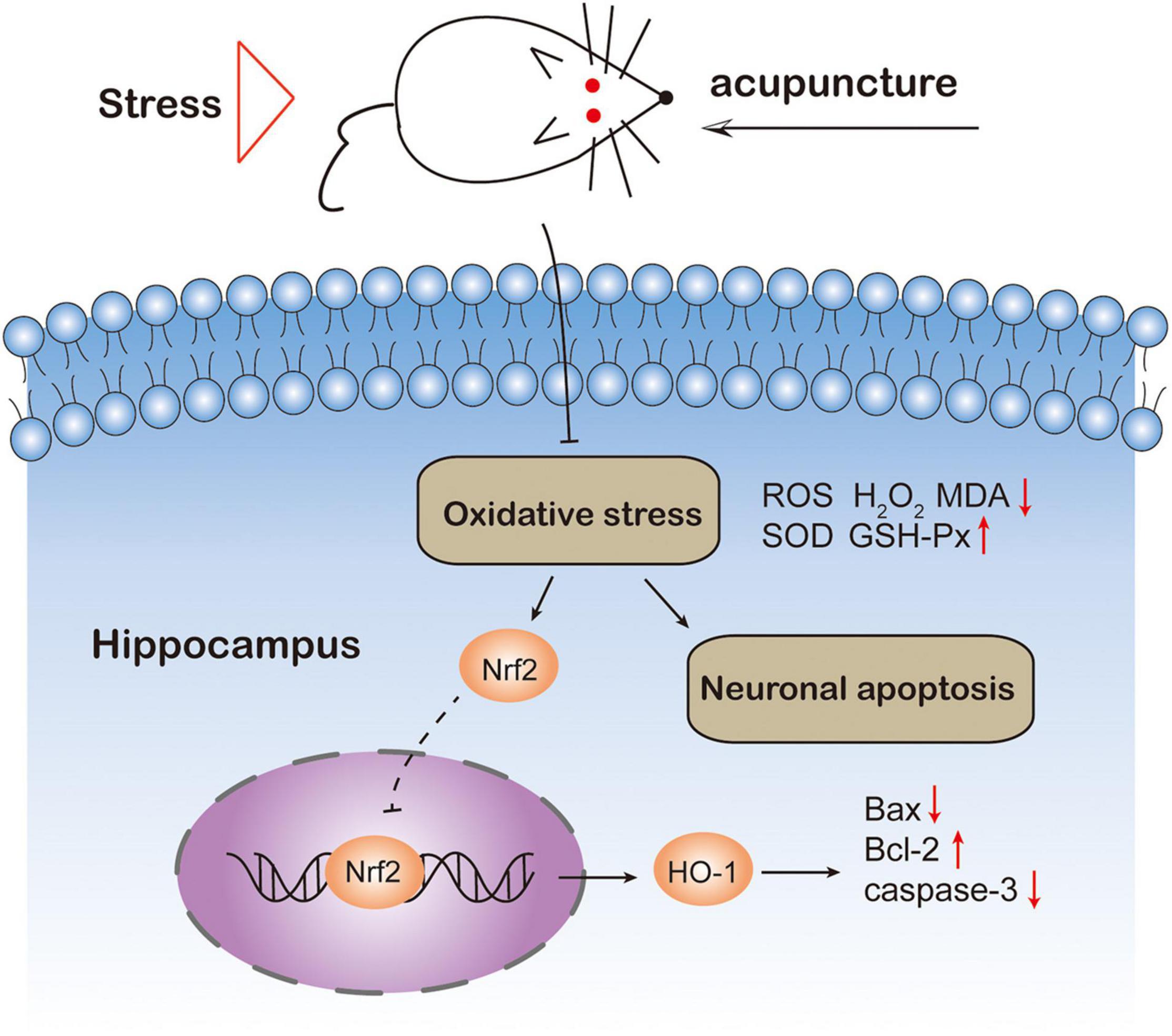
Figure 7. Schematic diagram of the mechanism of acupuncture preventing depression-like behavior in rats. Acupuncture ShangXing (GV23) and Fengfu (GV16) acupoints could suppress oxidative stress and neuronal apoptosis.
To normalize the function of the body, acupuncture is used to stimulate specific sites in body to balance yin and yang in Traditional Chinese Medicine (TCM) theory (Wen et al., 2021). Based on this feature, some people in Southeast Asia apply acupuncture to treat mental illnesses such as depression (Seo et al., 2019). Indeed, according to a review on the effectiveness of acupuncture in treating depression, acupuncture could reduce the severity of depression (Smith et al., 2018). A clinical study reported that manual acupuncture enhanced therapeutic effectiveness and potentially reduced adverse effects caused by selective serotonin reuptake inhibitors (SSRIs) antidepressants (Zhao et al., 2019). Nevertheless, the molecular mechanism of the antidepressant effect of acupuncture needs to be explored. A latest report indicated that the specificity of acupoints is that they could drive a specific autonomic pathway: the vagal–adrenal axis (Liu et al., 2021). And in a previous study, acupuncture increased dendritic plasticity (Davila-Hernandez et al., 2018) and modulate BDNF expression (Davila-Hernandez et al., 2021) in hippocampus of rats with chronic social isolation. Besides, acupuncture altered hepatic lipid metabolism and inflammatory makers in the chronic restraint stress (CRS) induced depression model (Jung et al., 2021). Chronic unpredictable mild stress (CUMS) is currently the most common model of depression in that chronic external stressful life events are the crucial cause of depression (Willner, 2017). Previous studies have shown that acupuncture ameliorates inflammatory response in a rat model of CUMS (Lu et al., 2017). And electroacupuncture could reverse depression-like behavior and regulate astrocytes in the hippocampus in rats exposed to CUMS (Yao et al., 2021). In this study, a fluoxetine group was selected as the positive control group to evaluate the effect of acupuncture on CUMS rats. We found that acupuncture could prevent the depression-like behaviors in CUMS rats, showing a similar effect to the classic antidepressant fluoxetine. It is important to note that acupuncture may be considered in some patients who are unwilling to take antidepressants with side effects.
Reports have shown that oxidative stress plays an important role in the development of depression (Ng et al., 2008). Because the brain tissue has a high metabolic rate of oxidative phosphorylation, a high concentration of unsaturated fatty acid content, and a relatively low antioxidant capacity. Thus the brain is very sensitive to oxidative stress damage, and the resulting oxygen free radicals can damage neurons (Wang et al., 2016). Previous clinical studies have indicated that levels of several enzymes involved in the production of reactive oxygen species rose in depressed patients compared with controls that were not depressed (Scapagnini et al., 2012). Meanwhile, the expression of reactive oxygen species (ROS) and the lipid peroxidation marker malondialdehyde (MDA) were significantly increased in the CUMS-induced rats’ hippocampus (Abd El-Fattah et al., 2018). Indeed, our study also demonstrated that oxidative stress products in the hippocampus such as MDA and H2O2 were significantly enhanced in rats exposed to CUMS model compared to the non-stressed group. In addition, acupuncture pre-treatment successfully reversed CUMS-induced elevation of the ROS in the hippocampus, suggesting that the antidepressant effect of acupuncture might be ascribed to its antioxidant properties.
Previous studies have shown that the Nrf2/HO-1 signaling pathway plays an important role in resisting oxidative stress damage (Suzuki and Yamamoto, 2015). In resting state, Nrf2 and its inhibitor Kelch-like epichlorohydrin-related protein-1 (Keap1) are coupled in the cytoplasm and are in a relatively inhibited state (Sandberg et al., 2014). When cells are ischemic and hypoxic, Nrf2 dissociates from Keap1, and its nuclear translocates and binds to HO-1 which plays an antioxidant role in the process of cell stress (Bocci and Valacchi, 2015). In our present study, the results showed that chronic stimulation could significantly reduce the mRNA level of Nrf2 and HO-1. On the contrary, acupuncture could reverse these changes. The above results indicate that CUMS inhibited the Nrf2 signaling pathway. However, acupuncture pre-treatment increases the expression of antioxidant enzymes in the Nrf2 signaling pathway, thereby protecting the brain from CUMS-induced depression.
Depression could cause changes in the activity of enzymes related to oxidative stress. Compared with non-depressed controls, the levels of several enzymes involved in ROS production in depression patients increased (Scapagnini et al., 2012). Due to the peroxidation of cell membrane lipids, the increase of reactive oxygen species leads to the increase of MDA level. A clinical study revealed that the levels of MDA were higher in patients with depression than in healthy controls (Sarandol et al., 2007). Moreover, there was a significant reduction in some important ROS scavengers including superoxide dismutases (SOD), catalase (CAT), and glutathione peroxidase (GSH-Px) in rodent models of depression. Whereas treatments with some potential antidepressant drugs elevated the levels of these antioxidants to varying degree while also alleviated depression-like behavior (Hu et al., 2020; Sousa et al., 2021). Consequent to the increasing the marker of oxidative stress, the activation of pro-inflammatory correlation factors also contributes to the pathogenesis of depression (Bakunina et al., 2015). Taken together, these findings support a possible link between depression and oxidative stress injury. Studies also have shown that increased ROS activates various signal transduction pathways, leading to apoptosis and caspase cleavage (Wei et al., 2017; Yang et al., 2019). In addition, animal reports have indicated that the hippocampus is closely related to the occurrence of depression (Micheli et al., 2018). Hence, we used immunohistochemistry to detect the expression of apoptotic factors in the hippocampus of each group of rats. The results showed that, compared with the normal group, the expression of Bax in the hippocampus of the model group increased, and the expression of Bcl-2 decreased. However, acupuncture could relieve hippocampal cell apoptosis in CUMS rats, which is consistent with previous results (Guo et al., 2021).
In this study, the regulation of acupuncture intervention on oxidative stress and its influence on the Nrf2 signal pathway were preliminarily discussed. The present study provides new insights into the antidepressant effects of acupuncture underlying oxidative stress and the related signaling pathway. However, there are some limitations to be improved: considering the principle of animal welfare and reducing the number of experimental animals. In future experiments, in order to further study the intervention mechanism of acupuncture, we can choose to study the effect of antioxidant factors on apoptosis in vivo.
All in all, the current research suggests that acupuncture is effective to treat the CUMS induced depressive rats, and the effect of acupuncture on depression-like behaviors induced by CUMS might be mediated by resisting oxidative stress response and anti-apoptosis.
The original contributions presented in the study are included in the article/supplementary material, further inquiries can be directed to the corresponding author.
The animal study was reviewed and approved by the Ethics Committee of Animal Care of the Xiamen University, Xiamen, China (License No. XMULAC202110062).
X-JM: study design. W-JnC and PL: experimental filmmaker. W-JnC and W-JeC: manuscript writing. Y-PC and YH: statistical analysis. J-KC and J-LS: data analysis. N-SL and W-YH: manuscript review. All authors approved the final version of the article.
This study was supported by the Shenzhen Science and Technology Program (No. JCYJ20210324121800002) and the Innovation and Entrepreneurship Club of the Department of Traditional Chinese Medicine of Xiamen University.
The authors declare that the research was conducted in the absence of any commercial or financial relationships that could be construed as a potential conflict of interest.
All claims expressed in this article are solely those of the authors and do not necessarily represent those of their affiliated organizations, or those of the publisher, the editors and the reviewers. Any product that may be evaluated in this article, or claim that may be made by its manufacturer, is not guaranteed or endorsed by the publisher.
CUMS, chronic unpredictable mild stress; Nrf2, Nuclear factor erythroid 2-like; HO-1, Heme oxygenase-1; OFT, open field test; SPT, sucrose preference test; FST, forced swimming test; SSRIs, selective serotonin reuptake inhibitors; ROS, reactive oxygen species; MDA, malondialdehyde; H2O2, hydrogen peroxide; SOD, superoxide dismutases; CAT, catalase; GSH-Px, glutathione peroxidase.
Abd El-Fattah, A. A., Fahim, A. T., Sadik, N. A. H., and Ali, B. M. (2018). Resveratrol and dimethyl fumarate ameliorate depression-like behaviour in a rat model of chronic unpredictable mild stress. Brain Res. 1701, 227–236. doi: 10.1016/j.brainres.2018.09.027
Bakunina, N., Pariante, C. M., and Zunszain, P. A. (2015). Immune mechanisms linked to depression via oxidative stress and neuroprogression. Immunology 144, 365–373. doi: 10.1111/imm.12443
Berk, M., Williams, L. J., Jacka, F. N., O’Neil, A., Pasco, J. A., Moylan, S., et al. (2013). So depression is an inflammatory disease, but where does the inflammation come from? BMC Med. 11:200. doi: 10.1186/1741-7015-11-200
Black, C. N., Bot, M., Scheffer, P. G., Cuijpers, P., and Penninx, B. W. (2015). Is depression associated with increased oxidative stress? A systematic review and meta-analysis. Psychoneuroendocrinology 51, 164–175. doi: 10.1016/j.psyneuen.2014.09.025
Bocci, V., and Valacchi, G. (2015). Nrf2 activation as target to implement therapeutic treatments. Front. Chem. 3:4. doi: 10.3389/fchem.2015.00004
Can, N., Can, Ö. D., Osmaniye, D., and Demir Özkay, Ü. (2018). Synthesis of some novel thiadiazole derivative compounds and screening their antidepressant-like activities. Molecules 23:716. doi: 10.3390/molecules23040716
Chang, D. Y., Zhao, J. H., Zhang, X. T., Lian, H. M., Du, X. M., Yuan, R., et al. (2019). Effect of ketamine combined with DHA on lipopolysaccharide-induced depression-like behavior in rats. Int. Immunopharmacol. 75:105788. doi: 10.1016/j.intimp.2019.105788
Chhillar, R., and Dhingra, D. (2013). Antidepressant-like activity of gallic acid in mice subjected to unpredictable chronic mild stress. Fundam. Clin. Pharmacol. 27, 409–418. doi: 10.1111/j.1472-8206.2012.01040.x
Dantzer, R., O’Connor, J. C., Freund, G. G., Johnson, R. W., and Kelley, K. W. (2008). From inflammation to sickness and depression: when the immune system subjugates the brain. Nat. Rev. Neurosci. 9, 46–56. doi: 10.1038/nrn2297
Davila-Hernandez, A., Gonzalez-Gonzalez, R., Guzman-Velazquez, S., Hernandez, O. T. H., Zamudio, S. R., and Martinez-Mota, L. (2021). Antidepressant-like effects of acupuncture via modulation of corticosterone, sex hormones, and hippocampal BDNF expression in male rats. Brain Res. Bull. 173, 53–65. doi: 10.1016/j.brainresbull.2021.05.007
Davila-Hernandez, A., Zamudio, S. R., Martinez-Mota, L., Gonzalez-Gonzalez, R., and Ramirez-San Juan, E. (2018). Antidepressant effects of acupoint stimulation and fluoxetine by increasing dendritic arborization and spine density in CA1 hippocampal neurons of socially isolated rats. Neurosci. Lett. 675, 48–53. doi: 10.1016/j.neulet.2018.03.057
Fang, Z. (1993). The atlas of acupuncture and moxibustion at Shu points and acupuncture techniques. J. Nanjing Railway Med. Coll. 12, 19–21.
Forbes, N. F., Stewart, C. A., Matthews, K., and Reid, I. C. (1996). Chronic mild stress and sucrose consumption: validity as a model of depression. Physiol. Behav. 60, 1481–1484. doi: 10.1016/S0031-9384(96)00305-8
Guo, Q., Lin, X. M., Di, Z., Zhang, Q. A., and Jiang, S. (2021). Electroacupuncture Ameliorates CUMS-induced Depression-like Behavior: involvement of the glutamatergic system and apoptosis in rats. Comb. Chem. High. Throughput Screen. 24, 996–1004. doi: 10.2174/1386207323666201027121423
Hu, Y., Liu, X., Zhang, T., Chen, C., Dong, X., Can, Y., et al. (2020). Behavioral and biochemical effects of KXS on postmyocardial infarction depression. Front. Pharmacol. 11:561817. doi: 10.3389/fphar.2020.561817
Jun, J. H., Lee, J. A., Choi, T. Y., Yun, K. J., Lim, H. J., and Lee, M. S. (2014). Herbal medicine (Gan Mai Da Zao decoction) for depression: a systematic review protocol. BMJ Open 4:e003690. doi: 10.1136/bmjopen-2013-003690
Jung, J., Lee, S. M., Lee, M. J., Ryu, J. S., Song, J. H., Lee, J. E., et al. (2021). Lipidomics reveals that acupuncture modulates the lipid metabolism and inflammatory interaction in a mouse model of depression. Brain Behav. Immun. 94, 424–436. doi: 10.1016/j.bbi.2021.02.003
Khan, M. S., Muhammad, T., Ikram, M., and Kim, M. O. (2019). Dietary supplementation of the antioxidant curcumin halts systemic LPS-Induced neuroinflammation-associated neurodegeneration and memory/synaptic impairment via the JNK/NF-kappaB/Akt Signaling Pathway in Adult Rats. Oxid. Med. Cell. Longev. 2019:7860650. doi: 10.1155/2019/7860650
Li, K., Yan, L., Zhang, Y., Yang, Z., Zhang, C., Li, Y., et al. (2020). Seahorse treatment improves depression-like behavior in mice exposed to CUMS through reducing inflammation/oxidants and restoring neurotransmitter and neurotrophin function. J. Ethnopharmacol. 250:112487. doi: 10.1016/j.jep.2019.112487
Li, P., Huang, W. Y., Yan, Y. N., Cheng, W. J., Liu, S. Y., Huang, Y., et al. (2021). Acupuncture can play an antidepressant role by regulating the intestinal microbes and neurotransmitters in a rat model of depression. Med. Sci. Monit. 27:e929027. doi: 10.12659/MSM.929027
Li, S. Y., Wang, Y., Gao, G. J., Guo, X., Zhang, Y., Zhang, Z. X., et al. (2020). Transcutaneous auricular vagus nerve stimulation at 20 Hz improves depression-like behaviors and down-regulates the hyperactivity of hpa axis in chronic unpredictable mild stress model rats. Front. Neurosci. 14:680. doi: 10.3389/fnins.2020.00680
Lindqvist, D., Dhabhar, F. S., James, S. J., Hough, C. M., Jain, F. A., Bersani, F. S., et al. (2017). Oxidative stress, inflammation and treatment response in major depression. Psychoneuroendocrinology 76, 197–205. doi: 10.1016/j.psyneuen.2016.11.031
Liu, S., Wang, Z., Su, Y., Qi, L., Yang, W., Fu, M., et al. (2021). A neuroanatomical basis for electroacupuncture to drive the vagal-adrenal axis. Nature 598, 641–645. doi: 10.1038/s41586-021-04001-4
Lu, J., Shao, R. H., Jin, S. Y., Hu, L., Tu, Y., and Guo, J. Y. (2017). Acupuncture ameliorates inflammatory response in a chronic unpredictable stress rat model of depression. Brain Res. Bull. 128, 106–112. doi: 10.1016/j.brainresbull.2016.11.010
Luo, T., Tian, H. L., Song, H. T., Zhao, J., Ai, L. Y., Fang, Y. M., et al. (2020). Possible involvement of tissue plasminogen activator/brain-derived neurotrophic factor pathway in anti-depressant effects of electroacupuncture in chronic unpredictable mild stress-induced depression in rats. Front. Psychiatry 11:63. doi: 10.3389/fpsyt.2020.00063
MacPherson, H., Richmond, S., Bland, M., Brealey, S., Gabe, R., Hopton, A., et al. (2013). Acupuncture and counselling for depression in primary care: a randomised controlled trial. Plos Medicine 10:e1001518. doi: 10.1371/journal.pmed.1001518
Maes, M., Galecki, P., Chang, Y. S., and Berk, M. (2011). A review on the oxidative and nitrosative stress (O&NS) pathways in major depression and their possible contribution to the (neuro)degenerative processes in that illness. Prog. Neuropsychopharmacol. Biol. Psychiatry 35, 676–692. doi: 10.1016/j.pnpbp.2010.05.004
Micheli, L., Ceccarelli, M., D’Andrea, G., and Tirone, F. (2018). Depression and adult neurogenesis: positive effects of the antidepressant fluoxetine and of physical exercise. Brain Res. Bull. 143, 181–193. doi: 10.1016/j.brainresbull.2018.09.002
Naeem, K., Tariq Al Kury, L., Nasar, F., Alattar, A., Alshaman, R., Shah, F. A., et al. (2021). Natural dietary supplement, carvacrol, alleviates lps-induced oxidative stress, neurodegeneration, and depressive-like behaviors via the Nrf2/HO-1 Pathway. J. Inflamm. Res. 14, 1313–1329. doi: 10.2147/JIR.S294413
Ng, F., Berk, M., Dean, O., and Bush, A. I. (2008). Oxidative stress in psychiatric disorders: evidence base and therapeutic implications. Int. J. Neuropsychopharmacol. 11, 851–876. doi: 10.1017/s1461145707008401
Porsolt, R. D. (1993). Behavioral despair revisited – a citation-classic commentary on depression – a new animal-model sensitive to antidepressant treatments. Curr. contents. Life sci. 20, 9–9.
Reiter, A., Bengesser, S. A., Hauschild, A. C., Birkl-Toglhofer, A. M., Fellendorf, F. T., Platzer, M., et al. (2020). Interleukin-6 Gene Expression Changes after a 4-week intake of a multispecies probiotic in major depressive disorder-preliminary results of the PROVIT study. Nutrients 12:2575. doi: 10.3390/nu12092575
Salim, S. (2017). Oxidative stress and the central nervous system. J. Pharmacol. Exp. Ther. 360, 201–205. doi: 10.1124/jpet.116.237503
Sandberg, M., Patil, J., D’Angelo, B., Weber, S. G., and Mallard, C. (2014). NRF2-regulation in brain health and disease: implication of cerebral inflammation. Neuropharmacology 79, 298–306. doi: 10.1016/j.neuropharm.2013.11.004
Sarandol, A., Sarandol, E., Eker, S. S., Erdinc, S., Vatansever, E., and Kirli, S. (2007). Major depressive disorder is accompanied with oxidative stress: short-term antidepressant treatment does not alter oxidative-antioxidative systems. Hum. Psychopharmacol. 22, 67–73. doi: 10.1002/hup.829
Scapagnini, G., Davinelli, S., Drago, F., De Lorenzo, A., and Oriani, G. (2012). Antioxidants as antidepressants: fact or fiction? CNS Drugs 26, 477–490. doi: 10.2165/11633190-000000000-00000
Seo, S. Y., Kang, S. Y., Kwon, O. S., Bang, S. K., Kim, S. P., Choi, K. H., et al. (2019). A mechanical acupuncture instrument mitigates the endoplasmic reticulum stress and oxidative stress of ovariectomized rats. Integr. Med. Res. 8, 187–194. doi: 10.1016/j.imr.2019.07.001
Singh, V. P., Patil, C. S., Jain, N. K., and Kulkarni, S. K. (2004). Tramadol interaction with fluoxetine: An isobolographic analysis. Drug Dev. Res. 61, 79–85. doi: 10.1002/ddr.10338
Si-yu, L., Wen-ya, H., An-ning, Z., Yu, W., Yang, H., Peng, L., et al. (2020). Analysis of the treatment of mental illness by the thirteen ghost points in Qianjin Fang. China J. Trad. Chin. Med. Pharm. 35, 1395–1398.
Slavich, G. M., and Irwin, M. R. (2014). From stress to inflammation and major depressive disorder: a social signal transduction theory of depression. Psychol. Bull. 140, 774–815. doi: 10.1037/a0035302
Smith, C. A., Armour, M., Lee, M. S., Wang, L. Q., and Hay, P. J. (2018). Acupuncture for depression. Cochrane Database Syst. Rev. 3:Cd004046. doi: 10.1002/14651858.CD004046.pub4
Sousa, M. S. B., Alves, D. V. S., Monteiro, H. M. C., Gomes, D. A., Lira, E. C., and Amancio-Dos-Santos, A. (2021). Sepsis impairs the propagation of cortical spreading depression in rats and this effect is prevented by antioxidant extract. Nutr. Neurosci. 24, 130–139. doi: 10.1080/1028415x.2019.1602987
Suzuki, T., and Yamamoto, M. (2015). Molecular basis of the Keap1-Nrf2 system. Free Radic. Biol. Med. 88(Pt B), 93–100. doi: 10.1016/j.freeradbiomed.2015.06.006
Wang, Q., Timberlake, M. A. II, Prall, K., and Dwivedi, Y. (2017). The recent progress in animal models of depression. Prog. Neuropsychopharmacol. Biol. Psychiatry 77, 99–109. doi: 10.1016/j.pnpbp.2017.04.008
Wang, X., Wang, Z., Liu, J., Chen, J., Liu, X., Nie, G., et al. (2016). Repeated acupuncture treatments modulate amygdala resting state functional connectivity of depressive patients. Neuroimage Clin. 12, 746–752. doi: 10.1016/j.nicl.2016.07.011
Wei, H., Gao, Z., Zheng, L., Zhang, C., Liu, Z., Yang, Y., et al. (2017). Protective effects of fucoidan on Abeta25-35 and d-Gal-Induced Neurotoxicity in PC12 Cells and d-Gal-Induced Cognitive Dysfunction in Mice. Mar. Drugs 15:77. doi: 10.3390/md15030077
Wen, J., Chen, X., Yang, Y., Liu, J., Li, E., Liu, J., et al. (2021). Acupuncture medical therapy and its underlying mechanisms: a systematic review. Am. J. Chin. Med. 49, 1–23. doi: 10.1142/S0192415X21500014
Willner, P. (2017). The chronic mild stress (CMS) model of depression: history, evaluation and usage. Neurobiol. Stress 6, 78–93. doi: 10.1016/j.ynstr.2016.08.002
Yang, L., Guan, G., Lei, L., Liu, J., Cao, L., and Wang, X. (2019). Oxidative and endoplasmic reticulum stresses are involved in palmitic acid-induced H9c2 cell apoptosis. Biosci. Rep. 39:BSR20190225. doi: 10.1042/bsr20190225
Yao, Z. Y., Zhang, Z. N., Zhang, J. P., Cai, X. W., Zhong, Z., Huang, Y., et al. (2021). Electroacupuncture alleviated the depression-like behavior by regulating FGF2 and astrocytes in the hippocampus of rats with chronic unpredictable mild stress. Brain Res. Bull. 169, 43–50. doi: 10.1016/j.brainresbull.2021.01.005
Zhao, B. C., Li, Z. G., Wang, Y. Z., Ma, X. H., Wang, X. Q., Wang, X. Q., et al. (2019). Manual or electroacupuncture as an add-on therapy to SSRIs for depression: a randomized controlled trial. J. Psychiatr. Res. 114, 24–33. doi: 10.1016/j.jpsychires.2019.04.005
Keywords: depression, acupuncture, oxidative stress, Nrf2/HO-1 signaling pathway, neuronal apoptosis
Citation: Cheng W-J, Li P, Huang W-Y, Huang Y, Chen W-J, Chen Y-P, Shen J-L, Chen J-K, Long N-S and Meng X-J (2022) Acupuncture Relieves Stress-Induced Depressive Behavior by Reducing Oxidative Stress and Neuroapoptosis in Rats. Front. Behav. Neurosci. 15:783056. doi: 10.3389/fnbeh.2021.783056
Received: 25 September 2021; Accepted: 30 November 2021;
Published: 04 January 2022.
Edited by:
Pietro Maria Chagas, Faculdade da Serra Gaúcha, BrazilReviewed by:
Marcel Henrique Sari, Federal University of Santa Maria, BrazilCopyright © 2022 Cheng, Li, Huang, Huang, Chen, Chen, Shen, Chen, Long and Meng. This is an open-access article distributed under the terms of the Creative Commons Attribution License (CC BY). The use, distribution or reproduction in other forums is permitted, provided the original author(s) and the copyright owner(s) are credited and that the original publication in this journal is cited, in accordance with accepted academic practice. No use, distribution or reproduction is permitted which does not comply with these terms.
*Correspondence: Xian-Jun Meng, bWVuZ3hpYW5qdW5AeG11LmVkdS5jbg==
†These authors have contributed equally to this work and share first authorship
Disclaimer: All claims expressed in this article are solely those of the authors and do not necessarily represent those of their affiliated organizations, or those of the publisher, the editors and the reviewers. Any product that may be evaluated in this article or claim that may be made by its manufacturer is not guaranteed or endorsed by the publisher.
Research integrity at Frontiers
Learn more about the work of our research integrity team to safeguard the quality of each article we publish.