
94% of researchers rate our articles as excellent or good
Learn more about the work of our research integrity team to safeguard the quality of each article we publish.
Find out more
ORIGINAL RESEARCH article
Front. Aging Neurosci., 28 June 2022
Sec. Neurocognitive Aging and Behavior
Volume 14 - 2022 | https://doi.org/10.3389/fnagi.2022.908063
This article is part of the Research TopicThe Many Faces of Brain AgingView all 8 articles
 Gaétan Zimmermann1Laure Joly2,3Pauline Schoepfer2
 Matthieu Doyen4Veronique Roch1Rachel Grignon1
 Paolo Salvi5
 Pierre-Yves Marie1,3Athanase Benetos2,3
 Antoine Verger1,4*
Brain 18F-FDG PET imaging is useful to characterize accelerated brain aging at a pre-symptomatic stage. This study aims to examine the interactions between brain glycolytic metabolism and hemodynamic parameters in different age groups.
Methods: A total of 72 patients (from 23 to 88 years of age, 38 women) without any cerebral diseases but with available cardiac, arterial peripheral, and central blood pressure measurements as well as arterial stiffness parameters obtained from brachial pressure and applanation tonometry and a brain 18F-FDG PET scan were prospectively included into this study. Quantitative voxel-to-voxel analyses were carried out to test for negative associations between brain glycolytic metabolism and individual hemodynamic parameters (p-voxel of <0.001 for the whole population and <0.005 for age groups).
Results: The heart rate parameter of the whole population showed the most extensive associations with brain metabolism (15,857 mm3, T-score: 5.1), predominantly affecting the frontal and temporal regions (69% of the volume). Heart rate for the younger age group, systolic and pulse pressure for the 41–60-year-old group, and diastolic pressure for the older group were most extensively associated with brain metabolism and mainly involved the fronto-temporal lobes (respective involvement of 52.8%, 60.9%, and 65.5%) which are also the regions implicated in accelerated brain aging.
Conclusion: This cross-sectional prospective study identified extensive associations between cerebral metabolism and hemodynamic parameters, indicating common aging mechanisms. Heart rate throughout adult life, systolic and pulse pressure parameters around middle age, and diastolic pressure parameters in older patients, suggest the existence of potentially therapeutic targets to prevent accelerated brain aging.
Associations between hemodynamic parameters and accelerated cerebral aging are now widely established, in particular chronic high blood pressure, which is the most important risk factor for cognitive decline in older adults (Gąsecki et al., 2013). In our previous study, we reported that impaired cerebral metabolism was significantly associated with a central pulse pressure greater than 50 mmHg in cognitively asymptomatic hypertensive older adults, highlighting the importance of measuring central hemodynamic parameters when investigating brain metabolism (Verger et al., 2015). Resting heart rate, a marker of cardiac autonomic function, is related to cardiovascular disease and atherosclerosis through sympathetic overactivity and pulsatile shear stress. Moreover, autonomic regulation seems to have consequences on cognitive processes (Forte et al., 2019). Among the hemodynamic parameters, those targeting arterial stiffness, a typical marker of arterial aging, can be appreciated directly by measuring the pulse wave velocity (PWV; Salvi et al., 2004; Laurent et al., 2006), which is associated with cerebral small vessel disease and an accelerated decline of cognitive functions even in very old adults (Kearney-Schwartz et al., 2009; Singer et al., 2014; Watfa et al., 2015; Pase et al., 2016).
18F-FluoroDesoxyGlucose Positron Emission Tomography (18F-FDG PET), which allows to investigate brain glycolytic metabolism (Guedj et al., 2022), helps to characterize accelerated brain aging at the pre-symptomatic stage. Previous studies have suggested that elevated peripheral arterial pressure levels (Langbaum et al., 2012) and even more so elevated central arterial pressure levels (Verger et al., 2015) lead to local reductions in cerebral metabolism in cognitively non-affected patients. These data illustrate the impact of hemodynamic parameters on brain function, at the preclinical remodeling stage. In addition, a large epidemiological study correlated elevated central arterial pressure levels with an increased risk of cardiac and/or cerebral stroke (Roman et al., 2009). To the best of our knowledge, there are however no currently available data that show the impact of hemodynamic parameters, including central arterial pressure, on the cerebral metabolism of participants at different ages of life.
This cross-sectional prospective study, therefore, aims to investigate associations between brain glycolytic metabolism, determined by 18F-FDG PET imaging, and hemodynamic parameters, as a function of age, in a population with no cerebral pathologies.
One hundred participants referred to the University Hospital of Nancy (France) for an 18F-FDG PET between August 2018 and November 2020, but with no oncological or neurological indications, were prospectively included in the PACTEP (Central Arterial Pressure and Positron Emission Tomography) study (Number Clinical Trials.gov: NCT03345290). Participants were age-stratified, at their time of inclusion into the study, and consisted of a 25% distribution of 18- to 40-year-old participants, 25% of 41- to 60-year-old participants, and 50% over 60-year-old participants. Pregnant women, minors, persons under judicial protection, participants with a history of psychiatric or neurological cerebral pathologies, or patients taking psychotropic drug treatments, participants who received chemotherapy or encephalic radiotherapy, and participants with atrial fibrillation or severe carotid stenosis were not included. Main indications for the 18F-FDG PET were: (1) an initial assessment or follow-up of inflammatory diseases (44%) such as granulomatosis (18%), vasculitis (8%), and others (18%; i.e., lupus, Sjögren’s syndrome, polymyalgia rheumatic, and spondyloarthropathy), with all these PET indications retained as covariates in our voxel-to-voxel quantitative analyses; (2) an etiological assessment of hyperthermia, decline in general condition (17%); (3) an initial assessment of cancer or suspected cancer, before any treatment (20%, including 13% in thoracic oncology); (4) an assessment of adenopathy or investigation of a hematological syndrome prior to any treatment (11%); and (5) an initial assessment or follow-up of an infectious pathology (8%), mainly alveolar echinococcosis.
On the day of the 18F-FDG PET scan patients, included in the study, initially performed a battery of neuropsychological tests: Mini-Mental State Examination (MMSE; Folstein et al., 1975), the Frontal Assessment Battery (FAB; Dubois et al., 2000), and the Mini International Neuropsychologic Interview (MINI; Sheehan et al., 1998). Any patients diagnosed with a cognitive disorder (MMSE < 27, FAB < 15) or a major depressive syndrome (as defined by the MINI-test) were subsequently excluded from the study.
This cross-sectional study was approved by the local ethics committee (CPP accreditation N°2018/26). All participants signed an informed consent form before enrollment. This research complied with the principles of the Declaration of Helsinki. This clinical trial was reported in the Clinical Trials database (NCT03345290).
Before their 18F-FDG PET scan and after resting 15 min in the supine position, participants were set up in a quiet room and three peripheral arterial pressure measurements were taken with an automated monitor (DINAMAP v100, General Electric®) and averaged to improve representativity (peripheral systolic blood pressure, pSBP and peripheral diastolic blood pressure, pDBP). Peripheral pulse pressure (pPP) was obtained by subtracting pDBP from pSBP.
Central pulse parameters were measured by two experienced physicians (PSc, LJ) using applanation tonometry (Pulsepen DiaTecne®, San Donato Milanese, Italy; Salvi et al., 2004). All results were expressed as an average of 10 measurements obtained from 10 cardiac cycles. The central systolic blood pressure (cSBP), central diastolic blood pressure (cDBP) parameters were derived from the analysis of the carotid pulse wave. The central pulse pressure (cPP) was determined by subtracting cDBP from cSBP, and the heart rate (HR) was established from the ECG. Carotid-femoral pulse wave velocity (cf-PWV) was obtained by applying the tonometer to the carotid and femoral arteries as previously reported (Van Bortel et al., 2012).
Brain PET acquisitions were performed on a digital camera (Vereos, Philips®), after a neurosensory rest of 45 min following the intravenous injection of 3.5 MBq of 18F-FDG. All participants had fasted at least 6 h prior to receiving the injection and had blood glucose levels < 10 mmol/L. Acquisitions were recorded over a 15 min single bed position and all brain PET scans were reconstructed with the OSEM iterative reconstruction algorithm, as used in clinical practice, and corrected for PSF (Point Spread Function), attenuation, random coincidences, and scatter. Reconstruction parameters were based on two iterations of 10 subsets, displayed in a 256 × 256 matrix with 1 × 1 × 1 mm3 (Doyen et al., 2021). The acquisition was then followed by the whole-body 18F-FDG PET for which the patient was referred. All brain PET images were carefully visually reviewed by nuclear physician specialists to exclude any patients with potential brain abnormalities.
18F-FDG PET brain images were processed using the Statistical Parametric Mapping, SPM 12, software (running on Matlab 2020b (MathWorks Inc., Sherborn, MA). An initial spatial normalization step was carried out with the low-dose scanner which is required for attenuation correction using the adaptive template registered to the Montreal National Institute (MNI) space. Transformations applied to the CT images of each patient were then applied to the co-registered PET images. The size of the scan voxels was set to 1 × 1 × 1 mm3. Intensity was normalized to the pons, a reference region with a glycolytic metabolism that is only weakly influenced by age (Verger et al., 2021) or by the presence of neurodegenerative pathologies (Minoshima et al., 1995). All images were subsequently smoothed with a 4 mm full-width at half-maximum Gaussian-filter to limit individual variations in gyral anatomy. Due to the advanced age of our population (>50% of patients over 60 years of age), partial volume effect corrections were applied, based on the Müller-Gärtner method (Müller-Gärtner et al., 1992) using the PETPVE12 toolkit (Greve et al., 2016). For this, a segmentation of white and gray matter was carried out on CT scans using the SPM tools after a visual analysis to check the accuracy of this segmentation.
Statistical analyses were performed at group-level and voxel-to-voxel using linear regression models of previously detailed hemodynamic parameters with an exclusive white matter mask. Covariates evaluated included age, considered as a continuous variable as well as sex, education level, the presence/absence of anti-hypertensive therapy, and the presence/absence of an inflammatory disease that may affect brain metabolism (Kalpouzos et al., 2009; Lee et al., 2012; Kim et al., 2015). These analyzes were initially performed on the entire population using a p-voxel value of 0.001 and subsequently on the three separate age groups (18–40, 41–60, and >60 years) using a less-restrictive p-voxel value of 0.005. All reported clusters were corrected for cluster volume based on Monte-Carlo simulations to avoid type II errors (Lieberman and Cunningham, 2009). Only negative associations between brain metabolism and hemodynamic parameters were retained. Specific cluster locations in the MNI space were obtained by using the SPM xjView toolbox1. For linear regression analyses, only clusters with a cumulative volume over 10 cm3 (i.e., at least 1% of the total encephalic volume) were considered as significant. Images were visually inspected at different stages of the pre-processing procedure to ensure the quality and convergence of the different methods applied (MD).
Categorical variables are expressed as percentages and continuous variables as medians and interquartile ranges. Because variables were not normally distributed, Chi-2 and Kruskal-Wallis tests were performed to compare categorical and continuous variables, respectively. A p-value < 0.05 was considered to be significant. All tests were performed in SPSS (SPSS Statistics for Windows, Version 20.0. Armonk, NY: IBM Corp). The statistical analysis for SPM is detailed in the previous section.
The SPM analysis was finally performed for 72 patients. Of the 100 candidates initially identified for inclusion in the study, 28 were subsequently excluded due to: (i) abnormal neuropsychiatric tests (four participants with an MMSE < 27 and/or MINI < 15 and/or major depressive syndrome), (ii) the identification of unsuspected brain lesions (one ischemic stroke and one cerebral tuberculosis), (iii) technical difficulties relating to applanation tonometry (eight participants), especially in patients with high body mass index (mean body mass index of these participants of 31.6 ± 6.2 kg.m−2) (Joly et al., 2010) or to the tomoscintigraphic acquisitions (three participants), and (iv) issues relating to the segmentation of white/gray matter on the low-dose CT scan which impeded corrections for the partial volume effect (11 participants). A flowchart for the final population used for the analyses is displayed in Figure 1.
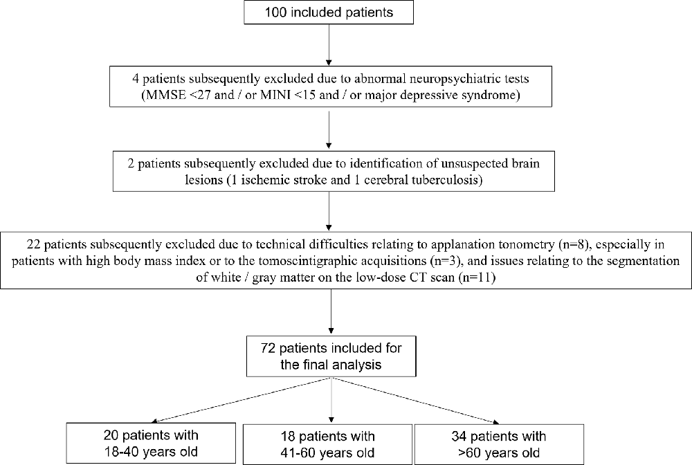
Figure 1. Flowchart of patients included in the final analysis.
Patients ranged from 23 to 88 years of age (mean age = 55.4 years ± 17.1) and were stratified into three age groups (18–40 years, n = 20; 41–60 years, n = 18; > 60 years, n = 34). There were no significant differences between the three age groups in terms of the sex distribution and the presence/absence of an inflammatory disease. As expected, there were significant differences in blood pressure (pSBP, cSBP, pPP) and arterial stiffness (cf-PWV) between the younger and older patients. Clinical and hemodynamic characteristics of the whole population and of the three age groups are given in Table 1.
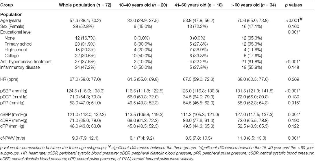
Table 1. Population characteristics.
Table 2 summarizes analyses of the whole population. The HR parameter showed the most extensive interactions with cerebral metabolism (37 clusters, cumulative volume of clusters: 15,857 mm3, T-score peak: 5.1) specifically with the frontal (35.8% of cumulative volume) and temporal lobe (33.2% of cumulative volume) regions. The T-maps projected onto two-dimensional slices of axial T1-weighted MRIs are given in Figure 2 and the spatial locations of abnormalities are detailed in the Supplementary Material.
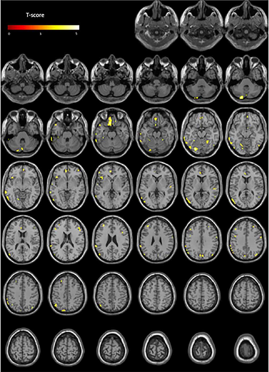
Figure 2. Anatomical localizations of significant brain metabolic clusters associated with HR (negative association) in the whole population (p < 0.001, uncorrected) projected onto two-dimensional slices of T1-weighted MRIs (from the base to the top of the skull).

Table 2. Results of the quantitative voxel-to-voxel analyses for the negative linear regression analyses between hemodynamic parameters and brain glycolytic metabolism on the whole population (p-voxel = 0.001, uncorrected, corrected for cluster volume).
We then evaluated the relative associations of each hemodynamic parameter with cerebral metabolism within each of the three age groups.
In the youngest age group, HR showed the most extensive associations with cerebral metabolism (29 clusters, cumulative volume of the clusters: 12,032 mm3, T-score peak: 10.5) with fronto-temporal predominant localizations (52.8% of the cumulative volume of the clusters).
In the 41- to 60-year-old patient group, systolic and pulse pressure parameters exhibited the most extensive associations with cerebral metabolism (cPP, cSBP, pPP, pSBP). This effect was particularly prominent for the central hemodynamic parameters: cPP (61 clusters, cumulative volume: 33,418 mm3; T-score peak: 14.9) and cSBP (49 clusters, cumulative volume: 24,597 mm3, T-score peak: 8.6), and to a lesser extent for the peripheral parameters: pPP (47 clusters, cumulative volume: 22,186 mm3; T-score peak: 8.7) and pSBP (34 clusters, cumulative volume: 16,231 mm3; T-score peak: 9.8). Spatial localization analyses showed diffuse metabolic abnormalities related to these parameters, with fronto-temporal predominance in particular for cPP and for cSBP (respectively 60.9% and 42.1% of the total of clusters identified).
In addition to associations between cerebral metabolism and HR in the oldest age group, cerebral metabolism showed the most extensive associations with the cDBP parameter (26 clusters, cumulative volume: 20,728 mm3, T-score peak: 5.7). As depicted in Figure 3, these associations predominantly involved the frontal and the temporal cortex: 65.5% of cumulative volume of the significant clusters were localized in the frontal or temporal lobe for cDBP.
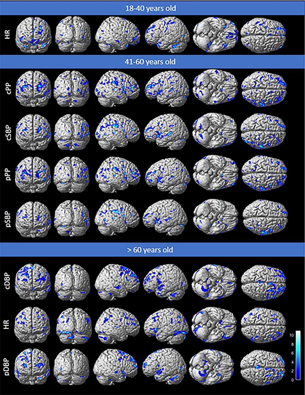
Figure 3. Anatomical localization of clusters associated with parameters that have a significant impact on brain metabolism with negative associations (p < 0.005, uncorrected), projected onto 3D volume-rendered images, spatially normalized, and smoothed into the standard SPM template. Parameters classified according to the cumulative significant cluster volumes for the three age groups.
Analyses of all parameters for the different age groups are given in Table 3, T-maps projected onto 3D volume-rendered images are shown in Figure 3 and the spatial location of abnormalities is detailed in the Supplementary Material.
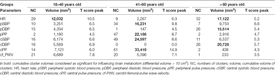
Table 3. Results of the quantitative voxel-to-voxel analyses for the linear regression analyses between hemodynamic parameters and brain glycolytic metabolism on the three age groups (p-voxel = 0.005, uncorrected, corrected for the cluster volume).
Our results demonstrate the existence of associations between hemodynamic parameters and cerebral glycolytic metabolism, with HR exhibiting the most extensive interactions with cerebral metabolism in the whole population. Interestingly, systolic and pulse pressure parameters showed the most extensive associations involving the 41-to-60-year age group whereas diastolic pressure was associated with cerebral metabolism in older participants, suggesting that these parameters may represent potential therapeutic targets. These differences among age groups could be explained by physiological hemodynamics modifications related to aging. The effect of hypertension on cognitive impairment, mainly related to systolic and pulse pressure parameters, has been reported in midlife subjects (Knopman et al., 2001). In older subjects, a continuously high level of pressure related to the diastolic level of pressure impacts consistently the brain metabolism, since diastolic compliance decreases with age (Wolsk et al., 2017).
In terms of the whole population, the number of associations of anomalous brain metabolism with hemodynamic parameters was particularly extensive for HR, suggesting an interaction of heart function and brain metabolism which may point to a common pre-symptomatic aging mechanism. To the best of our knowledge, very few studies have to date investigated the impact of resting HR on brain and cognitive decline. The European guidelines identify an HR of greater than 80 bpm as a parameter which impacts cardiovascular risk in hypertensive patients (Williams et al., 2018), since HR has been shown to be a determinant of accelerated progression of arterial stiffness in hypertensive patients (Benetos et al., 2002). Moreover, a high HR is an independent predictor of cognitive decline in patients at high cardiovascular risk (Böhm et al., 2015). This HR effect was also identified in the younger and older age groups of our age subgroup analysis, confirming the premise that HR influences brain metabolism throughout life. It is interesting that our study associated HR with fronto-temporal hypometabolism because fronto-temporal hypometabolism is a hallmark of accelerated brain aging (Verger et al., 2015).
Interactions between brain metabolism and hemodynamic parameters need to be investigated at all stages of life to identify potential targets for therapy. The most extensive associations in younger patients were obtained for HR. This parameter has previously been shown to influence brain metabolism in younger participants. Our data suggest that reducing the HR may decelerate the risk of developing brain abnormalities. One way to reduce HR is to practice a regular aerobic physical activity which has been demonstrated to impart positive effects on cognitive functions in young and middle-aged healthy participants (Cox et al., 2016). Several other studies have shown a positive impact of physical activity on executive functions (Kamijo and Takeda, 2009, 2010), which are localized to the frontal cortex, the region predominantly affected in our voxel-wise analyses (35.5% of the cumulative cluster volume).
In our middle age group, systolic and pulse pressures were the parameters that were most extensively associated with cerebral metabolism, suggestive of hypertension during middle adulthood causing pre-symptomatic brain remodeling, which may be predictive of future cognitive function. These data are consistent with several longitudinal studies which showed that midlife arterial hypertension is a risk factor for cognitive decline and dementia (Launer et al., 2000; Knopman et al., 2001). For peripheral pressure parameters, our results are consistent with the Langbaum et al. study which showed that systolic and pulse pressure are associated with frontal and temporal hypometabolism determined by 18F-FDG PET in middle-aged participants (Langbaum et al., 2012). It is also important to underline that central pressure (cPP, cSBP) parameter interactions involved more extensive brain volumes than peripherical parameter (pPP, pSBP) associations, suggesting that data obtained from carotid applanation tonometry are more representative of vascular stresses exerted on the brain. This is consistent with our previous work in older adults with hypertension (Verger et al., 2015). Although the role of reducing blood pressure to prevent the incidence of dementia is still controversial the latest meta-analysis of a large number of interventional trials confirms a significant effect of antihypertensive treatment in older adults (Hughes et al., 2020).
For older participants, cDBP exhibited the most extensive associations with brain metabolism (20.7 cm3). Several studies have shown significant associations between high pDBP values and cognitive decline in a middle-aged population (Tsivgoulis et al., 2009) or between high pDBP values and the progression of white matter lesions on MRI in older adults (McNeil et al., 2018), but to the best of our knowledge, no data are currently available for diastolic central parameters even if diastolic peripheral and central pressure values are close to each other. The pulse wave velocity, albeit associated with brain metabolism in older adults, was not an extensive association. This may be explained by 40% of our older adult population being non-hypertensive, and all of our hypertensive participants receiving successful antihypertensive treatments.
From a physio-pathological perspective, associations between hemodynamic parameters and brain metabolism were predominantly localized to the fronto-temporal regions which may correspond to the site of accelerated, vascular origin, cerebral aging. Indeed, hypertension—a risk factor for accelerated brain aging- is predominantly associated with disorders in the frontal and striatal regions and an impairment of executive functions (Buckner, 2004), which is consistent with our data. The potential impact of hemodynamic parameters measured in our study may be related to cerebral small vessel disease. Indeed, MRI hypersignals in white matter—which are evidence of small vessel disease—have been shown to be associated with a significant decrease in gray matter metabolism in older hypertensive adults, specifically involving the frontal cortex (Chetouani et al., 2017).
Our study has several limitations relating to its monocentric nature and its lack of longitudinal follow-up data. In addition, for ethical reasons, the patients included in this study presented with extracerebral pathologies, which motivated the examination. This may have influenced the results, especially for systemic pathologies such as inflammatory diseases, which may have subclinical repercussions on brain imaging. This potentially confounding effect was nevertheless considered in our analyses. By the same token, only a minority of our population was hypertensive (37.5%) and all hypertensive participants were receiving anti-hypertensive treatments, but this is quite representative of the proportion of hypertensive patients in France, based on the latest available epidemiological data (SPF, 2020).
To conclude, we report extensive associations between hemodynamic parameters, specifically HR, and cerebral metabolism throughout adult life. This suggests that brain metabolism and heart function may share common pre-symptomatic aging mechanisms. While systolic and pulse pressure parameters were more prominently involved around middle age, the number of interactions with diastolic pressure parameters were more extensive in older patients. These data suggest the presence of potentially useful therapeutic targets which may be exploited, during specific periods of life, to prevent accelerated brain aging.
The raw data supporting the conclusions of this article will be made available by the authors, without undue reservation.
The studies involving human participants were reviewed and approved by the ethics committee of CHRU of Nancy (CPP accreditation N°2018/26). The patients/participants provided their written informed consent to participate in this study.
AV, LJ, and VR: conceptualization. PSc, GZ, and RG: collection of clinical data. PSc, LJ, and PSa: measurements/analysis of haemodynamic parameters. RG: data curation. GZ, AV, and MD: SPM analysis. GZ, AV, LJ, AB, P-YM, and PSa: redaction—review and editing. All authors contributed to the article and approved the submitted version.
We thank the Nancyclotep platform for the financial support of this study. Study partially supported by the Italian Ministry of Health.
The authors declare that the research was conducted in the absence of any commercial or financial relationships that could be construed as a potential conflict of interest.
All claims expressed in this article are solely those of the authors and do not necessarily represent those of their affiliated organizations, or those of the publisher, the editors and the reviewers. Any product that may be evaluated in this article, or claim that may be made by its manufacturer, is not guaranteed or endorsed by the publisher.
HR, heart rate; cPP, central pulse pressure; cSBP, central systolic blood pressure; pPP, peripheral pulse pressure; pSBP, peripheral systolic blood pressure; cDBP, central diastolic blood pressure; pDBP, peripheral diastolic blood pressure.
The Supplementary Material for this article can be found online at: https://www.frontiersin.org/articles/10.3389/fnagi.2022.908063/full#supplementary-material.
Benetos, A., Adamopoulos, C., Bureau, J.-M., Temmar, M., Labat, C., Bean, K., et al. (2002). Determinants of accelerated progression of arterial stiffness in normotensive subjects and in treated hypertensive subjects over a 6-year period. Circulation 105, 1202–1207. doi: 10.1161/hc1002.105135
Böhm, M., Schumacher, H., Leong, D., Mancia, G., Unger, T., Schmieder, R., et al. (2015). Systolic blood pressure variation and mean heart rate is associated with cognitive dysfunction in patients with high cardiovascular risk. Hypertension 65, 651–661. doi: 10.1161/HYPERTENSIONAHA.114.04568
Buckner, R. L. (2004). Memory and executive function in aging and AD: multiple factors that cause decline and reserve factors that compensate. Neuron 44, 195–208. doi: 10.1016/j.neuron.2004.09.006
Chetouani, A., Chawki, M. B., Hossu, G., Kearney-Schwartz, A., Chauveau-Beuret, F., Bracard, S., et al. (2017). Cross-sectional variations of white and grey matter in older hypertensive patients with subjective memory complaints. Neuroimage Clin. 17, 804–810. doi: 10.1016/j.nicl.2017.12.024
Cox, E. P., O’Dwyer, N., Cook, R., Vetter, M., Cheng, H. L., Rooney, K., et al. (2016). Relationship between physical activity and cognitive function in apparently healthy young to middle-aged adults: a systematic review. J. Sci. Med. Sport 19, 616–628. doi: 10.1016/j.jsams.2015.09.003
Doyen, M., Mairal, E., Bordonne, M., Zaragori, T., Roch, V., Imbert, L., et al. (2021). Effect of point spread function deconvolution in reconstruction of brain 18F-FDG PET images on the diagnostic thinking efficacy in Alzheimer’s disease. Front. Med. (Lausanne) 8:721551. doi: 10.3389/fmed.2021.721551
Dubois, B., Slachevsky, A., Litvan, I., and Pillon, B. (2000). The FAB: a frontal assessment battery at bedside. Neurology 55, 1621–1626. doi: 10.1212/wnl.55.11.1621
Folstein, M. F., Folstein, S. E., and McHugh, P. R. (1975). “Mini-mental state”. A practical method for grading the cognitive state of patients for the clinician. J. Psychiatr. Res. 12, 189–198. doi: 10.1016/0022-3956(75)90026-6
Forte, G., Favieri, F., and Casagrande, M. (2019). Heart rate variability and cognitive function: a systematic review. Front. Neurosci. 13:710. doi: 10.3389/fnins.2019.00710
Gąsecki, D., Kwarciany, M., Nyka, W., and Narkiewicz, K. (2013). Hypertension, brain damage and cognitive decline. Curr. Hypertens. Rep. 15, 547–558. doi: 10.1007/s11906-013-0398-4
Greve, D. N., Salat, D. H., Bowen, S. L., Izquierdo-Garcia, D., Schultz, A. P., Catana, C., et al. (2016). Different partial volume correction methods lead to different conclusions: an 18F-FDG-PET study of aging. Neuroimage 132, 334–343. doi: 10.1016/j.neuroimage.2016.02.042
Guedj, E., Varrone, A., Boellaard, R., Albert, N. L., Barthel, H., van Berckel, B., et al. (2022). EANM procedure guidelines for brain PET imaging using [18F]FDG, version 3. Eur. J. Nucl. Med. Mol. Imaging 49, 632–651. doi: 10.1007/s00259-021-05603-w
Hughes, D., Judge, C., Murphy, R., Loughlin, E., Costello, M., Whiteley, W., et al. (2020). Association of blood pressure lowering with incident dementia or cognitive impairment: a systematic review and meta-analysis. JAMA 323, 1934–1944. doi: 10.1001/jama.2020.4249
Joly, L., Kearney-Schwartz, A., Salvi, P., Mandry, D., Watfa, G., Karcher, G., et al. (2010). 235 Pulse wave velocity assessment by external non-invasive devices and phase contrast magnetic resonance imaging in obese subjects. Arch. Cardiovasc. Dis. Suppl. 2:75. doi: 10.1016/S1878-6480(10)70237-8
Kalpouzos, G., Chételat, G., Baron, J.-C., Landeau, B., Mevel, K., Godeau, C., et al. (2009). Voxel-based mapping of brain gray matter volume and glucose metabolism profiles in normal aging. Neurobiol. Aging 30, 112–124. doi: 10.1016/j.neurobiolaging.2007.05.019
Kamijo, K., and Takeda, Y. (2009). General physical activity levels influence positive and negative priming effects in young adults. Clin. Neurophysiol. 120, 511–519. doi: 10.1016/j.clinph.2008.11.022
Kamijo, K., and Takeda, Y. (2010). Regular physical activity improves executive function during task switching in young adults. Int. J. Psychophysiol. 75, 304–311. doi: 10.1016/j.ijpsycho.2010.01.002
Kearney-Schwartz, A., Rossignol, P., Bracard, S., Felblinger, J., Fay, R., Boivin, J.-M., et al. (2009). Vascular structure and function is correlated to cognitive performance and white matter hyperintensities in older hypertensive patients with subjective memory complaints. Stroke 40, 1229–1236. doi: 10.1161/STROKEAHA.108.532853
Kim, J., Chey, J., Kim, S.-E., and Kim, H. (2015). The effect of education on regional brain metabolism and its functional connectivity in an aged population utilizing positron emission tomography. Neurosci. Res. 94, 50–61. doi: 10.1016/j.neures.2014.12.009
Knopman, D., Boland, L. L., Mosley, T., Howard, G., Liao, D., Szklo, M., et al. (2001). Cardiovascular risk factors and cognitive decline in middle-aged adults. Neurology 56, 42–48. doi: 10.1212/wnl.56.1.42
Langbaum, J. B. S., Chen, K., Launer, L. J., Fleisher, A. S., Lee, W., Liu, X., et al. (2012). Blood pressure is associated with higher brain amyloid burden and lower glucose metabolism in healthy late middle-age persons. Neurobiol. Aging 33, 827.e11–827.e19. doi: 10.1016/j.neurobiolaging.2011.06.020
Launer, L. J., Ross, G. W., Petrovitch, H., Masaki, K., Foley, D., White, L. R., et al. (2000). Midlife blood pressure and dementia: the Honolulu-Asia aging study. Neurobiol. Aging 21, 49–55. doi: 10.1016/s0197-4580(00)00096-8
Laurent, S., Cockcroft, J., Van Bortel, L., Boutouyrie, P., Giannattasio, C., Hayoz, D., et al. (2006). Expert consensus document on arterial stiffness: methodological issues and clinical applications. Eur. Heart J. 27, 2588–2605. doi: 10.1093/eurheartj/ehl254
Lee, S.-W., Park, M.-C., Lee, S.-K., and Park, Y.-B. (2012). The efficacy of brain (18)F-fluorodeoxyglucose positron emission tomography in neuropsychiatric lupus patients with normal brain magnetic resonance imaging findings. Lupus 21, 1531–1537. doi: 10.1177/0961203312459104
Lieberman, M. D., and Cunningham, W. A. (2009). Type I and Type II error concerns in fMRI research: re-balancing the scale. Soc. Cogn. Affect. Neurosci. 4, 423–428. doi: 10.1093/scan/nsp052
McNeil, C. J., Myint, P. K., Sandu, A.-L., Potter, J. F., Staff, R., Whalley, L. J., et al. (2018). Increased diastolic blood pressure is associated with MRI biomarkers of dementia-related brain pathology in normative ageing. Age Ageing 47, 95–100. doi: 10.1093/ageing/afx102
Minoshima, S., Frey, K. A., Foster, N. L., and Kuhl, D. E. (1995). Preserved pontine glucose metabolism in Alzheimer disease: a reference region for functional brain image (PET) analysis. J. Comput. Assist. Tomogr. 19, 541–547. doi: 10.1097/00004728-199507000-00006
Müller-Gärtner, H. W., Links, J. M., Prince, J. L., Bryan, R. N., McVeigh, E., Leal, J. P., et al. (1992). Measurement of radiotracer concentration in brain gray matter using positron emission tomography: MRI-based correction for partial volume effects. J. Cereb. Blood Flow Metab. 12, 571–583. doi: 10.1038/jcbfm.1992.81
Pase, M. P., Himali, J. J., Mitchell, G. F., Beiser, A., Maillard, P., Tsao, C., et al. (2016). Association of aortic stiffness with cognition and brain aging in young and middle-aged adults: the framingham third generation cohort study. Hypertension 67, 513–519. doi: 10.1161/HYPERTENSIONAHA.115.06610
Roman, M. J., Devereux, R. B., Kizer, J. R., Okin, P. M., Lee, E. T., Wang, W., et al. (2009). High central pulse pressure is independently associated with adverse cardiovascular outcome the strong heart study. J. Am. Coll. Cardiol. 54, 1730–1734. doi: 10.1016/j.jacc.2009.05.070
Salvi, P., Lio, G., Labat, C., Ricci, E., Pannier, B., and Benetos, A. (2004). Validation of a new non-invasive portable tonometer for determining arterial pressure wave and pulse wave velocity: the PulsePen device. J. Hypertens. 22, 2285–2293. doi: 10.1097/00004872-200412000-00010
Sheehan, D. V., Lecrubier, Y., Sheehan, K. H., Amorim, P., Janavs, J., Weiller, E., et al. (1998). The mini-international neuropsychiatric interview (M.I.N.I.): the development and validation of a structured diagnostic psychiatric interview for DSM-IV and ICD-10. J. Clin. Psychiatry 59, 22–33.
Singer, J., Trollor, J. N., Baune, B. T., Sachdev, P. S., and Smith, E. (2014). Arterial stiffness, the brain and cognition: a systematic review. Ageing Res. Rev. 15, 16–27. doi: 10.1016/j.arr.2014.02.002
SPF. (2020). Bulletin épidémiologique hebdomadaire, 29 septembre 2020, n°24 Journée mondiale du coeur, 29 septembre 2020. Available online at: https://www.santepubliquefrance.fr/import/bulletin-epidemiologique-hebdomadaire-29-septembre-2020-n-24-journee-mondiale-du-coeur-29-septembre-2020. Accessed December 11, 2021.
Tsivgoulis, G., Alexandrov, A. V., Wadley, V. G., Unverzagt, F. W., Go, R. C. P., Moy, C. S., et al. (2009). Association of higher diastolic blood pressure levels with cognitive impairment. Neurology 73, 589–595. doi: 10.1212/WNL.0b013e3181b38969
Van Bortel, L. M., Laurent, S., Boutouyrie, P., Chowienczyk, P., Cruickshank, J. K., De Backer, T., et al. (2012). Expert consensus document on the measurement of aortic stiffness in daily practice using carotid-femoral pulse wave velocity. J. Hypertens. 30, 445–448. doi: 10.1097/HJH.0b013e32834fa8b0
Verger, A., Doyen, M., Campion, J. Y., and Guedj, E. (2021). The pons as reference region for intensity normalization in semi-quantitative analysis of brain 18FDG PET: application to metabolic changes related to ageing in conventional and digital control databases. EJNMMI Res. 11:31. doi: 10.1186/s13550-021-00771-0
Verger, A., Gucht, A., Guedj, E., Marie, P., Hossu, G., Mandry, D., et al. (2015). Central pulse pressure is a determinant of heart and brain remodeling in the elderly: a quantitativeMRI and PET pilot study. J. Hypertens. 33, 1378–1385. doi: 10.1097/HJH.0000000000000566
Watfa, G., Benetos, A., Kearney-Schwartz, A., Labat, C., Gautier, S., Hanon, O., et al. (2015). Do arterial hemodynamic parameters predict cognitive decline over a period of 2 years in individuals older than 80 years living in nursing homes? The PARTAGE study. J. Am. Med. Dir. Assoc. 16, 598–602. doi: 10.1016/j.jamda.2015.01.098
Williams, B., Mancia, G., Spiering, W., Rosei, E. A., Azizi, M., Burnier, M., et al. (2018). 2018 ESC/ESH Guidelines for the management of arterial hypertension: the task force for the management of arterial hypertension of the European Society of Cardiology (ESC) and the European Society of Hypertension (ESH). Eur. Heart J. 39, 3021–3104. doi: 10.1093/eurheartj/ehy339
Keywords: brain 18F-FDG-PET, hemodynamics, cerebral aging, central arterial pressure, arterial stiffness
Citation: Zimmermann G, Joly L, Schoepfer P, Doyen M, Roch V, Grignon R, Salvi P, Marie P-Y, Benetos A and Verger A (2022) Interactions Between Brain 18F-FDG PET Metabolism and Hemodynamic Parameters at Different Ages of Life: Results From a Prospective Cross-Sectional Study. Front. Aging Neurosci. 14:908063. doi: 10.3389/fnagi.2022.908063
Received: 30 March 2022; Accepted: 06 June 2022;
Published: 28 June 2022.
Edited by:
Luca Marsili, University of Cincinnati, United StatesReviewed by:
Cyrille Blondet, Hôpitaux Universitaires de Strasbourg, FranceCopyright © 2022 Zimmermann, Joly, Schoepfer, Doyen, Roch, Grignon, Salvi, Marie, Benetos and Verger. This is an open-access article distributed under the terms of the Creative Commons Attribution License (CC BY). The use, distribution or reproduction in other forums is permitted, provided the original author(s) and the copyright owner(s) are credited and that the original publication in this journal is cited, in accordance with accepted academic practice. No use, distribution or reproduction is permitted which does not comply with these terms.
*Correspondence: Antoine Verger, YS52ZXJnZXJAY2hydS1uYW5jeS5mcg==
Disclaimer: All claims expressed in this article are solely those of the authors and do not necessarily represent those of their affiliated organizations, or those of the publisher, the editors and the reviewers. Any product that may be evaluated in this article or claim that may be made by its manufacturer is not guaranteed or endorsed by the publisher.
Research integrity at Frontiers
Learn more about the work of our research integrity team to safeguard the quality of each article we publish.