 Sophia Rogozinski1*
 Martin Klietz1
 Gesine Respondek1,2Wolfgang H. Oertel3
 Michel J. Grothe4
 Joana B. Pereira5,6Günter U. Höglinger1,2
- 1Department of Neurology, Hanover Medical School, Hanover, Germany
- 2German Center for Neurodegenerative Diseases (DZNE), Munich, Germany
- 3Department of Neurology, Philipps University of Marburg, Marburg, Germany
- 4Unidad de Trastornos del Movimiento, Servicio de Neurología y Neurofisiología Clínica, Instituto de Biomedicina de Sevilla, Hospital Universitario Virgen del Rocío/CSIC/Universidad de Sevilla, Seville, Spain
- 5Division of Clinical Geriatrics, Department of Neurobiology, Care Sciences and Society, Karolinska Institute, Stockholm, Sweden
- 6Clinical Memory Research Unit, Department of Clinical Sciences, Lund University, Lund, Sweden
Objectives: To study in vivo gray matter (GM) volumes of the nucleus basalis of Meynert (nbM) in different parkinsonian syndromes and assess their relationship with clinical variables.
Methods: T1-weighted magnetic resonance images from patients with progressive supranuclear palsy (PSP, N = 43), multiple system atrophy (MSA, N = 23), Parkinson’s disease (PD, N = 26), and healthy controls (HC, N = 29) were included. T1-weighted images were analyzed using a voxel-based morphometry approach implemented in the VBM8 toolbox, and nbM volumes were extracted from the spatially normalized GM images using a cyto-architectonically-defined nbM mask in stereotactic standard space. NbM volumes were compared between groups, while controlling for intracranial volume. Further, within each group correlation analyses between nbM volumes and the Mini Mental Status Examination (MMSE), Hoehn and Yahr stage, PSP Rating Scale, Unified Parkinson’s Disease Rating Scale part III and Frontal Assessment Battery scores were performed.
Results: Significantly lower nbM volumes in patients with PSP and PD compared to HC or patients with MSA were found. No significant correlations between MMSE and nbM volumes were detected in any of the subgroups. No significant correlations were found between clinical scores and nbM volumes in PSP or other groups.
Conclusion: nbM volumes were reduced both in PD and PSP but not in MSA. The lack of significant correlations between nbM and cognitive measures suggests that other factors, such as frontal atrophy, may play a more important role than subcortical cholinergic atrophy in PSP patients. These results may indicate that other drug-targets are needed to improve cognitive function in PSP patients.
Introduction
The nucleus basalis of Meynert (nbM) is located in the basal forebrain and consists of acetylcholine (ACh)–releasing neurons, which provide major cholinergic projections to cortical brain areas and the amygdala (Liu et al., 2015). The nbM plays an important role in memory, attention, perception, and arousal and has been shown to degenerate in multiple neuropsychiatric diseases (Bartus et al., 1982). In fact, Lewy bodies, the most characteristic histopathological feature of Parkinson’s disease (PD) and dementia with Lewy bodies (DLB), were first described in the nbM (Lewy, 1913). Based on post-mortem brain analyses, marked to moderate atrophy of the nbM was established in the 1980ies in a number of neurodegenerative diseases, including Alzheimer’s disease (AD) (Arendt et al., 1983; Doucette et al., 1986; Allen et al., 1988), PD (Rogers et al., 1985), and DLB (Arendt et al., 1983Rogers et al., 1985), as well as in a few cases with progressive supranuclear palsy (PSP) (Tagliavini et al., 1984; Rogers et al., 1985; Kasashima and Oda, 2003). Unfortunately, post mortem studies are mostly limited to advanced disease stages and do not allow performing clinico-pathological correlations during earlier disease stages. New methods using magnetic resonance imaging (MRI) now provide the means to evaluate gray matter volume changes of the nbM in living patients and correlate them with concomitant clinical data. For instance, previous volumetric MRI studies showed nbM volume loss was a predictor for the later occurrence of cognitive deficits in PD (Schulz et al., 2018; Pereira et al., 2020) and can be useful to predict cognitive outcome after deep brain stimulation in PD patients (Kubler et al., 2021). In other parkinsonian syndromes, including PSP and multisystem atrophy (MSA), nbM volumes have not been studied in living patients yet, and the relationship between nbM degeneration and clinical variables is still unclear in these diseases. While cognitive and behavioral changes are usually less dominant in patients with MSA (Ou et al., 2017; Santangelo et al., 2018), they are very common in PSP patients and usually occur early in the disease course (Hoglinger et al., 2017) being referred to as “subcortical dementia” (Albert et al., 1974). More specifically, executive functions, behavior, verbal fluency, visuospatial functions, and memory are largely affected in patients with PSP (Bak et al., 2006; Gerstenecker et al., 2013; Rittman et al., 2013; Burrell et al., 2014), significantly affecting the quality of life of the patients and their caregivers (Pekmezoviæ et al., 2015). Thus, treatment approaches for cognitive and behavioral deficits are highly needed for patients with PSP, and the nbM with its cholinergic projections could be a potential target.
The aim of the study was to apply MRI volumetric analyses in a large and clinically well characterized cohort of patients with PSP, MSA, PD, and in healthy controls (HC), to assess the amount of nbM atrophy in relation to clinical variables. To our knowledge, this is the first cross-sectional study to describe the relationship between nbM volumes in atypical parkinsonian syndromes and collected clinical parameters in patients with PSP and MSA.
Materials and Methods
Participants
Participants were recruited from 2009 to 2013 at the Department of Neurology in Marburg, Germany. Participants with a clinical diagnosis of probable PSP (Litvan et al., 1996), MSA (Gilman et al., 2008), and PD (Gibb, 1988), as well as healthy controls (HC) without neurological or psychiatric disease were included. Ethics approval was obtained at each site from the local ethics committee of the University of Marburg, and all participants gave written informed consent.
Clinical Assessments
Clinical assessments were performed by experienced movement disorder specialized neurologists from the Department of Neurology at University of Marburg, Germany in all study participants at the time of brain imaging. They included the following scales: Unified Parkinson’s Disease Rating Scale (UPDRS) part III (Goetz et al., 2008), Hoehn and Yahr (H&Y) stage (Martinez-Martin et al., 2018), Mini-Mental State Examination (MMSE) (Folstein et al., 1975) and Frontal Assessment Battery (FAB) (Dubois et al., 2000). In addition, PSP patients also underwent the Progressive Supranuclear Palsy Rating Scale (PSPRS) (Golbe and Ohman-Strickland, 2007), whereas MSA patients also underwent the Unified Multiple System Atrophy Rating Scale (UMSARS) (Wenning et al., 2004). These assessments were performed in the “on” state of the individual patient.
Magnetic Resonance Imaging Acquisition
MRI was performed in all participants at the time of clinical assessments at University of Marburg, Germany using a 3T Magnetom Trio with a standard CP head coil. T1-weighted 3D magnetization prepared rapid gradient echo (MPRAGE) sequences [Repetition time (TR) 1,900 ms, echo time (TE) 2.52 ms, 1 mm slice thickness, one number of signal averages (NSA) and field of view (FOV) 250 mm2, 1 × 1 × 1 mm3 resolution], T2- weighted axial and sagittal images, and axial fluid-attenuated inversion recovery images were included in the MRI-protocol.
The MPRAGE sequences were converted to ANALYZE 7.5 format, before pseudonymizing the file names for further processing.
Magnetic Resonance Imaging Preprocessing
T1-weighted images were preprocessed using the VBM8 toolbox1 running under the statistical parametric mapping software (SPM8)2 and MATLAB R2016a (The MathWorks, Natick, MA, United States). After reorientation, all images were segmented into gray matter, white matter and cerebrospinal fluid. The resulting gray and white matter tissues of each subject were registered to MNI space using DARTEL (Ashburner and Friston, 2011). Finally, the gray matter tissues were warped using the individual flow fields resulting from the DARTEL registration, and voxel values were modulated for volumetric changes introduced by the normalization. The volumes of two basal forebrain subregions corresponding to Ch1/Ch2 (medial septum/diagonal band of Broca) and Ch4 (nbM) were calculated from the normalized images using the masks of a stereotactic map obtained by combining post-mortem MRI with subsequent histological analysis (Kilimann et al., 2014). In addition, the total intracranial volume (ICV) of each subject was calculated as the sum of gray matter, white matter and cerebrospinal fluid segmentations in order to account for differences in head size in the statistical analyses.
Statistical Analysis
All statistical calculations were performed with the SPSS 26.0 program (IBM, Armonk, United States). Descriptive data were reported as means, standard deviations and ranges. Analyses of variance and Bonferroni post-hoc test were used to compare nbM volumes between groups, while adjusting for ICV. Kruskal-Wallis H-test was used to compare FAB, MMSE, disease duration and H and Y between groups. The significance level was set at α < 0.05. Pearson correlations were used to examine the association between nbM volumes and age, PSPRS and UPDRS III, whereas non-parametric Spearman’s rho correlations were used for the following non-normally distributed variables: FAB, MMSE, disease duration, and H&Y. To compensate for multiple comparison (m = 16) in these results Bonferroni correction was applied (significance level α = 0.05/16 = 0.003).
Results
Study Participants
In total, clinical and imaging data of 121 participants were included in the study. Of these, 26 (21.5%) had a clinical diagnosis of probable PD (Gibb, 1988), 43 (35.5%) a clinical diagnosis of probable PSP (Litvan et al., 1996), 23 (19%) a clinical diagnosis of MSA (Gilman et al., 2008), and 29 (24%) were best possible age-matched individuals, without known neurologic or psychiatric diseases as healthy controls (HC). Demographic and clinical data of all participants are shown in Table 1. Mean age at examination was 63.9 ± 7.5 years, with PSP patients having a significantly higher mean age at examination compared to MSA patients and HC (P < 0.05). Mean age at disease onset among all patients was 61.2 ± 6.9 years, with PSP patients having a significantly higher mean age at onset compared to MSA patients (P < 0.001). Mean disease duration at the time of examination was 3.7 ± 2.4 years.
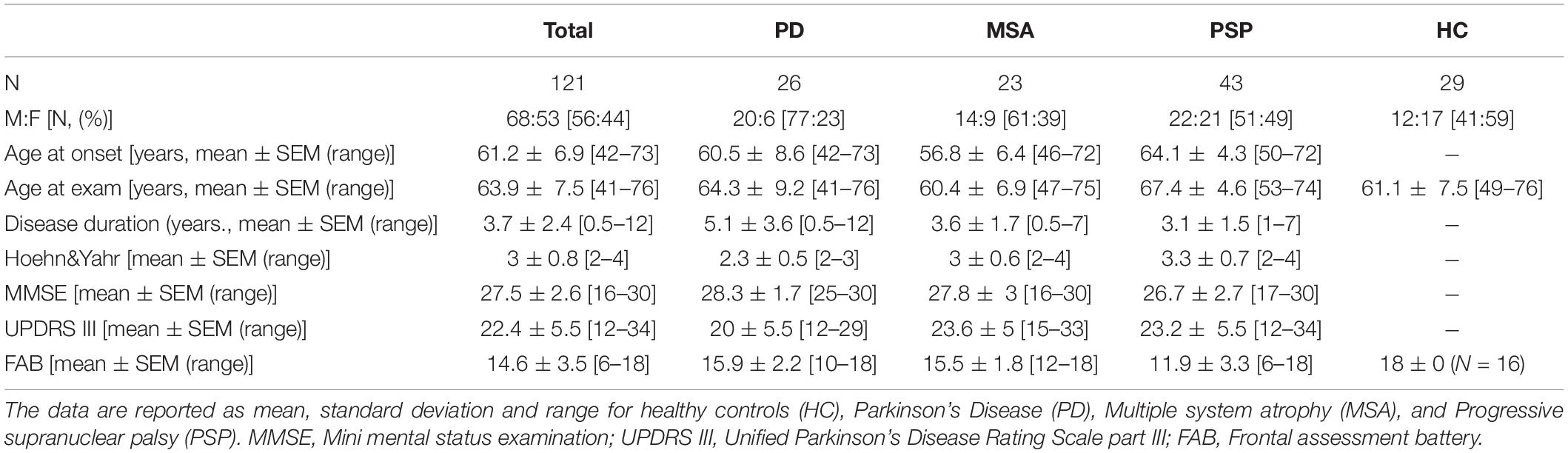
Table 1. Demographic and clinical data of the sample.
Clinical Scales
Clinical data of all participants are summarized in Table 1. The mean FAB score among all participants was 14.6 ± 3.5. In the PSP group, the FAB score was significantly lower compared to the PD, MSA and HC groups (Kruskal-Wallis test, p < 0.0001, post hoc Bonferroni PSP vs. PD p = 0.0003, PSP vs. MSA p = 0.048, PSP vs. HC P < 0.0001). In the PD and the MSA groups, the FAB scores were significantly lower compared to the healthy controls (Kruskal-Wallis-Test, p < 0.0001, post hoc Bonferroni PD vs. HC p = 0.027, MSA vs. HC p = 0.027).
The median H&Y stage among all participants was 3. The median H&Y stage was significantly lower in the PD group compared to the PSP and MSA groups (Kruskal-Wallis test, p < 0.0001, post hoc Bonferroni PD vs. MSA p = 0.006, PD vs. PSP p < 0001).
The mean MMSE score was 27.5 ± 2.6. In the PSP group, the MMSE score was significantly lower compared to the PD group (Kruskal-Wallis test, p = 0.01, post hoc Bonferroni p = 0.033). The mean score in the UPDRS part III among all patients was 27.5 (± 2.6). There were no significant differences in the subgroup analyses (Kruskal-Wallis test, p = 0.074). In the PSP group the mean PSPRS score was 42.3 (±8), in the MSA group the mean UMSARS score was 22.3 (±5.6).
Nucleus Basalis of Meynert Volumes
Figure 1 shows the anatomical localization of the nbM on a structural MRI template. The nbM volumes significantly differed between groups (ANOVA, p < 0.001) while controlling for intracranial volume (Figure 2). In PSP patients, nbM volumes were significantly lower compared to HC (Bonferroni post-hoc test, p < 0.001). Likewise, in PD patients, nbM volumes were significantly lower compared to HC (Bonferroni post-hoc test, p = 0.023). There was no significant difference in the nbM volumes between PSP and PD patients (Bonferroni post-hoc test, p = 1.000).
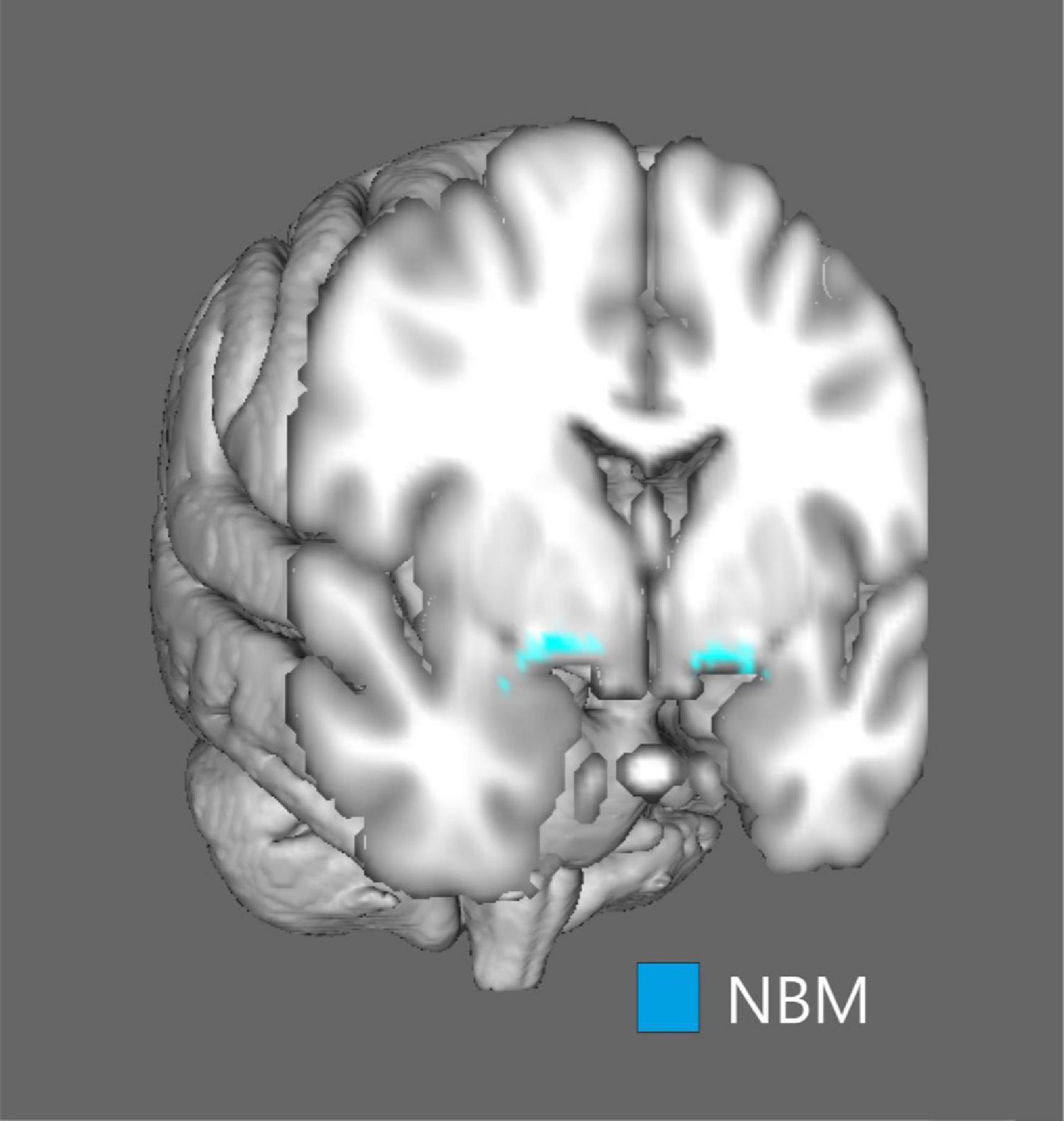
Figure 1. Visual representation of the nucleus basalis of Meynert. The mask of the nucleus basalis of Meynert was overlaid on a structural MRI surface template provided by the fMRI Software Library (FSL) software.
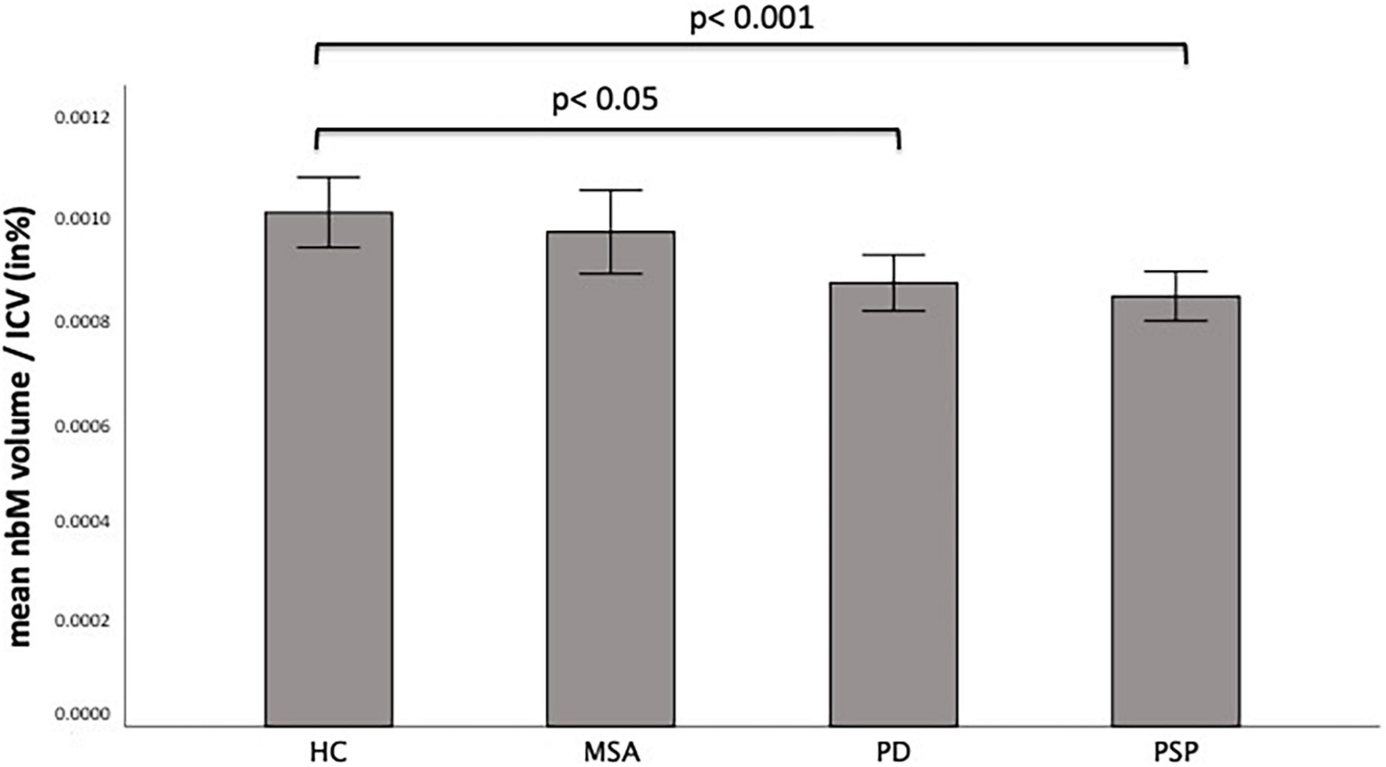
Figure 2. Mean nucleus of basalis Meynert (nbM) volumes corrected for intracranial volume (ICV) per diagnosis. The bar chart displays the nbM volume corrected for the individual ICV in percent in healthy controls (HC) and patients with Parkinson’s disease (PD), Multiple system atrophy (MSA), and Progressive supranuclear palsy (PSP). Significant differences are marked with the corresponding p-values.
Volumes of Other Brain Regions
The whole-brain gray matter (GM) volume significantly differed between all subgroups (ANOVA, p = 0.001) while controlling for intracranial volume (ICV) (Figure 3). Gray matter volume loss was largest in PSP patients compared to HC (Bonferroni post-hoc test, p = 0.0004). There was a significant difference in midbrain (MB) volume between all subgroups (ANOVA, p < 0.0001) while controlling for ICV.
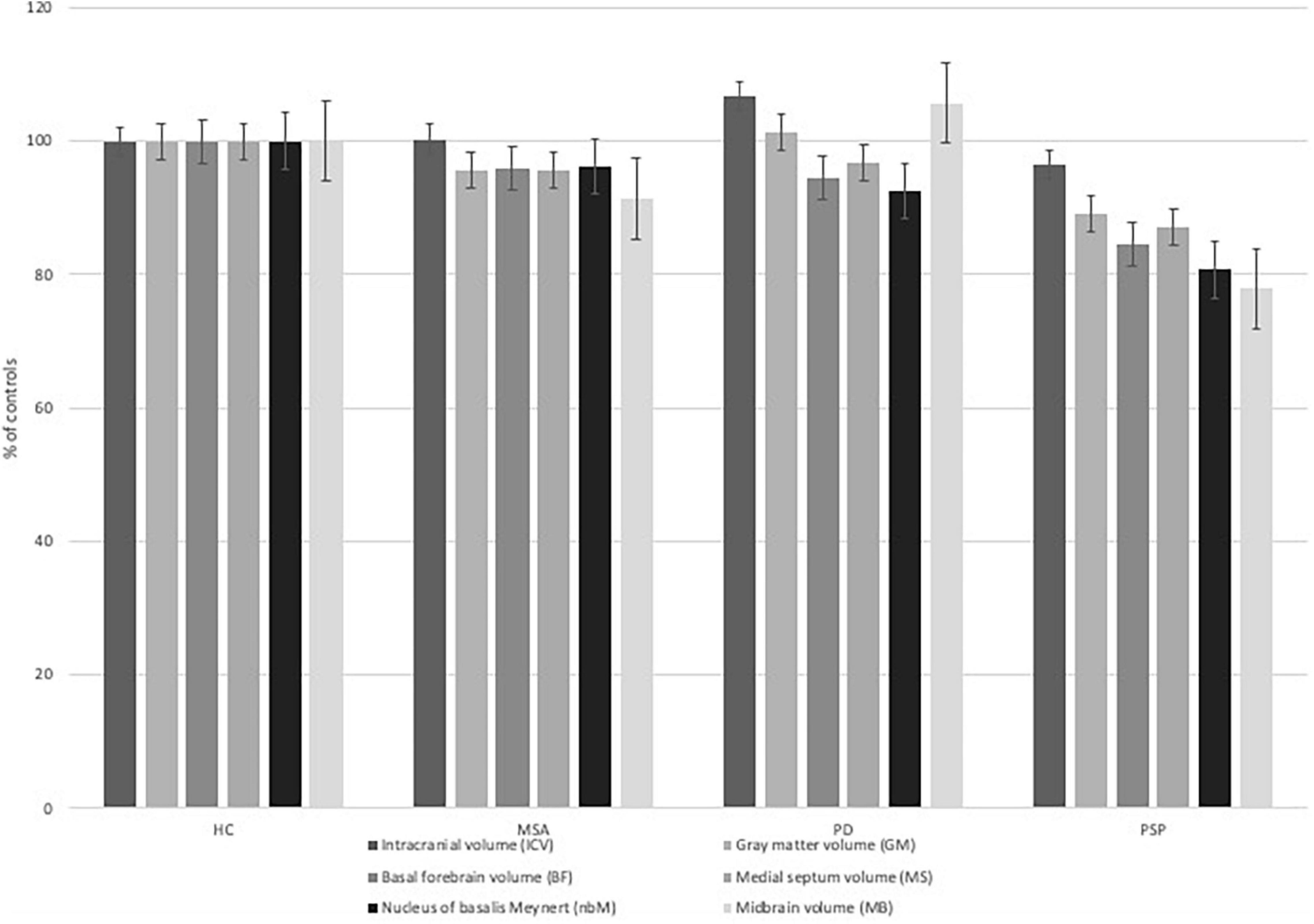
Figure 3. Brain volumes of interest per diagnosis in relation to healthy controls (HC). The bar chart displays different volumes of interest [Intracranial volume (ICV), gray matter volume (GM), basal forebrain volume (BF), medial septum volume (MS), nucleus of Meynert volume (nbM), midbrain volume (MB)] in relations to the volumes measured in healthy controls in percent.
The MB volume was significantly lower in MSA patients compared to HC (Bonferroni post-hoc test, p = 0.0005) and PD patients (Bonferroni post-hoc test, p = 0.003). The MB volume was significantly lower in PSP patients compared HC (Bonferroni post-hoc test, p < 0.0001), MSA patients (Bonferroni post-hoc test, p = 0.008), and PD patients (Bonferroni post-hoc test, p < 0.0001).
Correlations of Nucleus Basalis of Meynert Volume With Clinical Parameters
All correlations of nbM volume and clinical features are displayed in Table 2. Each individual hypothesis was tested with Bonferroni correction at α < 0.003. There was a trend of a negative correlation between age at examination and nbM volume in the PD group (Pearson correlation coefficient = −0.540, p = 0.004), although it did not reach statistical significance after Bonferroni correction. Also, there was no significant correlation between nbM volume and age at examination in the other subgroups (HC: Pearson correlation coefficient = −0.220, p = 0.252, MSA: Pearson correlation coefficient = −0.294, p = 0.174, PSP: Pearson correlation coefficient = −0.261, p = 0.92). There was no significant correlation between disease duration and nbM volume in PSP patients (Spearman’s rho = 0.285, p = 0.071).
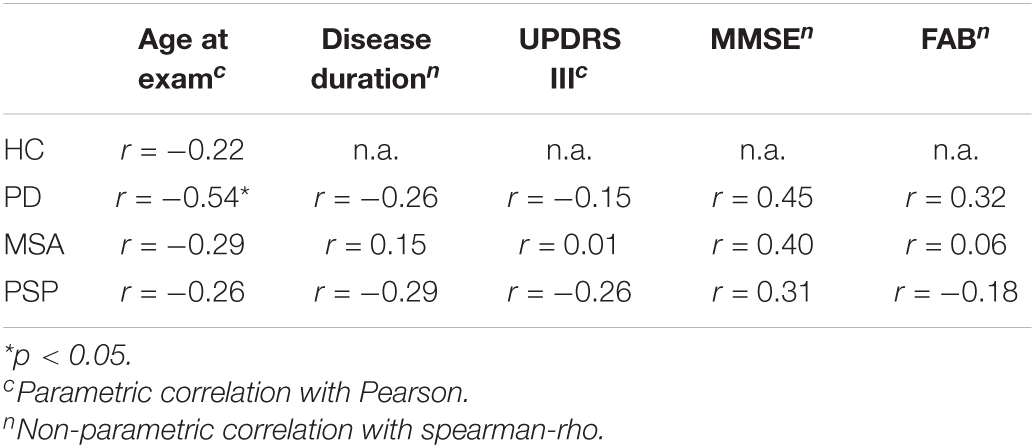
Table 2. Correlation of nbM volume and clinical features [healthy controls (HC), Parkinson’s Disease (PD), Multi system atrophy (MSA), Progressive supranuclear palsy (PSP), Mini mental status examination (MMSE), Unified Parkinson’s Disease Rating Scale part III (UPDRS III), Frontal assessment battery (FAB)].
There was no significant correlation between nbM volumes and FAB scores in any patient group (MSA: Spearman’s rho = 0.057, p = 0.861, PD: Spearman’s rho = 0.318, p = 0.139, PSP: Spearman’s rho = −0.180, p = 0.292).
There was no significant correlation between nbM volumes and MMSE in any patient group (MSA: Spearman’s rho = 0.396, p = 0.068 PD: Spearman’s rho = 0.448, p = 0.032 PSP: Spearman’s rho = 0.314, p = 0.111).
There was no significant correlation between nbM volumes and any of the motor scales, including H&Y stage (MSA: Spearman’s rho = −0.118, p = 0.59, PD: Spearman’s rho = −0.96, p = 0.663, PSP: Spearman’s rho = −0.66, p = 0.676), UPDRS III scores (MSA: Pearson correlation coefficient = −0.08, p = 0.972, PD: Pearson correlation coefficient = −0.149, p = 0.497, PSP: Pearson correlation coefficient = −0.257, p = 0.131), and PSPRS scores (Pearson correlation coefficient = −0.204, p = 0.19) as well as UMSARS scores (Pearson correlation coefficient = −0.208, p = 0.293).
Discussion
In this study, we investigated gray matter volumes of the nbM in patients with PSP, MSA, and PD as well as in HC, using volumetry in T1-weighted MRI, and correlated them with demographic data as well as with motor and cognitive symptoms. While robust MRI data on nbM atrophy in patients with PD were previously published (Ray et al., 2018; Gang et al., 2020), to our knowledge no studies had investigated nbM gray matter volumes in patients with PSP or MSA using MRI. Gray matter volume of the nbM was significantly lower in PSP and PD patients compared to controls. However, in neither subgroup a significant correlation with cognitive or other clinical scores could be detected.
In PD, MSA and PSP patients no significant correlation of disease duration an nbM volume was detected. These findings are in contrast to previously published neuropathological studies where a negative correlation of neuronal loss in the nbM and age at death was found (Tagliavini et al., 1984; Kasashima and Oda, 2003) suggesting that nbM atrophy may not reach significant dimensions in late onset patients. Complementary studies are necessary to confirm this hypothesis. There was no correlation between age and nbM volume in any patient subgroup or HC. But there was a strong trend for a negative correlation in the PD group without a negative correlation with disease duration. Further prospective studies with a larger number of patients are necessary to investigate other factors that may influence nBM atrophy in PD patients, such as age.
In previous studies from our group, gray matter atrophy in PSP was demonstrated predominantly in the midbrain, thalamus and basal ganglia, cerebellum and the frontal cortex. These results were consistent with the literature (Boxer et al., 2017), while in PD there was no pronounced atrophy (Fu et al., 2020). By means of histopathological examinations, a few studies previously demonstrated atrophy of the nbM in small case numbers of deceased PSP patients (Tagliavini et al., 1983, 1984; Rogers et al., 1985; Fu et al., 2020). Thanks to new MRI techniques and voxel-based volumetry it is now possible to perform an in vivo examination of the nbM. The nbM is subdivided into the anterior, the intermediate and the posterior nbM and previous studies demonstrated differential atrophy patterns of these subregions of the nbM in PD and AD. Some authors demonstrated a caudal-rostral pattern of cell loss in the nbM in AD (Liu et al., 2015), whereas in PD the intermediate part was most affected by cell loss (Liu et al., 2015). Most neuropathological studies have not investigated the relation between pattern of neuropsychological deficits and atrophy of the different parts of the nbM due to the lack of pre-mortem neuropsychological assessments. In our current study there was no correlation between nbM volume and cognition. However, only the MMSE was used as a screening tool for the detection of cognitive deficits and the FAB as a screening tool for executive dysfunctions. Some authors suggest a relation between pathologies in the anterior part of the nbM and executive dysfunction due to limbic and cortical innervations originating from this part (Liu et al., 2015). Since the occurrence of memory deficits is more pronounced in AD patients than in PD patients, it could be concluded that the posterior part of the nbM is more relevant for the occurrence of memory deficits (Liu et al., 2015). In our analysis, we did not differentiate between the different subregions of the nbM.
Overall, the cholinergic system and associated brain regions have only been investigated by a few studies in PSP patients before (Asahina et al., 1998; Warren et al., 2007a,b). Using PET-CT imaging with a ligand to muscarinic ACh receptors in a cohort of 7 PSP patients, 12 PD patients, and 8 HC, (Asahina et al., 1998) showed no evidence of relevant cholinergic cortical denervation in PSP patients. Using in vitro receptor autoradiography Warren et al. (2007b) demonstrated a reduction of M2 and M4 receptors in the posterior caudate and putamen, which could indicate a loss of posterior striatal cholinergic interneurons in PSP patients. Chui et al. (1986) investigated the role of non-dopaminergic systems in particular in the mid-cingulate cortex. In contrast to the previously mentioned studies, they did not find altered M2 or M4 receptor density in the caudate nuclei (Chui et al., 1986).
Unfortunately, trials with cholinesterase inhibitors (Litvan et al., 1989; Fabbrini et al., 2001) have not been successful for PSP patients so far. However, the present study underlines that impairment of the nbM occurs in PSP, which could potentially induce a decrease of ACh in the frontal cortex. However, since cognition did not correlate with the extent of atrophy of the nbM it seems reasonable that these impairments were more likely explained by atrophy of the frontal cortex itself. In contrast to these data, cholinesterase inhibitors have robust effects on cognition in PD patients, where nbM pathology is typically not accompanied by marked frontal cortex atrophy (Hanagasi et al., 2017). Further studies are necessary to confirm this hypothesis. Besides treatment with cholinesterase inhibitors, deep brain stimulation of the nbM came into focus as potentially promising treatment option for cognitive symptoms in patients with AD (Luo et al., 2021), LBD (Maltete et al., 2021), and PDD (Gratwicke et al., 2018). In relation to the data of this study, it can be assumed that this will not be a promising approach in PSP.
Limitations
In this study patients were included based only on clinical diagnosis and not confirmed by neuropathological examination. Magnet resonance microstructural analysis of the nbM may also help to elucidate pathological processes in this nucleus in vivo (Klietz et al., 2021). Further, longitudinal studies are needed to investigate nbM atrophy in PSP patients in the course of the disease.
Because only the MMSE and FAB were available in our cohort, we cannot draw conclusions about the spectrum of cognitive deficits. These screening tools cannot replace extensive neuropsychological testing (Tombaugh and McIntyre, 1992) in particular for executive functions, such as CERAD plus or Wisconsin Card Sorting Test (Lange et al., 2018; Lillig et al., 2021). A relevant limitation of this study is the age at examination. This parameter differs within the subgroups and is significantly higher in PSP patients compared to MSA and HC. An age-matched control group would be necessary to further clarify the findings of this study.
Conclusion
In conclusion, this study demonstrated a significant reduction of nbM volumes in PSP patients compared to a healthy control group. To our knowledge, this is the first in vivo MRI study showing this. A lower nbM volume was not significantly correlated with markers of cognitive performance (MMSE, FAB) in PSP patients. This suggests that atrophy of the nbM, and concomitant reductions in cholinergic innervation of forebrain structures, will only have a minor impact on cognition in PSP patients. Future neuropathological studies should address different pathologic processes in the nbM in different Parkinsonian syndromes and investigate regional specific atrophy in sub-segments of the nbM. Moreover, future imaging studies should use higher field strengths and correlate their findings with more extensive neuropsychological testing.
Data Availability Statement
The raw data supporting the conclusions of this article will be made available by the authors, without undue reservation.
Ethics Statement
The studies involving human participants were reviewed and approved by the Ethics committee, University of Marburg. The patients/participants provided their written informed consent to participate in this study.
Author Contributions
GH and JP: conception and design of the study. MK, GR, WO, and GH: acquisition of the data. SR, GR, MK, JP, and GH: analysis and interpretation of the data. SR, GR, and MK: drafting the manuscript. WO, MG, JP, and GH: revision of the manuscript for important intellectual content. All authors approved to the final version of the manuscript before submission.
Funding
MG was supported by the “Miguel Servet” program (CP19/00031) and a research grant (PI20/00613) of the Instituto de Salud Carlos III-Fondo Europeo de Desarrollo Regional (ISCIII-FEDER).
Conflict of Interest
The authors declare that the research was conducted in the absence of any commercial or financial relationships that could be construed as a potential conflict of interest.
Publisher’s Note
All claims expressed in this article are solely those of the authors and do not necessarily represent those of their affiliated organizations, or those of the publisher, the editors and the reviewers. Any product that may be evaluated in this article, or claim that may be made by its manufacturer, is not guaranteed or endorsed by the publisher.
Acknowledgments
We thank all participants for taking part in this study.
Footnotes
References
Albert, M. L., Feldman, R. G., and Willis, A. L. (1974). The ‘subcortical dementia’ of progressive supranuclear palsy. J. Neurol. Neurosurg. Psychiatry 37, 121–130. doi: 10.1136/jnnp.37.2.121
Allen, S. J., Dawbarn, D., and Wilcock, G. K. (1988). Morphometric immunochemical analysis of neurons in the nucleus basalis of Meynert in Alzheimer’s disease. Brain Res. 454, 275–281. doi: 10.1016/0006-8993(88)90827-x
Arendt, T., Bigl, V., Arendt, A., and Tennstedt, A. (1983). Loss of neurons in the nucleus basalis of Meynert in Alzheimer’s disease, paralysis agitans and Korsakoff’s Disease. Acta Neuropathol. 61, 101–108. doi: 10.1007/BF00697388
Asahina, M., Suhara, T., Shinotoh, H., Inoue, O., Suzuki, K., and Hattori, T. (1998). Brain muscarinic receptors in progressive supranuclear palsy and Parkinson’s disease: a positron emission tomographic study. J. Neurol. Neurosurg. Psychiatry 65, 155–163. doi: 10.1136/jnnp.65.2.155
Ashburner, J., and Friston, K. J. (2011). Diffeomorphic registration using geodesic shooting and Gauss-Newton optimisation. Neuroimage 55, 954–967. doi: 10.1016/j.neuroimage.2010.12.049
Bak, T. H., Caine, D., Hearn, V. C., and Hodges, J. R. (2006). Visuospatial functions in atypical parkinsonian syndromes. J. Neurol. Neurosurg. Psychiatry 77, 454–456. doi: 10.1136/jnnp.2005.068239
Bartus, R. T., Dean, R. L. III, Beer, B., and Lippa, A. S. (1982). The cholinergic hypothesis of geriatric memory dysfunction. Science 217, 408–414. doi: 10.1126/science.7046051
Boxer, A. L., Yu, J. T., Golbe, L. I, Litvan, I., Lang, A. E., and Höglinger, G. U. (2017). Advances in progressive supranuclear palsy: new diagnostic criteria, biomarkers, and therapeutic approaches. Lancet Neurol. 16, 552–563. doi: 10.1016/S1474-4422(17)30157-6
Burrell, J. R., Hodges, J. R., and Rowe, J. B. (2014). Cognition in corticobasal syndrome and progressive supranuclear palsy: a review. Mov. Disord. 29, 684–693. doi: 10.1002/mds.25872
Chui, H. C., Mortimer, J. A., Slager, U., Zarow, C., Bondareff, W., and Webster, D. D. (1986). Pathologic correlates of dementia in Parkinson’s disease. Arch. Neurol. 43, 991–995. doi: 10.1001/archneur.1986.00520100013007
Doucette, R., Fisman, M., Hachinski, V. C., and Mersky, H. (1986). Cell loss from the nucleus basalis of Meynert in Alzheimer’s disease. Can. J. Neurol. Sci. 13, 435–440. doi: 10.1017/s0317167100037070
Dubois, B., Slachevsky, A. I, Litvan, I., and Pillon, B. (2000). The FAB: a frontal assessment battery at bedside. Neurology 55, 1621–1626. doi: 10.1212/wnl.55.11.1621
Fabbrini, G., Barbanti, P., Bonifati, V., Colosimo, C., Gasparini, M., Vanacore, N., et al. (2001). Donepezil in the treatment of progressive supranuclear palsy. Acta Neurol. Scand. 103, 123–125. doi: 10.1034/j.1600-0404.2001.103002123.x
Folstein, M. F., Folstein, S. E., and McHugh, P. R. (1975). “Mini-mental state”. a practical method for grading the cognitive state of patients for the clinician. J. Psychiatr. Res. 12, 189–198.
Fu, T., Klietz, M., Nosel, P., Wegner, F., Schrader, C., Hoglinger, G. U., et al. (2020). Brain morphological alterations are detected in early-stage Parkinson’s disease with MRI morphometry. J. Neuroimaging 30, 786–792. doi: 10.1111/jon.12769
Gang, M., Baba, T., Hosokai, Y., Nishio, Y., Kikuchi, A., Hirayama, K., et al. (2020). Clinical and cerebral metabolic changes in parkinson’s disease with basal forebrain atrophy. Mov. Disord. 35, 825–832. doi: 10.1002/mds.27988
Gerstenecker, A., Mast, B., Duff, K., Ferman, T. J. I, Litvan, I., and Engene-Psp Study Group[Corporate Author] (2013). Executive dysfunction is the primary cognitive impairment in progressive supranuclear palsy. Arch. Clin. Neuropsychol. 28, 104–113. doi: 10.1093/arclin/acs098
Gibb, W. R. (1988). Accuracy in the clinical diagnosis of parkinsonian syndromes. Postgrad. Med. J. 64, 345–351. doi: 10.1136/pgmj.64.751.345
Gilman, S., Wenning, G. K., Low, P. A., Brooks, D. J., Mathias, C. J., Trojanowski, J. Q., et al. (2008). Second consensus statement on the diagnosis of multiple system atrophy. Neurology 71, 670–676. doi: 10.1212/01.wnl.0000324625.00404.15
Goetz, C. G., Tilley, B. C., Shaftman, S. R., Stebbins, G. T., Fahn, S., Martinez-Martin, P., et al. (2008). Movement disorder society-sponsored revision of the unified parkinson’s disease rating scale (MDS-UPDRS): scale presentation and clinimetric testing results. Mov. Disord. 23, 2129–2170. doi: 10.1002/mds.22340
Golbe, L. I., and Ohman-Strickland, P. A. (2007). A clinical rating scale for progressive supranuclear palsy. Brain 130, 1552–1565. doi: 10.1093/brain/awm032
Gratwicke, J., Zrinzo, L., Kahan, J., Peters, A., Beigi, M., Akram, H., et al. (2018). Bilateral deep brain stimulation of the nucleus basalis of meynert for parkinson disease dementia: a randomized clinical trial. JAMA Neurol. 75, 169–178. doi: 10.1001/jamaneurol.2017.3762
Hanagasi, H. A., Tufekcioglu, Z., and Emre, M. (2017). Dementia in Parkinson’s disease. J. Neurol. Sci. 374, 26–31.
Hoglinger, G. U., Respondek, G., Stamelou, M., Kurz, C., Josephs, K. A., Lang, A. E., et al. (2017). Movement disorder society-endorsed, clinical diagnosis of progressive supranuclear palsy: the movement disorder society criteria. Mov. Disord. 32, 853–864.
Kasashima, S., and Oda, Y. (2003). Cholinergic neuronal loss in the basal forebrain and mesopontine tegmentum of progressive supranuclear palsy and corticobasal degeneration. Acta Neuropathol. 105, 117–124. doi: 10.1007/s00401-002-0621-x
Kilimann, I., Grothe, M., Heinsen, H., Alho, E. J., Grinberg, L., Amaro, E. Jr., et al. (2014). Subregional basal forebrain atrophy in Alzheimer’s disease: a multicenter study. J. Alzheimers Dis. 40, 687–700. doi: 10.3233/JAD-132345
Klietz, M., Elaman, M. H., Mahmoudi, N., Nösel, P., Ahlswede, M., Wegner, F., et al. (2021). Cerebral microstructural alterationsin patients with early parkinson’sdisease detected with quantitativemagnetic resonance measurements. Front. Aging Neurosci. 13:763331. doi: 10.3389/fnagi.2021.763331
Kubler, D., Wellmann, S. K., Kaminski, J., Skowronek, C., Schneider, G. H., Neumann, W. J., et al. (2021). Nucleus basalis of Meynert predicts cognition after deep brain stimulation in Parkinson’s disease. Parkinsonism Relat. Disord. 94, 89–95. doi: 10.1016/j.parkreldis.2021.12.002
Lange, F., Bruckner, C., Knebel, A., Seer, C., and Kopp, B. (2018). Executive dysfunction in Parkinson’s disease: a meta-analysis on the wisconsin card sorting test literature. Neurosci. Biobehav. Rev. 93, 38–56. doi: 10.1016/j.neubiorev.2018.06.014
Lewy, F. (1913). Zur pathologischen anatomie der paralysis agitans. Dtsch Z Nervenheilkd. 50, 50–55.
Lillig, R., Ophey, A., Schulz, J. B., Reetz, K., Wojtala, J., Storch, A. I, et al. (2021). A new CERAD total score with equally weighted z-scores and additional executive and non-amnestic “CERAD-Plus” tests enhances cognitive diagnosis in patients with Parkinson’s disease: evidence from the LANDSCAPE study. Parkinsonism Relat. Disord. 90, 90–97. doi: 10.1016/j.parkreldis.2021.07.034
Litvan, I., Agid, Y., Calne, D., Campbell, G., Dubois, B., Duvoisin, R. C., et al. (1996). Clinical research criteria for the diagnosis of progressive supranuclear palsy (Steele-Richardson-Olszewski syndrome): report of the NINDS-SPSP international workshop. Neurology 47, 1–9. doi: 10.1212/wnl.47.1.1
Litvan, I., Gomez, C., Atack, J. R., Gillespie, M., Kask, A. M., Mouradian, M. M., et al. (1989). Physostigmine treatment of progressive supranuclear palsy. Ann. Neurol. 26, 404–407. doi: 10.1002/ana.410260318
Liu, A. K., Chang, R. C., Pearce, R. K., and Gentleman, S. M. (2015). Nucleus basalis of Meynert revisited: anatomy, history and differential involvement in Alzheimer’s and Parkinson’s disease. Acta Neuropathol. 129, 527–540. doi: 10.1007/s00401-015-1392-5
Luo, Y., Sun, Y., Tian, X., Zheng, X., Wang, X., Li, W., et al. (2021). Deep brain stimulation for alzheimer’s disease: stimulation parameters and potential mechanisms of action. Front. Aging Neurosci. 13:619543. doi: 10.3389/fnagi.2021.619543
Maltete, D., Wallon, D., Bourilhon, J., Lefaucheur, R., Danaila, T., Thobois, S., et al. (2021). Nucleus basalis of meynert stimulation for lewy body dementia: a phase i randomized clinical trial. Neurology 96, e684–e697. doi: 10.1212/WNL.0000000000011227
Martinez-Martin, P., Skorvanek, M., Rojo-Abuin, J. M., Gregova, Z., Stebbins, G. T., and Goetz, C. G. (2018). Validation study of the hoehn and yahr scale included in the MDS-UPDRS. Mov. Disord. 33, 651–652. doi: 10.1002/mds.27242
Ou, R., Liu, H., Hou, Y., Wei, Q., Cao, B., Zhao, B., et al. (2017). Executive dysfunction, behavioral changes and quality of life in Chinese patients with progressive supranuclear palsy. J. Neurol. Sci. 380, 182–186. doi: 10.1016/j.jns.2017.07.033
Pekmezoviæ, T., Jeèmenica-Lukiæ, M., Petroviæ, I., Špica, V., Tomiæ, A., Kostiæ, V. S., et al. (2015). Quality of life in patients with progressive supranuclear palsy: one-year follow-up. J. Neurol. 262, 2042–2048. doi: 10.1007/s00415-015-7815-3
Pereira, J. B., Hall, S., Jalakas, M., Grothe, M. J., Strandberg, O., Stomrud, E., et al. (2020). Longitudinal degeneration of the basal forebrain predicts subsequent dementia in Parkinson’s disease. Neurobiol. Dis. 139:104831. doi: 10.1016/j.nbd.2020.104831
Ray, N. J., Bradburn, S., Murgatroyd, C., Toseeb, U., Mir, P., Kountouriotis, G. K., et al. (2018). In vivo cholinergic basal forebrain atrophy predicts cognitive decline in de novo Parkinson’s disease. Brain 141, 165–176. doi: 10.1093/brain/awx310
Rittman, T., Ghosh, B. C., McColgan, P., Breen, D. P., Evans, J., Williams-Gray, C. H., et al. (2013). The addenbrooke’s cognitive examination for the differential diagnosis and longitudinal assessment of patients with parkinsonian disorders. J. Neurol. Neurosurg. Psychiatry 84, 544–551. doi: 10.1136/jnnp-2012-303618
Rogers, J. D., Brogan, D., and Mirra, S. S. (1985). The nucleus basalis of Meynert in neurological disease: a quantitative morphological study. Ann. Neurol. 17, 163–170. doi: 10.1002/ana.410170210
Santangelo, G., Cuoco, S., Pellecchia, M. T., Erro, R., Barone, P., and Picillo, M. (2018). Comparative cognitive and neuropsychiatric profiles between Parkinson’s disease, multiple system atrophy and progressive supranuclear palsy. J. Neurol. 265, 2602–2613. doi: 10.1007/s00415-018-9038-x
Schulz, J., Pagano, G., Fernandez Bonfante, J. A., Wilson, H., and Politis, M. (2018). Nucleus basalis of Meynert degeneration precedes and predicts cognitive impairment in Parkinson’s disease. Brain 141, 1501–1516. doi: 10.1093/brain/awy072
Tagliavini, F., Pilleri, G., Bouras, C., and Constantinidis, J. (1984). The basal nucleus of Meynert in patients with progressive supranuclear palsy. Neurosci. Lett. 44, 37–42. doi: 10.1016/0304-3940(84)90217-9
Tagliavini, F., Pilleri, G., Gemignani, F., and Lechi, A. (1983). Neuronal loss in the basal nucleus of Meynert in progressive supranuclear palsy. Acta Neuropathol. 61, 157–160. doi: 10.1007/BF00697397
Tombaugh, T. N., and McIntyre, N. J. (1992). The mini-mental state examination: a comprehensive review. J. Am. Geriatr. Soc. 40, 922–935. doi: 10.1111/j.1532-5415.1992.tb01992.x
Warren, N. M., Piggott, M. A., Greally, E., Lake, M., Lees, A. J., and Burn, D. J. (2007a). Basal ganglia cholinergic and dopaminergic function in progressive supranuclear palsy. Mov. Disord. 22, 1594–1600. doi: 10.1002/mds.21573
Warren, N. M., Piggott, M. A., Lees, A. J., and Burn, D. J. (2007b). The basal ganglia cholinergic neurochemistry of progressive supranuclear palsy and other neurodegenerative diseases. J. Neurol. Neurosurg. Psychiatry 78, 571–575. doi: 10.1136/jnnp.2006.099937
Keywords: progressive supranuclear palsy, cholinergic innervation, Parkinson’s disease, nucleus basalis of Meynert, voxel-based morphometry, subcortical dementia, multiple system atrophy
Citation: Rogozinski S, Klietz M, Respondek G, Oertel WH, Grothe MJ, Pereira JB and Höglinger GU (2022) Reduction in Volume of Nucleus Basalis of Meynert Is Specific to Parkinson’s Disease and Progressive Supranuclear Palsy but Not to Multiple System Atrophy. Front. Aging Neurosci. 14:851788. doi: 10.3389/fnagi.2022.851788
Received: 10 January 2022; Accepted: 24 February 2022;
Published: 01 April 2022.
Edited by:
Atsushi Takeda, National Hospital Organization Sendai Nishitaga Hospital, JapanCopyright © 2022 Rogozinski, Klietz, Respondek, Oertel, Grothe, Pereira and Höglinger. This is an open-access article distributed under the terms of the Creative Commons Attribution License (CC BY). The use, distribution or reproduction in other forums is permitted, provided the original author(s) and the copyright owner(s) are credited and that the original publication in this journal is cited, in accordance with accepted academic practice. No use, distribution or reproduction is permitted which does not comply with these terms.
*Correspondence: Sophia Rogozinski, cm9nb3ppbnNraS5zb3BoaWFAbWgtaGFubm92ZXIuZGU=