- 1Department of Chemistry and Biochemistry, The Ohio State University, Columbus, OH, United States
- 2The Comprehensive Cancer Center, The Ohio State University, Columbus, OH, United States
Proteomics, the study of the complete protein composition of a sample, is an important field for cancer research. Changes in the proteome can serve as a biomarker of cancer or lead to the development of a targeted therapy. This minireview will focus on mass spectrometry-based proteomics studies applied specifically to colorectal cancer, particularly the variety of cancer model systems used, including tumor samples, two-dimensional (2D) and three-dimensional (3D) cell cultures such as spheroids and organoids. A thorough discussion of the application of these systems will accompany the review of the literature, as each provides distinct advantages and disadvantages for colorectal cancer research. Finally, we provide conclusions and future perspectives for the application of these model systems to cancer research as a whole.
Introduction
Colorectal cancer is the third-most prevalent, and second-most deadly, cancer worldwide (Global Cancer Observatory, 2018). In the United States, it is estimated there will be 147,950 new cases of colorectal cancer and will claim over 50,000 lives in 2020 alone (American Cancer Society, 2020). Early diagnosis and various medical treatments can reduce the fatality rate, but it requires further understanding the intricacies of cancer formation, survival, and spread at the molecular level. As seen in a 2010 review, the field of omics has tremendous potential for new molecular discoveries that change the way we treat colorectal cancer (Nambiar et al., 2010). Omics research analyzes cancer at the molecular level. For example, a genomics study of colorectal cancer patients may discover a recurring mutation that could be a factor in tumor formation, while a metabolomics study has the potential to discover a small molecule secreted into the bloodstream that could be used as a diagnostic biomarker for the presence of colorectal cancer.
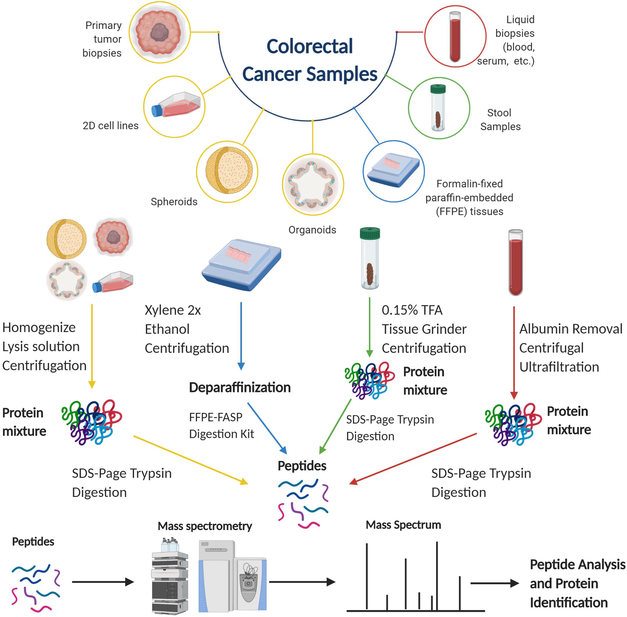
Proteomics is the study of proteins and can encompass the identification, quantification, localization, turnover, and regulation in a biological system. Liquid chromatography-mass spectrometry (LC-MS) has seen substantial use for proteomics in recent decades in which it has been utilized to obtain a global proteomic profile of a biological sample (Cravatt et al., 2007). In a mass spectrometry-based proteomics experiment, one method to obtain the identification and quantification of proteins is through bottom-up proteomics. As seen in Figure 1, proteins are extracted from a biological sample; the sample undergoes several steps to unfold all the proteins, reduce and alkylate the cysteine residues, before digestion into peptides by a proteolytic enzyme. The lysate undergoes chromatographic separation, typically by low-pH reversed-phase

Figure 1. Proteomics workflows for different colorectal cancer samples (Tirumalai et al., 2003; Ang et al., 2010; Quesada-Calvo et al., 2017). Figure created with BioRender.com.
chromatography before being analyzed by mass spectrometry. As peptides elute out of the column, the MS fragments the peptides to generate sequence-specific peaks. The methodology is outside the scope of this review; however, for a detailed explanation, Zhang et al. (2013) published an excellent overview of this approach.
Proteomics can be used in many facets of cancer research, beginning by being able to analyze the protein expressions in healthy and cancerous cells to identify differences in the protein abundance between the two populations. This information can be important to understand potential therapeutic targets in cancer cells. In addition to obtaining the basal proteome, mass spectrometry-based proteomics experiments can also be used to further evaluate the effect of a perturbation (adding a drug, knocking out a gene, etc.) by observing any proteomic alterations between control and treated groups. However, tumor heterogeneity is a major obstacle that has to be factored into any proteomics experiment due to the composition of cancer cells. Imperial et al. (2018) previously showed that the proteome throughout an individual tumor, as well between tumors at different locations, may be different. For a more detailed discussion of tumor heterogeneity, Lim and Lim (2018) provided an excellent review for solutions to overcoming this common problem. Additionally, it can be difficult to procure primary tumor samples and patient approval has to be obtained for their use. Due to these obstacles, it is often more favorable to use models that simulate the colorectal cancer proteome. With this in mind, researchers have developed several in vitro models, with varying degrees of complexity to study colorectal cancer.
This minireview highlights the use of mass spectrometry-based proteomics for primary colorectal tumor tissues studies and various models of colorectal cancer that are currently available. Since colorectal cancer proteomics is a rapidly evolving area of research, we will also provide our perspective on the future of this field in the new decade.
In vivo Colorectal Cancer Proteomics
In vivo colorectal cancer samples, samples obtained from either a patient or mouse model, are the most desirable source of proteomic information and have been used for colorectal cancer research for a number of years. Patient-derived in vivo samples can include primary tumor tissue, formalin-fixed paraffin-embedded (FFPE) tissue, liquid biopsies (plasma, serum, etc.), and stool samples. Much of the literature from the last 20 years focuses on these samples and has been previously reviewed (Tjalsma, 2010; Lee et al., 2018; Martins et al., 2019). In this section, we will briefly focus on previous studies using specifically primary tumor tissue samples, then more thoroughly discuss why in vitro model systems are useful and seeing increased popularity in the last decade.
Primary Tumors
For colorectal cancer research, no sample is more valuable than tissue directly from patients, as these samples provide the most accurate representation of the cancer’s proteome. Previously, the major focus of colorectal cancer proteomics research is the search for protein biomarkers for colorectal cancer, elucidated by comparing the proteomes of cancerous and non-cancerous tissue.
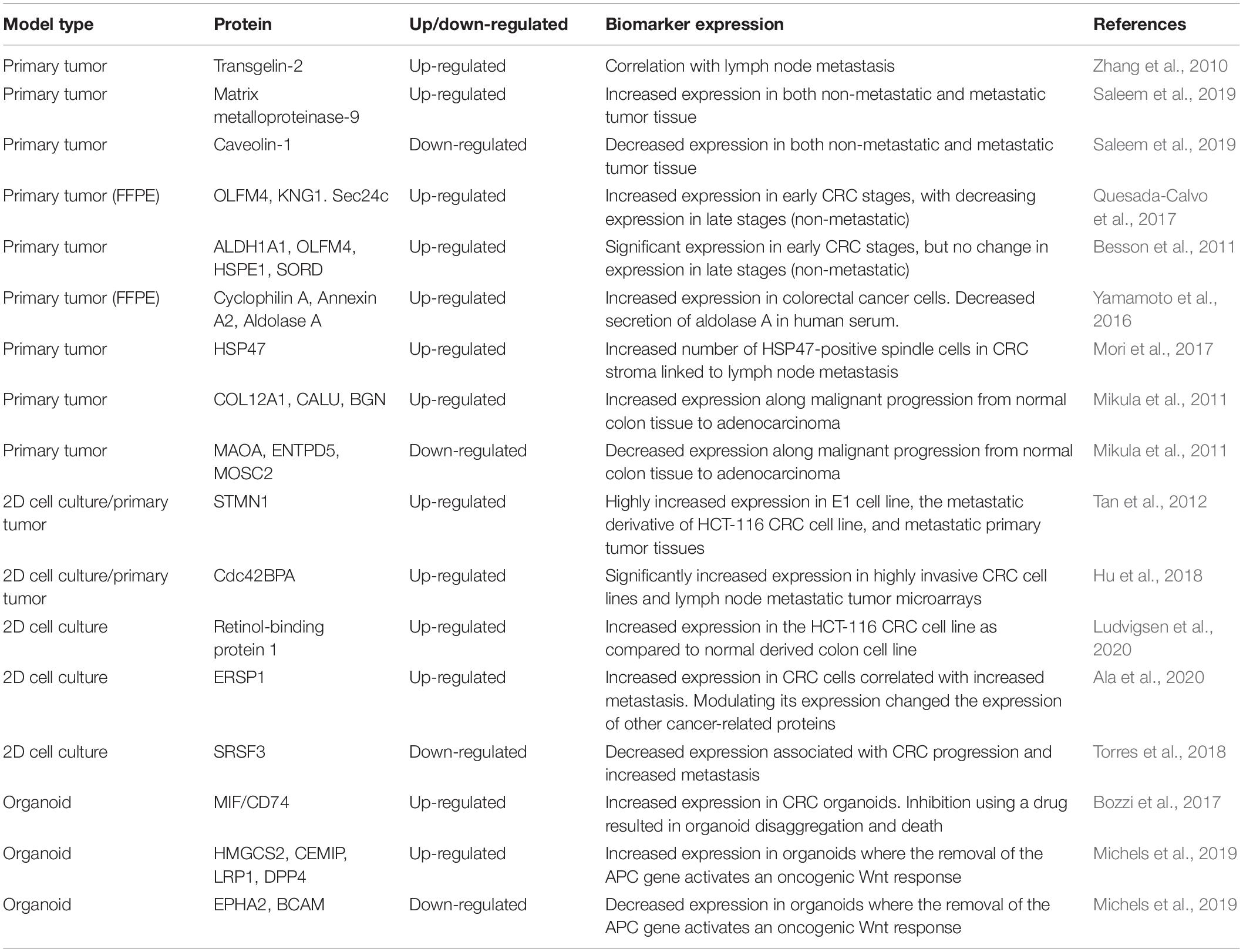
Protein biomarkers are proteins that are notably up- or down-regulated in the cancer proteome as compared to the normal proteome. Ideally, these proteins can serve as diagnostic markers of the presence of cancer, provide us with more information on cancer formation or survival, or possibly lead us to treatments that target these proteins. Using comparative proteomics with healthy and cancerous tissues, recent approaches show recognizable differences between these populations. The differences in protein abundance result in the identification of protein biomarkers, which are detailed in Table 1.
Examples of protein biomarkers include the protein OLFM4, found by two separate labs as a biomarker for colorectal cancer using bottom-up proteomics (Besson et al., 2011; Quesada-Calvo et al., 2017). Zhang et al. (2010) identified transgelin-2 as a biomarker of colorectal cancer, while Yamamoto et al. (2016) identified aldolase A. Mori et al. (2017) discovered shock protein 47 as a biomarker for the metastasis of colorectal cancer to lymph nodes. Mikula et al. (2011) used a combination of proteomics and transcriptomics to search for new protein biomarkers. Hao et al. (2017) used a novel algorithm to identify changes in abundance of entire protein pathways. They found elevated expression of proteins associated with chromatin modification and gene expression, but decreased expression of proteins responsible for core matrix architecture. These results would support the high mutation rate observed in cancer. Saleem et al. (2019) compared the proteomes of healthy tissue, non-adenomatous colon polyps, non-metastatic tumors, and metastatic tumors to identify proteins with elevated expression in the cancerous samples, such as matrix metalloproteinase-9, and decreased expression, such as caveolin-1. Similarly, Knol et al. (2014) compared benign and malignant tumor proteomes by enriching the protein fractions for chromatin-binding proteins, which had shown a difference previously (Albrethsen et al., 2010). Wisniewski et al. (2012) found that the proteome of colon adenocarcinoma tissue was significantly remodeled in comparison to normal tissue, which led them to compare the proteomes of colorectal mucosa, adenoma, and cancer (Wiśniewski et al., 2015). They noted significant changes in fatty acid metabolism and plasma membrane transporter proteins. New workflows using iTRAQ labeling (Jankova et al., 2011) or a centrifugal proteomic reactor (Liu et al., 2016) improve the data acquired from proteomic experiments. For further information on protein biomarkers, we suggest the following reviews: Tjalsma (2010); Lee et al. (2018), and Martins et al. (2019), which provide more information on recent advances and discoveries.
In contrast with the previously mentioned comparative studies, other cancer proteomics studies have utilized different approaches, for example, comparing the proteome with the genome or transcriptome. The relationship between the genome and proteome provides a more holistic view of colorectal cancer (Kang et al., 2012; Zhang et al., 2014; Vasaikar et al., 2019; Li et al., 2020).
In vitro Colorectal Cancer Proteomics
Primary human samples are the most valuable and accurate representations of human cancer, but they are a limited resource. A solution to this problem is the use of cultured models, which offer greater reproducibility and simplicity, and still have proteomes that are representative of primary colorectal cancer (Wang et al., 2017). The most common models for cancer research are cell lines. Cell lines have seen consistent use (Dumont et al., 2016) since the development of the HeLa cell line from cervical cancer in 1951 (Gey et al., 1952). While two-dimensional (2D) cell culture, where the cells are grown as a monolayer adhered to the bottom of the incubation flask, has been the most common use of cell lines, three-dimensional (3D) cell culture of multicellular spheroids is also possible. These cell cultures are called spheroids due to their shape, and for information on methodology, we would recommend a recent review from Chatzinikolaidou (2016). Even more recently, a new in vitro model called an organoid has become popular in cancer research. Several reviews on organoids exist (Clevers, 2016; Drost and Clevers, 2018; Xu et al., 2018), but their potential for proteomics is still relatively unexplored. In this section, we will review recent colorectal cancer proteomics research for all three in vitro models and discuss the inverse relationship between simplicity and proteomic accuracy as seen between them.
2D Cell Culture
Over the years, 2D cell culture has provided a simple, cost-effective model for cancer research and many cell lines have been developed. Importantly, 2D cell culture can be used to answer fundamental questions about cancer cells. Nusinow et al. (2020) recently used quantitative proteomics to screen the cell lines in the Cancer Cell Line Encyclopedia (CCLE). The CCLE is a large-scale database of over 1,000 cancer cell lines and includes information regarding gene expression, genome sequencing, metabolite profiling, drug sensitivity screens, and targeted protein quantification. The CCLE did not have deep proteome profiling until this study. In addition, similar to primary tumor samples, colorectal proteomics using 2D cell culture has recently focused on utilizing a comparison discovery-based approach to identifying protein biomarkers. Tan et al. (2012) used the colon cancer cell line, HCT-116, to identify Stathmin-1 as a protein biomarker of cancer cell migration. Ala et al. (2020) identified the pro-oncogenic role of ERSP1 in the colorectal cell lines HCA24 and COLO320DM. Hu et al. (2018) used HCT-116 and RKO, selectively cultured to be highly invasive, to identify the Cdc42-Cdc42BPA signaling pathway as a marker for colon cancer invasion. Ludvigsen et al. (2020) compared a normal derived colon mucosa cell line (NCM460) and a colon cancer cell line (HCT116) to identify protein biomarkers with increased expression in colorectal cancer, including retinol-binding protein 1. 2D cell culture has also been used to compare non-metastatic (SW480) and metastatic colon cancer cell lines (SW620) (Zhao et al., 2007; Schunter et al., 2017; Torres et al., 2018). These two cell lines derive from the same patient (Stage II for SW480 and Stage III for SW620). Although these two cell lines form poor spheroids (Stadler et al., 2018), they can provide valuable insight into proteins that may determine whether a cancer will metastasize.
One feature of cell lines not available for primary tumor samples is in vitro manipulation. These manipulations include dosing with anticancer drugs (Bauer et al., 2012; Valdes et al., 2017; Raimondo et al., 2018; Schroll et al., 2018), inducing autophagy using hypoxia (Lai et al., 2016), and binding growth inhibitors (Michalak et al., 2016), followed by downstream proteomics, an analysis that looks for differences in the proteome after the manipulation. Fanayan et al. (2013) took a different approach and performed a bottom-up proteomics experiment using chromosome-based data analysis. The proteomic data were organized into clusters based on the MS-based data and the chromosomal location of genes. One cluster identified with this method included several tumor suppressor proteins, including caveolin-1 and caveolin-2, associated with a region on chromosome 7 often deleted in the pathogenesis of colorectal cancer.
As seen above, there are many aspects of 2D cell culture that make them desirable for colorectal cancer proteomics. However, due to a lack of 3D structure, vasculature, and other in vivo tumor properties, information obtained from 2D cell culture is incomplete. With the introduction of 3D spheroids and organoids, their ability to provide a more biologically accurate environment (reviewed by Yamada and Cukierman, 2007) helps to fill this gap in the knowledgebase. Furthermore, most of the in vitro manipulations performed above can also be used for these models as well. Although 2D cell culture will never become obsolete due to its ease and simplicity, 3D cell cultures have increasingly become more common as models for colorectal cancer.
3D Cell Culture
Since 2D cell cultures do not have the same structural features as tumors, there is an inherent loss of spatial information and the proteome will not be truly representative of a tumor. With the introduction of spheroids, the cellular environments of tumors are more accurately replicated in vitro, while still providing many of the same advantages of 2D cell culture, for instance, ease of use, versatility, and lower experimental cost. Alternatively, a shared disadvantage between 2D cell culture and spheroids is the presence of only cancer cells, so information on the interaction between healthy and cancerous cells that would be present in vivo is lost. Another in vitro model, organoids, can be grown to contain both populations, solving this issue and providing a more accurate mimetic (Johnson et al., 2020). This section will review recent proteomic literature for both spheroids and organoids.
Spheroids
The first proteomics investigation of spheroids compared the spheroids’ proteome with their 2D counterparts. Yue et al. (2016) compared both the proteome and phosphoproteome between spheroids and 2D cell cultures of HT29, a colon cell line. Observation of reproducible changes in abundance for several proteins between the populations suggested that spheroids may provide more accurate proteomic information compared to 2D monolayer cultures. Certain proteins and pathways that showed a change in the spheroids were associated with slower growth and decreased cell and DNA replication, mimicking in vivo conditions. In a subsequent study, Kim et al. (2018) compared the proteomes of 3D and 2D SW480 cell cultures after the addition of an enzyme inhibitor. Another colon cell line frequently used for spheroids is HCT-116, which Feist et al. (2015) used to detect over 1350 proteins with very little variation between the replicate spheroids. McMahon et al. (2012) characterized the differences in the proteome between the three cellular populations in spheroids, the proliferating outer layer, the senescent middle layer, and the necrotic core, for the HT29 cell line. These cellular populations are also present in in vivo conditions, making this study particularly valuable for comparisons between in vivo and in vitro models. In addition to spheroids grown using immortalized cell lines, Rajcevic et al. (2014) used patient tumor material to produce spheroids with more in vivo tissue characteristics than cell line-based spheroids.
Recent studies in our laboratory have used spheroids to test chemotherapy drugs and the effects of glucose. LaBonia et al. (2018) tested the proteomic changes that occur with treatment of FOLFIRI (folinic acid, 5-fluorouracil, and irinotecan), a combination chemotherapy, on HCT-116 spheroids and observed that folinic acid penetrated into the core of the spheroids and several cancer-associated protein pathways were enriched. Schroll et al. (2016) showed that restricting glucose or serum from the growing spheroids resulted in similar proteomic changes. The effect of combining glucose restriction with chemotherapies has also been examined using spheroids (Schroll et al., 2017, 2018). Feist et al. (2017) used spheroids to evaluate differences in histone post-translational modifications and the effect of an epigenetic drug that targets these modifications. The use of spheroids in the proteomics studies above provides important information about how a treatment might affect an in vivo tumor, without testing the conditions on actual patients. These data are the first step toward verifying possible treatments for cancer that could be explored in clinical studies.
Organoids
Organoids are a relatively recent development in comparison to the other models discussed in this minireview. Xu et al. (2018) define organoids as “3D constructs” that can be developed from “embryonic stem cells, induced pluripotent stem cells, somatic cells, and cancer cells.” The use of stem cells is especially important for this technology, in that these cells can be used to make a variety of organoids, including intestinal organoids. Colorectal tumor organoids have very high potential for screening and thus the possibility for the development of organoid libraries has been explored (van de Wetering et al., 2015; Fujii et al., 2016). Another benefit to organoids is that non-cancerous organoids can be grown, so comparisons between healthy and cancer proteomes can be made, something not possible for 2D cell culture and spheroids.
One major disadvantage for organoid research is the use of a substance called Matrigel, an extracellular matrix necessary for preparing and embedding organoids. Unfortunately, Matrigel contains a variety of growth factor proteins that cause severe ion suppression (Abe et al., 2018). In their research, Abe and his colleagues performed phosphoproteomics analysis on colorectal cancer spheroids and organoids embedded in Matrigel. To avoid the ion suppression effects, they incorporated an acetone precipitation of the digested peptides in their workflow, which increased the percentage of the MS/MS spectra associated with peptides from 8.8 to 26.9%.
Gonneaud et al. (2017) wrote an excellent review of mass spectrometry-based proteomics using colon organoids. Bozzi et al. (2017) identified the MIF/CD74 axis as a target for therapeutics in colorectal cancer, in that its inhibition caused cancer cells to be vulnerable to oxidative stress-induced death. More recently, Lindeboom et al. (2018) used a multi-omics (metabolomics, proteomics, transcriptomics, epigenomics, and genomics) analysis to obtain a complete intramolecular view of intestinal organoids. Michels et al. (2019) used proteomic and transcriptomic profiling of colon organoids to observe physiologic and oncogenic responses to Wnt signaling, such as the up-regulated proteins HMGCS2 and CEMIP. Noticeably, proteomics data from colorectal organoids have been limited in the last couple of years, opening the door for novel developments going forward.
Future Directions/Conclusion
In this review, the various sample types used for recent colorectal cancer proteomics are described. In vivo and in vitro samples offer different advantages for research, and within the in vitro samples, differing degrees of complexity are also present. After reviewing the recent literature, the authors offer their thoughts on the future of colorectal cancer research. We believe that although primary tumor samples will continue to be valuable research tools, the application of proteomics to spheroids and organoids will continue to see increased representation as cell culture technology progresses and proteomics methodologies improve. These in vitro models demonstrate greater statistical reproducibility, due to the lack of tumor heterogeneity, and still maintain a relatively representative proteome. In addition, as these models are grown, they are more readily available than tumor samples that need to be collected from patients. While there are still challenges that need to be overcome, in vitro models provide a viable alternative to primary tumor samples, not only for colorectal cancer, but many other cancers as well.
Author Contributions
PL wrote the manuscript. AH helped revise the manuscript. Both authors contributed to the article and approved the submitted version.
Funding
PL was supported by a University Fellowship from The Ohio State University. AH was supported by R01-GM110406 from the NIH.
Conflict of Interest
The authors declare that the research was conducted in the absence of any commercial or financial relationships that could be construed as a potential conflict of interest.
Acknowledgments
We thank Drs. Ralph Tobias and Emily Sekera, as well as Ariana E. Shannon and Nicole C. Beller for their careful reading of the manuscript and thoughtful suggestions.
References
Abe, Y., Tada, A., Isoyama, J., Nagayama, S., Yao, R., Adachi, J., et al. (2018). Improved phosphoproteomic analysis for phosphosignaling and active-kinome profiling in Matrigel-embedded spheroids and patient-derived organoids. Sci. Rep. 8:11401. doi: 10.1038/s41598-018-29837-1
Ala, U., Manco, M., Mandili, G., Tolosano, E., Novelli, F., Provero, P., et al. (2020). Proteomics-Based Evidence for a Pro-Oncogenic Role of ESRP1 in Human Colorectal Cancer Cells. Int. J. Mol. Sci. 21:575. doi: 10.3390/ijms21020575
Albrethsen, J., Knol, J., Piersma, S., Pham, T., de Wit, M., Mongera, S., et al. (2010). Subnuclear proteomics in colorectal cancer: identification of proteins enriched in the nuclear matrix fraction and regulation in adenoma to carcinoma progression. Mol. Cell. Proteomics 9, 988–1005. doi: 10.1074/mcp.M900546-MCP200
American Cancer Society (2020). Key Statistics for Colorectal Cancer. Available online at https://www.cancer.org/cancer/colon-rectal-cancer/about/key-statistics.html#:~:text=Excluding%20skin%20cancers%2C%20colorectal%20cancer,new%20cases%20of%20rectal%20cancer [Accessed July 1, 2020]
Ang, C., Rothacker, J., Patsiouras, H., Burgess, A., and Nice, E. (2010). Murine fecal proteomics: A model system for the detection of potential biomarkers for colorectal cancer. J. Chromatogr. A. 1217, 3330–3340. doi: 10.1016/j.chroma.2009.10.007
Bauer, K. M., Lambert, P. A., and Hummon, A. B. (2012). Comparative label-free LC -MS/MS analysis of colorectal adenocarcinoma and metastatic cells treated with 5-fluorouracil. Proteomics 12, 1928–1937. doi: 10.1002/pmic.201200041
Besson, D., Pavageau, A., Valo, I., Bourreau, A., Belanger, A., Eymerit-Morin, C., et al. (2011). A Quantitative Proteomic Approach of the Different Stages of Colorectal Cancer Establishes OLFM4 as a New Nonmetastatic Tumor Marker. Mol. Cell. Proteomics 10:9712. doi: 10.1074/mcp.M111.009712
Bozzi, F., Mogavero, A., Varinelli, L., Belfiore, A., Manenti, G., Caccia, C., et al. (2017). MIF/CD74 axis is a target for novel therapies in colon carcinomatosis. J. Exp. Clin. Cancer Res. 36:16. doi: 10.1186/s13046-016-0475-z
Chatzinikolaidou, M. (2016). Cell spheroids: the new frontiers in in vitro models for cancer drug validation. Drug Discov. Today 21, 1553–1560. doi: 10.1016/j.drudis.2016.06.024
Clevers, H. (2016). Modeling development and disease with organoids. Cell 165, 1586–1597. doi: 10.1016/j.cell.2016.05.082
Cravatt, B., Simon, G., and Yates, J. III (2007). The biological impact of mass-spectrometry-based proteomics. Nature 450, 991–1000. doi: 10.1038/nature06525
Drost, J., and Clevers, H. (2018). Organoids in cancer research. Nat Rev Cancer 18, 407–418. doi: 10.1038/s41568-018-0007-6
Dumont, J., Euwart, D., Mei, B., Estes, S., and Kshirsagar, R. (2016). Human cell lines for biopharmaceutical manufacturing: history, status, and future perspectives. Crit. Rev. Biotechnol. 36, 1110–1122. doi: 10.3109/07388551.2015.1084266
Fanayan, S., Smith, J., Sethi, M., Cantor, D., Goode, R., Simpson, R., et al. (2013). Chromosome 7-Centric Analysis of Proteomics Data from a Panel of Human Colon Carcinoma Cell Lines. J. Proteom. Res. 12, 89–96. doi: 10.1021/pr300906y
Feist, P., Sidoli, S., Liu, X., Schroll, M., Rahmy, S., Fujiwara, R., et al. (2017). Multicellular Tumor Spheroids Combined with Mass Spectrometric Histone Analysis To Evaluate Epigenetic Drugs. Anal. Chem. 89, 2773–2781. doi: 10.1021/acs.analchem.6b03602
Feist, P., Sun, L., Liu, X., Dovichi, N., and Hummon, A. (2015). Bottom-up proteomic analysis of single HCT 116 colon carcinoma multicellular spheroids. Rapid Commun. Mass Spectrom 29, 654–658. doi: 10.1002/rcm.7150
Fujii, M., Shimokawa, M., Date, S., Takano, A., Matano, M., Nanki, K., et al. (2016). A Colorectal Tumor Organoid Library Demonstrates Progressive Loss of Niche Factor Requirements during Tumorigenesis. Cell Stem Cell 18, 827–838. doi: 10.1016/j.stem.2016.04.003
Gey, G., Coffman, W., and Kubicek, M. (1952). Tissue culture studies of the proliferative capacity of cervical carcinoma and normal epithelium. Cancer Res. 12, 264–265.
Global Cancer Observatory (2018). Colorectal Cancer. Available online at https://gco.iarc.fr/today/data/factsheets/cancers/10_8_9-Colorectum-fact-sheet.pdf [Accessed July 1, 2020].
Gonneaud, A., Asselin, C., Boudreau, F., and Boisvert, F. (2017). Phenotypic Analysis of Organoids by Proteomics. Proteomics 17:1700023. doi: 10.1002/pmic.201700023
Hao, J., Zhi, X., Wang, Y., Zhang, Z., Hao, Z., Ye, R., et al. (2017). Comprehensive Proteomic Characterization of the Human Colorectal Carcinoma Reveals Signature Proteins and Perturbed Pathways. Sci. Rep. 7:42436. doi: 10.1038/srep42436
Hu, H., Xu, W., Wang, Y., Zheng, C., Zhang, W., Li, B., et al. (2018). Comparative Proteomics Analysis Identifies Cdc42-Cdc42BPA Signaling as Prognostic Biomarker and Therapeutic Target for Colon Cancer Invasion. J. Proteom. Res. 17, 265–275. doi: 10.1021/acs.jproteome.7b00550
Imperial, R., Ahmed, Z., Toor, O., Erdogan, C., Khaliq, A., Case, P., et al. (2018). Comparative proteogenomic analysis of right-sided colon cancer, left-sided colon cancer and rectal cancer reveals distinct mutational profiles. Mol. Cancer 17:177. doi: 10.1186/s12943-018-0923-9
Jankova, L., Chan, C., Fung, C., Song, X., Kwun, S., Cowley, M., et al. (2011). Proteomic comparison of colorectal tumors and non-neoplastic mucosa from paired patient samples using iTRAQ mass spectrometry. Mol. Biosyst. 7, 2997–3005. doi: 10.1039/C1MB05236E
Johnson, K., DeStefanis, R., Emmerich, P., Grogan, P., Kratz, J., Makkar, S., et al. (2020). Human Colon Organoids and Other Laboratory Strategies to Enhance Patient Treatment Selection. Curr. Treat. Options Oncol. 21:35. doi: 10.1007/s11864-020-00737-9
Kang, U., Yeom, J., Kim, H., Kim, H., and Lee, C. (2012). Expression profiling of more than 3500 proteins of MSS-type colorectal cancer by stable isotope labeling and mass spectrometry. J. Proteom. 75, 3050–3062. doi: 10.1016/j.jprot.2011.11.021
Kim, Y., Jeon, H., Kim, D., Lee, S., Kim, K., Hong, J., et al. (2018). Quantitative Proteomic Analysis of 2D and 3D Cultured Colorectal Cancer Cells: Profiling of Tankyrase Inhibitor XAV939-Induced Proteome. Sci. Rep. 8:13255. doi: 10.1038/s41598-018-31564-6
Knol, J., de Wit, M., Albrethson, J., Piersma, S., Pham, T., Mongera, S., et al. (2014). Proteomics of differential extraction fractions enriched for chromatin-binding proteins from colon adenoma and carcinoma tissues. BBA Protein Proteom. 1844, 1034–1043. doi: 10.1016/j.bbapap.2013.12.006
LaBonia, G., Ludwig, K., Mousseau, C. B., and Hummon, A. (2018). iTRAQ Quantitative Proteomic Profiling and MALDI–MSI of Colon Cancer Spheroids Treated with Combination Chemotherapies in a 3D Printed Fluidic Device. Anal. Chem. 90, 1423–1430. doi: 10.1021/acs.analchem.7b04969
Lai, M., Chang, C., and Sun, H. (2016). Hypoxia Induces Autophagy through Translational Up-Regulation of Lysosomal Proteins in Human Colon Cancer Cells. PLoS One. 11:e0153627. doi: 10.1371/journal.pone.0153627
Lee, P. Y., Chin, S., Low, T. Y., and Jamal, R. (2018). Probing the colorectal cancer proteome for biomarkers: Current status and perspectives. J. Prot. 187, 93–105. doi: 10.1016/j.jprot.2018.06.014
Li, C., Sun, Y., Yu, G., Cui, J., Lou, Z., Zhang, H., et al. (2020). Integrated Omics of Metastatic Colorectal Cancer. Cancer Cell 38, 1–14. doi: 10.1016/j.ccell.2020.08.002
Lim, L. C., and Lim, Y. M. (2018). Proteome heterogeneity in colorectal cancer. Proteomics 18:1700169. doi: 10.1002/pmic.201700169
Lindeboom, R., van Voorthuijsen, L., Oost, K., Rodriguez-Colman, M., Luna-Velez, M., Furlan, C., et al. (2018). Integrative multi-omics analysis of intestinal organoid differentiation. Mol. Syst. Biol. 14:e8227. doi: 10.15252/msb.20188227
Liu, X., Xu, Y., Meng, Q., Zheng, Q., Wu, J., Wang, C., et al. (2016). Proteomic analysis of minute amounts of colonic biopsies by enteroscopy sampling. Biochem. Biophys. Res. Commun. 476, 286–292. doi: 10.1016/j.bbrc.2016.05.114
Ludvigsen, M., Thorlacius-Ussing, L., Vorum, H., Moyer, M., Stender, M., Thorlacius-Ussing, O., et al. (2020). Proteomic characterization of colorectal cancer cells versus normal-derived colon mucosa cells: approaching identification of novel diagnostic protein biomarkers in colorectal cancer. Int. J. Mol. Sci. 21:3466. doi: 10.3390/ijms21103466
Martins, B., de Bulhões, G., Cavalcanti, I., Matins, M., de Oliveira, P., and Martins, A. (2019). Biomarkers in Colorectal Cancer: The Role of Translational Proteomics Research. Front. Oncol. 9:1284. doi: 10.3389/fonc.2019.01284
McMahon, K., Volpato, M., Chi, H., Musiwaro, P., Poterlowicz, K., Peng, Y., et al. (2012). Characterization of Changes in the Proteome in Different Regions of 3D Multicell Tumor Spheroids. J. Proteom. Res. 11, 2863–2875. doi: 10.1021/pr2012472
Michalak, M., Warnken, U., Andre, S., Schnolzer, M., Gabius, H., and Kopitz, J. (2016). Detection of Proteome Changes in Human Colon Cancer Induced by Cell Surface Binding of Growth-Inhibitory Human Galectin-4 Using Quantitative SILAC-Based Proteomics. J. Proteom. Res. 15, 4412–4422. doi: 10.1021/acs.jproteome.6b00473
Michels, B., Mosa, M., and Grebbin, B. (2019). Human colon organoids reveal distinct physiologic and oncogenic Wnt responses. J. Exp. Med. 216, 704–720. doi: 10.1084/jem.20180823
Mikula, M., Rubel, T., Karczmarski, J., Goryca, K., Dadlez, M., and Ostrowski, J. (2011). Integrating proteomic and transcriptomic high-throughput surveys for search of new biomarkers of colon tumors. Funct. Integr. Genomics 11, 215–224. doi: 10.1007/s10142-010-0200-5
Mori, K., Toiyama, Y., Otake, K., Fujikawa, H., Saigusa, S., Hiro, J., et al. (2017). Proteomics analysis of differential protein expression identifies heat shock protein 47 as a predictive marker for lymph node metastasis in patients with colorectal cancer. Int. J. Cancer 140, 1425–1435. doi: 10.1002/ijc.30557
Nambiar, P., Gupta, R., and Misra, V. (2010). An “Omics” based survey of human colon cancer. Mutat. Res.-Fund. Mol. M. 693, 3–18. doi: 10.1016/j.mrfmmm.2010.07.008
Nusinow, D., Szpyt, J., Ghandi, M., Rose, C., McDonald, E. R. III, Kalocsay, M., et al. (2020). Quantitative proteomics of the cancer cell line encyclopedia. Cell 180, 387–402. doi: 10.1016/j.cell.2019.12.023
Quesada-Calvo, F., Massot, C., Bertrand, V., Longuespee, R., Bletard, N., Somja, J., et al. (2017). OLFM4, KNG1 and Sec24C identified by proteomics and immunohistochemistry as potential markers of early colorectal cancer stages. Clin. Proteom. 14:9. doi: 10.1186/s12014-017-9143-3
Raimondo, S., Saieva, L., Cristaldi, M., Monteleone, F., Fontana, S., and Alessandro, R. (2018). Label-free quantitative proteomic profiling of colon cancer cells identifies acetyl-CoA carboxylase alpha as antitumor target of Citrus limon-derived nanovesicles. J. Proteom. 173, 1–11. doi: 10.1016/j.jprot.2017.11.017
Rajcevic, U., Knol, J., Piersma, S., Bougnaud, S., Fack, F., Sundlisaeter, E., et al. (2014). Colorectal cancer derived organotypic spheroids maintain essential tissue characteristics but adapt their metabolism in culture. Proteom. Sci. 12:39. doi: 10.1186/1477-5956-12-39
Saleem, S., Tariq, S., Aleem, I., Shaheed, S., Tahseen, M., and Atiq, A. (2019). Proteomics analysis of colon cancer progression. Clin. Proteom. 16:44. doi: 10.1186/s12014-019-9264-y
Schroll, M., LaBonia, G., Ludwig, K., and Hummon, A. (2017). Glucose Restriction Combined with Autophagy Inhibition and Chemotherapy in HCT 116 Spheroids Decreases Cell Clonogenicity and Viability Regulated by Tumor Suppressor Genes. J. Proteom. Res. 16, 3009–3018. doi: 10.1021/acs.jproteome.7b00293
Schroll, M., Liu, X., Herzog, S., Skube, S., and Hummon, A. (2016). Nutrient restriction of glucose or serum results in similar proteomic expression changes in 3D colon cancer cell cultures. Nutr. Res. 36, 1068–1080. doi: 10.1016/j.nutres.2016.08.002
Schroll, M., Ludwig, K., Bauer, K., and Hummon, A. (2018). Calcitriol Supplementation Causes Decreases in Tumorigenic Proteins and Different Proteomic and Metabolomic Signatures in Right versus Left-Sided Colon Cancer. Metabolites 8:5. doi: 10.3390/metabo8010005
Schroll, M., Ludwig, K., LaBonia, G., Herring, E., and Hummon, A. (2018). Combined Short-Term Glucose Starvation and Chemotherapy in 3D Colorectal Cancer Cell Culture Decreases 14-3-3 Family Protein Expression and Phenotypic Response to Therapy. J. Am. Soc. Mass Spectrom 29, 2012–2022. doi: 10.1021/jasms.8b05688
Schunter, A. J., Yue, X., and Hummon, A. B. (2017). Phosphoproteomics of colon cancer metastasis: comparative mass spectrometric analysis of the isogenic primary and metastatic cell lines SW480 and SW620. Anal. Bioanal. Chem. 409, 1749–1763. doi: 10.1007/s00216-016-0125-5
Stadler, M., Scherzer, M., Walter, S., Holzner, S., Pudelko, K., Riedl, A., et al. (2018). Exclusion from spheroid formation identifies loss of essential cell-cell adhesion molecules in colon cancer cells. Sci. Rep. 8:1151. doi: 10.1038/s41598-018-19384-0
Tan, H., Wu, W., Ng, Y., Zhang, X., Yan, B., Ong, C., et al. (2012). Proteomic Analysis of Colorectal Cancer Metastasis: Stathmin-1 Revealed as a Player in Cancer Cell Migration and Prognostic Marker. J. Proteom. Res. 11, 1433–1445. doi: 10.1021/pr2010956
Tirumalai, R., Chan, K., Prieto, D., Issaq, H., Conrads, T., and Veenstra, T. (2003). Characterization of the Low Molecular Weight Human Serum Proteome. Mol. Cell Proteom. 2, 1096–1103. doi: 10.1074/mcp.M300031-MCP200
Tjalsma, H. (2010). Identification of biomarkers for colorectal cancer through proteomics-based approaches. Exp. Rev. of Proteom. 7, 879–895. doi: 10.1586/epr.10.81
Torres, S., Garcia-Palmero, I., Marin-Vincente, C., Bartolome, R., Calvino, E., Fernandez-Acenero, M., et al. (2018). Proteomic Characterization of Transcription and Splicing Factors Associated with a Metastatic Phenotype in Colorectal Cancer. J. Proteom. Res. 17, 252–264. doi: 10.1021/acs.jproteome.7b00548
Valdes, A., Garcia-Canas, V., Artemenko, K., Simo, C., Bergquist, J., and Cifuentes, A. (2017). Nano-liquid Chromatography-orbitrap MS-based Quantitative Proteomics Reveals Differences Between the Mechanisms of Action of Carnosic Acid and Carnosol in Colon Cancer Cells. Mol. Cell. Proteom. 16, 8–22. doi: 10.1074/mcp.M116.061481
van de Wetering, M., Francies, H., Francis, J., Bounova, G., Iorio, F., Pronk, A., et al. (2015). Prospective Derivation of a Living Organoid Biobank of Colorectal Cancer Patients. Cell 161, 933–945. doi: 10.1016/j.cell.2015.03.053
Vasaikar, S., Huang, C., Wang, X., Petyuk, V., Savage, S., Wen, B., et al. (2019). Proteogenomic analysis of human colon cancer reveals new therapeutic opportunities. Cell 177, 1035–1049. doi: 10.1016/j.cell.2019.03.030
Wang, J., Mouradov, D., Wang, X., Jorissen, R., Chambers, M., Zimmerman, L., et al. (2017). Colorectal cancer cell line proteomes are representative of primary tumors and predict drug sensitivity. Gastroenterology 153, 1082–1095. doi: 10.1053/j.gastro.2017.06.008
Wiśniewski, J., Duś-Szachniewicz, K., Ostasiewicz, P., Ziółkowski, P., Rakus, D., and Mann, M. (2015). Absolute Proteome Analysis of Colorectal Mucosa, Adenoma, and Cancer Reveals Drastic Changes in Fatty Acid Metabolism and Plasma Membrane Transporters. J. Proteome. Res. 14, 4005–4018. doi: 10.1021/acs.jproteome.5b00523
Wisniewski, J., Ostasiewicz, P., Dus, K., Zielinska, D. F., Gnad, F., and Mann, M. (2012). Extensive quantitative remodeling of the proteome between normal colon tissue and adenocarcinoma. Mol. Syst. Biol. 8:611. doi: 10.1038/msb.2012.44
Xu, H., Lyu, X., Yi, M., Zhao, M., Song, Y., and Wu, K. (2018). Organoid technology and applications in cancer research. J. Hematol. Oncol. 11:116. doi: 10.1186/s13045-018-0662-9
Yamada, K. M., and Cukierman, E. (2007). Modeling tissue morphogenesis and cancer in 3D. Cell 130, 601–610. doi: 10.1016/j.cell.2007.08.006
Yamamoto, T., Kudo, M., Peng, W., Takata, H., Takakura, H., Teduka, K., et al. (2016). Identification of aldolase A as a potential diagnostic biomarker for colorectal cancer based on proteomic analysis using formalin-fixed paraffin-embedded tissue. Tumor Biol. 37, 13595–13606. doi: 10.1007/s13277-016-5275-8
Yue, X., Lukowski, J., Weaver, E., Skube, S., and Hummon, A. (2016). Quantitative Proteomic and Phosphoproteomic Comparison of 2D and 3D Colon Cancer Cell Culture Models. J. Proteom. Res. 15, 4265–4276. doi: 10.1021/acs.jproteome.6b00342
Zhang, B., Wang, J., Wang, X., Zhu, J., Liu, Q., Shi, Z., et al. (2014). Proteogenomic characterization of human colon and rectal cancer. Nature 513, 382–387. doi: 10.1038/nature13438
Zhang, Y., Fonslow, B., Shan, B., Baek, M., and Yates, J. III (2013). Protein analysis by shotgun/bottom-up proteomics. Chem. Rev. 113, 2343–2394. doi: 10.1021/cr3003533
Zhang, Y., Ye, Y., Shen, D., Jiang, K., Zhang, H., Sun, W., et al. (2010). Identification of transgelin-2 as a biomarker of colorectal cancer by laser capture microdissection and quantitative proteome analysis. Cancer Sci. 101, 523–529. doi: 10.1111/j.1349-7006.2009.01424.x
Keywords: colorectal cancer, proteomics, mass spectrometry, organoids, cell culture, tumors, biomarkers
Citation: Lindhorst PH and Hummon AB (2020) Proteomics of Colorectal Cancer: Tumors, Organoids, and Cell Cultures—A Minireview. Front. Mol. Biosci. 7:604492. doi: 10.3389/fmolb.2020.604492
Received: 10 September 2020; Accepted: 20 November 2020;
Published: 10 December 2020.
Edited by:
Fangjun Wang, Chinese Academy of Sciences, ChinaReviewed by:
Hongqiang Qin, Chinese Academy of Sciences, ChinaHua Xiao, Shanghai Jiao Tong University, China
Copyright © 2020 Lindhorst and Hummon. This is an open-access article distributed under the terms of the Creative Commons Attribution License (CC BY). The use, distribution or reproduction in other forums is permitted, provided the original author(s) and the copyright owner(s) are credited and that the original publication in this journal is cited, in accordance with accepted academic practice. No use, distribution or reproduction is permitted which does not comply with these terms.
*Correspondence: Amanda B. Hummon, aHVtbW9uLjFAb3N1LmVkdQ==
 Philip H. Lindhorst
 Amanda B. Hummon