95% of researchers rate our articles as excellent or good
Learn more about the work of our research integrity team to safeguard the quality of each article we publish.
Find out more
CORRECTION article
Front. Microbiol. , 16 October 2024
Sec. Virology
Volume 15 - 2024 | https://doi.org/10.3389/fmicb.2024.1488274
This article is part of the Research Topic Interferon response against viral infections View all 9 articles
This article is a correction to:
Isolation of Limosilactobacillus mucosae G01 with inhibitory effects on porcine epidemic diarrhea virus in vitro from Bama pig gastroenteritis
 Bin Zhang1,2†
 Haiyan Shen1†
 Hongchao Gou1†
 Nile Wuri1,2
 Chunhong Zhang1
 Zhicheng Liu1,2
 Haiyan He1,2
 Jingjing Nie1
 Yunzhi Qu1
 Letu Geri2*
 Jianfeng Zhang1*
A corrigendum on
Isolation of Limosilactobacillus mucosae G01 with inhibitory effects on porcine epidemic diarrhea virus in vitro from Bama pig gastroenteritis
by Zhang, B., Shen, H., Gou, H., Wuri, N., Zhang, C., Liu, Z., He, H., Nie, J., Qu, Y., Geri, L., and Zhang, J. (2024). Front. Microbiol. 15:1360098. doi: 10.3389/fmicb.2024.1360098
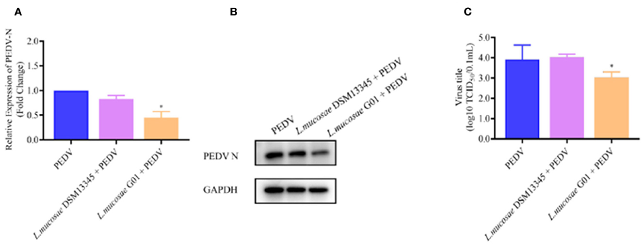
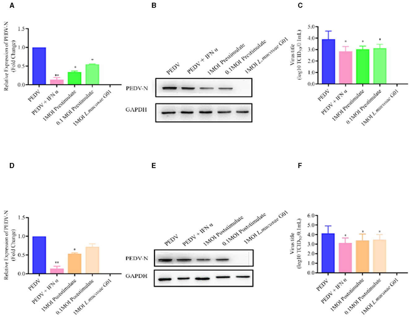
In the published article, there were two errors in Figures 4C, 5C as published. In Figures 4C, 5C, due to too many pictures in the data processing process, some pictures were repeated. The corrected Figures 4C, 5C appears below.

Figure 4. Depicts a comparison of the anti-PEDV effects of 1 MOI L. mucosae DSM13345 and L. mucosae G01 strains on IPEC-J2 cells. The method was utilized to evaluate the expression levels of the N protein in L. mucosae DSM13345 and L. mucosae G01 following 2 h interaction with IPEC-J2 cells, subsequent addition of PEDV-HZ, and after 24 h cell supernatants were harvested. Cell lysate containing PMSF was added for nucleic acid extraction and Western Blot assay. (A) RT-qPCR method was used to detect L. mucosae DSM13345 and L. mucosae G01 samples PEDV N mRNA expression level. (B) The quantification of N protein expression levels was performed through Western blot analysis. PEDV N is the viral content of the N nucleocapsid, and GAPDH is an internal reference for harvested cells. (C) TCID50 analysis cell supernatant titer. The purple color is L. mucosae DSM13345, orange color is L. mucosae G01, and blue color is PEDV control group. All assays were performed in triplicates, with three replicates per experiment, and each bar is the mean ± SEM. The asterisk indicates significant differences compared to the control group (p < 0.05).

Figure 5. L. mucosae G01 inhibits PEDV replication with both prestimulate and post stimulate. Depicts the prestimulation and poststimulation effects of L. mucosae G01 strain were demonstrated in IPEC-J2 cells after 24 h with 1 and 0.1 MOI and expression level of PEDV N protein and treat vero cells with a 10-fold dilution of viral supernatant for 72 h. (A) Expression content of mRNA levels of PEDV N nucleocapsid protein in response to prestimulation at 1 and 0.1 MOI. (B) Expression levels of PEDV N protein in response to prestimulation with at 1 and 0.1 MOI. (C) TCID50 analysis cell supernatant titer were measured with prestimulation at 1 and 0.1 MOI. (D) Expression content of mRNA levels of PEDV N nucleocapsid protein in response to poststimulation at 1 and 0.1 MOI. (E) Expression levels of PEDV N protein in response to poststimulation with at 1 and 0.1 MOI. (F) TCID50 analysis cell supernatant titer were measured with prestimulation at 1 and 0.1 MOI. The experimental groups included PEDV-infected, virus infected after IFN treated cells, cells infected with L. mucosae G01 1 MOI and then treated with PEDV, cells infected with L. mucosae G01 0.1 MOI and then treated with PEDV, and cells infected with L. mucosae G01. Cells infected 0.1 MOI PEDV then treated with L. mucosae G01 at 1 and 0.1 MOI. Pink color is PEDV and IFN-α as positive control. Green is the prestimulation group: 1 MOI L. mucosae G01 interacted with IPEC-J2 cells followed by 0.1 MOI PEDV, light green is the prestimulation group: 0.1 MOI L. mucosae G01 interacted with IPEC-J2 cells followed by 0.1 MOI PEDV, and orange is the treatment group: 0.1 MOI PEDV interacted with IPEC-J2 cells before interacting with 1 MOI L. mucosae G01, light yellow is 0.1 MOI PEDV first interacting with IPEC-J2 cells before interacting with 0.1 MOI L. mucosae G01 and blue color is PEDV control group. All assays were performed in triplicates, with three replicates per experiment, and each bar is the mean ± SEM. Statistically significant differences between groups are indicated by *p < 0.05 or **p < 0.01.
The authors apologize for this error and state that this does not change the scientific conclusions of the article in any way. The original article has been updated.
All claims expressed in this article are solely those of the authors and do not necessarily represent those of their affiliated organizations, or those of the publisher, the editors and the reviewers. Any product that may be evaluated in this article, or claim that may be made by its manufacturer, is not guaranteed or endorsed by the publisher.
Keywords: PEDV, antiviral activity, L. mucosae G01, IFN, Bama pig
Citation: Zhang B, Shen H, Gou H, Wuri N, Zhang C, Liu Z, He H, Nie J, Qu Y, Geri L and Zhang J (2024) Corrigendum: Isolation of Limosilactobacillus mucosae G01 with inhibitory effects on porcine epidemic diarrhea virus in vitro from Bama pig gastroenteritis. Front. Microbiol. 15:1488274. doi: 10.3389/fmicb.2024.1488274
Received: 29 August 2024; Accepted: 27 September 2024;
Published: 16 October 2024.
Approved by:
Frontiers Editorial Office, Frontiers Media SA, SwitzerlandCopyright © 2024 Zhang, Shen, Gou, Wuri, Zhang, Liu, He, Nie, Qu, Geri and Zhang. This is an open-access article distributed under the terms of the Creative Commons Attribution License (CC BY). The use, distribution or reproduction in other forums is permitted, provided the original author(s) and the copyright owner(s) are credited and that the original publication in this journal is cited, in accordance with accepted academic practice. No use, distribution or reproduction is permitted which does not comply with these terms.
*Correspondence: Letu Geri, Z2VyaWxldHVzeUBpbWF1LmVkdS5jbg==; Jianfeng Zhang, MTM2Njg5MzkyOThAMTM5LmNvbQ==
†These authors have contributed equally to this work and share first authorship
Disclaimer: All claims expressed in this article are solely those of the authors and do not necessarily represent those of their affiliated organizations, or those of the publisher, the editors and the reviewers. Any product that may be evaluated in this article or claim that may be made by its manufacturer is not guaranteed or endorsed by the publisher.
Research integrity at Frontiers

Learn more about the work of our research integrity team to safeguard the quality of each article we publish.