 Jianzhen Zhuo
 Ke Wang
 Zijun Shi3
95% of researchers rate our articles as excellent or good
Learn more about the work of our research integrity team to safeguard the quality of each article we publish.
Find out more
ORIGINAL RESEARCH article
Front. Microbiol. , 09 May 2023
Sec. Virology
Volume 14 - 2023 | https://doi.org/10.3389/fmicb.2023.1191004
This article is part of the Research Topic Computational Drug Discovery for Emerging Viral Infections View all 13 articles
Immunogenic cell death (ICD) serves a critical role in regulating cell death adequate to activate an adaptive immune response, and it is associated with various inflammation-related diseases. However, the specific role of ICD-related genes in COVID-19 remains unclear. We acquired COVID-19-related information from the GEO database and a total of 14 ICD-related differentially expressed genes (DEGs) were identified. These ICD-related DEGs were closely associated with inflammation and immune activity. Afterward, CASP1, CD4, and EIF2AK3 among the 14 DEGs were selected as feature genes based on LASSO, Random Forest, and SVM-RFE algorithms, which had reliable diagnostic abilities. Moreover, functional enrichment analysis indicated that these feature genes may have a potential role in COVID-19 by being involved in the regulation of immune response and metabolism. Further CIBERSORT analysis demonstrated that the variations in the immune microenvironment of COVID-19 patients may be correlated with CASP1, CD4, and EIF2AK3. Additionally, 33 drugs targeting 3 feature genes had been identified, and the ceRNA network demonstrated a complicated regulative association based on these feature genes. Our work identified that CASP1, CD4, and EIF2AK3 were diagnostic genes of COVID-19 and correlated with immune activity. This study presents a reliable diagnostic signature and offers an overview to investigate the mechanism of COVID-19.
Coronavirus infectious disease 2019 (COVID-19), caused by severe acute respiratory syndrome coronavirus 2 (SARS-CoV-2), is a public crisis worldwide since 2019 (Zhong et al., 2020; Kai-Wang To et al., 2021). Due to the mutable nature of SARS-CoV-2, numerous countries are suffering multi-dominant viral infections (Araf et al., 2022; Hadj, 2022). To control its spread, it is crucial to diagnose COVID-19 at an early stage. Accurate molecular diagnostic tests are necessary and valuable to confirm the rapid diagnosis of COVID-19, thus providing valid information for decision-making by the patient, healthcare facilities, and public health organizations (Islam and Iqbal, 2020; Safiabadi Tali et al., 2021; Yuce et al., 2021). Notably, large-scale genome-wide association studies have demonstrated specific disease-related elements in the population (Badua et al., 2021; Robishaw et al., 2021), and there are numerous reports on genes and the prevalence of COVID-19, improving the early diagnosis and clinical management of COVID-19.
Typical pathophysiological mechanisms of COVID-19 include cell death, immune response, oxidative stress, and metabolic activity (Tay et al., 2020; Gattinoni et al., 2021; van Eijk et al., 2021). Cell death and immune infiltration serve a critical role in the formation and progression of COVID-19 (Lee et al., 2020), and immunogenic cell death (ICD) is regarded as a regulative process that could affect cell death and immune infiltration simultaneously (Kroemer et al., 2022). ICD facilitates the activation and recruitment of antigen-presenting cells, thus activating innate and adaptive immune responses (Galluzzi et al., 2020; Minute et al., 2020). Initiating adaptive immunity in ICD is not only enhancing antitumor benefits but is essential for the optimal elimination of infectious etiologies (Galluzzi et al., 2017; Li et al., 2021). Therefore, ICD regulators may have potential diagnostic and therapeutic applications for COVID-19. However, earlier reports focused on only some immune cells and immunological molecules in COVID-19, lacking a perspective on the ICD in COVID-19.
To date, there is growing evidence that multiple cell death modalities, such as ferroptosis, NETosis, and necroptosis, play an important role in the development of COVID-19 (Kang and Wang, 2022; Nishiga et al., 2022; Zhang et al., 2023). Likewise, ICD may also regulate the progression of COVID-19. However, no similar studies have previously explored this specific process, and this study is the first exploration to analyze the relationship between ICD and COVID-19.
In this work, we aimed to determine the vital ICD regulators in the development of COVID-19, thus identifying valuable biomarkers for COVID-19 diagnosis. Subsequently, the correlation between ICD regulators and the infiltrating immune landscape was investigated. Based on the diagnosis biomarkers, the ceRNA network was established, and targeted small molecular agents for COVID-19 were explored. Finally, we validated the identified diagnosis biomarkers with the external dataset. This work offers an in-depth understanding of the developmental mechanisms of COVID-19 at the molecular level and identifies valuable biomarkers.
The gene expression profiles for COVID-19 and control samples were downloaded from the GEO databases (Barrett et al., 2013). The GSE157103 dataset comprised 100 COVID-19 samples and 26 control samples (Overmyer et al., 2021), and this dataset was deemed to be the training cohort for the principal analysis of this research. The GSE171110 dataset comprising 44 COVID-19 samples and 10 control samples was applied to validate the expression of the feature genes (Levy et al., 2021). Also, the 34 ICD-related genes included in this research were acquired from the previous report (Wang et al., 2021).
The expression profiles of ICD-related genes in the samples were extracted from the GSE157103 dataset. Next, the limma package was utilized to identify the ICD-related differentially expressed genes (DEGs) between different types of samples (Ritchie et al., 2015). Genes with value of p < 0.05 were deemed significant.
The expression data were analyzed with functional enrichment to evaluate the potential functionality of promising targets. Gene ontology (GO) is a common approach for identifying the functions of genes, including molecular functions, biological pathways, and cellular components (The Gene Ontology C, 2019; Warwick Vesztrocy and Dessimoz, 2020). Kyoto Encyclopedia of Genes and Genomes (KEGG) pathway enrichment was performed to investigate the genomic information of the DEGs (Kanehisa et al., 2017). Reactome enrichment analysis is also applied to explore gene functions, similar to GO and KEGG analysis (Good et al., 2021). These enrichment approaches were performed with the cluster profiler package (Yu et al., 2012). Additionally, disease ontology was utilized to annotate genes from a disease perspective (Schriml et al., 2022).
The ICD-related DEGs were further utilized to identify significant feature genes, thus diagnosing COVID-19. The feature identification approach is a procedure of limiting the number of factors, specifically vital for establishing a predictive model (Lee et al., 2021). LASSO regression, Random Forest (RF) algorithm, and SVM-RFE were included in this study to explore feature genes. The “glmnet” package was applied to conduct minimum LASSO regression, thus choosing the linear model and keeping the reliable variables (Engebretsen and Bohlin, 2019). Binomial distribution variables were further presented in the LASSO categorization, with a standard error value as the minimum parameter. Next, according to various dependent decision trees from a training pool, the RF algorithm promotes the precision of the model by randomly limiting the overfitting of individual decision trees (Yang et al., 2020). SVM-RFE can identify the optimal parameters by removing the SVM-derived eigenvectors (Sanz et al., 2018). An SVM module based on the “e1071” package was created to further evaluate the diagnostic value of the selected biomarker in COVID-19 (Jiang et al., 2020). The intersected genes, as the most significant feature genes from these three algorithms, were identified for subsequent analysis. Meanwhile, the predictive reliability of feature genes was evaluated by the receiver operating characteristic (ROC) curve, and the area under the curve (AUC) was further obtained (Park et al., 2004). A logistic regression signature with these feature genes was also established to assess diagnostic ability, and ROC curve was utilized to present this result.
To further investigate the potential pathways of the feature genes, single-gene gene set enrichment analysis (GSEA) and gene set variation analysis (GSVA) were performed with the GSEA package and GSVA package, respectively (Subramanian et al., 2005; Hanzelmann et al., 2013). GSEA was applied to evaluate the distribution landscape of the feature genes of a predetermined collection to explore their attribution to the phenotype. For GSVA, we also applied the KEGG pathway set to conduct enrichment analysis for each feature gene. Next, the limma package was utilized to discuss the difference in the GSVA score of the feature gene’s up- and down-regulated groups (Ritchie et al., 2015).
The CIBERSORT algorithm was applied to analyze the normalized gene expression data in the GSE157103 dataset, and the fraction of immune cells was identified (Chen et al., 2018; Overmyer et al., 2021). Violin plots were displayed to present the expressional difference of the immune infiltrating cells. And Spearman correlation analysis was executed to investigate the association between diverse immune infiltrating cells (Fujita et al., 2009). Meanwhile, the correlation between feature genes and immune cells was also explored with a similar method. These results were visualized with the “ggplot2” package (Ito and Murphy, 2013). p-value < 0.05 demonstrated statistical significance.
Drug Gene Interaction Database (DGIdb1) integrates existing literature on drug-gene interactions to provide clinical guidance for personalized treatment of disease. In this report, DGIdb was applied to identify gene-targeted drugs, and DrugBank database was further applied to identify drugs’ structural elements (Wagner et al., 2016; Wishart et al., 2018).
Three databases (miRanda, miRDB, TargetScan) were utilized to predict miRNAs (Enright et al., 2003; Agarwal et al., 2015; Chen and Wang, 2020), and the intersections of the predicted miRNAs were identified as the target miRNAs. Afterward, spongeScan was applied to identify the corresponding lncRNAs for miRNAs (Furio-Tari et al., 2016). A ceRNA network using Cytoscape was established predicated on the cross-talk between mRNAs, miRNAs, and lncRNAs (Shannon et al., 2003; Wu et al., 2020).
Based on the GSE157103 dataset, 14 ICD-related differentially expressed genes (DEGs) were identified between COVID-19 and control samples. Detailed information on these DEGs were presented in Table 1. Among them, the expression levels of ENTPD1, HMGB1, HSP90AA1, ATG5, CASP8, EIF2AK3, PIK3CA, CASP1, MYD88, and TLR4 were up-regulated, whereas BAX, TNF, CD4, and FOXP3 were down-regulated in COVID-19 samples than that in non-COVID-19 controls (Figure 1A). Next, the correlation between these genes was displayed in Figures 1B,C, and the significant interaction of most genes was observed. Furthermore, Figure 1D also indicated the pattern of the CNV alterations in ICD-related DEGs on their respective chromosomes.
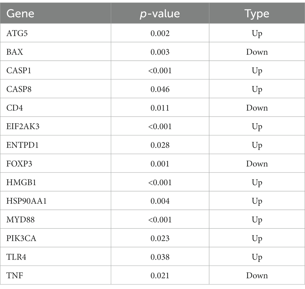
Table 1. ICD-related DEGs between COVID-19 samples and control samples.
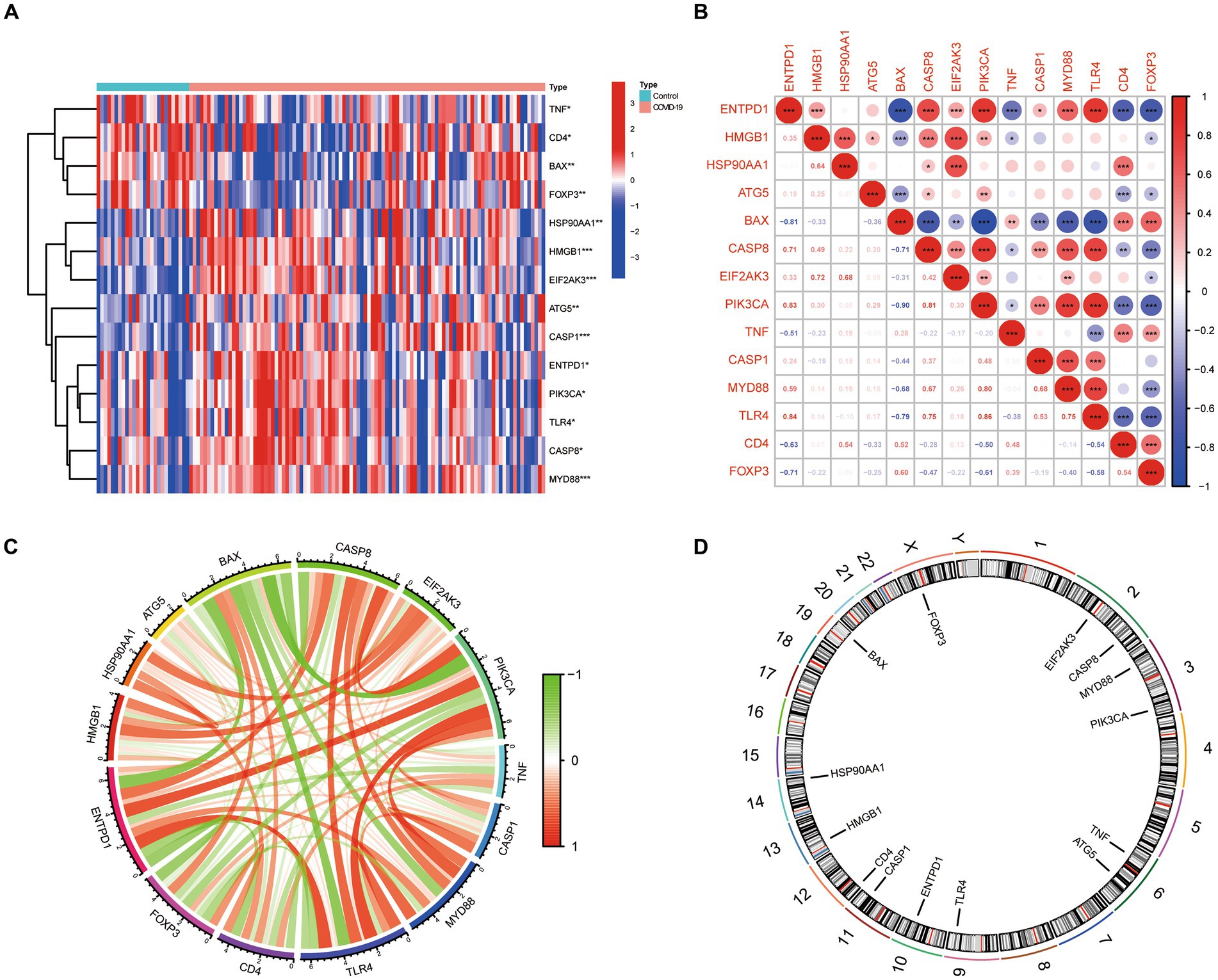
Figure 1. The expression landscape of 14 ICD-related DEGs. (A) Heatmap of DEGs between the COVID-19 samples and control samples. (B,C) The correlation diagram of DEGs. (D) Chromosomal positions of DEGs.
We conducted GO, KEGG, Reactome, and DO enrichment analysis on ICD-related DEGs, thus discovering the potential molecular biological characteristics of COVID-19. The GO enrichment analyses indicated that these genes were largely involved in the regulation of cytokines (Figure 2A). KEGG pathway analyses showed that the positive regulation of cytokine production, response to lipopolysaccharide, response to molecules of bacterial origin, and T cell activation were enriched (Figure 2B). Moreover, Reactome terms were enriched in signaling by interleukins, programmed cell death, and regulated necrosis (Figure 2C). Interestingly, the ICD-related DEGs were also markedly enriched in inflammation and tumor-related signatures (Figure 2D). These findings demonstrated that ICD-related DEGs may serve a critical role in the etiology of COVID-19 by engaging in the regulation of cytokines and diverse cell death processes.
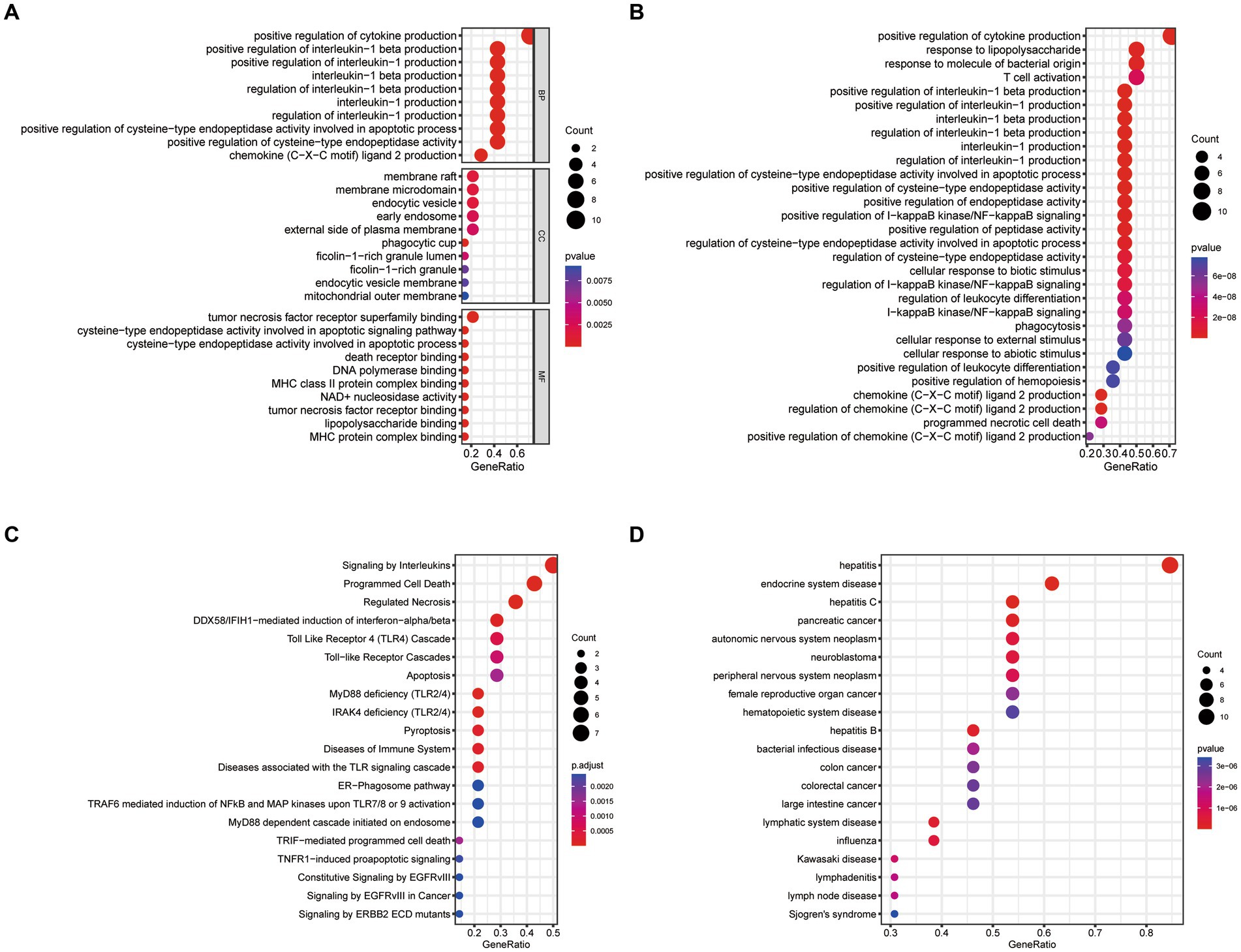
Figure 2. Functional enrichment analysis of ICD-related DEGs. (A) GO, (B) KEGG, (C) Reactome, (D) DO enrichment analysis for DEGs.
For exploring the potential pathogenesis of COVID-19, we further evaluated the diagnostic values of ICD-related DEGs. Machine learning algorithms (LASSO, RF, and SVM-RFE) were selected and executed to identify the significant ICD-related DEGs, thus distinguishing COVID-19 from control samples. LASSO regression analysis was performed to select nine feature genes with statistically significant univariate parameters (Figures 3A,B). RF incorporating feature selection was utilized to investigate the relationship between error rate, number of classification trees, and 14 genes with relative importance (Figure 3C). Meanwhile, the respective importance of these genes was also presented in Figure 3D. Next, SVM-RFE analysis indicated that the SVM model based on five feature genes had an optimum generalization performance (Figures 3E,F). The feature genes from the abovementioned algorithms were intersected, and three feature genes (CASP1, CD4, and EIF2AK3) were determined for subsequent analysis (Figure 3G). To reveal the diagnostic accuracy of feature genes in distinguishing COVID-19 from non-COVID-19 controls, ROC curves were plotted for the three feature genes. As depicted in Figure 3H, the AUCs for the three feature genes were higher than 0.6, indicating that these genes have good diagnostic performance. Furthermore, we established a logistic regression model based on the three feature genes, and the ROC curves suggested that the logistic regression model differentiated COVID-19 and control samples with AUC = 0.899 (Figure 3I). This evidence demonstrated that the three feature genes can be considered valuable diagnostic genes for COVID-19, and the diagnostic model incorporating the three feature genes may have greater accuracy than individual feature genes.
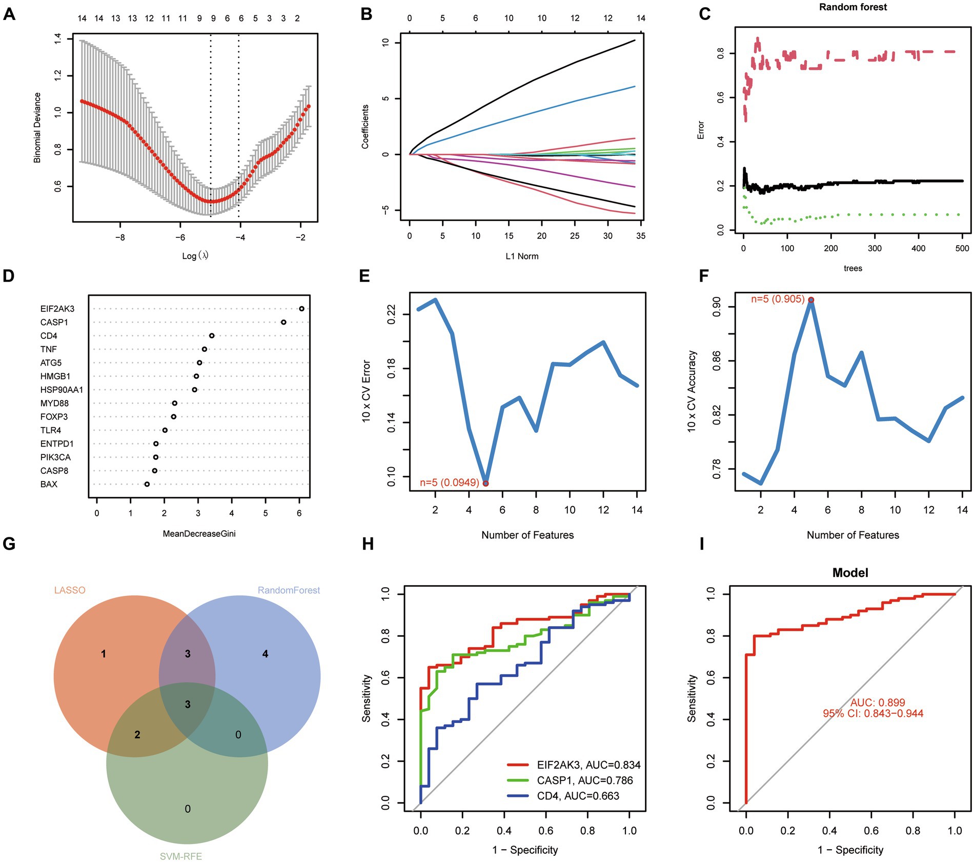
Figure 3. Identification of diagnostic genes for COVID-19. (A,B) Establishment of the LASSO model. (C,D) Screening biomarkers based on random forest (RF) algorithm. (E,F) Results of screening biomarkers based on SVM-RFE algorithm. (G) Venn diagram showing the intersected genes. (H) ROC curves for the feature genes. (I) Logistic regression model to identify the AUC of disease samples.
To further determine the underlying role of feature genes to identify characteristic differences between COVID-19 samples and control samples, a single-gene GSEA-KEGG pathway analysis was performed. The top six pathways enriched for individual feature genes were presented in Figures 4A–C. After a systematic analysis, we observed that CASP1 and CD4 were activated in immune response (such as antigen processing and presentation) and RIG-I-like receptor signaling pathway. Meanwhile, EIF2AK3 was closely associated with the metabolism-related activity. Afterward, we found the differentially enriched GSVA pathways between the high- and low-expression groups of each feature gene (Figure 5). The findings indicated that the high expression of CASP1 in COVID-19 may induce this disease by “tyrosine metabolism,” “base excision repair,” and “glyoxylate and dicarboxylate metabolism,” while down-regulation of CASP1 promoted “DORSO_VENTRAL_AXIS_FORMATION,” “O_GLYCAN_BIOSYNTHESIS,” and “VALINE_LEUCINE_AND_ISOLEEUCINE_BIOSYNTHESIS.” CD4, whose expression was limited in COVID-19 samples, was significantly correlated with the immune response (“AUTOIMMUNE THYROID DISEASE,” “GRAFT VERSUS HOST DISEASE,” and “ALLOGRAFT REJECTION”). Moreover, the high expression of CD4 activated the pathways such as “GLYCINE SERINE AND THREONINE METABOLISM,” and “PANTOTHENATE AND COA BIOSYNTHESIS.” Similarly, in the EIF2AK3 over-expression group, “GLYCOSAMINOGLYCAN DEGRADATION,” and metabolism-related pathways (“TAURINE AND HYPOTAURINE METABOLISM,” “PHENYLALANINE METABOLISM,” “TYROSINE METABOLISM”) were enriched. The low-expressed EIF2AK3 served a critical function in the protein export, and biosynthesis process (such as N-GLYCAN biosynthesis and AMINOACYL TRNA biosynthesis).
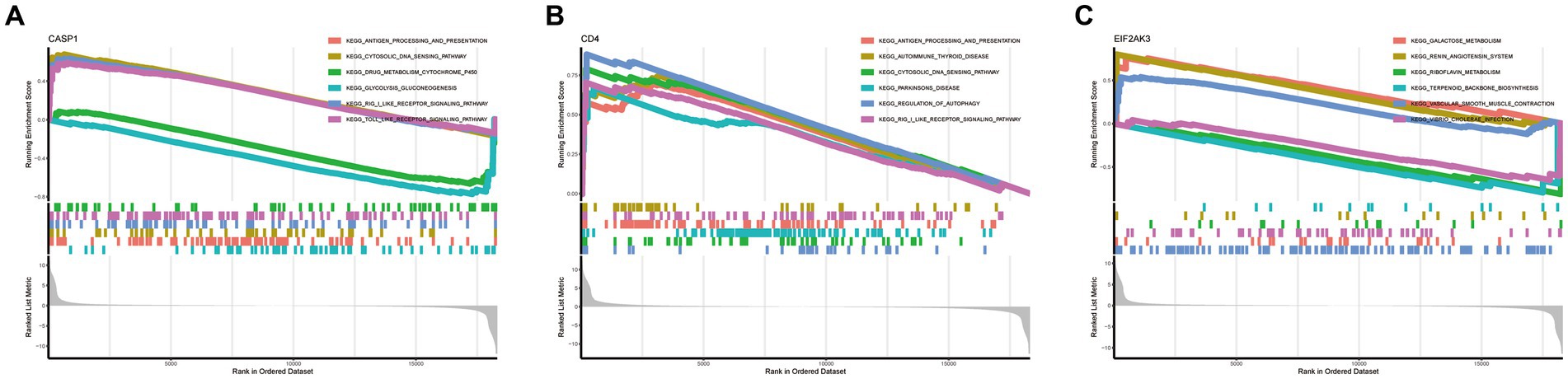
Figure 4. Single-gene GSEA-KEGG pathway analysis in CASP1 (A), CD4 (B), and EIF2AK3 (C).
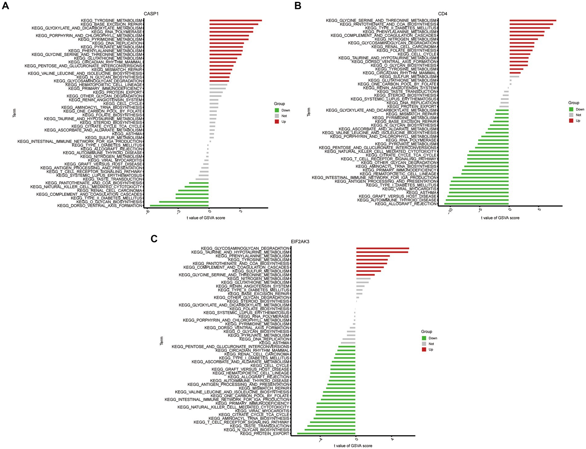
Figure 5. High- and low-expression groups based on the expression levels of each feature gene combined with GSVA in CASP1 (A), CD4 (B), and EIF2AK3 (C).
Based on the CIBERSORT algorithm, the infiltration levels of 22 types of immune cells between COVID-19 and control samples were evaluated. As presented in Figure 6A, compared with the control samples, naïve B cells, follicular helper T cells, Tregs, activated NK cells, monocytes, and activated mast cells were less enriched, while plasma cells, CD4 naïve T cells, activated CD4 memory T cells, γδ T cells, resting dendritic cells, and activated dendritic cells were more enriched in COVID-19 sample. Subsequently, after performing a correlation analysis of infiltrating immune cells, we identified diverse pairs of interacting immune cells (Figure 6B). Similarly, the proportions of different immune cells between the COVID-19 and control samples had a significant difference (Figure 6C). Furthermore, we also explored the relevance of three diagnostic genes to immune cells. As shown in Figure 6D, Pearson correlation analysis suggested that activated dendritic cells, neutrophils, activated NK cells, activated CD4 memory T cells and γδ T cells were all associated with three feature genes (CASP1, CD4, and EIF2AK3). These results suggested that alterations in the immune microenvironment of COVID-19 samples correlated with these three genes.
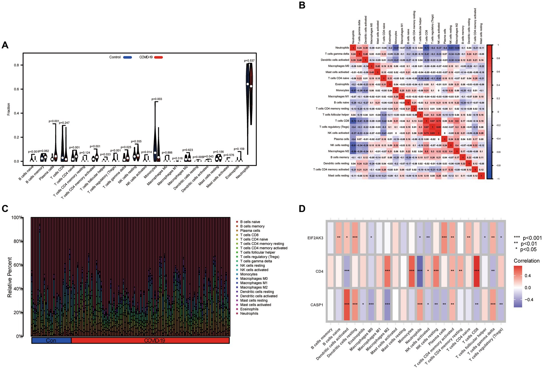
Figure 6. Immune landscape analysis. (A) The differences in immune cells from the immune microenvironment between COVID-19 patients and normal samples. (B) The correlation diagram of immune cells. (C) The bar plot presenting the proportion of infiltrated immune cells calculated by the CIBERSORT algorithm. (D) The correlation between feature genes and immune cells.
DGIdb was utilized to identify the underlying drug or molecular compounds that could regulate the expression of feature genes in the setting of COVID-19. As demonstrated in the drug-gene interaction network (Figure 7), 33 drugs or molecular compounds targeting feature genes were identified, including 23 for CASP1, 9 for CD4, and 1 for EIF2AK3. The results suggest that CASP1 is more likely to be a valuable target for drug development against COVID-19. Interestingly, the drugs that interacted with different genes were not observed.
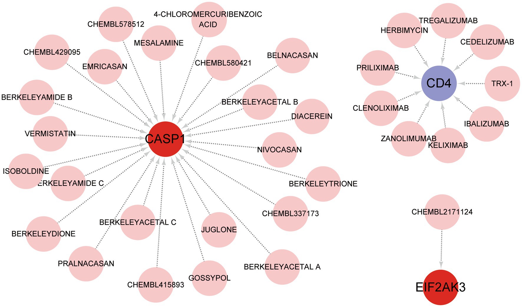
Figure 7. Prediction of feature gene-targeted drugs based on DGIdb database.
Recently, the ceRNA network is emerging as a hot topic and serves a pivotal role in controlling gene activity. The ceRNA network was established with miRNA as a bridge to determine the relationship between target gene, mRNA and lncRNA by interacting miRNA response factors. Referring to epigenetic regulators, miRNAs are considered valuable therapeutic options for diverse diseases. The miRNAs associated with 3 feature genes were explored from three online databases, and the interaction demonstrated by these databases were also identified. A total of 63 miRNAs and 3 mRNAs formed 67 interaction pairs. Additionally, we investigated the lncRNAs that corresponded to regulator miRNAs based on the spongeScan database and further established a ceRNA network of lncRNA-miRNA-mRNA. Finally, this network was comprised of 145 nodes (3 genes, 63 miRNAs, and 75 lncRNAs) and 151 edges (Figure 8).
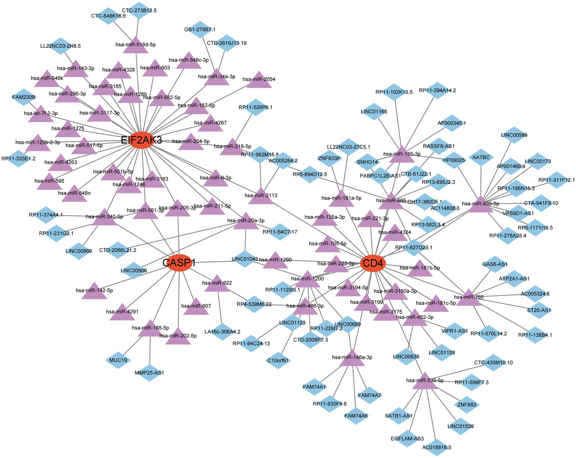
Figure 8. A ceRNA network based on three feature genes.
Additionally, the expression of three diagnostic genes (CASP1, CD4, and EIF2AK3) was also explored in the GSE171110 dataset. We observed that the expression levels of these diagnostic genes were consistent with the GSE157103 dataset (Figure 9). Among them, the expression of CD4 (p < 0.001) in COVID-19 samples was lower than that of control samples, while CASP1 (p = 0.018) and EIF2AK3 (p = 0.012) were higher in COVID-19 samples.
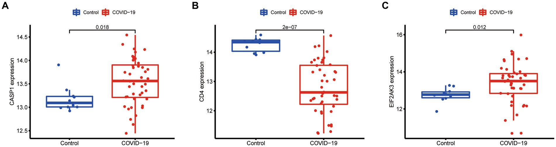
Figure 9. Expression difference of the feature genes (A, CASP1; B, CD4; C, EIF2AK3) between COVID-19 and normal samples in the validation cohort (GSE171110 dataset).
COVID-19 has been ravaging the world for years, and its specific cause remains unclear. The mutational and variable nature of COVID-19 renders it impossible to explain it with changes in a single gene. Hence, the management of COVID-19 patients cannot be implemented by a method, instead diverse methods, including multi-targeted agents and multi-modalities treatment (Fahmi et al., 2021; Rantam et al., 2021). It is essential to explore novel and promising biomarkers for the early diagnosis and management of COVID-19 (Zhong et al., 2020; Gattinoni et al., 2021). Notably, virous cell death forms have been observed to be significantly correlated with the progression of COVID-19, but the association between COVID-19 and ICD has not yet been clarified. Herein, we attempted to identify diagnostic biomarkers pertaining to COVID-19 and delve into the role exerted by ICD within COVID-19.
In this work, we identified 14 ICD-related DEGs between COVID-19 and normal samples, including 10 up-regulated and 4 down-regulated genes. Afterward, functional enrichment analysis of these DEGs was executed, and these genes presented close associations to the regulation of cytokines and cell death signals (such as regulation of cysteine-type endopeptidase activity involved in the apoptotic process, programmed cell death, and regulated necrosis). According to these findings, COVID-19 displayed immunological processes and cell death involvement, thus inducing inflammation and infection.
We applied three different machine learning algorithms (LASSO, RF, and SVM-RFE), each with its advantages. LASSO regression identifies parameters by finding the variable with the lowest incidence of categorical error (Tibshirani, 1997). RF algorithm is an integrative algorithm consisting of decision trees, which is applied to train and predict samples (Simsekler et al., 2021). SVM-RFE has acquired extensive attention in ranking traits and selecting important traits for categorization (Sanz et al., 2018). Finally, CASP1, CD4, and EIF2AK3 were identified and were valuable for further explorations, which demonstrated that our prediction displayed reliability with the integrated approaches.
CASP1, a member of the caspase family, serves a critical role in the execution process of cell apoptosis (Kapplusch et al., 2019). The expression of CASP1 was regulated, and the inflammasome activated by CASP1-NLRP3 dependent pathway can subsequently secrete IL-1β and IL-18, thus leading to dysfunctional autophagy (Wang et al., 2017). Upregulation of CASP1 in COVID-19 was reported in a prior report, consistent with our findings (Yang et al., 2022). CD4 encodes the CD4 membrane glycoprotein of T lymphocytes, and CD4+ T cell plays a critical role in anti-viral immunity (Matthias et al., 2002; Sharma et al., 2005). CD4 mediates various biological processes of immune cells. Meanwhile, immune cells, primarily comprising T cells, B cells, and macrophages, crucially affect the pathogenesis of COVID-19 (Tay et al., 2020). EIF2AK3, as a metabolic stress-sensing protein kinase, inhibits protein translation and regulated pro-survival autophagy (You et al., 2021). These genes displayed significantly different expressions between COVID-19 and normal samples, and this result was also observed in the validation cohort. Furthermore, GSEA and GSVA for three feature genes were performed, and we observed that these genes were significantly associated with immune response and metabolism-related activities. Based on the abovementioned findings, CASP1, CD4, and EIF2AK3 have the potential to influence the progression of COVID-19 and serve as diagnostic biomarkers, whereas this finding remains to be confirmed by further clinical trials.
To more comprehensively investigate the effects played by the abundance of immune cells in COVID-19, this research conducted CIBERSORT for evaluating the infiltrating immune status within COVID-19. The abundance of naïve B cells, follicular helper T cells, Tregs, activated NK cells, monocytes, and activated mast cells decreased, while the abundance of plasma cells, CD4 naïve T cells, activated CD4 memory T cells, γδ T cells, resting dendritic cells, and activated dendritic cells increased, potentially demonstrating the relevance of COVID-19 initiation and development. It is common knowledge that these cells are an essential part of human adaptive immunity, and these cells may be potential factors for COVID-19 pathogenesis (Sette and Crotty, 2021). Meanwhile, three feature genes presented a significant correlation with diverse immune cells, which also confirmed the autoimmunogenicity of these genes. Although this is widely recognized, the molecular mechanisms and functions of immune cell infiltration in COVID-19 need to be further investigated urgently.
Finally, the feature gene for gene-targeted drugs and the ceRNA network were explored. A diagram of 33 drugs with promising therapeutic efficacy against COVID-19 was displayed. The performance of the selected drugs was validated for diverse diseases; however, the mechanisms of many drugs were unclear. Further molecular experiments and clinical investigations are necessary to identify the drugs that are valuable for COVID-19 treatment. The target miRNAs and the target lncRNAs were predicted for CASP1, CD4, and EIF2AK3, and a ceRNA network was established with Cytoscape. This network proposes a potential mechanism for selected genes to be regulated at the transcriptome level.
Inevitably, these are some restrictions of this study. Firstly, this study was performed on public databases, and additional laboratory experiments are necessary. Secondly, the association between these results in this study and clinical variables was worthy of future analysis and validation. However, this study could still reflect convincing evidence to further investigate the significance of ICD-related genes in the treatment and diagnosis of COVID-19.
Taken together, we presented that differentially expressed genes: CASP1, CD4, and EIF2AK3 might be considered as the diagnostic biomarker for COVID-19 and characteristic immunological infiltration could influence the systemic immune microenvironment in the pathogenesis of COVID-19. This work may reveal potential biomarkers related to immune regulation that ultimately led to COVID-19 infection. Additional leading-edge approaches, such as single-cell sequencing method, will further offer promising perspectives into COVID-19 development and drug targets.
The original contributions presented in the study are included in the article/supplementary material, further inquiries can be directed to the corresponding author.
Ethical review and approval was not required for the study on human participants in accordance with the local legislation and institutional requirements. Written informed consent for participation was not required for this study in accordance with the national legislation and the institutional requirements.
JZ and CY: conceptualization. JZ and KW: methodology. ZS: software and data curation. JZ, KW, and ZS: validation. CY: formal analysis, writing—review and editing, and project administration. JZ: investigation, writing original draft preparation, visualization, and supervision. KW: resources. All authors contributed to the article and approved the submitted version.
The authors declare that the research was conducted in the absence of any commercial or financial relationships that could be construed as a potential conflict of interest.
All claims expressed in this article are solely those of the authors and do not necessarily represent those of their affiliated organizations, or those of the publisher, the editors and the reviewers. Any product that may be evaluated in this article, or claim that may be made by its manufacturer, is not guaranteed or endorsed by the publisher.
Agarwal, V., Bell, G. W., Nam, J. W., and Bartel, D. P. (2015). Predicting effective microRNA target sites in mammalian mRNAs. elife 4:e05005:4. doi: 10.7554/eLife.05005
Araf, Y., Akter, F., Tang, Y. D., Fatemi, R., Parvez, M. S. A., Zheng, C., et al. (2022). Omicron variant of SARS-CoV-2: genomics, transmissibility, and responses to current COVID-19 vaccines. J. Med. Virol. 94, 1825–1832. doi: 10.1002/jmv.27588
Badua, C., Baldo, K. A. T., and Medina, P. M. B. (2021). Genomic and proteomic mutation landscapes of SARS-CoV-2. J. Med. Virol. 93, 1702–1721. doi: 10.1002/jmv.26548
Barrett, T., Wilhite, S. E., Ledoux, P., Evangelista, C., Kim, I. F., Tomashevsky, M., et al. (2013). NCBI GEO: archive for functional genomics data sets--update. Nucleic Acids Res. 41, D991–D995. doi: 10.1093/nar/gks1193
Chen, B., Khodadoust, M. S., Liu, C. L., Newman, A. M., and Alizadeh, A. A. (2018). Profiling tumor infiltrating immune cells with CIBERSORT. Methods Mol. Biol. 1711, 243–259. doi: 10.1007/978-1-4939-7493-1_12
Chen, Y., and Wang, X. (2020). miRDB: an online database for prediction of functional microRNA targets. Nucleic Acids Res. 48, D127–D131. doi: 10.1093/nar/gkz757
Engebretsen, S., and Bohlin, J. (2019). Statistical predictions with glmnet. Clin. Epigenet. 11:123. doi: 10.1186/s13148-019-0730-1
Enright, A. J., John, B., Gaul, U., Tuschl, T., Sander, C., and Marks, D. S. (2003). MicroRNA targets in Drosophila. Genome Biol. 5:R1. doi: 10.1186/gb-2003-5-1-r1
Fahmi, M., Kharisma, V. D., Ansori, A. N. M., and Ito, M. (2021). Retrieval and investigation of data on SARS-CoV-2 and COVID-19 using bioinformatics approach. Adv. Exp. Med. Biol. 1318, 839–857. doi: 10.1007/978-3-030-63761-3_47
Fujita, A., Sato, J. R., Demasi, M. A., Sogayar, M. C., Ferreira, C. E., and Miyano, S. (2009). Comparing Pearson, spearman and Hoeffding's D measure for gene expression association analysis. J. Bioinform. Comput. Biol. 7, 663–684. doi: 10.1142/s0219720009004230
Furio-Tari, P., Tarazona, S., Gabaldon, T., Enright, A. J., and Conesa, A. (2016). spongeScan: a web for detecting microRNA binding elements in lncRNA sequences. Nucleic Acids Res. 44, W176–W180. doi: 10.1093/nar/gkw443
Galluzzi, L., Buque, A., Kepp, O., Zitvogel, L., and Kroemer, G. (2017). Immunogenic cell death in cancer and infectious disease. Nat. Rev. Immunol. 17, 97–111. doi: 10.1038/nri.2016.107
Galluzzi, L., Vitale, I., Warren, S., Adjemian, S., Agostinis, P., Martinez, A. B., et al. (2020). Consensus guidelines for the definition, detection and interpretation of immunogenic cell death. J. Immunother. Cancer 8:e000337. doi: 10.1136/jitc-2019-000337
Gattinoni, L., Gattarello, S., Steinberg, I., Busana, M., Palermo, P., Lazzari, S., et al. (2021). COVID-19 pneumonia: pathophysiology and management. Eur. Respir. Rev. 30:210138. doi: 10.1183/16000617.0138-2021
Good, B. M., Van Auken, K., Hill, D. P., Mi, H., Carbon, S., Balhoff, J. P., et al. (2021). Reactome and the gene ontology: digital convergence of data resources. Bioinformatics 37, 3343–3348. doi: 10.1093/bioinformatics/btab325
Hadj, H. I. (2022). Covid-19 vaccines and variants of concern: a review. Rev. Med. Virol. 32:e2313. doi: 10.1002/rmv.2313
Hanzelmann, S., Castelo, R., and Guinney, J. (2013). GSVA: gene set variation analysis for microarray and RNA-seq data. BMC Bioinform. 14:7. doi: 10.1186/1471-2105-14-7
Islam, K. U., and Iqbal, J. (2020). An update on molecular diagnostics for COVID-19. Front. Cell. Infect. Microbiol. 10:560616. doi: 10.3389/fcimb.2020.560616
Ito, K., and Murphy, D. (2013). Application of ggplot2 to Pharmacometric graphics. CPT Pharmacometrics Syst. Pharmacol. 2:e79. doi: 10.1038/psp.2013.56
Jiang, H., Gu, J., Du, J., Qi, X., Qian, C., and Fei, B. (2020). A 21-gene support vector machine classifier and a 10-gene risk score system constructed for patients with gastric cancer. Mol. Med. Rep. 21, 347–359. doi: 10.3892/mmr.2019.10841
Kai-Wang To, K., Sridhar, S., Chiu, K. H., Hung, D. L., Li, X., Hung, I. F., et al. (2021). Lessons learned 1 year after SARS-CoV-2 emergence leading to COVID-19 pandemic. Emerg. Microbes. Infect. 10, 507–535. doi: 10.1080/22221751.2021.1898291
Kanehisa, M., Furumichi, M., Tanabe, M., Sato, Y., and Morishima, K. (2017). KEGG: new perspectives on genomes, pathways, diseases and drugs. Nucleic Acids Res. 45, D353–D361. doi: 10.1093/nar/gkw1092
Kang, Y., and Wang, Q. (2022). Potential therapeutic value of necroptosis inhibitor for the treatment of COVID-19. Eur. J. Med. Res. 27:283. doi: 10.1186/s40001-022-00913-7
Kapplusch, F., Schulze, F., Rabe-Matschewsky, S., Russ, S., Herbig, M., Heymann, M. C., et al. (2019). CASP1 variants influence subcellular caspase-1 localization, pyroptosome formation, pro-inflammatory cell death and macrophage deformability. Clin. Immunol. 208:108232. doi: 10.1016/j.clim.2019.06.008
Kroemer, G., Galassi, C., Zitvogel, L., and Galluzzi, L. (2022). Immunogenic cell stress and death. Nat. Immunol. 23, 487–500. doi: 10.1038/s41590-022-01132-2
Lee, S., Channappanavar, R., and Kanneganti, T. D. (2020). Coronaviruses: innate immunity, Inflammasome activation, inflammatory cell death, and cytokines. Trends Immunol. 41, 1083–1099. doi: 10.1016/j.it.2020.10.005
Lee, Y. W., Choi, J. W., and Shin, E. H. (2021). Machine learning model for predicting malaria using clinical information. Comput. Biol. Med. 129:104151. doi: 10.1016/j.compbiomed.2020.104151
Levy, Y., Wiedemann, A., Hejblum, B. P., Durand, M., Lefebvre, C., Surenaud, M., et al. (2021). CD177, a specific marker of neutrophil activation, is associated with coronavirus disease 2019 severity and death. iScience 24:102711. doi: 10.1016/j.isci.2021.102711
Li, Y., Liu, X., Zhang, X., Pan, W., Li, N., and Tang, B. (2021). Immunogenic cell death inducers for enhanced cancer immunotherapy. Chem. Commun. (Camb.) 57, 12087–12097. doi: 10.1039/d1cc04604g
Matthias, L. J., Yam, P. T., Jiang, X. M., Vandegraaff, N., Li, P., Poumbourios, P., et al. (2002). Disulfide exchange in domain 2 of CD4 is required for entry of HIV-1. Nat. Immunol. 3, 727–732. doi: 10.1038/ni815
Minute, L., Teijeira, A., Sanchez-Paulete, A. R., Ochoa, M. C., Alvarez, M., Otano, I., et al. (2020). Cellular cytotoxicity is a form of immunogenic cell death. J. Immunother. Cancer 8:e000325. doi: 10.1136/jitc-2019-000325
Nishiga, M., Jahng, J. W. S., and Wu, J. C. (2022). Ferroptosis of pacemaker cells in COVID-19. Circ. Res. 130, 978–980. doi: 10.1161/CIRCRESAHA.122.320951
Overmyer, K. A., Shishkova, E., Miller, I. J., Balnis, J., Bernstein, M. N., Peters-Clarke, T. M., et al. (2021). Large-scale multi-omic analysis of COVID-19 severity. Cell Syst. 12, 23–40.e7. doi: 10.1016/j.cels.2020.10.003
Park, S. H., Goo, J. M., and Jo, C. H. (2004). Receiver operating characteristic (ROC) curve: practical review for radiologists. Korean J. Radiol. 5, 11–18. doi: 10.3348/kjr.2004.5.1.11
Rantam, F. A., Kharisma, V. D., Sumartono, C., Nugraha, J., Wijaya, A. Y., Susilowati, H., et al. (2021). Molecular docking and dynamic simulation of conserved B cell epitope of SARS-CoV-2 glycoprotein Indonesian isolates: an immunoinformatic approach. F1000Res 10:813. doi: 10.12688/f1000research.54258.1
Ritchie, M. E., Phipson, B., Wu, D., Hu, Y., Law, C. W., Shi, W., et al. (2015). Limma powers differential expression analyses for RNA-sequencing and microarray studies. Nucleic Acids Res. 43:e47. doi: 10.1093/nar/gkv007
Robishaw, J. D., Alter, S. M., Solano, J. J., Shih, R. D., DL, D. M., Maki, D. G., et al. (2021). Genomic surveillance to combat COVID-19: challenges and opportunities. Lancet Microbe 2, e481–e484. doi: 10.1016/S2666-5247(21)00121-X
Safiabadi Tali, S. H., LeBlanc, J. J., Sadiq, Z., Oyewunmi, O. D., Camargo, C., Nikpour, B., et al. (2021). Tools and techniques for severe acute respiratory syndrome coronavirus 2 (SARS-CoV-2)/COVID-19 detection. Clin. Microbiol. Rev. 34:e00228-20. doi: 10.1128/CMR.00228-20
Sanz, H., Valim, C., Vegas, E., Oller, J. M., and Reverter, F. (2018). SVM-RFE: selection and visualization of the most relevant features through non-linear kernels. BMC Bioinform. 19:432. doi: 10.1186/s12859-018-2451-4
Schriml, L. M., Munro, J. B., Schor, M., Olley, D., McCracken, C., Felix, V., et al. (2022). The human disease ontology 2022 update. Nucleic Acids Res. 50, D1255–D1261. doi: 10.1093/nar/gkab1063
Sette, A., and Crotty, S. (2021). Adaptive immunity to SARS-CoV-2 and COVID-19. Cells 184, 861–880. doi: 10.1016/j.cell.2021.01.007
Shannon, P., Markiel, A., Ozier, O., Baliga, N. S., Wang, J. T., Ramage, D., et al. (2003). Cytoscape: a software environment for integrated models of biomolecular interaction networks. Genome Res. 13, 2498–2504. doi: 10.1101/gr.1239303
Sharma, D., Balamurali, M. M., Chakraborty, K., Kumaran, S., Jeganathan, S., Rashid, U., et al. (2005). Protein minimization of the gp120 binding region of human CD4. Biochemistry 44, 16192–16202. doi: 10.1021/bi051120s
Simsekler, M. C. E., Alhashmi, N. H., Azar, E., King, N., Luqman, R., and Al, M. A. (2021). Exploring drivers of patient satisfaction using a random forest algorithm. BMC Med. Inform. Decis. Mak. 21:157. doi: 10.1186/s12911-021-01519-5
Subramanian, A., Tamayo, P., Mootha, V. K., Mukherjee, S., Ebert, B. L., Gillette, M. A., et al. (2005). Gene set enrichment analysis: a knowledge-based approach for interpreting genome-wide expression profiles. Proc. Natl. Acad. Sci. U. S. A. 102, 15545–15550. doi: 10.1073/pnas.0506580102
Tay, M. Z., Poh, C. M., Renia, L., MacAry, P. A., and Ng, L. F. P. (2020). The trinity of COVID-19: immunity, inflammation and intervention. Nat. Rev. Immunol. 20, 363–374. doi: 10.1038/s41577-020-0311-8
The Gene Ontology C (2019). The gene ontology resource: 20 years and still GOing strong. Nucleic Acids Res. 47, D330–D338. doi: 10.1093/nar/gky1055
Tibshirani, R. (1997). The lasso method for variable selection in the cox model. Stat. Med. 16, 385–395. doi: 10.1002/(sici)1097-0258(19970228)16:4<385::aid-sim380>3.0.co;2-3
van Eijk, L. E., Binkhorst, M., Bourgonje, A. R., Offringa, A. K., Mulder, D. J., Bos, E. M., et al. (2021). COVID-19: immunopathology, pathophysiological mechanisms, and treatment options. J. Pathol. 254, 307–331. doi: 10.1002/path.5642
Wagner, A. H., Coffman, A. C., Ainscough, B. J., Spies, N. C., Skidmore, Z. L., Campbell, K. M., et al. (2016). DGIdb 2.0: mining clinically relevant drug-gene interactions. Nucleic Acids Res. 44, D1036–D1044. doi: 10.1093/nar/gkv1165
Wang, X., Wu, S., Liu, F., Ke, D., Wang, X., Pan, D., et al. (2021). An immunogenic cell death-related classification predicts prognosis and response to immunotherapy in head and neck squamous cell carcinoma. Front. Immunol. 12:781466. doi: 10.3389/fimmu.2021.781466
Wang, D., Zhang, J., Jiang, W., Cao, Z., Zhao, F., Cai, T., et al. (2017). The role of NLRP3-CASP1 in inflammasome-mediated neuroinflammation and autophagy dysfunction in manganese-induced, hippocampal-dependent impairment of learning and memory ability. Autophagy 13, 914–927. doi: 10.1080/15548627.2017.1293766
Warwick Vesztrocy, A., and Dessimoz, C. (2020). Benchmarking gene ontology function predictions using negative annotations. Bioinformatics 36, i210–i218. doi: 10.1093/bioinformatics/btaa466
Wishart, D. S., Feunang, Y. D., Guo, A. C., Lo, E. J., Marcu, A., Grant, J. R., et al. (2018). DrugBank 5.0: a major update to the DrugBank database for 2018. Nucleic Acids Res. 46, D1074–D1082. doi: 10.1093/nar/gkx1037
Wu, X., Sui, Z., Zhang, H., Wang, Y., and Yu, Z. (2020). Integrated analysis of lncRNA-mediated ceRNA network in lung adenocarcinoma. Front. Oncol. 10:554759. doi: 10.3389/fonc.2020.554759
Yang, C. A., Huang, Y. L., and Chiang, B. L. (2022). Innate immune response analysis in COVID-19 and Kawasaki disease reveals MIS-C predictors. J. Formos. Med. Assoc. 121, 623–632. doi: 10.1016/j.jfma.2021.06.009
Yang, L., Wu, H., Jin, X., Zheng, P., Hu, S., Xu, X., et al. (2020). Study of cardiovascular disease prediction model based on random forest in eastern China. Sci. Rep. 10:5245. doi: 10.1038/s41598-020-62133-5
You, K., Wang, L., Chou, C. H., Liu, K., Nakata, T., Jaiswal, A., et al. (2021). QRICH1 dictates the outcome of ER stress through transcriptional control of proteostasis. Science 371:eabb6896. doi: 10.1126/science.abb6896
Yu, G., Wang, L. G., Han, Y., and He, Q. Y. (2012). clusterProfiler: an R package for comparing biological themes among gene clusters. OMICS 16, 284–287. doi: 10.1089/omi.2011.0118
Yuce, M., Filiztekin, E., and Ozkaya, K. G. (2021). COVID-19 diagnosis -a review of current methods. Biosens. Bioelectron. 172:112752. doi: 10.1016/j.bios.2020.112752
Zhang, R., Sun, C., Han, Y., Huang, L., Sheng, H., Wang, J., et al. (2023). Neutrophil autophagy and NETosis in COVID-19: perspectives. Autophagy 19, 758–767. doi: 10.1080/15548627.2022.2099206
Keywords: COVID-19, immunogenic cell death, biomarker, diagnosis, machine learning
Citation: Zhuo J, Wang K, Shi Z and Yuan C (2023) Immunogenic cell death-led discovery of COVID-19 biomarkers and inflammatory infiltrates. Front. Microbiol. 14:1191004. doi: 10.3389/fmicb.2023.1191004
Edited by:
Arli Aditya Parikesit, Indonesia International Institute for Life-Sciences (i3L), IndonesiaReviewed by:
Arif Ansori, Airlangga University, IndonesiaCopyright © 2023 Zhuo, Wang, Shi and Yuan. This is an open-access article distributed under the terms of the Creative Commons Attribution License (CC BY). The use, distribution or reproduction in other forums is permitted, provided the original author(s) and the copyright owner(s) are credited and that the original publication in this journal is cited, in accordance with accepted academic practice. No use, distribution or reproduction is permitted which does not comply with these terms.
*Correspondence: Chunlei Yuan, MTM4MDI2OTk2NDBAMTM5LmNvbQ==
Disclaimer: All claims expressed in this article are solely those of the authors and do not necessarily represent those of their affiliated organizations, or those of the publisher, the editors and the reviewers. Any product that may be evaluated in this article or claim that may be made by its manufacturer is not guaranteed or endorsed by the publisher.
Research integrity at Frontiers
Learn more about the work of our research integrity team to safeguard the quality of each article we publish.