
94% of researchers rate our articles as excellent or good
Learn more about the work of our research integrity team to safeguard the quality of each article we publish.
Find out more
ORIGINAL RESEARCH article
Front. Microbiol., 12 May 2023
Sec. Food Microbiology
Volume 14 - 2023 | https://doi.org/10.3389/fmicb.2023.1165771
This article is part of the Research TopicInteractions between Bioactive Food Ingredients and Intestinal Microbiota, volume IIView all 16 articles
 Carlotta Vizioli1,2
 Rosario Jaime-Lara2,3,4
 Scott G. Daniel5
 Alexis Franks3
 Ana F. Diallo6
 Kyle Bittinger5
 Tina P. Tan7
 Daniel J. Merenstein7
 Brianna Brooks3
 Paule V. Joseph2,3
*
†
 Katherine A. Maki8
†
Katherine A. Maki8
†Introduction: The consumption of probiotics may influence children’s gut microbiome and metabolome, which may reflect shifts in gut microbial diversity composition and metabolism. These potential changes might have a beneficial impact on health. However, there is a lack of evidence investigating the effect of probiotics on the gut microbiome and metabolome of children. We aimed to examine the potential impact of a two (Streptococcus thermophilus and Lactobacillus delbrueckii; S2) vs. three (S2 + Bifidobacterium animalis subsp. lactis strain BB-12) strain-supplemented yogurt.
Methods: Included in this study were 59 participants, aged one to five years old, recruited to phase I of a double-blinded, randomized controlled trial. Fecal samples were collected at baseline, after the intervention, and at twenty days post-intervention discontinuation, and untargeted metabolomics and shotgun metagenomics were performed.
Results: Shotgun metagenomics and metabolomic analyses showed no global changes in either intervention group’s gut microbiome alpha or beta diversity indices, except for a lower microbial diversity in the S2 + BB12 group at Day 30. The relative abundance of the two and three intervention bacteria increased in the S2 and S2 + BB12 groups, respectively, from Day 0 to Day 10. In the S2 + BB12 group, the abundance of several fecal metabolites increased at Day 10, including alanine, glycine, lysine, phenylalanine, serine, and valine. These fecal metabolite changes did not occur in the S2 group.
Discussion: In conclusion, there were were no significant differences in the global metagenomic or metabolomic profiles between healthy children receiving two (S2) vs. three (S2 + BB12) probiotic strains for 10 days. Nevertheless, we observed a significant increase (Day 0 to Day 10) in the relative abundance of the two and three probiotics administered in the S2 and S2 + BB12 groups, respectively, indicating the intervention had a measurable impact on the bacteria of interest in the gut microbiome. Future research using longer probiotic intervention durations and in children at risk for gastrointestinal disorders may elucidate if functional metabolite changes confer a protective gastrointestinal effect.
The gut microbiome is comprised of the entire gastrointestinal (GI) microbial community, including bacteria, fungi, viruses, and their genes. Metagenomic analysis captures a comprehensive summary of the microbiome, i.e., microbial diversity and their ecological niches (microbial function) (Wang et al., 2015). Gut colonization starts prenatally and continues after birth. The gut microbiome in early infancy begins to stabilize early in life (Bäckhed et al., 2015). Several mechanisms, including birth mode (Yuan et al., 2016), type of milk received (Mayer-Davis et al., 2006), and environmental factors (Yatsunenko et al., 2012), shape the development of the gut microbiome from infancy to adulthood. The environment and diet during the first two to five years turn an immature microbiota into a more stable, resilient, adult-like gut microbial community (Yatsunenko et al., 2012). The human gut microbiome influences nutritional absorption, immune health, and behavior (Jungersen et al., 2014). Pre-clinical and clinical studies suggest that the gut microbiota-immune system crosstalk may be responsible for long-term health (Kostic et al., 2015; Stiemsma and Michels, 2018). Disruptions to a healthy gut microbiome are observed during disease states and across chronic illnesses, including inflammatory and immune disorders (Neis et al., 2015; Boulangé et al., 2016; Stinson et al., 2017).
Probiotics are defined by the International Scientific Association for Probiotics and Prebiotics as “live microorganisms that, when administered in adequate amounts, confer a health benefit on the host (Hill et al., 2014).” Probiotics are increasingly used in commercial products because of their potential benefits on the gut microbiota that have been shown to exert positive effects on host physiology (Hill et al., 2014; Hojsak et al., 2018). The mechanism of action by which probiotics confer health benefits are diverse and include: colonization and normalization of perturbed intestinal microbial populations, competitive exclusion of pathogens, and modulation of the immune system via production of anti-inflammatory factors (Plaza-Diaz et al., 2019).
Probiotics have been used in treatment of GI symptoms and prevention or management of GI disorders. Several studies have shown that probiotic strains from the Bifidobacterium genus promote the growth of beneficial bacteria, inhibit pathogenic microorganisms by secreting antibacterial factors (Moroni et al., 2006), competitive adhesion to intestinal epithelial cells (Plaza-Diaz et al., 2019) improve GI barrier (Srutkova et al., 2015; Schroeder et al., 2018), promoting the formation of mucous layers maintaining of intestinal immune homeostasis (Artis, 2008), and lower inflammatory cytokines (Xue et al., 2017). Moreover, consumption of Lactobacillus and Bifidobacterium genera have been associated with improved mental health and memory function in pre-clinical and human studies (Savignac et al., 2015; Mörkl et al., 2020; Sharma et al., 2021; Griffin et al., 2022).
There is increasing interest in using probiotics as a tool to maintain and restore a healthy gut microbiota. While evidence supports their use in some GI diseases, the impact of probiotics on healthy gut microbiota and its metabolism is still unclear in both adults and children (Wilkins and Sequoia, 2017; Suez et al., 2018, 2019; Merenstein et al., 2021). Few studies have examined the effect of probiotics in healthy adults (McKean et al., 2017; Suez et al., 2018; Merenstein et al., 2021), and even fewer have studied the effect of probiotics in healthy children (Łukasik et al., 2018). Although Bifidobacterium animalis subsp. lactis BB-12 (BB-12) is among the most common probiotic supplements and has previously been demonstrated to be well-tolerated by healthy children (Tan et al., 2017), there are limited studies examining the effects of BB-12 on structural and functional characteristics of the gut microbiome in children ages one to five years old (Tan et al., 2017).
Metabolites produced by gut microorganisms have been identified in modulating human health, including the immune system, metabolic, and neurobehavioral traits (Arpaia et al., 2013; LeBlanc et al., 2017; McKean et al., 2017; Plaza-Diaz et al., 2019; Mörkl et al., 2020). Furthermore, emerging studies suggest that probiotics’ effects on intestinal metabolites may contribute to intestinal health and immune function (Dai et al., 2011; den Besten et al., 2013; Conlon and Bird, 2015; LeBlanc et al., 2017; Lee et al., 2018). Thus, there is growing interest in the link between probiotic administration and the subsequent impact on metabolite changes in the context of human health and disease. Shotgun metagenomics sequencing and untargeted metabolomics technologies have grown exponentially in the last decade providing a key tool to closer examine the microbial characterization, function, and metabolism in a sample (e.g., fecal or tissue samples). Therefore, metabolomics offers an efficient and accurate strategy of exploring the biological role of how probiotics may impact the pediatric gut microbiome, including how these metabolites respond to different combinations of symbiotic bacteria administration (i.e., Bifidobacterium vs. Lactobacillus spp. probiotic genera) (Riekeberg and Powers, 2017).
Exploring the effect of probiotics on the gut microbiome and metabolome in healthy children may provide more extensive insight into the relationship between probiotics, gut microbiota, metabolites, and human health. This may aid in developing more effective methods of assessing gut health by simultaneously characterizing the gut microbiome and functional impacts of microbiome community changes on the metabolome. Furthermore, GI disorders are among the most common ailments reported in pediatric primary care. Therefore, utilizing our approach including microbiome and metabolome analyses to characterize structural and functional responses of gut microbiota to two probiotics combinations (Streptococcus thermophilus and Lactobacillus delbrueckii [S2] vs. Streptococcus thermophilus, Lactobacillus delbrueckii, and Bifidobacterium animalis subsp. lactis strain BB-12 [S2 + BB12]) in children is an important first step to design future research evaluating the efficacy of probiotics in the prevention of dysbiosis–associated GI disorders.
To date, few studies have been conducted to examine the effect of probiotics on gut microbial compositional and functional structure combined with associated fecal metabolome changes in healthy children. Thus, this study uses shotgun metagenomic sequencing and untargeted fecal metabolomics to examine the effects induced by the consumption of yogurt with and without the BB-12 probiotic strain (BB12) in healthy children aged one to five years old. This study builds upon findings by Tan et al. (2017) using shotgun metagenomics sequencing for microbiome analysis and the integration of metabolomic data.
A total of 59 healthy participants between the ages of one and five years (mean age = 2.38 ± 1.22) were included in the study (Figure 1A). The yogurt was administered to the participants for ten consecutive days. Fecal samples were analyzed at baseline (Day 0), ten (Day 10), and 30 (Day 30) days from both S2 + BB12 (n = 25, 28, and 25, respectively) and S2 (n = 31, 31, and 30, respectively) groups (Figure 1B).
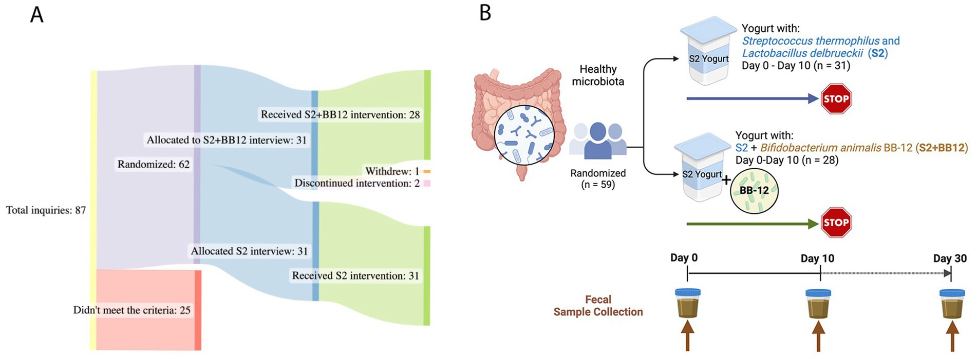
Figure 1. Study Design: a total of 59 children, aged one to five years old, were analyzed for this double-blinded, randomized controlled study (A). The participants consumed yogurt with two (S2 group) vs. three (S2 + BB12 group) probiotics’ strains (B). The participants consumed yogurt during ten consecutive days. Fecal samples were collected before the intervention (Day 0), after ten days of yogurt consumption (Day 10), and after 20 days of yogurt consumption discontinuation (Day 30). Created with BioRender.com and SankeyMATIC.com.
Participants were relatively divided equally between males and females (n = 28 males and n = 31 females). Most of the participants included were White (n = 40). Additional demographic information from the included participants is displayed in Table 1.
The administration of BB-12, in addition to S. thermophilus and L. delbrueckii, influenced the composition of the gut microbiome, although there were no overt global microbiome changes quantified by alpha and beta diversity indices. Species richness and Shannon indices were not significantly different between the S2 and S2 + BB12 groups at Day 10 (p = 0.65 and p = 0.24, respectively; Figures 2A,B). At Day 30, there were no significant differences in species richness between S2 and S2 + BB12 groups (p = 0.669), but Shannon diversity was significantly lower in the S2 + BB12 group compared to the S2 group (p = 0.044; Figures 2A,B). There were no between group differences in beta diversity (based on Bray–Curtis dissimilarity) at Day 10 (R2 = 0.02, p = 0.31; Figure 2C) or Day 30 (R2 = 0.02, p = 0.427; Supplementary Figure S1). Age was a significant predictor of alpha diversity measures in the Day 0 and Day 10 samples, irrespective of treatment group (species richness p = 0.001, Shannon index p = 0.045). These findings agree with previously reported results by Tan et al. (2017) using 16S rRNA analysis.
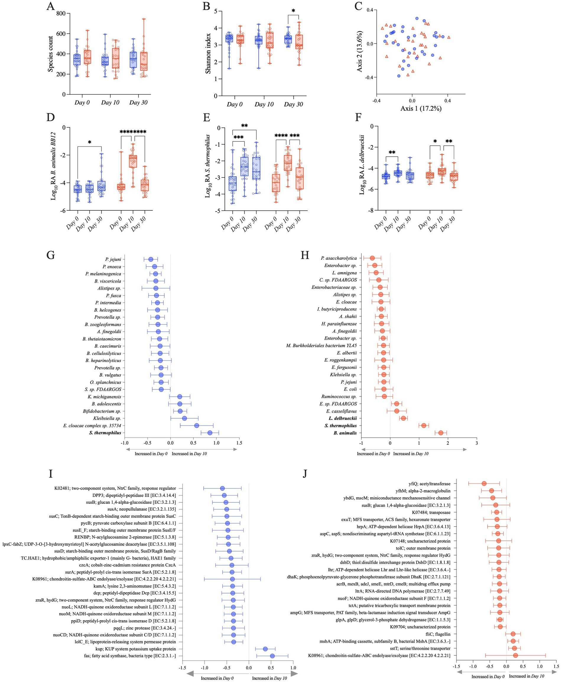
Figure 2. Observed species (A) and Shannon (B) alpha diversity indexes in S2 (blue) and S2 + BB12 (red) groups at Day 0, Day 10, and Day 30. PCoA based on Bray–Curtis dissimilarity followed by a PERMANOVA (p = 0.318) showing no separation among samples (C). Relative abundances of the three strains used in the S2 and S2 + BB12 interventions: B animalis (D), S. thermophilus (E), and L. delbrueckii (F) during time comparing S2 (blue) and S2 + BB12 groups (red). Top 25 changing taxa (G,H) and gene orthologs (I,J) between Day 0 and Day 10 in S2 (G–I) and S2 + BB12 (H–J) groups identified by linear model.
We built upon these findings by using shotgun metagenomics, which allowed us to observe differential abundant taxa at the species level. As expected, the metagenomic analysis showed an increase of B. animalis in the S2 + BB12 group. B. animalis abundance was significantly impacted by the administration of the BB12 supplemented yogurt (group × time p < 0.001; Figure 2D). We observed an effect of time, but not group, in L. delbrueckii (time p < 0.001; group p = 0.405). S. thermophilus’s relative abundance were significantly different in time and group × time interaction (time p < 0.001; group p = 0.349; group x time p = 0.020) (Figures 2E,F). As expected, post hoc testing showed in the S2 + BB12 group a statistically significant increase comparing Day 0 vs. Day 10 in BB-12 (p < 0.001), S. thermophilus (p < 0.001), and L. delbrueckii (p = 0.018) abundances. Similarly, Day 10 vs. Day 30 comparison showed a statistically significant decrease in BB-12 (p < 0.001), S. thermophilus (p = 0.001), and L. delbrueckii (p = 0.004) abundances (Figures 2D–F). In the S2 group, we found statistically significant differences in Day 0 vs. Day 10 comparison of S. thermophilus (p < 0.001) and L. delbrueckii (p = 0.005) abundances. Interestingly, BB-12 increased comparing Day 0 to Day 30 in the same group (p = 0.016).
We then focused our analyses on within-group differences in both groups from Day 0 to Day 10, as both groups received a probiotic intervention (S2 vs. S2 + BB12) and were healthy children. The 25 taxa with the greatest change in relative abundance between Day 0 and Day 10 were evaluated to observe the effect on individual microbial taxa following a ten-day administration of two (S2) vs. three (S2 + BB12) strains of probiotics. S. thermophilus, one of the intervention bacteria, was significantly increased in the S2 group at Day 10 compared to Day 0 (FDR p = 0.003; Figure 2G). Interestingly, the other intervention bacteria L. delbrueckii was absent among the 25 most changing taxa in the S2 group. In the S2 + BB12 group, all three intervention bacteria (i.e., B. animalis, S. thermophilus, and L. delbrueckii) were significantly increased at Day 10 (FDR p < 0.001, FDR p < 0.0001, and FDR p = 0.048, respectively), but the S2 + BB12 intervention did not significantly influence the abundance of any other taxa when FDR correction was applied (Figure 2G; Supplementary Table S1). In both groups most of the top changing taxa were decreased in Day 10 compared to Day 0 (19/25 and 20/25 taxa in S2 and S2 + BB12 groups respectively), but many of these differences were not significant before or after FDR correction (Figures 2G,H; Supplementary Table S1). We also saw an influence of race and ethnicity on within group taxonomic response to the S2 and S2 + BB12 interventions; E. cloacae complex sp. 35734 and K. michiganensis were both decreased in Asian children compared to Black or African American children (FDR p = 0.015 and 0.032, respectively, S2 group) and B. pseudocatenulatum was decreased in non-Hispanic children (FDR p = 0.022, S2 + BB12 group; Supplementary Table S2).
When we analyzed the differential abundance of gene orthologs, we found that the relative abundance of the glucan 1,4-alpha-glucosidase, chondroitin-sulfate-ABC endolyase/exolyase, and response regulator HydG genes decreased from Day 0 to Day 10 in both groups, (Figures 2I,J). These differences were also not statistically significant before or after FDR correction (Supplementary Table S1). Except for two orthologs, fatty acid synthase [K11533] and KUP potassium uptake protein, the 23 remaining orthologs among the 25 top changing ones between groups were decreased in the S2 group after ten days of probiotics administration, although these decreases were not statistically significant (Figure 2I; Supplementary Table S1). An overall trend of decreased orthologs relative abundance from Day 0 to Day 10 was similar in the S2 + BB12 group, except for four orthologs that had a non-statistically significant increase following BB12 administration, including fliC (flagellin), msbA; ATP-binding cassette, subfamily B, bacterial MsbA, sstT; serine/threonine transporter, and chondroitin-sulfate-ABC endolyase/exolyase (Figure 2J; Supplementary Table S1). There were two orthologs that changed significantly with age in the S2 + BB12 group; hexuronate transporter decreased (FDR p = 0.025) and aspartyl-tRNA synthetase increased (FDR p = 0.022; Supplementary Table S3). No significant changes were found with the other covariates in the S2 group.
We selected 18 known probiotic strains identified through literature search (Supplementary Table S4), to observe if the administration of two (S2) vs. three (S2 + BB12) probiotic strains would influence the abundance other known probiotics. Most of the identified probiotic-associated taxa belonged to Lactobacillus, Bacteroides, and Bifidobacterium genera, and were commonly used as probiotic supplements in the food industry or clinical trials (see Supplementary Table S4 for references). We observed that the abundance of targeted probiotic-associated bacteria increased from Day 0 to Day 10 in 61% (11/18) of selected taxa in the S2 group (Figure 3A) and 72% (13/18) of taxa in the S2 + BB12 (Figure 3B) group, but most of the differences did not reach statistical significance. Nevertheless, cumulative bacterial responses changes of probiotic-associated bacteria differed according to intervention group. For example, we observed no pattern in samples distribution in the S2 group (Figure 3C), while in the S2 + BB12 group we can observe a separation of Day 0 vs. Day 10 samples (Figure 3D). In the S2 + BB12 group, many of the Day 10 probiotic-associated bacteria clustered with B. animalis, indicating similar response to the probiotic (Figure 3D).
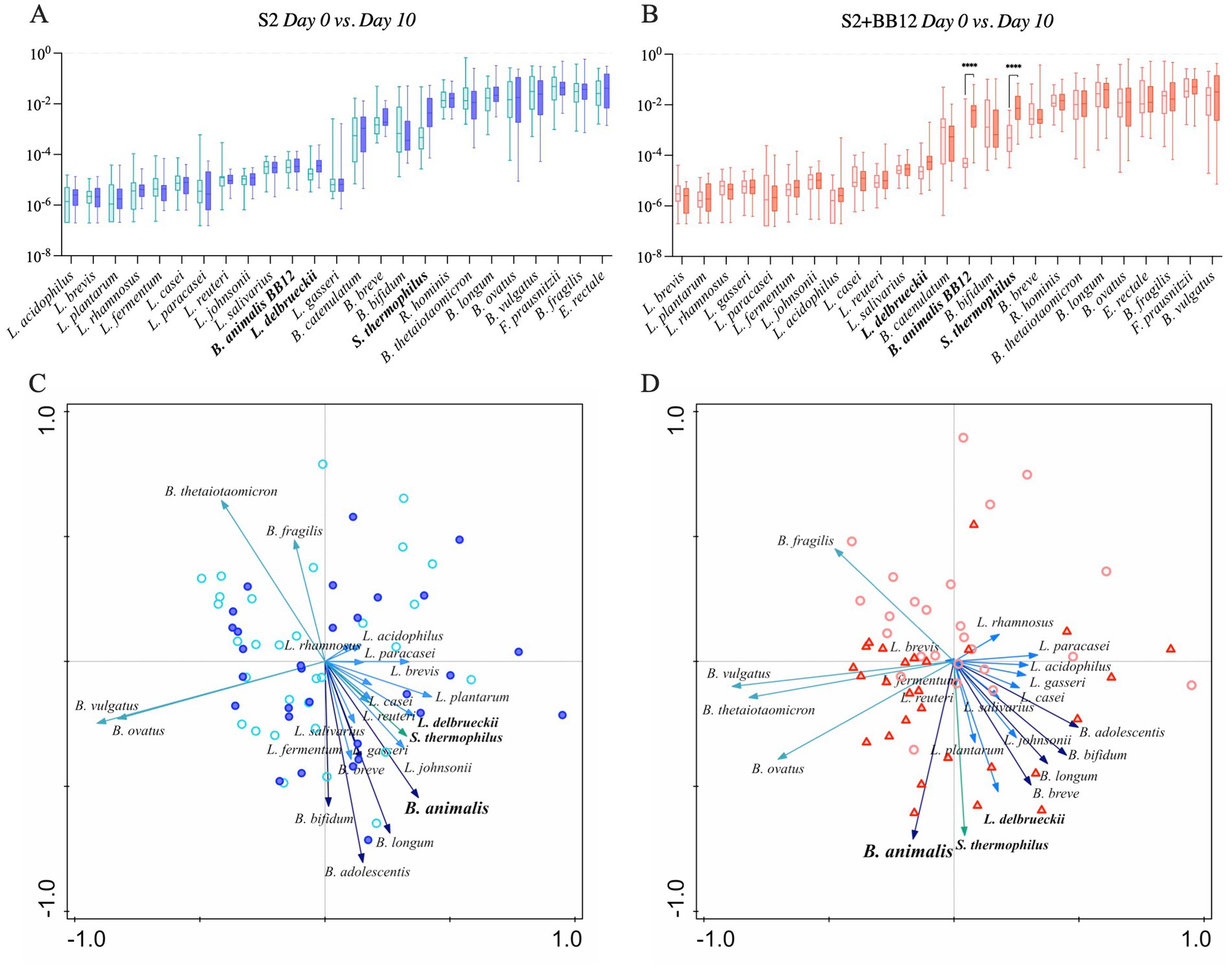
Figure 3. Relative abundances of selected probiotic taxa are plotted in S2 (A) and S2 + BB12 (B) groups after ten days of probiotic intervention (Day 0 vs. Day 10). Post-selection PCA shows selected probiotics following ten days of probiotics administration in S2 (C) and S2 + BB12 (D) groups. The difference in S. thermophilus and B. animalis between Day 0 and Day 10 abundances are statistically significant different (p < 0.0001 for both) in S2 + BB12 group. Day 10 is represented by the darkest color all plots. In PCA, different arrows colors refer to different genera: Blue, light blue, dark green, and green represent Bifidobacterium, Lactobacillus, Bacteroides, and Streptococcus genera, respectively. Each arrow points in the direction of the steepest increase of the values for the corresponding feature. The angle between arrows indicates the correlation between the different features (positive when the angle is sharp and negative when the angle is larger than 90 degrees). The length of the arrow is a measure of fit for the feature.
The untargeted metabolomic analysis identified 734 metabolites. To evaluate the impact of S2 vs. S2 + BB12 probiotics on the metabolome, we performed both exploratory analyses quantifying differences in all annotated metabolites and hypothesis-driven metabolomics analyses focused on amino acid-associated and short chain fatty acid metabolites.
After excluding xenobiotics, we conducted exploratory metabolomic analyses on 601 biochemicals. We analyzed the differences between Day 0 vs. Day 10, Day 10 vs. Day 30, and Day 0 vs. Day 30 within the S2 and S2 + BB12 groups (Supplementary Table S5). After correcting for multiple comparisons, we did not find statistically significant differences between groups for any metabolite evaluated. Nevertheless, there were clinically relevant metabolites that were differentially abundant during the probiotic intervention within groups, but did not survive correction for multiple testing (raw and FDR corrected p-values, along with FC response are listed in Supplementary Table S5). Therefore, metabolites with a FC response ≥ |2| and raw p-value <0.05 comparing Day 0 vs. Day 10 and Day 0 vs. Day 30 in S2 (Figures 4A,C,E) and S2 + BB12 (Figures 4B,D,F) groups are presented. We found greater changes in the S2 + BB12 group compared to the S2 group (14 vs. three metabolites). After ten days of yogurt consumption, we found an increase of N-acetylvaline (FDR p = 0.974, FC = 2.151) and a decrease of arachidoylcarnitine (C20) * (FDR p = 0.974, FC = 0.436). Day 0 vs. Day 30 comparison exhibited several changes in lipids decreased at Day 30 belonging to diacyglicerol metabolism: linoleoyl-linoleoyl-glycerol (18:2/18:2) (Wang et al., 2015)* (FDR p = 0.216, FC = 0.29), palmitoyl-linoleoyl-glycerol (16:0/18:2) (Bäckhed et al., 2015)* (FDR p = 0.188, FC = 0.311), palmitoyl-linoleoyl-glycerol (16:0/18:2) (Wang et al., 2015)* (FDR p = 0.216, FC = 0.263), oleoyl-linoleoyl-glycerol (18:1/18:2) (Bäckhed et al., 2015) (FDR p = 0.188, FC = 0.295), palmitoyl-oleoyl-glycerol (16:0/18:1) (Bäckhed et al., 2015)* (FDR p = 0.188, FC = 0.252), and oleoyl-linoleoyl-glycerol (18,1/18:2) (Wang et al., 2015) (FDR p = 0.166, FC = 0.221). The glycerolipids 2-palmitoyl-galactosylglycerol (16:0)* (FDR p = 0.166, FC = 2.358) and 1-palmitoyl-galactosylglycerol (16,0)* (FDR p = 0.166 FC = 2.319), were increased together with 3-hydroxybutyrate (BHBA) (FDR p = 0.906, FC = 2.821).
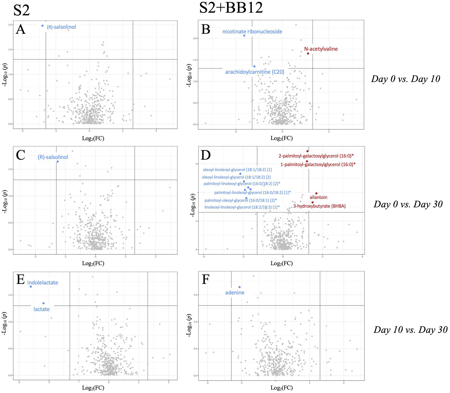
Figure 4. Volcano plot (p < 0.05, FC > 2) comparing Day 0 vs. Day 10 (A,B), Day 0 vs. Day 30 (C,D), and Day 10 vs. Day 30 (E,F) within S2 and S2 + BB12, respectively. Metabolites in red/blue represent metabolites increased/decreased at Day 10 (A,B) and Day 30 (C–F).
In the S2 group, the amino acid (R)-salsolinol was decreased after ten days of intervention (Day 0 vs. Day 10) (FDR p = 1.000, FC = 0.38) and 20 days after yogurt consumption discontinuation (Day 0 vs. Day 30) (FDR p = 0.995, FC = 0.423). Lactate and indolelactate were also decreased looking at Day 0 vs. Day 30 comparison (FC = 0.222, FDR p = 0.999 and FC = 0.165, FDR p = 0.999, respectively). PCA of untargeted metabolites showed no separation between the time points in any group (Supplementary Figures S2 A–F).
Next, we focused on evaluating differences in specific metabolites associated with amino acid metabolism or biosynthesis, as we hypothesized that they would be impacted by probiotic intervention and associated with the gut microbiome in both groups. There were several amino-acid associated metabolites that differed as a result of time in both groups (Supplementary Table S6) including 3-methyl-2-oxobutyrate (p = 0.004), 3-methyl-2-oxovalerate (p = 0.025), alanine (p = 0.001), glutamate (p = 0.034), isoleucine (p = 0.020), and valine (p = 0.004), among others. Conversely, glycine (p = 0.025), indolelactate (p = 0.034), N-acetylserine (p = 0.049), and pyroglutamine (p = 0.007) had significant group × time effects as a result of the S2 and S2 + BB12 probiotic interactions (Supplementary Table S6). In the S2 + BB12 group, average fecal metabolite abundances of 4-methyl-2-oxopentanoate, alanine, cysteine s-sulfate, glycine, histidine, lysine, N-acetylalanine, N-acetylglutamine, N-acetylleucine, phenylalanine, serine and valine were significantly higher at Day 10 (versus baseline Day 0; Figures 5A–N). Alanine, glycine, and N-acetylglutamine metabolite levels continued to be lower 20 days after the S2 + BB12 probiotic was discontinued (Day 30) vs. baseline (Figures 5B,D,H), while fecal cysteine s-sulfate levels increased back to baseline levels after probiotic discontinuation.
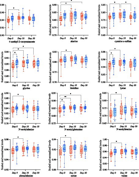
Figure 5. Targeted metabolomic analysis on amino acids was used to study the differences comparing Day 0 vs. Day 10, Day 0 vs. Day 30, and Day 10 vs. Day 30 within S2 (blue) and S2 + BB12 (red) groups. Linear mixed-effects model followed by post hoc pairwise testing with Tukey’s correction (when appropriate) were performed. All mixed model results of selected amino acid-associated metabolites and post hoc testing results (when group, time, or group * time model results were significant) are listed in Supplementary Table S6.
Microbe-metabolite interactions were tested through correlation matrices and visualized with network graphs. After filtering, 26 microbial taxa and 79 metabolites were tested for associations (Figure 6; Supplementary Tables S7, S8).
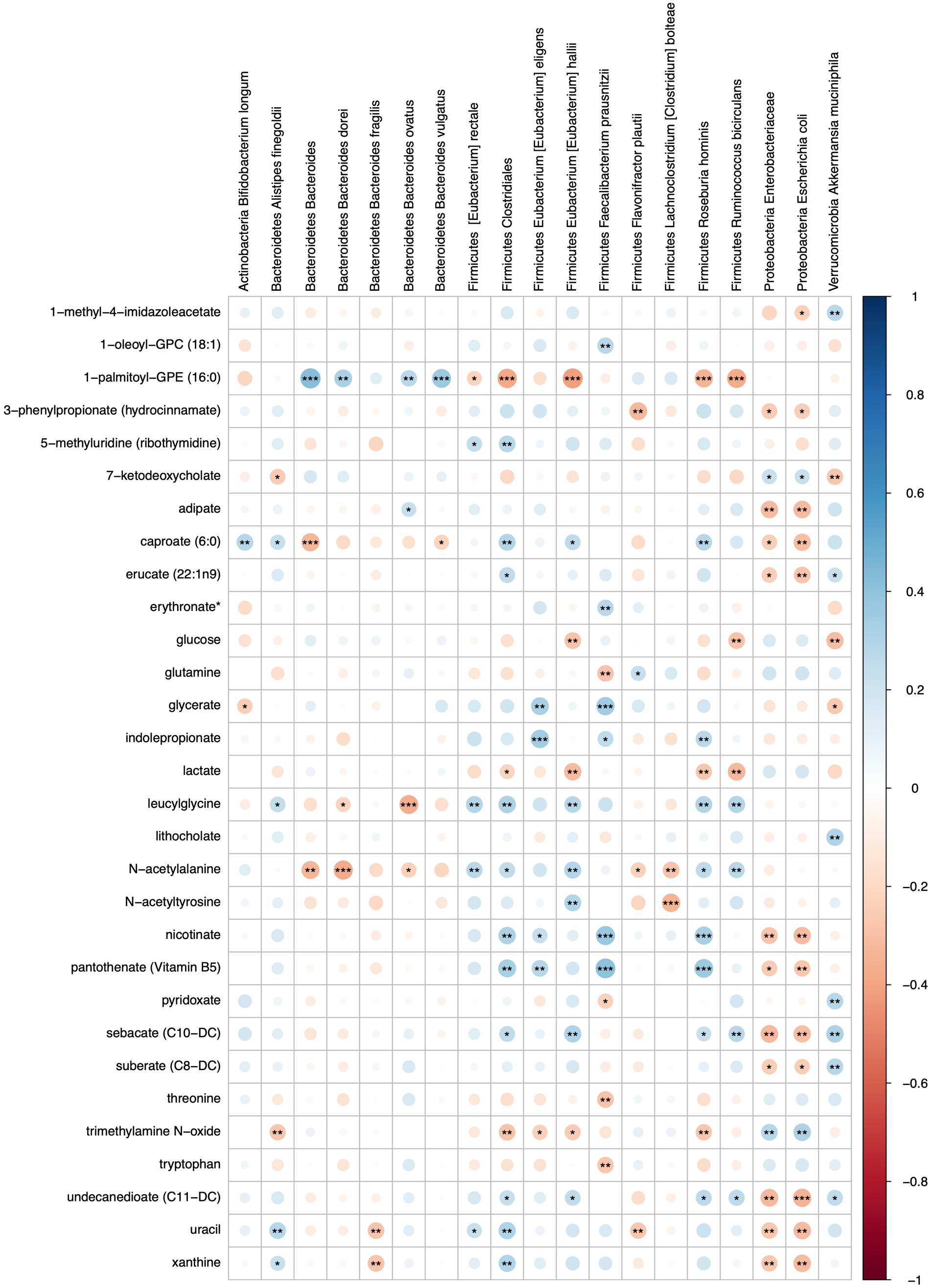
Figure 6. Heatmap representing FDR corrected correlations between microbial taxa and metabolites in S2 and S2 + BB12 groups. Red and blue represent negative and positive correlations, respectively. Stars indicate FDR values: * = FDR < 0.05, ** = FDR < 0.01, and *** = FDR < 0.001.
Metabolites that were significantly positively correlated with Enterobacteriaceae and Escherichia coli included trimethylamine N-oxide (TMAO), ursodeoxycholate, 7-ketodeoxycholate, and glycine (Figure 7). Other taxa that had multiple significant positive associations with metabolites included Akkermansia muciniphila, Eubacterium hallii, Roseburia hominis, and Clostridiales. Of the supplemented probiotic bacteria, B. animalis was positively associated with uracil, S. thermophilus with deoxycarnitine and phenylalanine, and L. delbrueckii with thymine. Known products of bacteria such as nicotinate (vitamin B3), pantothenate (vitamin B5) correlated positively with Clostridiales, R. hominis, F. prausnitzii, and E. eligens (Figure 7A).
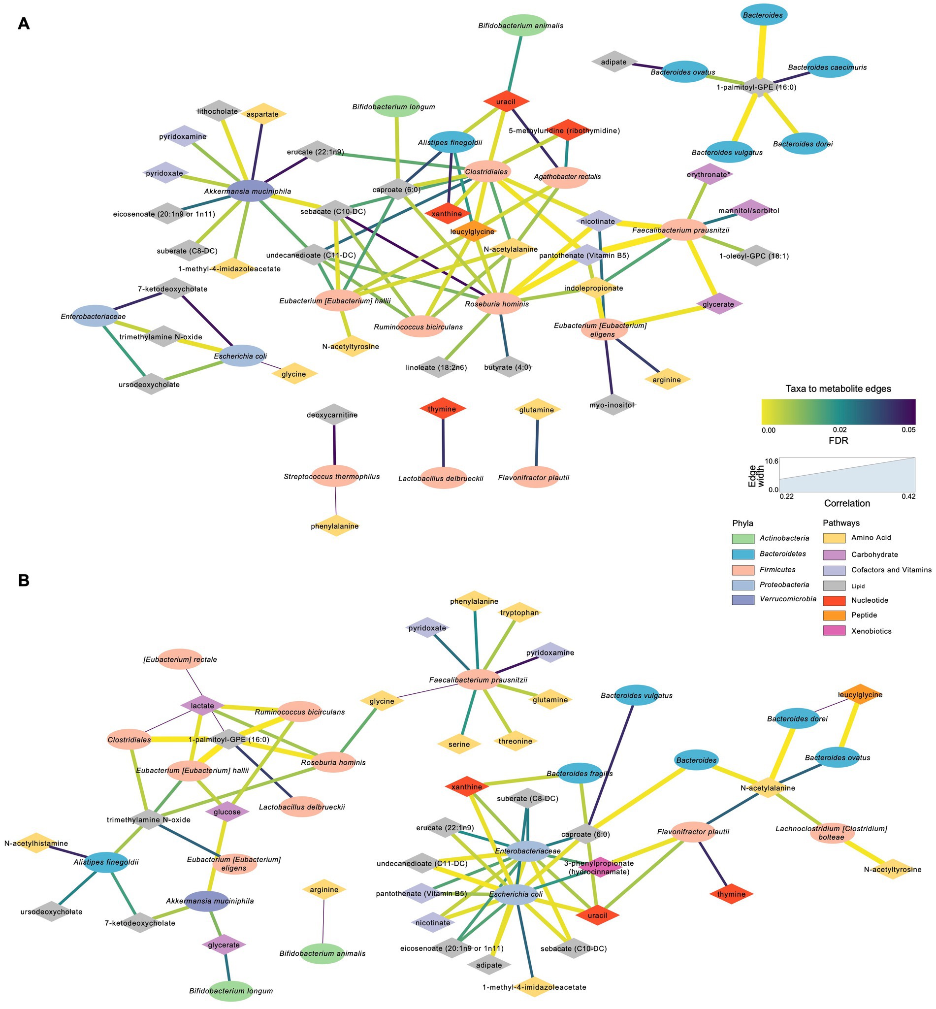
Figure 7. Significant microbial taxa and metabolite associations: Network analyses in samples across Day 0, Day 10, and Day 30 within S2 and S2 + BB12 groups. (A) Network generated from microbial taxa (ellipses) that passed filtering and microbial-associated metabolites (diamonds) identified from previous work (Tanes et al., 2021) showing only positive correlations. (B) Same network but showing only negative correlations. Width of edges is proportional to Spearman correlation coefficient and edge color is mapped to the FDR value or the correlation test. All correlations between selected taxa and metabolites and statistical significance testing results are listed in Supplementary Table S5.
Several amino acids correlated negatively with F. prausnitzii and R. hominis, including glutamine, lactate, threonine, tryptophan and TMAO (Figure 7B). Lactate was also negatively associated with A. rectalis, R. bicirculans, E. hallii, and Clostridiales; glucose with R. bicirculans, E. hallii, and A. muciniphila; and glycerate with A. muciniphila and B. longum. Among the probiotics of interest, B. animalis correlated negatively with arginine and L. delbrueckii with 1-palmitoyl-GPE (16:0). Uracil was positively associated with Clostridiales, A. finegoldii, and E. rectale. Uracil was also negatively correlated with B. dorei, F. plautii, Enterobacteriaceae and E. coli.
This randomized, controlled study aimed to observe the differences following the administration of yogurt supplemented with two (S2) vs. three (S2 + BB12) probiotic strains in healthy children. We studied probiotics consumption effects on the fecal microbiome and metabolome comparing baseline, post-ten days (Day 10), and post-30 days (Day 30) following yogurt administration. There were no significant differences in the global metagenomic or metabolomic profiles between healthy children receiving two (S2) vs. three (S2 + BB12) probiotic strains for 10 days. Nevertheless, we observed a significant increase (Day 0 to Day 10) in the relative abundance of the two and three probiotics administered in the S2 and S2 + BB12 groups, respectively, indicating the intervention had a measurable impact on the bacteria of interest in the gut microbiome. Interestingly, in the S2 group, S. thermophilus species appear to maintain a sustained increase over time, maintaining the significant increase in relative abundance from Day 0 vs. Day 30 As both cohorts of children received a probiotic cocktail and the intervention lasted over a short period of time, the lack of strong association between groups in global microbiome changes is not surprising and suggests a resilient and stable gut microbiota in this cohort of healthy children ages one to five. Nevertheless, existing literature reports about infant’s and children’s gut microbiota stability are conflicting (Bäckhed et al., 2015; Kumbhare et al., 2019). Infant and children’s gut microbiota are thought to be immature and therefore more susceptible to perturbations. In line with other studies (Laursen et al., 2017; Wang et al., 2021) our data show short-term effects of probiotic administrations in both groups on the gut microbiome and metabolome. The children’s gut microbiota stability observed in this study is consistent with the parent study’s findings (Tan et al., 2017).
Most studies examining the health benefits of probiotics have focused on people with pathologies. However, few studies have examined the effects of probiotics on healthy individuals and even fewer have examined the use of probiotics in children. A review of probiotic supplementation in healthy adults found that probiotic supplementation led to a transient increase in the concentration of supplement-specific bacteria but failed to support the ability of probiotics to cause persistent changes in gut microbiota (Khalesi et al., 2019). This is consistent with our results finding a significant increase in the concentration of the supplement-specific bacteria BB-12, following 10 days of BB-12 supplementation. Importantly, as reported in adults, these results were temporary. On Day 30 (20 days following the termination of the BB-12 supplementation), there were no significant differences in the concentration of BB-12 in the S2 + BB12 group.
As yogurts and other dairy products commonly supplemented with probiotics often possess other beneficial characteristics, such as a high calcium content, these properties of probiotics could allow consumers to benefit from the nutritional components without risking disruption to their microbiota and health. This potential beneficial shift to probiotic-associated bacteria that we demonstrated in the S2 + BB12 group (Figure 3) supports the theory that a probiotic intervention may provide a net positive contribution to the gut microbiome ecosystem without conferring strong effects on specific individual bacteria. These findings are also consistent with the concept of emergent properties which postulates that individual properties cannot entirely be explained by their individual components (Ponge, 2005). Therefore, in the context of the current study, although no significant differences following the interventions were observed, there was a global shift towards the increase of bacteria belonging to Bifidobacterium and Lactobacillus genera, commonly used probiotics. The combined effects of these bacteria could have more functional implications and protective effects than individual bacterial changes supporting the use of probiotics to maintain a healthy gut ecosystem in hopes to prevent gastrointestinal disorders in children, although more research is needed to directly test and validate this notion. Importantly, as this study was conducted on healthy children, these findings should not be extrapolated to children with gastrointestinal disorders or subjects seeking intervention for other diseases or disorders.
Species belonging to Bifidobacterium genus were highly abundant in both groups at the three time points. Bifidobacteria are highly represented in children, as they use milk oligosaccharides as a carbon source and to restrict human milk oligosaccharides availability to other microorganisms (Turroni et al., 2011). In addition to the parent study findings, our metagenomic approach allowed a low-level taxonomic affiliation, revealing differences among species belonging to the same genus as in the case of Bifidobacterium. It explains why Bifidobacterium genus was not among the differentially abundant species in the previous study, i.e., because different Bifidobacterium species were abundant in the experimental group (S2 + BB12) and in the control group S2 (e.g., B. catenulatum, B. pseudocatenulatum). Additionally, this metagenomic approach adds to the existing literature of the genomic potential of the microbial community underlying microbiome-host interactions.
Because the fecal metabolome is influenced by different factors, these changes could reflect shifts of dietary intake, digestion, microbial degradation, and host absorption. When we analyzed the fecal metabolome and microbial diversity using both participant groups, we found associations between metabolites (e.g., uracil, deoxycarnitine, and thymine) and microorganisms. Uracil was positively associated with Clostridiales family, A. finegoldii, B. animalis, and E. rectale. B. dorei, F. plautii, Enterobacteriaceae family, and E. coli were negatively correlated with uracil. During infections, the immune response triggered by uracil promotes pathogen bacteria elimination, intestinal cell repair, and host homeostasis (Lee et al., 2013). Thymine was positively associated with L. delbrueckii, the probiotic used in both interventions, and negatively correlated to F. plautii. Thymine was found to accelerate microbial metabolisms and ROS production improving antibiotic efficacy both in vitro and in vivo (Liu et al., 2021). Deoxycarnitine, positively associated with S. thermophilus, was linked to increased intestinal permeability (Ghonimy et al., 2018). This suggests that the fecal metabolome may influence gut immune function, permeability, and homeostasis.
A strength of our study is that it was a randomized, blinded controlled trial conducted on children ages one to five years old, an age group that is rarely studied in probiotic research. Additionally, we incorporated and integrated both metagenomic and metabolomic analyses to characterize the effect of BB-12 supplemented yogurt on the children’s gut-microbiome and metabolism. This unique integration allowed us to test more system-wide and functional effects on the gut microbiome as a result of two probiotic interventions, providing more comprehensive data on gut health. However, this study examined short-term changes of probiotics over a 10-day period, and more studies should be conducted to investigate the long-term effects on probiotic consumption in this age group. An additional limitation is that we did not incorporate information of dietary intake in the current analysis, as it was not collected in the original study procedures as described by Tan et al. (2017). Diet is an important driver of microbiome composition and an important covariate when evaluating the impact of an intervention, such as probiotics, on gut microbiome communities (Maki et al., 2019). Dietary intake recall and assessment has been historically challenging in children and ongoing work is being performing to improve the validity of dietary reporting in study cohorts with age ranges such as the ones in this study (Livingstone et al., 2004; Livingstone, 2022). Future work evaluating the impact of interventions on gut microbiome composition in children should aim to collect dietary intake should be performed, likely with the assistance of a caregiver or parent (Walker et al., 2018), in order to adequately control for the impact of food consumption on study measures. Novel metagenomics-based methods are being developed to validate self-report tools, such as using metabarcoding to quantify plant components of human diets (Reese et al., 2019). While these methods are outside of the scope of the current research, they will be useful tools to validate and quantify different dietary components in future microbiome studies.
More studies are needed to elucidate the mechanistic pathways by which probiotics such as BB-12 can affect mucosal barrier functions and innate immunity. Future studies should expand upon the findings presented in this double-blinded, randomized controlled trial and examine the interplay of diet and probiotics on metabolites and microbiota in children. Additionally, further research is needed to investigate environmental factors that influence the impacts of probiotics on children’s health status and behavior. The long-term supplementation BB-12 on this population and its longitudinal effects during development should also be examined. This would allow for more in-depth knowledge of the impact that probiotics have on gut microbial communities in developing children as they age. Additionally, future research in children at risk for gastrointestinal disorders may elucidate if these functional metabolite changes as a result of S2 + BB12 probiotic intervention confer a protective gastrointestinal effect.
In conclusion, the results from this deep metagenomic and metabolomic characterization of the gut microbiome and metabolome of children following BB-12 consumption did not show statistically significant differences between the groups, although net positive emergent property effects were witnessed in the S2 + BB12 group over time. The functional redundancy in healthy microbial systems and metabolic stability reflects no changes in the microbial diversity, although we did observe a separation effect in the S2 + BB12 group as a result of the three-strain intervention when we focused on probiotic-associated and beneficial bacteria reported the literature. Our study validated previous results from Tan et al. (2017) and allowed a more in-depth taxonomic characterization of the microorganisms, their genes, and their metabolites. We detected higher abundances of two of the probiotic intervention bacterial taxa (B. animalis and S. thermophilus) in study subjects receiving the based S2 probiotic intervention + BB12, but no individual taxonomic changes occurred in the S2 only group. Finally, although we did not see global fecal metabolome response to either probiotic, several fecal metabolites were increased in the S2 + BB12 group, indicating a net functional impact of the addition of BB12 to the probiotic intervention. Future research replicating these results across different patient populations will confirm the therapeutic use of BB12 as a probiotic intervention to exert beneficial impacts on the pediatric gastrointestinal system.
Participants ages one to five years old were recruited through the Capital Area Primary Care Research Network for phase I of a double-blinded, randomized controlled study (protocol NCT001652287). Participants included in this study were children whose parents/caregivers were able to read, write, and speak either English or Spanish and had access to a telephone and refrigerator. Eligible participants provided written informed consent were enrolled and randomized as described by Tan et al. to either the BB-12® or control yogurt drink by family cluster. The study protocol was approved by the Georgetown University Institutional Review Board (IRB No. 2012-1,112, Washington, DC). The independent Data Safety Monitoring Board reviewed the protocol before study initiation and checked adverse event data at approximately 33, 50 and 66% data completion. Additional monitoring was conducted by the FDA/CBER, under IND#13691 and the National Institutes of Health (NIH), National Center for Complementary and Integrative Health (NCCIH), including its Office of Clinical and Regulatory Affairs. Participants’ eligibility criteria are described in Tan et al. (2017) which included the absence of lactose intolerance and chronic conditions, such as diabetes and asthma. The participants were asked not to consume any products containing probiotics for 14 days before initiating the yogurt intervention and throughout the entire intervention period. The base yogurt drink was prepared with live yogurt starter cultures of Streptococcus thermophilus and Lactobacillus delbrueckii probiotics [referred to as the two strain [S2] yogurt group (Nagaoka, 2019)], as described in Tan et al. (2017). At baseline, the children were randomized into two groups called S2 or S2 + BB12. Participants in the S2 group (n = 31) were administered 112 g of the base yogurt beverage (containing Streptococcus thermophilus and Lactobacillus delbrueckii only) with no additions to the drink. In contrast, participants in the S2 + BB12 group (n = 28) were administered the base yogurt beverage that was supplemented with an additional 1 × 10¹⁰ colony-forming units of BB12 per serving per day. BB12 was chosen as the interventional probiotic of interest as it has been widely studied in infants and children (Merenstein et al., 2010, 2011; Goldenberg et al., 2015; Merenstein et al., 2021), and is associated with positive gastrointestinal protective mechanisms including maintenance of tight junction function and immune regulation in the gut (Uusitupa et al., 2020; Cheng et al., 2021). The yogurt was administered to the participants in both groups for ten consecutive days.
Fecal samples were collected at baseline (Day 0), following 10 days of yogurt consumption (Day 10), and 20 days following discontinuation of yogurt administration (Day 30), and immediately stored after collection at −80°C. Samples from days 0, 10 and 30 were then thawed, and approximately 100 mg of the samples were sent to Microbiome Center of the Children’s Hospital of Philadelphia (n = 169) for microbiome analysis and another 100 mg were sent to Metabolon Inc. (Morrisville, NC, United States; n = 174) for metabolomic analyses.
The DNA used for the metagenomic analysis was extracted using the DNeasy PowerSoil Kit (Qiagen, Hilden, Germany) and quantified with the Quant-iT PicoGreen Assay Kit (Molecular Probes). Shotgun libraries were generated from 0.5 ng DNA using the Nextera XT Library Prep Kit (Illumina, San Diego, CA, United States) and libraries were sequenced on an Illumina HiSeq 2500 in High Output mode to produce paired-end 125 bp sequence reads. Extraction blanks and nucleic acid-free water were processed along with experimental samples to empirically assess environmental and reagent contamination. A laboratory-generated mock community consisting of DNA from Vibrio campbellii and Lambda phage was included as a positive sequencing control.
The metabolomic analysis was performed using untargeted ultra-performance liquid chromatography-tandem mass spectrometry (UPLC/MS/MS, Waters ACQUITY, Milford, MA, United States), as described previously (Vizioli et al., 2021). Briefly, the fecal samples were prepared using the automated MicroLab STAR system (Hamilton Company, Franklin, MA, United States) and extracted at a constant per-mass basis. Proteins were removed using methanol precipitation (Glen Mills GenoGrinder 2000), followed by centrifugation. The samples were processed using four methods: reverse phase (RP)-UPLC/MS/MS with electrospray ionization (ESI), in both positive (optimized for hydrophilic and hydrophobic compounds, respectively) and negative modes, and hydrophilic interaction chromatography (HILIC)-UPLC/MS/MS-ESI in negative ion mode. The raw UPLC/MS/MS data were integrated into ion peaks organized by mass, retention time/index, and peak area. Metabolites were annotated by comparison of individual spectra to a standard reference library, and area-under-the-curve analysis was performed for peak quantification.
For gut microbiome and metabolome analyses, we studied within and between group differences after 10 days of probiotic consumption (Day 10) and 20 days post probiotic discontinuation (Day 30). Shotgun metagenomic data were analyzed using Sunbeam (Clarke et al., 2019). The abundance of bacteria was estimated using Kraken. (Wood and Salzberg, 2014). Taxa that were above 0.1% abundance in any sample were used for differential abundance testing, along with including the microbial taxa at the species level that were included in the probiotic interventions (i.e., L. delbrueckii, S. thermophilus, and B. animalis). Differential abundance analysis was performed using linear models of Log10 transformed relative abundances. Reads were mapped to the KEGG database (Ogata et al., 1999) using DIAMOND (Buchfink et al., 2015) to estimate the abundance of bacterial gene orthologs. Differences between groups for gene and pathways abundances were found by applying linear models to logit transformed relative abundances. Alpha diversity within samples in the S2 and S2 + BB12 groups were assessed by computing the expected number of species at a sequencing depth of 1,000 reads and the Shannon index. To evaluate community-level differences between S2 and S2 + BB12 group fecal samples, beta diversity was calculated using Bray–Curtis dissimilarity matrices, visualized using Principal Coordinates Analysis (PCoA) plots, and relationships within and between S2 and S2 + BB12 groups were compared using the PERMANOVA test.
The top 25 most abundant bacterial taxa and gene orthologs from shotgun metagenomics sequencing were selected using linear models to evaluate the taxa with greatest estimated change in Log10 transformed relative abundance for the given comparison. Probiotic-associated bacteria, previously demonstrated to be short chain fatty acid (SCFA)-producers and beneficial for GI health (Markowiak-Kopeć and Śliżewska, 2020), were determined from the literature (Supplementary Table S4). The impact of the S2 vs. S2 + BB12 probiotic strains on overall probiotic-associated bacterial relative abundance was additionally measured and visualized by CANOCO version 5 (Braak and Milauer, 1998) in a post-selection PCA to evaluate the effect of targeted probiotic strain administration on bacterial responses of taxa known to be linked to gut microbiome health.
Exploratory and hypothesis-driven metabolite analyses were performed with untargeted metabolite data processed by Metabolon Inc. using MetaboAnalyst 5.01 (Chong et al., 2018) and R. (R Development Core Team, 2013) Metabolites with 20% or more missing values were excluded from the exploratory analyses. Missing values, if any, were imputed as 1/5 of the minimum positive value of each feature. Metabolite values were median-scaled and Log10 transformed. Wilcoxon rank-sum test, fold change (FC) analyses (FC threshold = 2), and Principal Component Analysis (PCA) were performed to analyze differences between Day 0 vs. Day 10, Day 0 vs. Day 30, and Day 0 vs. Day 30 within the S2 and S2 + BB12 groups and between groups within each time point.
Hypothesis-driven metabolite analyses were additionally performed in metabolites associated with the amino acid super pathways (i.e., glycine, serine and threonine metabolism, alanine and aspartate metabolism, etc.), and SCFAs, as these metabolites are strongly associated with gut microbial community characteristics (Neis et al., 2015). Linear mixed-effects model followed by post hoc pairwise testing (when appropriate) and Tukey’s correction were performed in JMP statistical analysis platform (Ye et al., 2000).
To create a network of metabolite-taxa correlation pairs, filtering was applied to metabolites as above as well as restricting to bacterial substrates and products based on previous work (Tanes et al., 2021). Briefly, metabolite substrates were defined as those that were increased after treatment with antibiotics and products were those that decreased (Tanes et al., 2021). Microbial taxa at the species level were filtered to include taxa present at >0.01% mean relative abundance, along with including the microbial taxa at the species level that were included in the probiotic interventions (i.e., L. delbrueckii, S. thermophilus, and B. animalis). Spearman correlation testing was then performed on each microbe-metabolite pair with FDR correction applied to p-values. Network diagrams of bacteria and metabolites were generated using Cytoscape v3.9.1 (Shannon et al., 2003). Metabolites that significantly correlated with L. delbrueckii, S. thermophilus, and BB-12 were additionally tested for intervention-associated change over time. Statistical significance was defined as p-values or FDR corrected p-values <0.05 for all statistical analyses. All statistical tests were adjusted for the following covariates: age, race, gender, ethnicity, and total number of housemates except for correlations between genes and metabolites.
The content is solely the responsibility of the authors and does not necessarily represent the official views of the National Institutes of Health.
The datasets presented in this study can be found in the NCBI repository, accession number PRJNA929986: https://www.ncbi.nlm.nih.gov/bioproject/PRJNA929986.
The studies involving human participants were reviewed and approved by Georgetown University Institutional Review Board. Written informed consent to participate in this study was provided by the participants’ legal guardian/next of kin.
DM and PJ: concept and design, obtained funding, and study supervision. TT, DM, AF, and BB: acquisition of data and sample processing. CV, AF, SD, KB, RJ-L, KM, and PJ: statistical analysis or interpretation of data. CV, AD, AF, BB, RJ-L, KM, and PJ: drafting of the manuscript. AF, DM, TT, BB, PV, SD, and KB: critical revision of the manuscript for important intellectual content. PJ: administrative, technical, or material support. All authors contributed to the article and approved the submitted version.
PJ is supported by National Institute of Alcohol Abuse and Alcoholism under award number, Z01AA000135, the National Institute of Nursing Research and the Rockefeller University Heilbrunn Nurse Scholar Award. PJ is supported by the Office of Workforce Diversity, and the Office of Workforce Diversity, National Institutes of Health Distinguished Scholar Program. Intramural Research Training Award (to AF, RJ-L, and BB). RJ-L is supported by the Center of Compulsive Behaviors Fellowship, National Institutes of Health. Funding from Dannon to TT and DM, the Department of Family Medicine, Georgetown University Medical Center, Washington, DC. KM is supported by intramural research funds at the National Institutes of Health, Clinical Center.
The authors would like to thank Joan Austin for her review and edits and Jennifer J. Barb for bioinformatics consultative support. A special thanks to the participants of this study and the Georgetown Microbiome Group.
DM previously served as a paid expert at Howard University and Bayer. DM has done legal work for Visniome VSL#3, Golo for Life, and President of the International Scientific Association for Probiotics and Prebiotics (ISAPP) board.
The remaining authors declare that the research was conducted in the absence of any commercial or financial relationships that could be construed as a potential conflict of interest.
All claims expressed in this article are solely those of the authors and do not necessarily represent those of their affiliated organizations, or those of the publisher, the editors and the reviewers. Any product that may be evaluated in this article, or claim that may be made by its manufacturer, is not guaranteed or endorsed by the publisher.
The Supplementary material for this article can be found online at: https://www.frontiersin.org/articles/10.3389/fmicb.2023.1165771/full#supplementary-material
SUPPLEMENTARY FIGURE S1 | Metagenomics PCoA day 30.
SUPPLEMENTARY FIGURE S2 | Metabolite PCA.
SUPPLEMENTARY TABLE S1 | Top 25 taxa genes within group.
SUPPLEMENTARY TABLE S2 | Covariate results taxa.
SUPPLEMENTARY TABLE S3 | Covariate results gene.
SUPPLEMENTARY TABLE S4 | Probiotics selection.
SUPPLEMENTARY TABLE S5 | Metabolites and pathways volcano plot.
SUPPLEMENTARY TABLE S6 | Selected metabolite model results.
SUPPLEMENTARY TABLE S7 | 26 taxa to 479 metab correlations.
SUPPLEMENTARY TABLE S8 | 26 taxa to 79 metab network.
FC, Fold change; FDR, False discovery rate; GI, Gastrointestinal; PCA, Principal component analysis; PCoA, Principal coordinate analysis; PERMANOVA, Permutational analysis of variance; RA, Relative abundance; S2, Two-strain (Streptococcus thermophilus and Lactobacillus delbrueckii) yogurt group; S2 + BB12, Two-strain (Streptococcus thermophilus and Lactobacillus delbrueckii) plus Bifidobacterium animalis subsp. lactis strain BB-12 yogurt group; TMAO, Trimethylamine N-oxide.
Arpaia, N., Campbell, C., Fan, X., Dikiy, S., van der Veeken, J., deRoos, P., et al. (2013). Metabolites produced by commensal bacteria promote peripheral regulatory T-cell generation. Nature 504, 451–455. doi: 10.1038/nature12726
Artis, D. (2008). Epithelial-cell recognition of commensal bacteria and maintenance of immune homeostasis in the gut. Nat. Rev. Immunol. 8, 411–420. doi: 10.1038/nri2316
Bäckhed, F., Roswall, J., Peng, Y., Feng, Q., Jia, H., Kovatcheva-Datchary, P., et al. (2015). Dynamics and stabilization of the human gut microbiome during the first year of life. Cell Host Microbe 17, 690–703. doi: 10.1016/j.chom.2015.04.004
Boulangé, C. L., Neves, A. L., Chilloux, J., Nicholson, J. K., and Dumas, M. E. (2016). Impact of the gut microbiota on inflammation, obesity, and metabolic disease. Genome Med. 8:42. doi: 10.1186/s13073-016-0303-2
Braak, C.T . and Milauer, P. CANOCO Reference Manual and User’s Guide to Canoco for Windows: Software for Canonical Community Ordination (Version 4). (1998).
Buchfink, B., Xie, C., and Huson, D. H. (2015). Fast and sensitive protein alignment using DIAMOND. Nat. Methods 12, 59–60. doi: 10.1038/nmeth.3176
Cheng, J., Laitila, A., and Ouwehand, A. C. (2021). Bifidobacterium animalis subsp. lactis HN019 effects on gut health: a review. Front. Nutr. 8:790561. doi: 10.3389/fnut.2021.790561
Chong, J., Soufan, O., Li, C., Caraus, I., Li, S., Bourque, G., et al. (2018). MetaboAnalyst 4.0: towards more transparent and integrative metabolomics analysis. Nucleic Acids Res. 46, W486–w494. doi: 10.1093/nar/gky310
Clarke, E. L., Taylor, L. J., Zhao, C., Connell, A., Lee, J. J., Fett, B., et al. (2019). Sunbeam: an extensible pipeline for analyzing metagenomic sequencing experiments. Microbiome 7:46. doi: 10.1186/s40168-019-0658-x
Conlon, M. A., and Bird, A. R. (2015). The impact of diet and lifestyle on gut microbiota and human health. Nutrients 7, 17–44. doi: 10.3390/nu7010017
Dai, Z. L., Wu, G., and Zhu, W. Y. (2011). Amino acid metabolism in intestinal bacteria: links between gut ecology and host health. Front. Biosci. 16, 1768–1786. doi: 10.2741/3820
den Besten, G., van Eunen, K., Groen, A. K., Venema, K., Reijngoud, D. J., and Bakker, B. M. (2013). The role of short-chain fatty acids in the interplay between diet, gut microbiota, and host energy metabolism. J. Lipid Res. 54, 2325–2340. doi: 10.1194/jlr.R036012
Ghonimy, A., Zhang, D., Farouk, M., and Wang, Q. (2018). The impact of carnitine on dietary fiber and gut bacteria metabolism and their mutual interaction in monogastrics. Int. J. Mol. Sci. 19:1008. doi: 10.3390/ijms19041008
Goldenberg, J. Z., Goldenberg, J. Z., Humphrey, C., El Dib, R., and Johnston, B. C. (2015). Probiotics for the prevention of pediatric antibiotic-associated diarrhea. Cochrane Database Syst. Rev. 12:Cd004827. doi: 10.1002/14651858.CD004827.pub5
Griffin, S. M., Lehtinen, M. J., Meunier, J., Ceolin, L., Roman, F. J., and Patterson, E. (2022). Restorative effects of probiotics on memory impairment in sleep-deprived mice. Nutr. Neurosci. 26, 254–264. doi: 10.1080/1028415X.2022.2042915
Hill, C., Guarner, F., Reid, G., Gibson, G. R., Merenstein, D. J., Pot, B., et al. (2014). Expert consensus document. The International Scientific Association for Probiotics and Prebiotics consensus statement on the scope and appropriate use of the term probiotic. Nat. Rev. Gastroenterol. Hepatol. 11, 506–514. doi: 10.1038/nrgastro.2014.66
Hojsak, I., Fabiano, V., Pop, T. L., Goulet, O., Zuccotti, G. V., Çokuğraş, F. C., et al. (2018). Guidance on the use of probiotics in clinical practice in children with selected clinical conditions and in specific vulnerable groups. Acta Paediatr. 107, 927–937. doi: 10.1111/apa.14270
Jungersen, M., Wind, A., Johansen, E., Christensen, J., Stuer-Lauridsen, B., and Eskesen, D. (2014). The science behind the probiotic strain Bifidobacterium animalis subsp. lactis BB-12®. Microorganisms 2, 92–110. doi: 10.3390/microorganisms2020092
Khalesi, S., Bellissimo, N., Vandelanotte, C., Williams, S., Stanley, D., and Irwin, C. (2019). A review of probiotic supplementation in healthy adults: helpful or hype? Eur. J. Clin. Nutr. 73, 24–37. doi: 10.1038/s41430-018-0135-9
Kostic, A. D., Gevers, D., Siljander, H., Vatanen, T., Hyötyläinen, T., Hämäläinen, A. M., et al. (2015). The dynamics of the human infant gut microbiome in development and in progression toward type 1 diabetes. Cell Host Microbe 17, 260–273. doi: 10.1016/j.chom.2015.01.001
Kumbhare, S. V., Patangia, D. V., Patil, R. H., Shouche, Y. S., and Patil, N. P. (2019). Factors influencing the gut microbiome in children: from infancy to childhood. J. Biosci. 44:49. doi: 10.1007/s12038-019-9860-z
Laursen, M. F., Laursen, R. P., Larnkjær, A., Michaelsen, K. F., Bahl, M. I., and Licht, T. R. (2017). Administration of two probiotic strains during early childhood does not affect the endogenous gut microbiota composition despite probiotic proliferation. BMC Microbiol. 17:175. doi: 10.1186/s12866-017-1090-7
LeBlanc, J. G., Chain, F., Martín, R., Bermúdez-Humarán, L. G., Courau, S., and Langella, P. (2017). Beneficial effects on host energy metabolism of short-chain fatty acids and vitamins produced by commensal and probiotic bacteria. Microb. Cell Fact. 16:79. doi: 10.1186/s12934-017-0691-z
Lee, S. H., Joo, N. S., Kim, K. M., and Kim, K. N. (2018). The therapeutic effect of a multistrain probiotic on diarrhea-predominant irritable bowel syndrome: a pilot study. Gastroenterol. Res. Pract. 2018:8791916. doi: 10.1155/2018/8791916
Lee, K. A., Kim, S. H., Kim, E. K., Ha, E. M., You, H., Kim, B., et al. (2013). Bacterial-derived uracil as a modulator of mucosal immunity and gut-microbe homeostasis in Drosophila. Cells 153, 797–811. doi: 10.1016/j.cell.2013.04.009
Liu, Y., Yang, K., Jia, Y., Shi, J., Tong, Z., Wang, Z., et al. (2021). Thymine sensitizes gram-negative pathogens to antibiotic killing. Front. Microbiol. 12:622798. doi: 10.3389/fmicb.2021.622798
Livingstone, M. B. E. (2022). Issues in dietary intake assessment of children and adolescents. Br. J. Nutr. 127, 1426–1427. doi: 10.1017/S0007114522000770
Livingstone, M. B., Robson, P. J., and Wallace, J. M. (2004). Issues in dietary intake assessment of children and adolescents. Br. J. Nutr. 92, S213–S222. doi: 10.1079/BJN20041169
Łukasik, J., Salminen, S., and Szajewska, H. (2018). Rapid review shows that probiotics and fermented infant formulas do not cause d-lactic acidosis in healthy children. Acta Paediatr. 107, 1322–1326. doi: 10.1111/apa.14338
Maki, K. A., Diallo, A. F., Lockwood, M. B., Franks, A. T., Green, S. J., and Joseph, P. V. (2019). Considerations when designing a microbiome study: implications for nursing science. Biol. Res. Nurs. 21, 125–141. doi: 10.1177/1099800418811639
Markowiak-Kopeć, P., and Śliżewska, K. (2020). The effect of probiotics on the production of short-Chain fatty acids by human intestinal microbiome. Nutrients 12:1107. doi: 10.3390/nu12041107
Mayer-Davis, E. J., Rifas-Shiman, S. L., Zhou, L., Hu, F. B., Colditz, G. A., and Gillman, M. W. (2006). Breast-feeding and risk for childhood obesity: does maternal diabetes or obesity status matter? Diabetes Care 29, 2231–2237. doi: 10.2337/dc06-0974
McKean, J., Naug, H., Nikbakht, E., Amiet, B., and Colson, N. (2017). Probiotics and subclinical psychological symptoms in healthy participants: a systematic review and meta-analysis. J. Altern. Complement. Med. 23, 249–258. doi: 10.1089/acm.2016.0023
Merenstein, D., Fraser, C. M., Roberts, R. F., Liu, T., Grant-Beurmann, S., Tan, T. P., et al. (2021). Bifidobacterium animalis subsp. lactis BB-12 protects against antibiotic-induced functional and compositional changes in human fecal microbiome. Nutrients 13:2814. doi: 10.3390/nu13082814
Merenstein, D., Gonzalez, J., Young, A. G., Roberts, R. F., Sanders, M. E., and Petterson, S. (2011). Study to investigate the potential of probiotics in children attending school. Eur. J. Clin. Nutr. 65, 447–453. doi: 10.1038/ejcn.2010.290
Merenstein, D. J., Smith, K. H., Scriven, M., Roberts, R. F., Sanders, M. E., and Petterson, S. (2010). The study to investigate the potential benefits of probiotics in yogurt, a patient-oriented, double-blind, cluster-randomised, placebo-controlled, clinical trial. Eur. J. Clin. Nutr. 64, 685–691. doi: 10.1038/ejcn.2010.30
Mörkl, S., Butler, M. I., Holl, A., Cryan, J. F., and Dinan, T. G. (2020). Probiotics and the microbiota-gut-brain Axis: focus on psychiatry. Curr. Nutr. Rep. 9, 171–182. doi: 10.1007/s13668-020-00313-5
Moroni, O., Kheadr, E., Boutin, Y., Lacroix, C., and Fliss, I. (2006). Inactivation of adhesion and invasion of food-borne Listeria monocytogenes by bacteriocin-producing Bifidobacterium strains of human origin. Appl. Environ. Microbiol. 72, 6894–6901. doi: 10.1128/AEM.00928-06
Nagaoka, S. (2019). Yogurt production. Methods Mol. Biol. 1887, 45–54. doi: 10.1007/978-1-4939-8907-2_5
Neis, E. P., Dejong, C. H., and Rensen, S. S. (2015). The role of microbial amino acid metabolism in host metabolism. Nutrients 7, 2930–2946. doi: 10.3390/nu7042930
Ogata, H., Goto, S., Sato, K., Fujibuchi, W., Bono, H., and Kanehisa, M. (1999). KEGG: Kyoto Encyclopedia of Genes And Genomes. Nucleic Acids Res. 27, 29–34. doi: 10.1093/nar/27.1.29
Plaza-Diaz, J., Ruiz-Ojeda, F. J., Gil-Campos, M., and Gil, A. (2019). Mechanisms of action of probiotics. Adv. Nutr. 10, S49–S66. doi: 10.1093/advances/nmy063
Ponge, J. F. (2005). Emergent properties from organisms to ecosystems: towards a realistic approach. Biol. Rev. Camb. Philos. Soc. 80, 403–411. doi: 10.1017/S146479310500672X
R Development Core Team (2013). R: A Language and Environment for Statistical Computing R Foundation for Statistical Computing: Vienna, Austria.
Reese, A. T., Kartzinel, T. R., Petrone, B. L., Turnbaugh, P. J., Pringle, R. M., and David, L. A. (2019). Using DNA metabarcoding to evaluate the plant component of human diets: a proof of concept. mSystems 4:e00458. doi: 10.1128/mSystems.00458-19
Riekeberg, E., and Powers, R. (2017). New frontiers in metabolomics: from measurement to insight. F1000Res 6:1148. doi: 10.12688/f1000research.11495.1
Savignac, H. M., Tramullas, M., Kiely, B., Dinan, T. G., and Cryan, J. F. (2015). Bifidobacteria modulate cognitive processes in an anxious mouse strain. Behav. Brain Res. 287, 59–72. doi: 10.1016/j.bbr.2015.02.044
Schroeder, B. O., Birchenough, G. M. H., Ståhlman, M., Arike, L., Johansson, M. E. V., Hansson, G. C., et al. (2018). Bifidobacteria or Fiber protects against diet-induced microbiota-mediated colonic mucus deterioration. Cell Host Microbe 23, 27–40.e7. doi: 10.1016/j.chom.2017.11.004
Shannon, P., Markiel, A., Ozier, O., Baliga, N. S., Wang, J. T., Ramage, D., et al. (2003). Cytoscape: a software environment for integrated models of biomolecular interaction networks. Genome Res. 13, 2498–2504. doi: 10.1101/gr.1239303
Sharma, R., Gupta, D., Mehrotra, R., and Mago, P. (2021). Psychobiotics: the next-generation probiotics for the brain. Curr. Microbiol. 78, 449–463. doi: 10.1007/s00284-020-02289-5
Srutkova, D., Schwarzer, M., Hudcovic, T., Zakostelska, Z., Drab, V., Spanova, A., et al. (2015). Bifidobacterium longum CCM 7952 promotes epithelial barrier function and prevents acute DSS-induced colitis in strictly strain-specific manner. PLoS One 10:e0134050. doi: 10.1371/journal.pone.0134050
Stiemsma, L. T., and Michels, K. B. (2018). The role of the microbiome in the developmental origins of health and disease. Pediatrics 141:e20172437. doi: 10.1542/peds.2017-2437
Stinson, L. F., Payne, M. S., and Keelan, J. A. (2017). Planting the seed: origins, composition, and postnatal health significance of the fetal gastrointestinal microbiota. Crit. Rev. Microbiol. 43, 352–369. doi: 10.1080/1040841X.2016.1211088
Suez, J., Zmora, N., Segal, E., and Elinav, E. (2019). The pros, cons, and many unknowns of probiotics. Nat. Med. 25, 716–729. doi: 10.1038/s41591-019-0439-x
Suez, J., Zmora, N., Zilberman-Schapira, G., Mor, U., Dori-Bachash, M., Bashiardes, S., et al. (2018). Post-antibiotic gut mucosal microbiome reconstitution is impaired by probiotics and improved by autologous FMT. Cells 174, 1406–1423.e16. doi: 10.1016/j.cell.2018.08.047
Tan, T. P., Ba, Z., Sanders, M. E., D’Amico, F. J., Roberts, R. F., Smith, K. H., et al. (2017). Safety of Bifidobacterium animalis subsp. lactis (B. lactis) strain BB-12-supplemented yogurt in healthy children. J. Pediatr. Gastroenterol. Nutr. 64, 302–309. doi: 10.1097/MPG.0000000000001272
Tanes, C., Bittinger, K., Gao, Y., Friedman, E. S., Nessel, L., Paladhi, U. R., et al. (2021). Role of dietary fiber in the recovery of the human gut microbiome and its metabolome. Cell Host Microbe 29, 394–407.e5. doi: 10.1016/j.chom.2020.12.012
Turroni, F., van Sinderen, D., and Ventura, M. (2011). Genomics and ecological overview of the genus Bifidobacterium. Int. J. Food Microbiol. 149, 37–44. doi: 10.1016/j.ijfoodmicro.2010.12.010
Uusitupa, H. M., Rasinkangas, P., Lehtinen, M. J., Mäkelä, S. M., Airaksinen, K., Anglenius, H., et al. (2020). Bifidobacterium animalis subsp. lactis 420 for metabolic health: review of the research. Nutrients 12:892. doi: 10.3390/nu12040892
Vizioli, C., Jaime-Lara, R. B., Franks, A. T., Ortiz, R., and Joseph, P. V. (2021). Untargeted metabolomic approach shows no differences in subcutaneous adipose tissue of diabetic and non-diabetic subjects undergoing bariatric surgery: an exploratory study. Biol. Res. Nurs. 23, 109–118. doi: 10.1177/1099800420942900
Walker, J. L., Ardouin, S., and Burrows, T. (2018). The validity of dietary assessment methods to accurately measure energy intake in children and adolescents who are overweight or obese: a systematic review. Eur. J. Clin. Nutr. 72, 185–197. doi: 10.1038/s41430-017-0029-2
Wang, W. L., Xu, S. Y., Ren, Z. G., Tao, L., Jiang, J. W., and Zheng, S. S. (2015). Application of metagenomics in the human gut microbiome. World J. Gastroenterol. 21, 803–814. doi: 10.3748/wjg.v21.i3.803
Wang, S., Xun, Y., Ahern, G. J., Feng, L., Zhang, D., Xue, Y., et al. (2021). A randomized, double blind, parallel, placebo-controlled study to investigate the efficacy of Lactobacillus paracasei N1115 in gut development of young children. Food Sci. Nutr. 9, 6020–6030. doi: 10.1002/fsn3.2533
Wilkins, T., and Sequoia, J. (2017). Probiotics for gastrointestinal conditions: a summary of the evidence. Am. Fam. Physician 96, 170–178.
Wood, D. E., and Salzberg, S. L. (2014). Kraken: ultrafast metagenomic sequence classification using exact alignments. Genome Biol. 15:R46. doi: 10.1186/gb-2014-15-3-r46
Xue, L., He, J., Gao, N., Lu, X., Li, M., Wu, X., et al. (2017). Probiotics may delay the progression of nonalcoholic fatty liver disease by restoring the gut microbiota structure and improving intestinal endotoxemia. Sci. Rep. 7:45176. doi: 10.1038/srep45176
Yatsunenko, T., Rey, F. E., Manary, M. J., Trehan, I., Dominguez-Bello, M. G., Contreras, M., et al. (2012). Human gut microbiome viewed across age and geography. Nature 486, 222–227. doi: 10.1038/nature11053
Ye, C., Liu, J., Ren, F., and Okafo, N. (2000). Design of experiment and data analysis by JMP® (SAS institute) in analytical method validation. J. Pharm. Biomed. Anal. 23, 581–589. doi: 10.1016/S0731-7085(00)00335-6
Keywords: probiotics, gut microbiome, L. delbrueckii , B. animalis BB-12, metagenomics, S. thermophilus , children, metabolomics
Citation: Vizioli C, Jaime-Lara R, Daniel SG, Franks A, Diallo AF, Bittinger K, Tan TP, Merenstein DJ, Brooks B, Joseph PV and Maki KA (2023) Administration of Bifidobacterium animalis subsp. lactis strain BB-12® in healthy children: characterization, functional composition, and metabolism of the gut microbiome. Front. Microbiol. 14:1165771. doi: 10.3389/fmicb.2023.1165771
Received: 14 February 2023; Accepted: 17 April 2023;
Published: 12 May 2023.
Edited by:
Xiaodong Xia, Dalian Polytechnic University, ChinaReviewed by:
Chunlong Mu, University of Calgary, CanadaCopyright © 2023 Vizioli, Jaime-Lara, Daniel, Franks, Diallo, Bittinger, Tan, Merenstein, Brooks, Joseph and Maki. This is an open-access article distributed under the terms of the Creative Commons Attribution License (CC BY). The use, distribution or reproduction in other forums is permitted, provided the original author(s) and the copyright owner(s) are credited and that the original publication in this journal is cited, in accordance with accepted academic practice. No use, distribution or reproduction is permitted which does not comply with these terms.
*Correspondence: Paule V. Joseph, cGF1bGUuam9zZXBoQG5paC5nb3Y=
†These authors share senior authorship
Disclaimer: All claims expressed in this article are solely those of the authors and do not necessarily represent those of their affiliated organizations, or those of the publisher, the editors and the reviewers. Any product that may be evaluated in this article or claim that may be made by its manufacturer is not guaranteed or endorsed by the publisher.
Research integrity at Frontiers

Learn more about the work of our research integrity team to safeguard the quality of each article we publish.