- 1Laboratory, Nanshan Center for Disease Control and Prevention, Shenzhen, China
- 2Clinic, IVC Shenzhen Animal Hospital, Shenzhen, China
- 3State Key Laboratory of Infectious Disease Prevention and Control, Chinese Center for Disease Control and Prevention, Beijing, China
The prevalence of Campylobacter spp.in pets is a potential concern for human health. However, little is known about the pet-related Campylobacter spp. in China. A total of 325 fecal samples were collected from dogs, cats, and pet foxes. Campylobacter spp. were isolated by culture, and MALDI-TOF MS was used to identify 110 Campylobacter spp. isolates in total. C. upsaliensis (30.2%, 98/325), C. helveticus (2.5%, 8/325), and C. jejuni (1.2%, 4/325) were the three found species. In dogs and cats, the prevalence of Campylobacter spp. was 35.0% and 30.1%, respectively. A panel of 11 antimicrobials was used to evaluate the antimicrobial susceptibility by the agar dilution method. Among C. upsaliensis isolates, ciprofloxacin had the highest rate of resistance (94.9%), followed by nalidixic acid (77.6%) and streptomycin (60.2%). Multidrug resistance (MDR) was found in 55.1% (54/98) of the C. upsaliensis isolates. Moreover, 100 isolates, including 88 C. upsaliensis, 8 C. helveticus, and 4 C. jejuni, had their whole genomes sequenced. By blasting the sequence against the VFDB database, virulence factors were identified. In total, 100% of C. upsaliensis isolates carried the cadF, porA, pebA, cdtA, cdtB, and cdtC genes. The flaA gene was present in only 13.6% (12/88) of the isolates, while the flaB gene was absent. By analyzing the sequence against the CARD database, we found that 89.8% (79/88) of C. upsaliensis isolates had antibiotic target alteration in the gyrA gene conferring resistance to fluoroquinolone, 36.4% (32/88) had the aminoglycoside resistance gene, and 19.3% (17/88) had the tetracycline resistance gene. The phylogenetic analysis using the K-mer tree method obtained two major clades among the C. upsaliensis isolates. All eight isolates in subclade 1 possessed the gyrA gene mutation, the aminoglycoside and tetracycline resistance genes, and were phenotypically resistant to six classes of antimicrobials. It has been established that pets are a significant source of Campylobacter spp. strains and a reservoir for them. This study is the first to have documented the presence of Campylobacter spp. in pets in Shenzhen, China. In this study, C. upsaliensis of subclade 1 required additional attention due to its broad MDR phenotype and relatively high flaA gene prevalence.
1. Introduction
Campylobacteriosis is one of the most common bacterial foodborne illnesses worldwide (Campagnolo et al., 2018; Hudson et al., 2021). Although in most cases, campylobacteriosis is manifested as mild and self-limiting, in a small percentage of people, Campylobacter infection is a precursor of more serious illnesses, including Guillain–Barré syndrome (GBS) and Miller–Fisher Syndrome (MFS) (Moore et al., 2005; Finsterer, 2022; Latov, 2022).
Between 2012 and 2015, FoodNet identified 39,345 culture-confirmed Campylobacter infections; C. jejuni and C. coli were the main identified species in the USA (Patrick et al., 2018). Although C. upsaliensis is significantly less pathogenic than C. jejuni, it cannot be considered non-pathogenic (Bojanić et al., 2020). C. upsaliensis has been isolated from human blood, placental tissue, breast abscess, and stool (Couturier et al., 2012), and it was also isolated from an infected large hepatic cyst (Ohkoshi et al., 2020). Moreover, Nakamura et al. described a severe fatal infection caused by C. upsaliensis that killed a 70-year-old woman (Nakamura et al., 2015).
A known risk factor for human campylobacteriosis is contact with dogs and cats (Acke, 2018; Martinez-Anton et al., 2018). Ownership greatly raised the risk of pet-associated human C. jejuni or C. coli infection (Mughini et al., 2013; Campagnolo et al., 2018). For companion animals, C. upsaliensis, C. jejuni, and C. helveticus in dogs and C. helveticus, C. upsaliensis, and C. jejuni in cats are the Campylobacter spp. most frequently isolated from fecal samples (Acke, 2018). Flagellum, several flagellum-secreted components, protein adhesins, cytolethal distending toxin (CDT), lipooligosaccharide (LOS), and serine protease HtrA are the virulence-associated bacterial determinants in Campylobacter spp. (Tegtmeyer et al., 2021). These virulence factors may harm the intestine directly through cell invasion or toxin generation or indirectly through other means (Lopes et al., 2021). Analysis of the virulence factor is critical for improved diagnosis, surveillance, and control (Bolton, 2015).
A growing issue is that Campylobacter species are becoming more antibiotic-resistant, making therapy more challenging. In total, 83.1% of the Campylobacter spp. isolates from chickens and pigs were multidrug-resistant (MDR), and 99% were resistant to at least one antimicrobial agent (Choi et al., 2021). Nalidixic acid, ciprofloxacin, tetracycline, ampicillin, azithromycin, chloramphenicol, and gentamicin were typically the antimicrobials with the highest rate of resistance (Shakir et al., 2021; Asuming-Bediako et al., 2022; Behailu et al., 2022; Ščerbová et al., 2022).
Whole-genome sequencing (WGS) has become more widely used due to falling costs. WGS is the most informative approach for characterizing bacterial isolates (Llarena et al., 2017; Cantero et al., 2018; Rokney et al., 2020). It can provide a variety of information, including identifying genes encoding for antimicrobial resistance, detecting virulence factors, subtyping, and the source contribution to outbreaks (Bravo et al., 2021; Prendergast et al., 2022). Moreover, the plasmid-borne type VI secretion system in C. coli was discovered by WGS (Ghatak et al., 2017). Additionally, C. lari strain SCHS02 was shown to be genetically related to human clinical isolates and isolates from bird samples rather than isolates from other environmental sources (Song et al., 2020).
This study evaluated the prevalence and genetic characteristics of Campylobacter spp. in pets to assess the possibility of human infection with Campylobacter from pets in Shenzhen, China.
2. Materials and methods
The institutional review board of the Nanshan Center for Disease Control and Prevention approved the study.
2.1. Culture, isolation, and identification
A total of 325 feces samples from dogs (n = 217), cats (n = 103), and pet foxes (n = 5) were taken from 16 chain animal hospitals in Shenzhen, China, between September and November 2019. From the pet owners' responses to surveys, we gathered informed permission and data on the animal's age, sex, weight, clinical signs, and past usage of antimicrobials. We sorted the animals into groups based on their age, weight, and medical conditions and then compared the prevalence of Campylobacter in each group. SPSS26.0 (IBM Corp., Armonk, NY, United States) was used for statistical analysis, and the count data were compared between the groups using the chi-square test (χ2). The statistical significance of the difference was indicated by a statistical probability of <0.05 (P < 0.05).
The feces samples (~5 g) were taken in a 5 ml threaded pipe with sampling solution (brain heart infusion medium with 20% glycerin) and transferred to the laboratory at 4°C within 4 h for bacterial isolation. We isolated Campylobacter using Campylobacter isolation commercial kits (ZC-CAMPY-001, Qingdao Sinova Bio-technology Co. Ltd, Qingdao, China). In brief, we vortexed the sampling solution and inoculated it into 4 ml enrichment broth with approximately 0.5 ml of the solution. The main component of the enrichment broth was the modified Preston broth with vancomycin, trimethoprim, and amphotericin B. The broth was then incubated at 37°C for 24 h in a microaerobic atmosphere (5% O2, 10% CO2, and 85% N2), provided by Anoxomat Mark II, the Netherlands. Approximately 300 μl of the enrichment medium was spotted on the surface of a 0.45 μm cellulose membrane filter from the kit, which was pasted onto Karmali and Columbia agar plates. After being incubated for 24 h at 37°C in a microaerophilic environment, at least four small round and whitish colonies of 2 mm in diameter were streaked on blood agar plates to obtain the pure culture. Matrix-assisted laser desorption/ionization time-of-flight mass spectrometry (MALDI-TOF MS), as previously described (Hsieh et al., 2018; Lawton et al., 2018), was used to identify suspicious colonies using the RUO Bacterial Test Standard (bioMérieux).
2.2. Antimicrobial susceptibility testing
All Campylobacter isolates' minimum inhibitory concentration (MIC) was calculated and performed using commercial kits from Qingdao Sinova Bio-technology Co. Ltd, Qingdao, China. The agar dilution method was used, and the breakpoints used for 11 antimicrobials were as follows:
I. The Clinical and Laboratory Standards Institute (CLSI) M45 (3rd Edition, 2015) for C. jejuni was comprised as follows:
1. erythromycin (≥ 32 μg ml−1)
2. ciprofloxacin (≥ 4μg ml−1)
3. tetracycline (≥ 16μg ml−1).
II. The CLSI M100-M02-M07 (2015) for Enterobacteriaceae was included:
1. chloromycetin (≥ 32 μg ml−1)
2. gentamycin (≥ 16 μg ml−1)
3. azithromycin (≥ 32 μg ml−1)
4. nalidixic acid (≥ 32 μg ml−1)
5. streptomycin: there are no MIC interpretive standards.
III. Combined with NARMS 2015 for C. jejuni and consisting of
1. telithromycin (≥ 8μg ml−1)
2. florfenicol (≥ 8μg ml−1)
3. clindamycin (≥ 1μg ml−1).
IV. EUCAST for C. jejuni
streptomycin (≥ 4 μg ml−1).
In brief, to create a suspension of 0.5 McFarland turbidity, Campylobacter colonies were suspended in NaCl solution. Colony-forming units (CFU) per ml were determined by diluting 100 μl of the suspension in 900 μl of 0.9% NaCl solution. We employed a 96-well microplate covered with Mueller–Hinton agar, sheep blood, and 11 antimicrobials. Each well was dispensed with 2 μl of the suspension. The plates were incubated at 37°C for 48 h in a microaerophilic atmosphere. The MIC was defined as the lowest concentration of each antimicrobial that could prevent visible growth, and C. jejuni ATCC33560 was used as the control. In addition, this study defined multidrug resistance (MDR) as resistance to three or more classes of antimicrobials.
2.3. Whole-genome sequencing (WGS)
One or two blood agar plates (Huankai Biology, Guangzhou, China) of Campylobacter were needed to obtain sufficient material for DNA extraction. The colonies were resuspended in tubes containing 1 ml of phosphate-buffered saline (PBS). The tubes were centrifuged at 8,000 rpm for 15 min before removing the supernatant. Following the manufacturer's instructions, the resultant pellet was further processed for DNA recovery using the commercial nucleic acid extraction kit (Tianlong, China). The double-stranded DNA (dsDNA) concentration was examined with a microplate spectrophotometer (Epoch, BioTek, USA). Novo Source Technology Co., Ltd. (Beijing, China) used the NovaSeq PE150 (Illumina, San Diego, CA, USA) to perform WGS when the DNA mass was more than 1 μg.
After sequencing, the WGS raw data were trimmed and de novo assembled by CLC Genomics workbench 12 (QIAGEN Bioinformatics). In brief, the sample DNA was randomly interrupted to construct a DNA library for double-end sequencing. Quality trimming was based on quality scores with a limit cutoff of 0.05 and an ambiguity number of ≤ 2. Filter options were the minimum frequency of 2.0%, the minimum forward/reverse balance of 0.05, and the minimum average base quality of 20.0. Low-quality reads were removed if the quality scores of ≥ 3 consecutive bases were ≤ Q30, and then, the high-quality reads were assembled. The assembly parameters were the automatic word size of 45, the automatic bubble size of 98, and the minimum contig length of 500.
To identify the species, the sequence was blasted against the Campylobacter database downloaded from PubMed (2021-11-10). Next, the ResFinder database (2021-11-20) and Comprehensive Antibiotic Resistance Database (CARD, 2023-2-22) were used to acquire the antimicrobial resistance genes. Finally, the results from CARD were used. The setting options in CARD were “perfect and strict hits only”, “exclude nudge”, and “high quality/coverage”. We used the ≥ 87% identity of the matching region as the cutoff value. BLASTn was used to find a ciprofloxacin resistance-causing nucleotide mutation in the gyrA gene. The presence of virulence factors was determined by submitting the assembled genomes to the virulence factor database (VFDB, 2021-12-15) (http://www.mgc.ac.cn/cgi-bin/VFs/v5/main.cgi?func=VFanalyzer), and R language (4.1.2) was used to obtain the heatmap of a virulence factor of C. upsaliensis. Phylogenetic analysis was conducted by the K-mer tree method with the CLC Genomics workbench 12. K-mers are substrings of length K contained within a biological sequence. The K-mer length in this study was 16, the only index k-mers with the prefix ATGAC, and feature frequency profile (FFP) via Jensen–Shannon divergences was used to construct a phylogenetic tree by the neighbor-joining algorithm, which is a distance-based method that can create trees based on multiple single sequences. Reference strains were downloaded from PubMed, and a sequence length of > 1.5Mb was selected. Finally, we annotated the phylogenetic tree by the ITOL website (v6).
3. Results
3.1. Prevalence of Campylobacter spp. from pets
A total of 110 Campylobacter spp. isolates were identified by MALDI-TOF MS. Among the pets with Campylobacter spp., 99.1% (108/109) harbored a single type of Campylobacter, and only one dog (0.1%, 1/109) carried C. upsaliensis and C. jejuni concurrently. The overall prevalence of Campylobacter spp. in pets was 33.5% (109/325). C. upsaliensis (30.2%, 98/325) showed the highest prevalence, followed by C. helveticus (2.5%, 8/325) and C. jejuni (1.2%, 4/325). According to Table 1, the proportion of dogs and cats with Campylobacter spp. was 35.0% and 30.1%, respectively.
From the questionnaire, 20.7% (45/217) of dogs exhibited diarrheal symptoms, whereas 79.3% (172/217) did not. 51.6% (112/217) of dogs weighed over 5 kg, and 48.4% (105/217) of dogs weighed <5 kg. 67.7% (147/217) of the dogs were older than 3 years old compared to 32.3% (70/217) of dogs younger than 3 years old. For cats, there were symptoms of diarrhea in 29.1% (30/103) and none in 70.8% (70/103). 51.6% (75/103) weighed over 2 kg, whereas 48.4% (28/103) were under 2 kg. 9.2% (96/103) of the cats were younger than 3 years old, and 6.8% (7/103) were older than 3 years old. Three C. jejuni strains were found in two cats and one dog without diarrhea, and one C. jejuni strain was found in a cat with diarrhea.
Antimicrobials were used by 24.6% (80/325) of the pets in the past 6 months, and the most frequently used antimicrobials were beta-lactams (58.8%, 47/80).
There was no statistically significant difference in the prevalence of Campylobacter spp. between the diarrhea group and the non-diarrhea group among dogs or cats. No statistically significant difference was identified between dogs or cats that were younger (3 years old) or older (> 3 years old). Moreover, there was no statistically significant difference in the prevalence among the pets' weights.
3.2. Antimicrobial susceptibility
The antimicrobial susceptibility of all 110 isolates was determined by the agar dilution method, and the test was repeated twice in parallel. We present the results in Supplementary Table 1.
The MICs and the percentage of resistant isolates of C. upsaliensis are shown in Table 2. Ciprofloxacin had the highest rate of resistance (94.9%), followed by nalidixic acid (77.6%) and streptomycin (60.2%). 84.7% (83/98) of the C. upsaliensis isolates were resistant to at least two classes of antimicrobials. Multidrug resistance (MDR, resistant to at least three classes of antimicrobials) was found in 55.1% (54/98) of the isolates. The main MDR pattern (35.7%, 35/98) was aminoglycosides/quinolones/lincosamides. Furthermore, 12 isolates were resistant to six classes of antimicrobials, namely aminoglycosides,/ketolides,/macrolides,/quinolones,/lincosamides, and/tetracyclines.
All four C. jejuni isolates were MDR. The susceptible antimicrobials were erythromycin, azithromycin, gentamycin, and telithromycin.
One of the eight C. helveticus isolates was MDR, macrolides/quinolones/aminoglycosides/phenicols. All eight isolates were susceptible to telithromycin, clindamycin, tetracycline, and chloromycetin. Three C. helveticus isolates were susceptible to all the antimicrobials in this study.
3.3. Whole-Genome sequencing and virulence factor
A total of 100 isolates including 88 C. upsaliensis, 8 C. helveticus, and 4 C. jejuni were conducted with WGS successfully. However, four isolates cannot be revived, and six failed the WGS. The sequence has been deposited at GenBank under the accession SAMN31577778 to SAMN31577842 and SAMN31577844 to SAMN31577878.
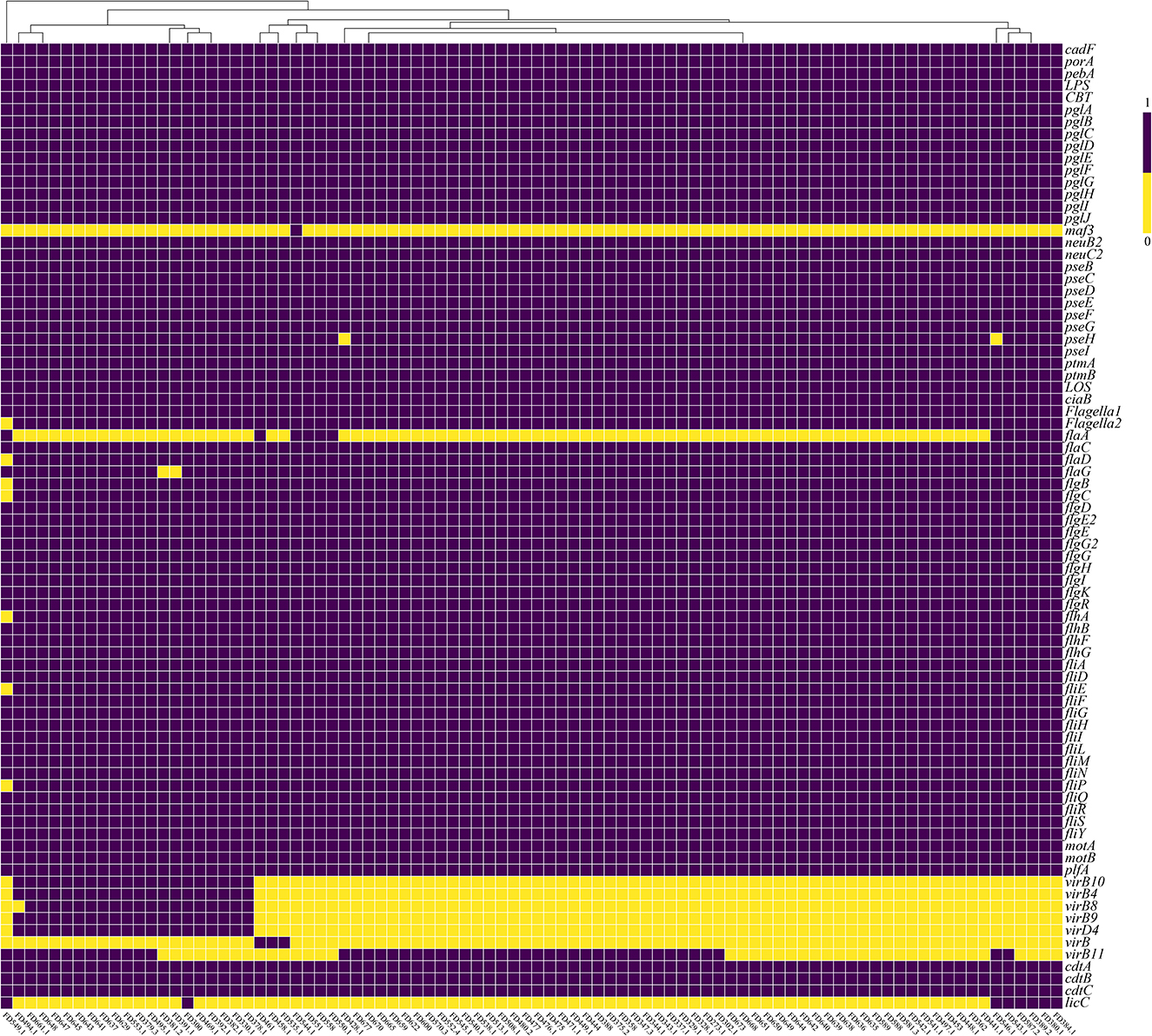
According to Figure 1, 100% of C. upsaliensis isolates carried the cadF, porA, and pebA genes, related to adhesion. In addition, all C. upsaliensis isolates carried genes associated with N-glycosylation, including the pglA, pglB, pglC, and pglD genes, and genes related to O-linked flagellar glycosylation, including the pseB, pseC, pseD, and pseE genes.

Figure 1. Heatmap of a virulence factor of 88 C. upsaliensis isolates from pets in Shenzhen, China. The labels on the X-axis represent the name of the isolates, while labels on the Y-axis correspond to the name of the virulence factor. Value “1” denotes the presence of the virulence factor, whereas value “0” denotes the absence of the virulence factor in the isolates.
Over 97.7% of the C. upsaliensis isolates had flagella-related genes, including the flaC, flaD, flaG, flgB, flgC, and flgD genes, which are involved in motility, chemotaxis, and invasion. However, the flaA gene was present in only 13.6% (12/88) of the isolates, while the flaB gene was absent.
The ciaB gene, which is connected to the invasion, was present in all C. upsaliensis isolates. The ciaC gene, however, was not found. Moreover, the cdtA, cdtB, and cdtC genes, which code for the cytolethal distending toxin(CDT), were present in all C. upsaliensis isolates. Additionally, 21.5% (19/88) of the isolates had the virB10, virB4, virB8, virB9, and virD4 genes related to the type IV secretion system (T4SS), which is involved in secretion and invasion. We present the results in Supplementary Table 2.
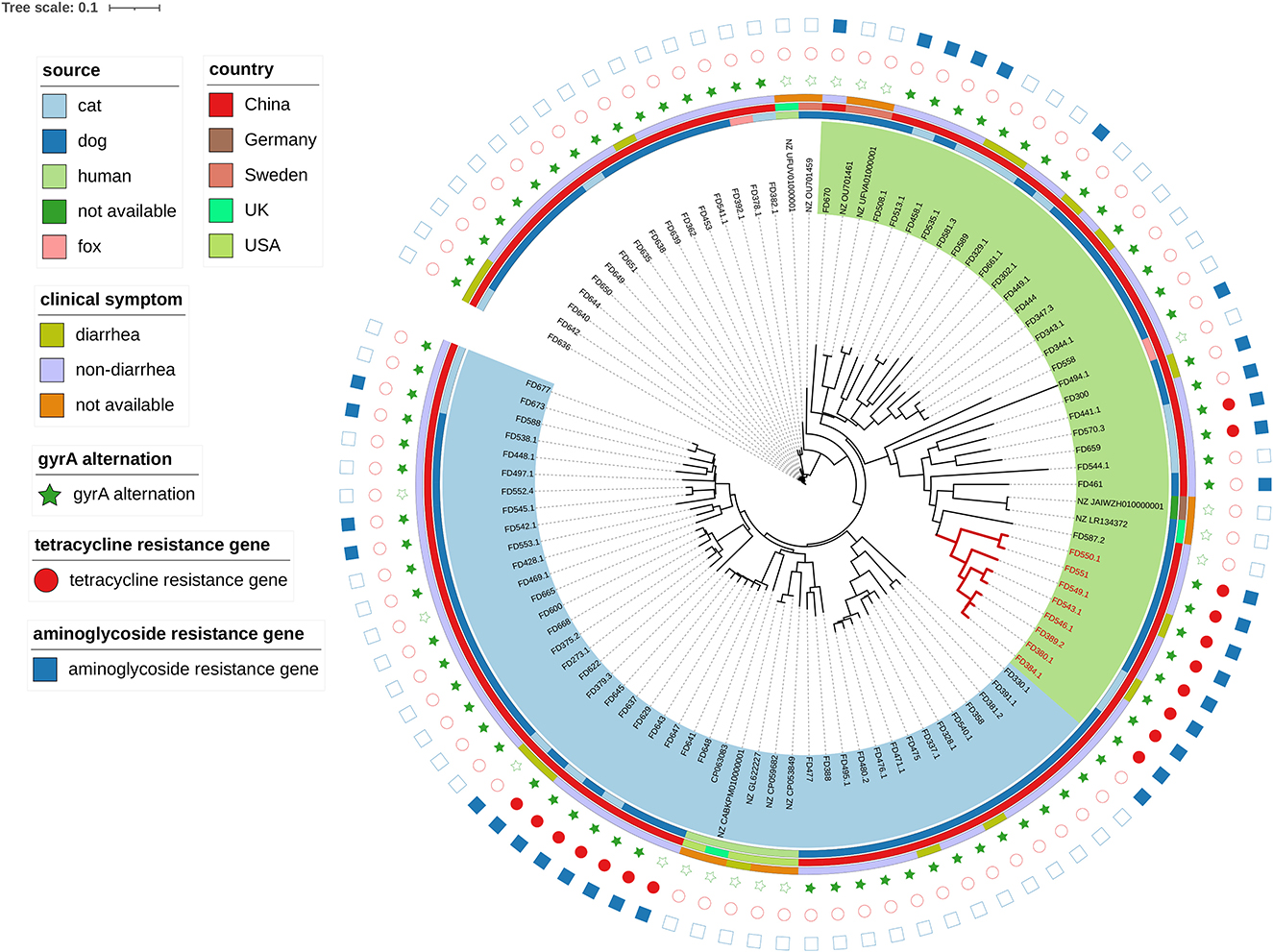
3.4. Phylogenetic analysis
The significant genetic diversity of C. upsaliensis isolates in the current study is shown by the phylogenetic analysis in Figure 2. Two major clades were obtained and labeled with green and blue shades. No apparent aggregation was detected in the diarrhea group or the non-diarrhea group. Eight strains in subclade 1 (FD550.1, FD551, FD549.1, FD543.1, FD546.1, FD389.2, FD380.1, and FD384.1) had tetracycline and aminoglycoside resistance genes and the gyrA alternation. Moreover, 100% of subclade 1 isolates harbored the flaA gene, compared to only 2% (4/80) of non-subclade 1 isolates. In addition, they were phenotypically resistant to six classes of antimicrobials, namely macrolides,/quinolones,/aminoglycosides,/tetracyclines,/lincosamides, and/ketolide.

Figure 2. Phylogenetic analysis of 88 C. upsaliensis isolates in this study and 11 reference strains by the K-mer tree method. The inner circle represents the source, the middle circle for the country, and the outside circle for the clinical symptom. The stars filled in green represent the gyrA gene alteration conferring resistance to fluoroquinolone, the circles filled in red represent the tetracycline resistance gene detected, and the rectangles filled in blue represent the aminoglycoside resistance gene detected. No resistance gene was found, as defined by the shapes without filled color. Two major clades were obtained and labeled with green and blue shades. Red branch isolates were attributed to subclade 1. The tree scale represents a 0.1 change per nucleotide position.
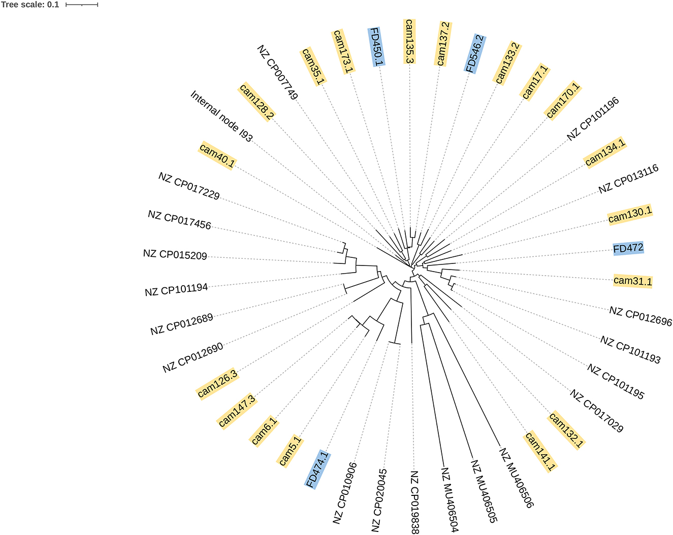
According to the phylogenetic analysis in Figure 3, two C. jejuni isolates (FD546.2 and FD450.1) in this study were related to the chicken source C. jejuni (cam133.2, cam 135.3, and cam 137.2) from 2018 in Shenzhen, China.

Figure 3. Phylogenetic analysis of four C. jejuni isolates in this study and 19 reference strains by the K-mer tree method. The labels in blue were C. jejuni isolates in this study, and the labels in yellow were chicken source isolates from 2018 in Shenzhen, China. Other isolates were C. jejuni reference strains downloaded from PubMed.
3.5. Resistance genotype and concordance with the phenotypic antimicrobial resistance
Blasting with the CARD database found three main genotypical antimicrobial resistance types, namely gyrA alternation, aminoglycoside, and tetracycline resistance, consistent with the result of phenotypical antimicrobial resistance. We found the nucleotide mutation C257T in the gyrA gene, causing the substitution T86I.
In C. upsaliensis, 89.7% (79/88) of the isolates had antibiotic target alteration (C257T) in the gyrA gene conferring resistance to fluoroquinolone. In total, 36.4% (32/88) of the isolates carried the aminoglycoside resistance gene and were phenotypically aminoglycoside resistant (gentamycin or streptomycin resistance), comprised of 21 isolates that had the aph (2″)-If gene, 10 isolates had the aac (6′)-aph (2″) gene, and 1 isolate simultaneously carried the aph (2″)-If and aac (6′)-aph (2″) genes. However, 20.5% (18/88) of isolates exhibited MIC ≥ 64 μg ml−1 against streptomycin while lacking the aminoglycoside resistance gene. In addition, 19.3% (17/88) of the isolates had the tetracycline resistance gene [tet (O) or tet (O/M/O)] and were phenotypically resistant to tetracycline, whereas 1.1% (1/88) of the isolates were phenotypically tetracycline resistant while lacked tetracycline resistance gene.
All four C. jejuni isolates (100%, 4/4) had antibiotic target alteration (C257T) in the gyrA gene conferring resistance to fluoroquinolone. In addition, four C. jejuni isolates carried the beta-lactam resistance gene; three of them had the OXA-193 gene, and one had the OXA-595 gene. In addition, all four isolates had the cmeR gene, related to antibiotic efflux pump belonging to resistance-nodulation-cell division (RND) antibiotic, causing resistance to macrolide antimicrobials, fluoroquinolone antimicrobials, cephalosporin, and fusidane antimicrobials. The cmeR gene was absent in C. upsaliensis and C. helveticus isolates in this study. Three C. jejuni isolates (75%, 3/4) harbored the tetracycline resistance gene and were phenotypically resistant.
Two C. helveticus isolates harbored the aminoglycoside resistance gene [aph (2″)-If)]and were phenotypically aminoglycoside resistant (gentamycin or streptomycin resistance). Moreover, two C. helveticus isolates had the gyrA gene alteration (C257T) conferring resistance to fluoroquinolone.
4. Discussion
Since dogs and cats are the primary hosts of C. upsaliensis and C. helveticus, these animals may be crucial in the epidemiology of these species and serve as potential reservoirs for human infection (Acke, 2018). However, little is known about the pet-related Campylobacter spp. in China. Our study demonstrated the prevalence and whole-genome sequence profile of Campylobacter spp. in pets and indicated the high antimicrobial resistance of the isolates, which was previously unreported in Shenzhen, China.
A meta-analysis research conducted in 2015 from 34 studies on the prevalence of Campylobacter spp. in domestic pets found that domestic cats and dogs had a mean prevalence of roughly 25% (Pintar et al., 2015), which is consistent with 33.5% of the pets in this study. Our findings, which agreed with Acke's review (Acke, 2018), showed that C. upsaliensis had the highest prevalence (30.2%, 98/325), followed by C. helveticus and C. jejuni.
Koláčková et al. reported that young dogs on homemade food who have diarrhea might be regarded as a risk group for the potential transfer of Campylobacter infections from pets to humans (Koláčková et al., 2015). Karama et al. also reported that age was the only risk factor linked with a higher likelihood of carrying C. upsaliensis, and older dogs had a significantly higher prevalence of Campylobacter spp. (Karama et al., 2019). In our study, however, no significant difference was found between the diarrhea group and the non-diarrhea group in terms of the prevalence of Campylobacter spp. in dogs or cats, younger dogs (3 years old), or older dogs (>3 years old). Additionally, three of the four C. jejuni isolates found in this study came from pets that did not have diarrhea. From our study, Campylobacter does not necessarily relate to diarrhea in pets.
Our study also demonstrated that Campylobacter spp. from pets exhibited similar high resistance to antimicrobials as isolates from the clinical patient. The high antimicrobial resistance rate of Campylobacter is an increasing problem in public health (Contreras-Omaña et al., 2021; Rahman et al., 2021; Wallace et al., 2021; Eryildiz et al., 2022; Sasaki et al., 2022). In this study, MDR was found in 55.1% of the Campylobacter isolates. The main MDR pattern was aminoglycosides/quinolones/lincosamides, which were comparable with the antimicrobial resistance profile of isolates from clinical patients (Wieczorek et al., 2018; Frazão et al., 2021; Mencía-Gutiérrez et al., 2021).
Moreover, our findings confirmed the variety of virulence factors that Campylobacter spp. carried. Virulence factors can be divided into bacterial chemotaxis, motility, attachment, invasion, survival, cellular transmigration, and spread to deeper tissue (Tegtmeyer et al., 2021). The primary flagellar genes associated with strain motility are the flaA and flaB genes (Lopes et al., 2021). Only 13.6% (12/88) of the isolates in this study had the flaA gene, and no isolate had the flaB gene. We used the Campylobacter isolation kit incorporating a membrane filter method, and only dynamic isolates can penetrate the filtration membrane. Based on the experimental results, we confirmed that the flaB gene is not essential for total motility in Campylobacter, which was reported previously (Koolman et al., 2015).
The most interesting finding was that subclade 1 contained eight isolates that displayed phenotypically resistance to six classes of antimicrobials and had resistance genes against tetracyclines and aminoglycosides and harbored the gyrA gene alteration. Additionally, all isolates in subclade 1 possessed the flaA gene, which was uncommon outside subclade 1, suggesting isolates in subclade 1 may have more advantage in mobility.
Goyal et al. demonstrated an outbreak of pet store puppy-associated extensively drug-resistant C. jejuni infections (Goyal et al., 2021). In addition, Thépault et al. reported that genotype comparison with previously characterized isolates revealed a partial overlap among C. jejuni isolates from pets, chickens, cattle, and clinical cases (Thépault et al., 2020). This overlap suggests the potential role of livestock and humans in pets' exposure to Campylobacter or vice versa.
Furthermore, concordance between the genotypically and phenotypically resistance against antimicrobials was observed in C. upsaliensis. The percentages of phenotypically resistant isolates against ciprofloxacin, gentamicin, and tetracycline were 94.9%, 43.9%, and 21.4%, respectively. Accordingly, the percentages of genotypically resistant isolates against fluoroquinolone, aminoglycosides, and tetracyclines were 89.7%, 36.4%, and 19.3%, respectively. Interestingly, 18 C. upsaliensis isolates showed high MICs (≥ 64 μg ml−1) against streptomycin without an aminoglycoside resistance gene detected. We referred that this aminoglycoside resistance is not conferred by known resistance genes. In addition, concordance between the genotype and phenotype was influenced by the computational pipelines, genome coverage, and the type of ARG but not by input data (Hodges et al., 2021).
Our study had limitations, including a lack of adequate geographic representation, a short sampling season, and a failure to isolate from the patient and pet simultaneously. These shortcomings led to an inadequate impact of the epidemiological analysis, which should be improved in future research.
Our analysis concluded that pets are a significant source of Campylobacter spp. strains and a potential reservoir for them. In particular, C. upsaliensis of subclade 1 in this study required additional investigation because of its broad MDR phenotype and relatively high prevalence of the flaA gene.
Data availability statement
The data presented in the study are deposited in the NCBI repository, BioProject accession number PRJNA897092.
Ethics statement
The animal study was reviewed and approved by the Institutional Review Board of the Nanshan Center for Disease Control and Prevention. Written informed consent was obtained from the owners for the participation of their animals in this study.
Author contributions
YD and MZ designed the experiments. CJ, YM, BZ, MY, and JH participated in the sample collection and performed the experiments. GZ and HW carried out the genome bioinformatics study. CJ and YM wrote the manuscript. The submitted manuscript was read and approved by all authors.
Funding
CJ was taking part in the Sanming Project of Medicine (SZSM201803081), the Key Discipline Construction Subsidy of Nanshan District, and the Nanshan District Science and Technology Plan Project (2020049). The study design, data collection, interpretation, and the choice to submit the study for publication were all independent of the funders.
Acknowledgments
We appreciate Academician Fu Gao from the Chinese Academy of Sciences, who helped with the study design. We also appreciate the staff of the 16 chain animal hospitals who helped with the collection of the pet stools.
Conflict of interest
The authors declare that the research was conducted in the absence of any commercial or financial relationships that could be construed as a potential conflict of interest.
Publisher's note
All claims expressed in this article are solely those of the authors and do not necessarily represent those of their affiliated organizations, or those of the publisher, the editors and the reviewers. Any product that may be evaluated in this article, or claim that may be made by its manufacturer, is not guaranteed or endorsed by the publisher.
Supplementary material
The Supplementary Material for this article can be found online at: https://www.frontiersin.org/articles/10.3389/fmicb.2023.1152719/full#supplementary-material
References
Acke, E. (2018). Campylobacteriosis in dogs and cats: a review. N. Z. Vet. J. 66, 221–228. doi: 10.1080/00480169.2018.1475268
Asuming-Bediako, N., Kunadu, A. P., Jordan, D., Abraham, S., and Habib, I. (2022). Prevalence and antimicrobial susceptibility pattern of Campylobacter jejuni in raw retail chicken meat in Metropolitan Accra, Ghana. Int. J. Food Microbiol. 376, 109760. doi: 10.1016/j.ijfoodmicro.2022.109760
Behailu, Y., Hussen, S., Alemayehu, T., Mengistu, M., and Fenta, D. A. (2022). Prevalence, determinants, and antimicrobial susceptibility patterns of Campylobacter infection among under-five children with diarrhea at Governmental Hospitals in Hawassa city, Sidama, Ethiopia. A cross-sectional study. PLoS ONE. 17, e0266976. doi: 10.1371/journal.pone.0266976
Bojanić, K., Acke, E., Roe, W. D., Marshall, J. C., Cornelius, A. J., Biggs, P. J., et al. (2020). Comparison of the pathogenic potential of Campylobacter jejuni, C. upsaliensis and C. helveticus and limitations of using larvae of galleria mellonella as an infection model. Pathogens. 9, 713. doi: 10.3390/pathogens9090713
Bolton, D. J. (2015). Campylobacter virulence and survival factors. Food Microbiol. 48, 99–108. doi: 10.1016/j.fm.2014.11.017
Bravo, V., Katz, A., Porte, L., Weitzel, T., Varela, C., Gonzalez-Escalona, N., et al. (2021). Genomic analysis of the diversity, antimicrobial resistance and virulence potential of clinical Campylobacter jejuni and Campylobacter coli strains from Chile. PLoS Negl. Trop. Dis. 15, e0009207. doi: 10.1371/journal.pntd.0009207
Campagnolo, E. R., Philipp, L. M., Long, J. M., and Hanshaw, N. L. (2018). Pet-associated Campylobacteriosis: A persisting public health concern. Zoonoses Public Health. 65, 304–311. doi: 10.1111/zph.12389
Cantero, G., Correa-Fiz, F., Ronco, T., Strube, M., Cerdà-Cuéllar, M., and Pedersen, K. (2018). Characterization of Campylobacter jejuni and Campylobacter coli broiler isolates by whole-genome sequencing. Foodborne Pathog. Dis. 15, 145–152. doi: 10.1089/fpd.2017.2325
Choi, J. H., Moon, D. C., Mechesso, A. F., Kang, H. Y., Kim, S. J., Song, H. J., et al. (2021). Antimicrobial resistance profiles and macrolide resistance mechanisms of campylobacter coli isolated from pigs and chickens. Microorganisms. 9, 1077. doi: 10.3390/microorganisms9051077
Contreras-Omaña, R., Escorcia-Saucedo, A. E., and Velarde-Ruiz, V. J. (2021). Prevalence and impact of antimicrobial resistance in gastrointestinal infections: A review. Rev. Gastroenterol. Mex. (Engl Ed). 86, 265–275. doi: 10.1016/j.rgmxen.2021.06.004
Couturier, B. A., Hale, D. C., and Couturier, M. R. (2012). Association of Campylobacter upsaliensis with persistent bloody diarrhea. J. Clin. Microbiol. 50, 3792–3794. doi: 10.1128/JCM.01807-12
Eryildiz, C., Sakru, N., and Kuyucuklu, G. (2022). Investigation of antimicrobial susceptibilities and resistance genes of campylobacter isolates from patients in Edirne, Turkey. Iran. J. Public Health 51, 569–577. doi: 10.18502/ijph.v51i3.8933
Finsterer, J. (2022). Triggers of Guillain-Barré syndrome: Campylobacter jejuni predominates. Int. J. Mol. Sci. 23. doi: 10.3390/ijms232214222
Frazão, M. R., Cao, G., Medeiros, M., Duque, S., Allard, M. W., and Falcão, J. P. (2021). Antimicrobial resistance profiles and phylogenetic analysis of Campylobacter jejuni strains isolated in Brazil by whole genome sequencing. Microb. Drug Resist. 27, 660–669. doi: 10.1089/mdr.2020.0184
Ghatak, S., He, Y., Reed, S., Strobaugh, T. J., and Irwin, P. (2017). Whole genome sequencing and analysis of Campylobacter coli YH502 from retail chicken reveals a plasmid-borne type VI secretion system. Genom Data. 11, 128–131. doi: 10.1016/j.gdata.2017.02.005
Goyal, D., Watkins, L., Montgomery, M. P., Jones, S., Caidi, H., and Friedman, C. R. (2021). Antimicrobial susceptibility testing and successful treatment of hospitalised patients with extensively drug-resistant Campylobacter jejuni infections linked to a pet store puppy outbreak. J. Glob. Antimicrob. Resist. 26, 84–90. doi: 10.1016/j.jgar.2021.04.029
Hodges, L. M., Taboada, E. N., Koziol, A., Mutschall, S., Blais, B. W., Inglis, G. D., et al. (2021). Systematic evaluation of whole-genome sequencing based prediction of antimicrobial resistance in Campylobacter jejuni and C. coli. Front. Microbiol. 12, 776967. doi: 10.3389/fmicb.2021.776967
Hsieh, Y. H., Wang, Y. F., Moura, H., Miranda, N., Simpson, S., Gowrishankar, R., et al. (2018). Application of MALDI-TOF MS Systems in the Rapid Identification of Campylobacter spp. of public health importance. J. AOAC Int. 101, 761–768. doi: 10.5740/jaoacint.17-0266
Hudson, L. K., Andershock, W. E., Yan, R., Golwalkar, M., M'Ikanatha, N. M., Nachamkin, I., et al. (2021). Phylogenetic analysis reveals source attribution patterns for Campylobacter spp. in Tennessee and Pennsylvania. Microorganisms. 9, 2300. doi: 10.3390/microorganisms9112300
Karama, M., Cenci-Goga, B. T., Prosperi, A., Etter, E., El-Ashram, S., McCrindle, C., et al. (2019). Prevalence and risk factors associated with Campylobacter spp. occurrence in healthy dogs visiting four rural community veterinary clinics in South Africa. Onderstepoort J. Vet. Res. 86, e1–e6. doi: 10.4102/ojvr.v86i1.1673
Koláčková, I., Dušková, M., Vojkovská, H., Bardon, J., Pudová, V., and Karpíšková, R. (2015). Dogs as a possible source of human Campylobacter infecfions. Klin Mikrobiol Infekc Lek. 21, 36–40.
Koolman, L., Whyte, P., Burgess, C., and Bolton, D. (2015). Distribution of virulence-associated genes in a selection of Campylobacter isolates. Foodborne Pathog. Dis. 12, 424–432. doi: 10.1089/fpd.2014.1883
Latov, N. (2022). Campylobacter jejuni infection, anti-ganglioside antibodies, and neuropathy. Microorganisms. 10, 2139. doi: 10.3390/microorganisms10112139
Lawton, S. J., Weis, A. M., Byrne, B. A., Fritz, H., Taff, C. C., Townsend, A. K., et al. (2018). Comparative analysis of Campylobacter isolates from wild birds and chickens using MALDI-TOF MS, biochemical testing, and DNA sequencing. J. Vet. Diagn. Invest. 30, 354–361. doi: 10.1177/1040638718762562
Llarena, A. K., Taboada, E., and Rossi, M. (2017). Whole-genome sequencing in epidemiology of campylobacter jejuni infections. J. Clin. Microbiol. 55, 1269–1275. doi: 10.1128/JCM.00017-17
Lopes, G. V., Ramires, T., Kleinubing, N. R., Scheik, L. K., Fiorentini, Â. M., and Padilha, D. S. W. (2021). Virulence factors of foodborne pathogen Campylobacterjejuni. Microb. Pathog. 161, 105265. doi: 10.1016/j.micpath.2021.105265
Martinez-Anton, L., Marenda, M., Firestone, S. M., Bushell, R. N., Child, G., Hamilton, A. I., et al. (2018). Investigation of the role of campylobacter infection in suspected acute polyradiculoneuritis in dogs. J. Vet. Intern. Med. 32, 352–360. doi: 10.1111/jvim.15030
Mencía-Gutiérrez, A., Martín-Maldonado, B., Pastor-Tiburón, N., Moraleda, V., González, F., García-Peña, F. J., et al. (2021). Prevalence and antimicrobial resistance of Campylobacter from wild birds of prey in Spain. Comp. Immunol. Microbiol. Infect. Dis. 79, 101712. doi: 10.1016/j.cimid.2021.101712
Moore, J. E., Corcoran, D., Dooley, J. S., Fanning, S., Lucey, B., Matsuda, M., et al. (2005). Campylobacter. Vet. Res. 36, 351–382. doi: 10.1051/vetres:2005012
Mughini, G. L., Smid, J. H., Wagenaar, J. A., Koene, M. G., Havelaar, A. H., Friesema, I. H., et al. (2013). Increased risk for Campylobacter jejuni and C. coli infection of pet origin in dog owners and evidence for genetic association between strains causing infection in humans and their pets. Epidemiol Infect 141, 2526–2535. doi: 10.1017/S0950268813000356
Nakamura, I., Omori, N., Umeda, A., Ohkusu, K., and Matsumoto, T. (2015). First case report of fatal sepsis due to Campylobacter upsaliensis. J. Clin. Microbiol. 53, 713–715. doi: 10.1128/JCM.02349-14
Ohkoshi, Y., Sato, T., Murabayashi, H., Sakai, K., Takakuwa, Y., Fukushima, Y., et al. (2020). Campylobacter upsaliensis isolated from a giant hepatic cyst. J. Infect. Chemother. 26, 752–755. doi: 10.1016/j.jiac.2020.02.015
Patrick, M. E., Henao, O. L., Robinson, T., Geissler, A. L., Cronquist, A., Hanna, S., et al. (2018). Features of illnesses caused by five species of Campylobacter, foodborne diseases active surveillance network (FoodNet) - 2010-2015. Epidemiol. Infect. 146, 1–10. doi: 10.1017/S0950268817002370
Pintar, K. D., Christidis, T., Thomas, M. K., Anderson, M., Nesbitt, A., Keithlin, J., et al. (2015). A systematic review and meta-analysis of the Campylobacter spp. prevalence and concentration in household pets and petting zoo animals for use in exposure assessments. PLoS ONE. 10, e0144976. doi: 10.1371/journal.pone.0144976
Prendergast, D. M., Lynch, H., Whyte, P., Golden, O., Murphy, D., Gutierrez, M., et al. (2022). Genomic diversity, virulence and source of Campylobacter jejuni contamination in Irish poultry slaughterhouses by whole genome sequencing. J. Appl. Microbiol. doi: 10.1111/jam.15753
Rahman, M. A., Paul, P. R., Hoque, N., Islam, S. S., Haque, A., Sikder, M. H., et al. (2021). Prevalence and antimicrobial resistance of campylobacter species in diarrheal patients in Mymensingh, Bangladesh. Biomed Res. Int. 2021, 9229485. doi: 10.1155/2021/9229485
Rokney, A., Valinsky, L., Vranckx, K., Feldman, N., Agmon, V., Moran-Gilad, J., et al. (2020). WGS-based prediction and analysis of antimicrobial resistance in Campylobacter jejuni isolates from Israel. Front. Cell. Infect. Microbiol. 10, 365. doi: 10.3389/fcimb.2020.00365
Sasaki, Y., Yonemitsu, K., Uema, M., Asakura, H., and Asai, T. (2022). Prevalence and antimicrobial resistance of Campylobacter and Salmonella in layer flocks in Honshu, Japan. J. Vet. Med. Sci. doi: 10.1292/jvms.22-0257
Ščerbová, J., Lauková, A., Losasso, C., and Barco, L. (2022). Antimicrobial susceptibility to natural substances of Campylobacter jejuni and Campylobacter coli isolated from Italian poultry. Foodborne Pathog. Dis. 19, 266–271. doi: 10.1089/fpd.2021.0085
Shakir, Z. M., Alhatami, A. O., Ismail, K. Y., and Muhsen, A. H. (2021). Antibiotic resistance profile and multiple antibiotic resistance index of campylobacter species isolated from poultry. Arch. Razi Inst. 76, 1677–1686. doi: 10.22092/ari.2021.356400.1837
Song, H., Kim, J., Guk, J. H., An, J. U., Lee, S., and Cho, S. (2020). Complete genome sequence and comparative genomic analysis of hyper-aerotolerant Campylobacter lari strain SCHS02 isolated from duck for its potential pathogenicity. Microb. Pathog. 142, 104110. doi: 10.1016/j.micpath.2020.104110
Tegtmeyer, N., Sharafutdinov, I., Harrer, A., Soltan, E. D., Linz, B., and Backert, S. (2021). Campylobacter virulence factors and molecular host-pathogen interactions. Curr. Top. Microbiol. Immunol. 431, 169–202. doi: 10.1007/978-3-030-65481-8_7
Thépault, A., Rose, V., Queguiner, M., Chemaly, M., and Rivoal, K. (2020). Dogs and cats: reservoirs for highly diverse campylobacter jejuni and a potential source of human exposure. Animals (Basel). 10, 838. doi: 10.3390/ani10050838
Wallace, R. L., Bulach, D., McLure, A., Varrone, L., Jennison, A. V., Valcanis, M., et al. (2021). Antimicrobial resistance of Campylobacter spp. causing human infection in australia: an international comparison. Microb. Drug Resist. 27, 518–528. doi: 10.1089/mdr.2020.0082
Keywords: Campylobacter upsaliensis, Campylobacter jejuni, Campylobacter helveticus, whole-genome sequencing, antimicrobial resistance, matrix-assisted laser desorption ionization-time of flight mass spectrum, pets, virulence-associated genes
Citation: Ju C, Ma Y, Zhang B, Zhou G, Wang H, Yu M, He J, Duan Y and Zhang M (2023) Prevalence, genomic characterization and antimicrobial resistance of Campylobacter spp. isolates in pets in Shenzhen, China. Front. Microbiol. 14:1152719. doi: 10.3389/fmicb.2023.1152719
Received: 28 January 2023; Accepted: 09 May 2023;
Published: 01 June 2023.
Edited by:
Michael Brouwer, Wageningen University and Research, NetherlandsReviewed by:
Heriberto Fernandez, Austral University of Chile, ChileHosny El-Adawy, Friedrich Loeffler Institut, Germany
Linda Van Der Graaf, Utrecht University, Netherlands
Copyright © 2023 Ju, Ma, Zhang, Zhou, Wang, Yu, He, Duan and Zhang. This is an open-access article distributed under the terms of the Creative Commons Attribution License (CC BY). The use, distribution or reproduction in other forums is permitted, provided the original author(s) and the copyright owner(s) are credited and that the original publication in this journal is cited, in accordance with accepted academic practice. No use, distribution or reproduction is permitted which does not comply with these terms.
*Correspondence: Maojun Zhang, emhhbmdtYW9qdW5AaWNkYy5jbg==; Yongxiang Duan, c3pkdWFueXhAMTYzLmNvbQ==
†These authors share first authorship
 Changyan Ju
 Yanping Ma
 Bi Zhang2
 Jiaoming He
 Yongxiang Duan
 Maojun Zhang
