- 1Division of Infectious Diseases, Children’s National Health System, Washington, DC, United States
- 2Department of Pediatrics, George Washington University School of Medicine and Health Sciences, Washington, DC, United States
- 3Computational Biology Institute, Milken Institute School of Public Health, The George Washington University, Washington, DC, United States
- 4Department of Microbiology, Immunology and Tropical Medicine, George Washington University School of Medicine and Health Sciences, Washington, DC, United States
- 5Division of Pulmonary and Sleep Medicine, Children’s National Health System, Washington, DC, United States
- 6Division of Genomic Medicine, The George Washington University, Washington, DC, United States
- 7Department of Medicine, George Washington University School of Medicine and Health Sciences, Washington, DC, United States
- 8Division of Emergency Medicine, Children’s National Health System, Washington, DC, United States
Cystic fibrosis (CF) is an autosomal recessive disease associated with recurrent lung infections that can lead to morbidity and mortality. The impact of antibiotics for treatment of acute pulmonary exacerbations on the CF airway microbiome remains unclear with prior studies giving conflicting results and being limited by their use of 16S ribosomal RNA sequencing. Our primary objective was to validate the use of true single molecular sequencing (tSMS) and PathoScope in the analysis of the CF airway microbiome. Three control samples were created with differing amounts of Burkholderia cepacia, Pseudomonas aeruginosa, and Prevotella melaninogenica, three common bacteria found in cystic fibrosis lungs. Paired sputa were also obtained from three study participants with CF before and >6 days after initiation of antibiotics. Antibiotic resistant B. cepacia and P. aeruginosa were identified in concurrently obtained respiratory cultures. Direct sequencing was performed using tSMS, and filtered reads were aligned to reference genomes from NCBI using PathoScope and Kraken and unique clade-specific marker genes using MetaPhlAn. A total of 180–518 K of 6–12 million filtered reads were aligned for each sample. Detection of known pathogens in control samples was most successful using PathoScope. In the CF sputa, alpha diversity measures varied based on the alignment method used, but similar trends were found between pre- and post-antibiotic samples. PathoScope outperformed Kraken and MetaPhlAn in our validation study of artificial bacterial community controls and also has advantages over Kraken and MetaPhlAn of being able to determine bacterial strains and the presence of fungal organisms. PathoScope can be confidently used when evaluating metagenomic data to determine CF airway microbiome diversity.
Introduction
Cystic fibrosis (CF) is an autosomal recessive disease that affects more than 30,000 people in the United States (MacKenzie et al., 2014). Patients suffer from recurrent and chronic pulmonary infections that are strongly associated with morbidity and mortality (Ramsey, 1996). Recent use of culture-independent next generation sequencing (NGS) has identified novel and diverse communities of microbes in the CF airway, leading to an alteration in the traditional understanding of the role of infection in progressive lung disease (Huang and LiPuma, 2016). Decreasing microbial diversity is clearly associated with the presence of Pseudomonas aeruginosa and increasing age (Cox et al., 2010; Klepac-Ceraj et al., 2010; Boutin et al., 2015; Coburn et al., 2015). Cross-sectional studies have shown a difference in the structure and composition of airway microbiota between stable patients and those experiencing severe pulmonary decline, with decreased diversity in those with more advanced disease (Coburn et al., 2015; Flight et al., 2015; Bacci et al., 2016).
Antibiotic use has dramatically improved the longevity of children with CF and are likely responsible for perturbations of the airway microbiome (VanDevanter and LiPuma, 2012). However, the impact of antibiotics for treatment of acute pulmonary exacerbations on the CF airway microbiome remains unclear. Some studies have found that antibiotics do not lead to significant changes within the airway microbiome. Specifically, Fodor et al. (2012) found small decreases in bacterial richness but minimal changes in the overall community structure. Price et al. (2013) also followed CF patients through acute pulmonary exacerbations and found that the total and relative abundance of bacterial genera were stable during the exacerbation and after antibiotic treatment. Cuthbertson et al. (2016) also found that certain core microbiota remained resilient, regardless of exacerbation or antibiotic treatment state, but that rare species had much greater variability over time. However, other studies have reported that microbial diversity and the proportion of pathogenic bacteria decreased following antibiotic treatment. Zemanick et al. (2013) and Smith et al. (2014) found a decrease in microbial diversity early in the treatment course (around 72 h), which was also associated with a high relative abundance of P. aeruginosa. Later in the antibiotic treatment course (>7 days), the diversity appeared to return (Smith et al., 2014).
Longitudinal studies in patients with CF have shown long-term antibiotic use has also been associated with decreasing microbial diversity over time (Klepac-Ceraj et al., 2010; Zhao et al., 2012). However, increased antibiotic exposure is also confounded by age and declining lung function, making the establishment of a direct link between antibiotic use and microbial diversity difficult (Zhao et al., 2012).
Most of these prior studies investigating the impact of antibiotics on changes in the CF airway microbiome used 16S ribosomal RNA (rRNA) sequencing. This may be a limitation, as this approach requires PCR amplification that may suffer from primer bias in terms of accurate assessment of relative frequencies of bacterial taxa. There may also be a lack of differentiation amongst species with highly similar 16S sequences (Hilton et al., 2016). Metagenomic studies of the CF airway can give an unbiased look at the microbiome, and can also provide details on pathogen strain types (Feigelman et al., 2017). Furthermore, metagenomics studies could be used investigate the presence of antibiotic resistance genes or other fitness-conferring mutations (Feigelman et al., 2017). Our primary objective with this study was to validate the use of a metagenomic sequencing approach using true single molecular sequencing (tSMS, SeqLL Inc.) technology and the PathoScope computational framework (Francis et al., 2013; Hong et al., 2014) in CF airway samples.
MATERIALS AND METHODS
Creation of Control Samples for Method Validation
Approximately 5 μg of dehydrated genomic bacterial DNA for P. aeruginosa (ATCC® 47085D-5, strain PAO1-LAC), B. cepacia (ATCC® 25416D-5), and Prevotella melaninogenica (ATCC® 25845D-5) were obtained from ATCC (Manassas, VA, United States). To re-suspend the genomic DNA, 60 μL of molecular grade water were added to each sample. The samples were centrifuged (2000 g × 10 s) and incubated while continuously rocking overnight at 4°C. They were then incubated at 65°C for 1 h and then measured using a NanoDropTM spectrophotometer. Measured DNA concentrations were 194.2 ng/μL for P. aeruginosa, 187.8 ng/μL for B. cepacia, and 147.8 ng/μL for P. melaninogenica. Different proportions of these bacterial DNA were mixed together to create artificial community controls. Each 100 ng of Control A contained 20.7 ng of P. aeruginosa, 40 ng of B. cepacia, and 39.3 ng of P. melaninogenica. Control B contained 36.7 ng of P. aeruginosa, 35.4 ng of B. cepacia, and 35.4 ng of P. melaninogenica per 100 ng. Control C contained 47.5 ng of P. aeruginosa, 34.4 ng of B. cepacia, and 18.1 ng of P. melaninogenica per 100 ng. These mixtures were then frozen at -80°C until sequencing was performed.
Patients and Sample Collection
The creation of a bio- and data repository was approved 08DEC2015 by the Institutional Review Board (Pro6781) at Children’s National Health System. Study subjects were consented for participation in the study prior to respiratory sample collection and extraction of data from electronic medical records. Paired sputa were obtained from three participants with documented antibiotic resistance for this study. Patient demographics and sample details are reported in Table 1.
Respiratory Sample Collection and Processing
Per the biorepository protocol, spontaneously expectorated sputum samples obtained for clinical care were collected from the microbiology laboratory within 24 h of the patient’s clinical visit. Sputum samples were stored in a 4°C refrigerator prior to processing. For processing, sputum samples were mixed with Sputasol (dithiothreitol, Fisher Healthcare, Houston TX, United States), vortexed, and placed in a 37°C heated bead bath to homogenize the sample. The homogenized sputum was pelleted through centrifugation (12,000 g × 10 min). Supernatants were removed and bacterial pellets were frozen at -80°C until they underwent DNA extraction.
Respiratory Culture Results
Clinical culture results within the electronic medical record were used to identify the pathogen and MICs for various antibiotics. The clinical microbiology laboratory uses MicroScan (Beckman Coulter, Brea, CA, United States) to determine identification and susceptibility of bacterial pathogens grown in culture and has an internally validated protocol it uses for mucoid Pseudomonas aeruginosa (Zimmer et al., 2004).
DNA Extraction
Pelleted bacterial cells were rapidly thawed and mixed with 1 mL of sterile phosphate buffered saline (PBS). Bacterial DNA was extracted using a QIAamp DNA Microbiome kit (Qiagen, Valencia, CA, United States), following the protocol as outlined by the company. This kit was chosen as it has been reported to increase the ratio of bacterial to human DNA extracted (Qiagen, 2016).
Metagenomic NGS
Metagenomic NGS was performed using tSMS (SeqLL Inc., Woburn, MA, United States). A starting amount of at least 300 ng of DNA (range 300–3000 ng) was used. Samples were prepared by first shearing to 100–200 nucleotides to create the appropriate sized fragments. This was followed by poly-A tailing and 3′ end blocking for capture on the flow cell surface. Two sequencing runs were performed, with the first loading 11.5 ng of DNA per sample and the second loading 16 ng of DNA per sample. The samples were then sequenced using 18 channels of a flow cell (two channels per sample). One channel was used for the run reference oligo. The instrument was operated at 550 field of view depth.
Bioinformatic and Statistical Analysis
Raw reads were filtered by SeqLL to those with a quality reference score at or above 4.4/5.0 and with a length cutoff of 24 bases. The quality score considers the length of the aligned read, number of matches, and number of errors when it is normalized to the length of each read. The formula used is score = (number of matches∗5-number of mismatches∗4)/read length (Kapranov et al., 2010). Filtered reads per channel ranged from between 7.3 million to 13.3 million. The internal control oligo generated an observed mean length that indicated operational performance that was consistent with optimal system operational specifications.
FASTQ files containing filtered reads were aligned to reference genomes using PathoScope (Hong et al., 2014), Kraken (Davis et al., 2013), and MetaPhlAn (Segata et al., 2012). PathoScope and Kraken attempt to remove human sequences before aligning to microbial reference genomes. The reference database for PathoScope was created using sequences identified in the National Center for Biotechnology Information (NCBI) Archaea, Bacteria, Virus, and Fungal reference and representative genome database, which contains at least one genome for each species in the Entrez genome collection that has assembly data. To this we added all complete genome assemblies for P. aeruginosa, B. cepacia, and Burkholderia cenocepacia, thus enabling strain-specific identification of these species. The Kraken reference database also included NCBI bacterial and viral reference genomes. PathoScope and Kraken were run using the Colonial One High-Performance Computing Cluster at GWU. Reference contigs with unusually high read counts were screened against the nt database using BLAST; contigs determined to be contaminants (e.g., human sequences) were removed before analysis. MetaPhlAn was run using bioBakery v1.7, a virtual environment operated by the Huttenhower Lab (bioBakery, 2017).
Alpha diversity was measured as the number of species identified, the Shannon-Weiner Index, and the Simpson’s Reciprocal Index. The Shannon-Weiner Index was calculated in Excel (Microsoft, Redmond, WA, United States) using the equation . The Simpson’s Reciprocal Index was calculated using the equation . Continuous variables were compared using t-test, while percentages of relative taxonomic abundance were compared using linear regression or McNemar’s test for correlated proportions. Taxonomy and metadata files were imported into phyloseq (McMurdie and Holmes, 2013) within R. Geometric means were used to estimate size factor and dispersion estimates, and differentially abundant species were identified using log2 fold change (adjusted p-value < 0.05) as implemented in DESeq2 (Love et al., 2014). PERMANOVA was also calculated to measure the differences in overall bacterial distribution using the adonis function of vegan in R (Oksanen et al., 2017). Lastly, principle coordinates analysis (PCoA) plots were generated using Bray–Curtis distance matrices with log transformed counts to visualize differences between computational frameworks.
Results
Control Sample Comparison
We analyzed the tSMS generated metagenomics data with PathoScope resulting in the identification of a range of 33–73 bacterial/viral strains per control sample. The Kraken analysis of the same data resulted in the identification of a range of 442–518 bacterial/viral strains per sample, and the MetaPhlAn analysis resulted in the identification of a range of 55–76 bacterial/viral strains per sample.
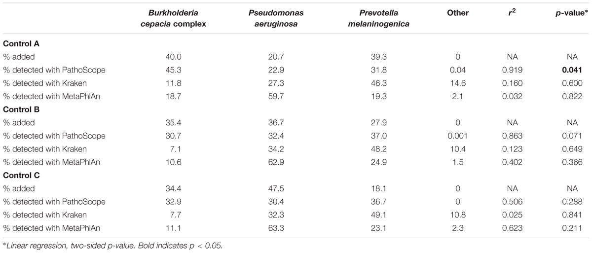
When looking individually at the proportions between each comparison, it can be appreciated that PathoScope was more representative of the true amounts of bacteria used to create the artificial communities than Kraken or MetaPhlAn (Table 2). These differences in proportions were measured using linear regression. PathoScope had higher r2-values than Kraken in all comparisons, and had higher r2-values than MetaPhlAn two out of three times. In fact, PathoScope was significantly similar to the added proportions in Control A (p = 0.041), and approached significance in Control B (p = 0.071).
Cystic Fibrosis Sample Comparison
Metagenomic Sequencing
Six sputum samples from three study subjects who experienced an acute pulmonary exacerbation and whose respiratory cultures grew antibiotic-resistant bacteria were sequenced (see Table 1). Across all six samples, a total of 36 million sequencing reads passed quality control filters (6–12 M reads per sample). The filtered reads were assigned taxonomic labels using three metagenomic taxonomic classifiers: PathoScope, Kraken, and MetaPhlAn. PathoScope and Kraken align against whole reference genomes, while MetaPhlAn uses a reference set of clade-specific marker genes. With PathoScope, 3.6% (range 2.7–4.4%) of the total reads were initially aligned to genomes within the bacterial and viral reference database. Of these reads, 66% (range 48–87%) of the reads were removed as they aligned to human genome sequences during the filtering process. Ultimately, 1.3% (range 0.5–1.8%) of the total sequences were aligned to bacterial and viral reference genomes. With Kraken, 13% (range 10–16%) of the reads were classified. After filters were applied for human reads, 2.4% (range 0.4–4.1%) of the classified reads were identified as microbial. Of the classified reads, 2.3% (range 0.3–4%) were identified as bacterial and 0.002% (0.001–0.003%) were identified as viral. MetaPhlAn output was reported as relative abundance of microbial species after filtering, so the above determinations of aligned/classified and human filtering was not possible. However, of the 100% microbial reads reported per sample, 54% (range 16–77%) were identified as bacterial and 40% (range 20–59%) were identified as viral. Kraken identified the most distinct bacterial and viral (including bacteriophage) species (n = 516), while MetaPhlAn identified the next most (n = 202) followed by PathoScope (n = 91). PathoScope was also able to provide strain level information, identifying 283 strains total; two strains of B. cepacia, 9 strains of B. cenocepacia, and 109 strains of P. aeruginosa were detected within the six sputum samples.
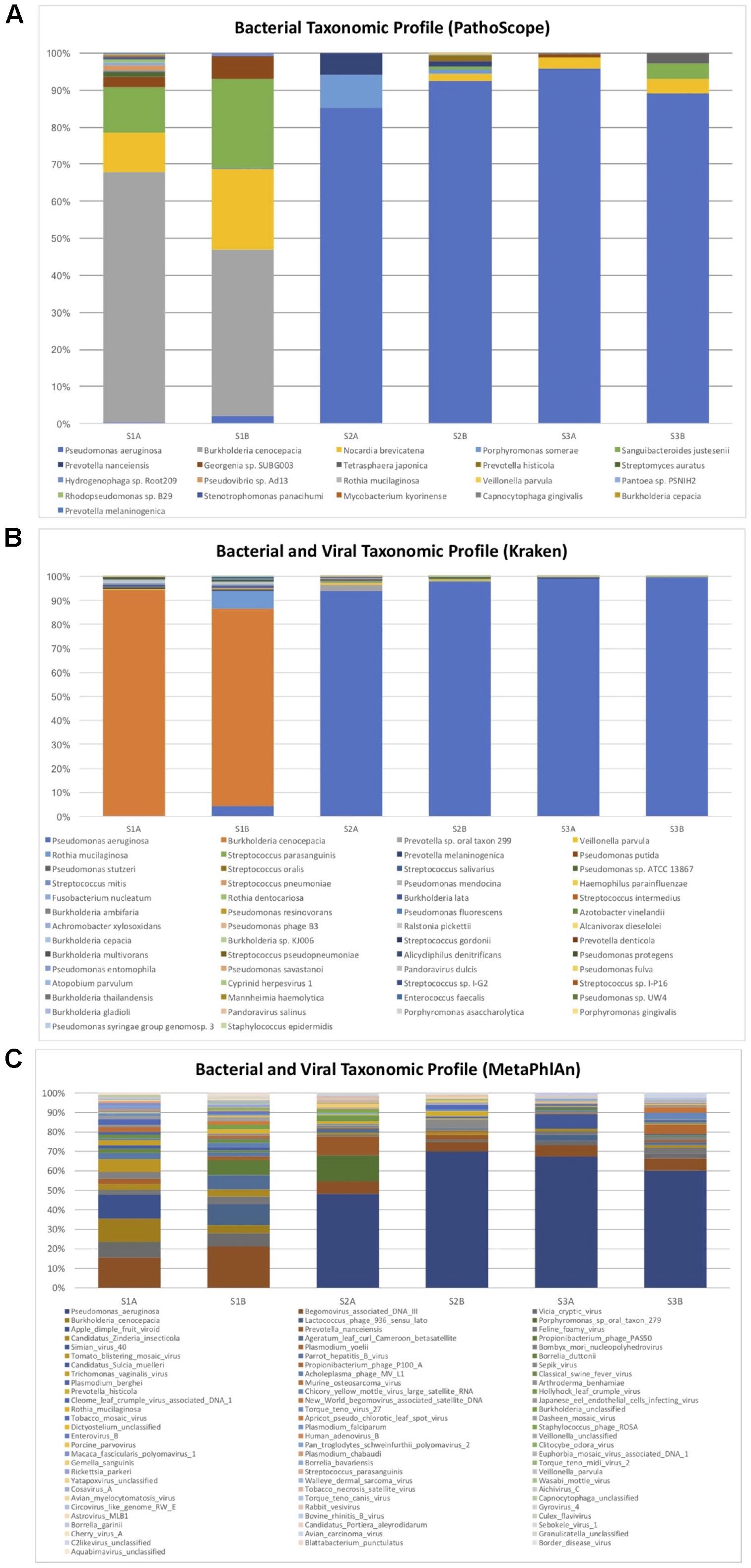
With PathoScope, fifty-one bacteria contributed to more than 0.01% of aligned reads per sample, and only 22 bacteria contributed to more than 0.01% of all total aligned reads. The bacterial taxonomic profile of each of the samples is showed over 83% of the total reads were aligned to P. aeruginosa and 4.7% aligned to B. cenocepacia (Figure 1A). The remaining reads that attributed to more than one percent of total aligned bacteria were Nocardia brevicatena (3.1%), Porphyromonas somerae (2.4%), Sanguibacteroides justesenii (2.2%), and Prevotella nanceiensis (1.7%). No viruses were detected with over 0.01% contribution to all total aligned reads.

FIGURE 1. Bacterial and viral taxonomic profile of pre- and post-antibiotic sputum samples in three subjects with cystic fibrosis who grew antibiotic resistant bacteria. Only bacterial and viral species with a minimum total observation count of 0.01% of total reads are shown for PathoScope (A) and Kraken (B). Only bacterial and viral species with a minimum total observation count of 0.1% of total reads are shown for MetaPhlAn (C). Burkholderia cepacia was the pathogen identified in culture for subject S1, but the majority of reads were attributed to Burkholderia cenocepacia. Pseudomonas aeruginosa was identified as the predominant bacteria for subjects S2 and S3, and was also identified in corresponding respiratory cultures.
With Kraken, 130 species contributed to at least 0.01% of the aligned reads per sample, and 54 species contributed to 0.01% of all aligned reads. The bacterial and viral taxonomic profile of each of the samples showed over 93% of the total reads aligned to P. aeruginosa, while 3.5% of total aligned reads were B. cenocepacia (Figure 1B). The remaining identified bacteria that contributed to more than 0.1% of total aligned reads were Prevotella sp. oral taxon 299 (0.7%), Veillonella parvula (0.3%), Rothia mucilaginosa (0.2%), Streptococcus parasanguinis (0.2%), and Prevotella melaninogenica (0.1%). Other bacteria identified within the B. cepacia complex include B. ambifaria (0.03%), B. lata (0.03%), B. cepacia (0.02%), and B. multivorans (0.01%). Pseudomonas phage B3 was detected with 0.02% contribution to all total aligned reads.
With MetaPhlAn, 201 species contributed to at least 0.01% of the aligned reads per sample, and 175 species contributed to 0.01% of all aligned reads. One hundred sixty three species contributed to at least 0.1% of aligned reads per sample, while 82 contributed to 0.1% of all aligned reads. The bacterial and viral taxonomic profile of each of the samples showed over 38% of the total reads aligned to P. aeruginosa, while 2.6% of reads aligned to B. cenocepacia (Figure 1C). Porphyromonas and Prevotella species, commonly identified in the CF lung, were identified at more than 1% of total aligned reads. The majority of other high contributors to the community identified were viruses and phages.
When comparing diversity indices at the species level there were no significant differences identified by the Shannon-Weiner index or the Simpson’s reciprocal index across all computational platforms (Table 3). Significant differences were seen with a decreased species count from pre- to post-antibiotics using Kraken (p = 0.023), and a decrease in the proportion of cultured bacteria using PathoScope (p = 0.016). We also measured bacterial distributions pre- versus post-antibiotics using Bray–Curtis distance matrices by PERMANOVA. There was no significant difference detected with either platform (Kraken p = 0.05, PathoScope p = 0.6, and MetaPhlAn p > 0.999).

TABLE 3. Alpha diversity indices and percentage of reads attributed to the cultured pathogen at the species level.
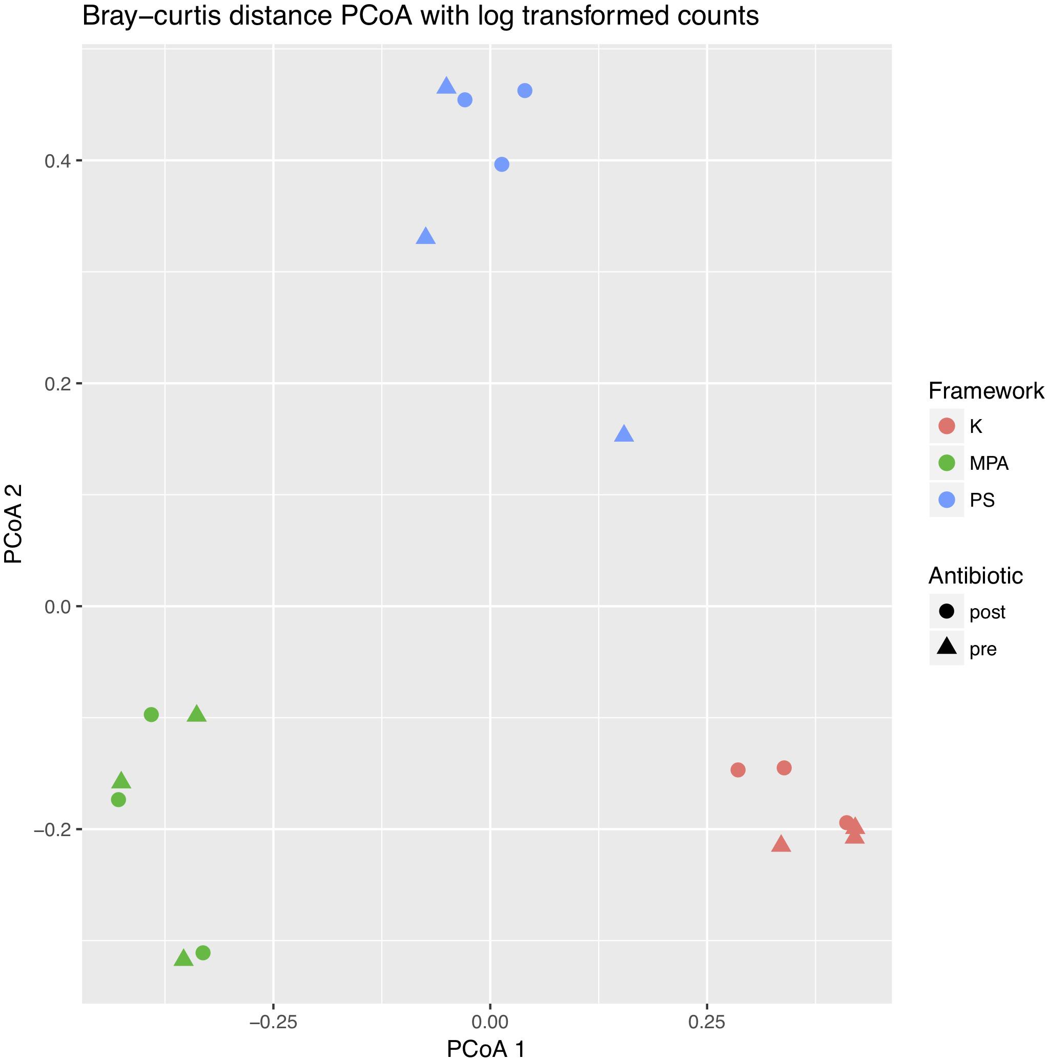
Next, to better evaluate potential differences by computational framework, we performed a Bray–Curtis PCoA plot using log transformed counts (Figure 2). PERMANOVA again revealed no difference by antibiotic timing (p = 0.993), but did detect a significant difference by computational framework (p = 0.001). The subsequent permutation test for homogeneity of multivariate dispersions was not significant (p = 0.989).

FIGURE 2. Two-dimensional principle coordinates analysis (PCoA) plot of pre- and post-antibiotic samples analyzed across computational frameworks. The PCoA was created using Bray–Curtis distance matrices with log transformed counts. Differences in sample types are shown by different shapes, while differences in computational framework are shown by different colors. K, Kraken, MPA, MetaPhlAn, PS, PathoScope.
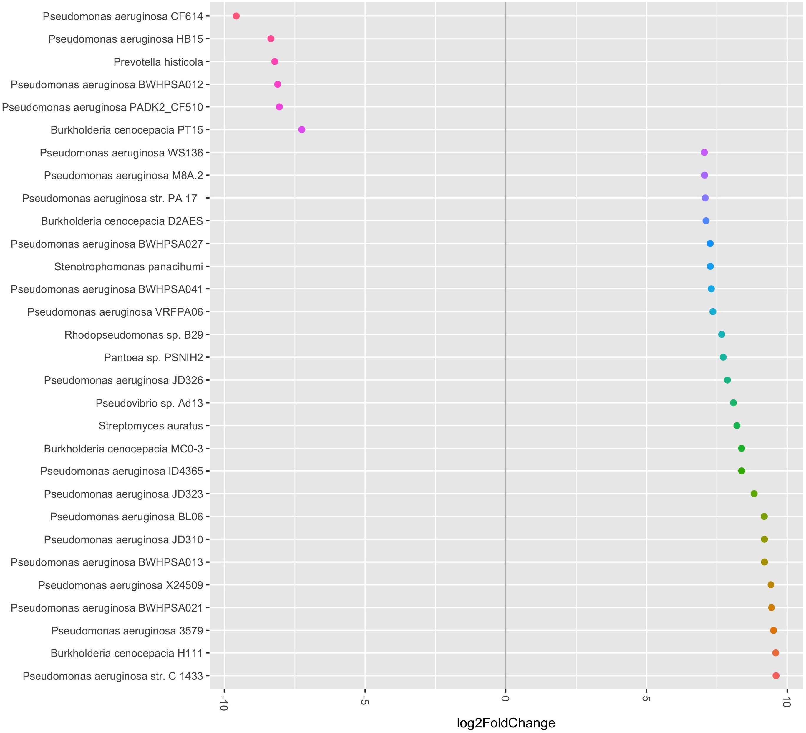
When evaluating the PathoScope data at the strain level, there were again no significant differences noted in alpha diversity pre- and post-antibiotic treatment. The pre- and post-antibiotic Shannon-Weiner diversity was 1.798 (0.433) vs. 1.464 (0.083), respectively (p = 0.310). The pre- and post-antibiotic Simpson’s reciprocal index was 4.256 (1.531) vs. 3.052 (0.274), respectively (p = 0.318). There was also no significant difference identified by PERMANOVA (p = 0.9). However, using phyloseq and DESeq2 to evaluate strain specific data generated in PathoScope, we found several significant differences pre- and post-antibiotics (see Figure 3). Prevotella histicola, one B. cenocepacia strain, and four P. aeruginosa strains were more abundant in the post-antibiotic samples.

FIGURE 3. Relative abundance of bacterial species and strains pre- and post-antibiotic treatment. The bacterial species and strains plotted on the left side of the graph were more abundant in the post-antibiotic samples, while the bacterial species and strains plotted on the right side of the graph were more abundant in the pre-antibiotic samples. All fold-changes are significant at p < 0.05.
Kraken and MetaPhlAn focus solely on bacterial and viral species identification and do not identify fungal sequences from metagenomic data. PathoScope allows for metagenomics data to be aligned to fungal reference genomes. Ninety-two fungal species were identified that contributed to at least >0.1% of total fungal reads amongst all six samples. Approximately 4 and 1.4% of total fungal reads were assigned Aspergillus and Candida species, respectively, which are both known fungal pathogens in CF (Delhaes et al., 2012; Willger et al., 2014).
Discussion
16S rRNA sequencing has traditionally been used to describe the airway microbiome in cystic fibrosis patients (Harris et al., 2007; Tunney et al., 2008; Fodor et al., 2012; Zhao et al., 2012; Zemanick et al., 2013; Carmody et al., 2013; Lim et al., 2014). There are many reasons for this, but part of it has to do with human DNA contamination within respiratory samples that makes sample processing complex (Lim et al., 2014). By limiting to 16S rRNA sequencing, however, the resolution for microbiome characterization is limited. If we do not identify bacteria to their species level, we may not discover the differing roles of organisms such as Prevotella based on their species or strain (Zemanick et al., 2013; Sherrard et al., 2014, 2016). Furthermore, metagenomic sequencing can also identify viruses, including bacteriophages, which can harbor antibiotic resistance genes or increase bacterial growth and virulence (Willner and Furlan, 2010; Willner et al., 2012). Thus, we sought to establish a technique of performing metagenomic sequencing of the cystic fibrosis airway microbiome using tSMS and PathoScope. By eliminating artificial bias, tSMS may has been successfully used in other areas but has not previously been used to study the CF airway microbiome (Orlando et al., 2011; Ginolhac et al., 2012; SEQLL, 2016). PathoScope, which also has not previously been used to study the CF lung, has successfully been used to filter out human reads and accurately identify pathogens within clinical samples (Francis et al., 2013; Hong et al., 2014; Byrd et al., 2014; Pérez-Losada et al., 2015). We compared our PathoScope results to results generated using Kraken (Davis et al., 2013) and MetaPhlAn (Segata et al., 2012).
The use of different NGS platforms and bioinformatic analysis techniques can impact both pathogen identification and diversity measures (Hahn et al., 2016). Our initial study of three control samples was encouraging that this combination of techniques would be successful in accurately detecting B. cepacia and P. aeruginosa. Control C showed much more variability than Controls A and B. This may be due to pipetting errors as this control sample was created last, or due to errors in sequencing as there were a large number of bacterial strains detected in this sample and almost 0.2% of taxonomic ID calls were for bacteria not added to the sample.
Our results demonstrate the ability to detect P. aeruginosa effectively using our metagenomic approach, which is a very important pathogen in CF (Harris et al., 2007; Carmody et al., 2013; Zemanick et al., 2013; Smith et al., 2014). This species grew in the respiratory cultures of two out of three study participants and was easily identified in those four samples. It was also detected to be part of the airway microbiome of the third subject, and the total number of reads aligned to P. aeruginosa was more than 47%. We were also able to easily identify B. cenocepacia and B. cepacia, which are also important pathogens within the CF airway (Fodor et al., 2012). It should be noted that Burkholderia cepacia complex includes at least 17 Burkholderia species, with B. cenocepacia being the one of the most common in CF (Drevinek and Mahenthiralingam, 2010). Other genera that have been previously described to be components of the CF airway microbiome include and were identified in our cohort include Porphyromonas spp., Prevotella spp., Rothia spp., Streptococcus spp., and Veillonella spp. (Harris et al., 2007; Tunney et al., 2008; Fodor et al., 2012; Zhao et al., 2012; Carmody et al., 2013; Zemanick et al., 2013; Lim et al., 2014). While PathoScope, Kraken, and MetaPhlAn all identified B. cenocepacia and P. aeruginosa as the dominant bacteria, lower abundance bacteria and the detection of viruses were not completely parallel. In addition, no bacteriophages were detected using PathoScope, but Pseudomonas phage B3 was detected using Kraken. Propionibacterium and Staphylococcus phages were also detected using MetaPhlAn. The limits in detection of bacteriophages in our samples are likely due in part to the smaller reference libraries for viruses and phages (Feigelman et al., 2017).
As PathoScope allowed for the detection of bacterial strain, it allowed us the opportunity to compare bacterial strains pre- and post-antibiotics. Interestingly, there was a shift in the relative abundance of a few strains of P. aeruginosa and B. cenocepacia. This might suggest that these strains possessed the necessary antibiotic resistance, while the other strains did not. Some prior studies demonstrated that P. aeruginosa decreased with antibiotic exposure during an acute pulmonary exacerbation (Zemanick et al., 2013). However, other studies have shown resilience of core bacteria within the CF airway microbiome with antibiotic use (Cuthbertson et al., 2016). Studies have microbial diversity following antibiotic use have also been mixed, with some showing decreased diversity (Zemanick et al., 2013; Smith et al., 2014), while other show no changes in diversity (Fodor et al., 2012; Price et al., 2013). The level of detail available using metagenomics and PathoScope could provide new insights into studies of individual bacterial abundance and microbial diversity of the CF airway in response to antibiotic use.
Using PathoScope, we were also able to evaluate the presence of fungal pathogens within the cystic fibrosis airway microbiome. Candida albicans and Aspergillus fumigatus are commonly detected in CF sputum cultures and have also been associated with acute pulmonary exacerbations (Willger et al., 2014). Sequencing studies of the CF lung mycobiome have also identified these pathogens. One study found that 74–99% of fungal reads were due to a mixture of Candida species and Malassezia (Willger et al., 2014). An earlier study found more diversity of fungal pathogens within four adult CF patients (Delhaes et al., 2012). In our study, we similarly identified the presence of several Aspergillus and Candida species. However, we also found more richness, with a total of 92 fungal species.
Our study has a few limitations. First, it is limited by the small number of subjects. Second, the contamination of human DNA in our sequencing may have affected our analysis. Our rates of 1–2% non-human reads are similar to other groups (Bacci et al., 2017). However, others have published that about a half a million reads are sufficient to provide a comprehensive metagenomic analysis of the taxa within the CF airway (Moran Losada et al., 2016).
Conclusion
PathoScope outperformed Kraken and MetaPhlAn in our validation study of artificial bacterial community controls. PathoScope also has advantages over Kraken and MetaPhlAn in being able to determine bacterial strains and the presence of fungal organisms. Thus, PathoScope can be confidently used when evaluating metagenomic data to determine CF airway microbiome diversity.
Availability of Data
The sequence data has been uploaded to NCBI under BioProject PRJNA422117.
Ethics Statement
The study protocol was approved by the Institutional Review Board at Children’s National Health System and was carried out in accordance with their recommendations. All subjects gave written informed consent in accordance with the Declaration of Helsinki.
Author Contributions
AH designed the study, performed the experiments, analyzed the data, and wrote the manuscript. MB contributed to study design, data analysis, and wrote sections of the manuscript. KG contributed to study design and data analysis. HC, IS, GP, and AK were all involved in study participant recruitment and sample collection. TM contributed to study design. RF and KC contributed to study design and interpretation of data analysis. All authors edited and approved the final manuscript.
Funding
AH was funded in part by a K12 Career Development Program K12HL119994 through the National Heart, Lung and Blood Institute and the Margaret Q. Landenberger Research Foundation MQLRF20170207. The sample collection for this study was performed with funding from the Clark Charitable Trust. The sequencing performed in this study was funded by The Frank and Nancy Parsons Foundation. This project was also partially supported by Award Number UL1TR000075 from the NIH National Center for Advancing Translational Sciences. Its contents are solely the responsibility of the authors and do not necessarily represent the official views of the National Institutes of Health.
Conflict of Interest Statement
The authors declare that the research was conducted in the absence of any commercial or financial relationships that could be construed as a potential conflict of interest.
Acknowledgments
The authors would like to acknowledge the GWU Colonial One High-Performance Computing Cluster for computational time. We would also like to acknowledge Anamaris M. Colberg-Poley, Ph.D., and Ian Toma, Ph.D., for insight into respiratory processing techniques of mixed microbial samples, Caroline Conlan for assistance in R programming, and Matthew Schwaberow, B.S, M.B.A., for assistance with MetaPhlAn. MB is a doctoral student in the Systems Biology Program of the Institute for Biomedical Sciences at The George Washington University. This work is from a dissertation to be presented to the above program in partial fulfillment of the requirements for the Ph.D. degree.
References
Bacci, G., Mengoni, A., Fiscarelli, E., Segata, N., Taccetti, G., Dolce, D., et al. (2017). A different microbiome gene repertoire in the airways of cystic fibrosis patients with severe lung disease. Int. J. Mol. Sci. 18:E1654. doi: 10.3390/ijms18081654
Bacci, G., Paganin, P., Lopez, L., Vanni, C., Dalmastri, C., Cantale, C., et al. (2016). Pyrosequencing unveils cystic fibrosis lung microbiome differences associated with a severe lung function design. PLoS One 11:e0156807. doi: 10.1371/journal.pone.0156807
bioBakery (2017). bioBakery. Available at: https://bitbucket.org/biobakery/biobakery/wiki/biobakery_basic on 2/22/18
Boutin, S., Graeber, S. Y., Weitnauer, M., Panitz, J., Stahl, M., Clausznitzer, D., et al. (2015). Comparison of microbiomes from different niches of upper and lower airways in children and adolescents with cystic fibrosis. PLoS One 10:e0116029. doi: 10.1371/journal.pone.0116029
Byrd, A. L., Perez-Rogers, J. F., Manimaran, S., Catro-Nallar, E., Toma, I., McCaffrey, T., et al. (2014). Clinical PathoScope: rapid alignment and filtration for accurate pathogen identification in clinical samples using unassembled sequencing data. BMC Bioinformatics 15:262. doi: 10.1186/1471-2015-15-262
Carmody, L. A., Zhao, J., Schloss, P. D., Petrosino, J. F., Murray, S., Young, V. B., et al. (2013). Changes in cystic fibrosis airway microbiota at pulmonary exacerbation. Ann. Am. Thorac. Soc. 10, 179–187. doi: 10.1513/AnnalsATS.201211-107OC
Coburn, B., Wang, P. W., Diaz Caballero, J., Clark, S. T., Brahma, V., Donaldson, S., et al. (2015). Lung microbiota across age and disease stage in cystic fibrosis. Sci. Rep. 5:10241. doi: 10.1038/srep10241
Cox, M. J., Allgaier, M., Taylor, B., Baek, M. S., Huang, Y. J., Daly, R. A., et al. (2010). Airway microbiota and pathogen abundance in age-stratified cystic fibrosis patients. PLoS One 5:e11044. doi: 10.1371/journal.pone.0011044
Cuthbertson, L., Rogers, G. B., Walker, A. W., Oliver, A., Green, L. E., Daniels, T. W. V., et al. (2016). Respiratory microbiota resistance and resilience to pulmonary exacerbation and subsequent antimicrobial intervention. ISME J. 10, 1081–1091. doi: 10.1038/ismej.2015.198
Davis, M. P. A., van Dongen, S., Abreu-Goodger, C., Bartonicek, N., and Enright, A. J. (2013). Kraken: a set of tools for quality control and analysis of high-throughput sequence data. Methods 63, 41–49. doi: 10.1016/j.ymeth.2013.06.027
Delhaes, L., Monchy, S., Fréalle, E., Hubans, C., Salleron, J., Leroy, S., et al. (2012). The airway microbiota in cystic fibrosis: a complex fungal and bacterial community—implications for therapeutic management. PLoS One 7:e36313. doi: 10.1371/journal.pone.0036313
Drevinek, P., and Mahenthiralingam, E. (2010). Burkholderia cenocepacia in cystic fibrosis: epidemiology and molecular mechanisms of virulence. Clin. Microbiol. Infect. 16, 821–830. doi: 10.1111/j.1469-0691.2010.03237.x
Feigelman, R., Kahlert, C. R., Baty, F., Rassouli, F., Kleiner, R. L., Kohler, P., et al. (2017). Sputum DNA sequencing in cystic fibrosis: non-invasive access to the lung microbiome and to pathogen details. Microbiome 5, 1–14. doi: 10.1186/s40168-017-0234-1
Flight, W. G., Smith, A., Paisey, C., Marchesi, J. R., Bull, M. J., Norville, P. J., et al. (2015). Rapid detection of emerging pathogens and loss of microbial diversity associated with severe lung disease in cystic fibrosis. J. Clin. Microbiol. 53, 2022–2029. doi: 10.1128/JCM.00432-15
Fodor, A. A., Klem, E. R., Gilpin, D. F., Elborn, J. S., Boucher, R. C., Tunney, M. M., et al. (2012). The adult cystic fibrosis airway microbiota is stable over time and infection type, and highly resilient to antibiotic treatment of exacerbations. PLoS One 7:e45001. doi: 10.1371/journal.pone.0045001
Francis, O. E., Bendall, M., Manimaran, S., Hong, C. J., Clement, N. L., Castro-Nallar, E., et al. (2013). Pathoscope: species identification and strain attribution with unassembled sequencing data. Genome Res. 23, 1721–1729. doi: 10.1101/gr.150151.112
Ginolhac, A., Vilstrup, J., Stenderup, J., Rasmussen, M., Stiller, M., Shapiro, B., et al. (2012). Improving the performance of true single molecule sequencing for ancient DNA. BMC Genomics 13:177. doi: 10.1186/1471-2164-13-177
Hahn, A., Sanyal, A., Perez, G. F., Colberg-Poley, A. M., Campos, J., Rose, M. C., et al. (2016). Different next generation sequencing platforms produce different microbial profiles and diversity in cystic fibrosis sputum. J. Microbial. Methods 130, 95–99. doi: 10.1016/j.mimet.2016.09.002
Harris, J. K., De Groote, M. A., Sagel, S. D., Zemanick, E. T., Kapsner, R., Penvari, C., et al. (2007). Molecular identification of bacteria in bronchoalveolar lavage fluid from children with cystic fibrosis. Proc. Natl. Acad. Sci. U.S.A. 104, 20529–20533. doi: 10.1073/pnas.0709804104
Hilton, S. K., Castro-Nallar, E., Perez-Losada, M., Toma, I., McCaffrey, T. A., Hoffman, E. P., et al. (2016). Metataxonomic and metagenomic approaches vs. culture-based techniques for clinical pathology. Front. Microbiol. 7:484. doi: 10.3389/fmicb.2016.00484
Hong, C., Manimaran, S., Shen, Y., Perez-Rogers, J. F., Byrd, A. L., Castro-Nallar, E., et al. (2014). PathoScope 2. 0: a complete computational framework for strain identification in environmental or clinical sequencing samples. Microbiome 2:33. doi: 10.1186/2049-2618-2-33
Huang, Y. J., and LiPuma, J. J. (2016). The microbiome in cystic fibrosis. Clin. Chest Med. 37, 59–67. doi: 10.1016/j.ccm.2015.10.003
Kapranov, P., St Laurent, G., Raz, T., Ozsolak, F., Reynolds, P., Sorensen, P. H. B., et al. (2010). The majority of total nuclear-encoded non-ribosomal RNA in a human cell is ‘dark matter’ un-annotated RNA. BMC Biol. 8:149. doi: 10.1186/1741-7007-8-149
Klepac-Ceraj, V., Lemon, K. P., Martin, T. R., Allgaier, M., Kembel, S. W., Knapp, A. A., et al. (2010). Relationship between cystic fibrosis respiratory tract bacterial communities and age, genotype, antibiotics, and Pseudomonas aeruginosa. Environ. Microbiol. 12, 1293–1303. doi: 10.1111/j.1462-2920.2010.02173.x
Lim, Y. W., Evangelista, J. S., Schmieder, R., Bailey, B., Haynes, M., Furlan, M., et al. (2014). Clinical insights from metagenomic analysis of sputum samples from patients with cystic fibrosis. J. Clin. Microbiol. 52, 425–437. doi: 10.1128/JCM.02204-13
Love, M. I., Huber, W., and Anders, S. (2014). Moderated estimation of fold change and dispersion for RNA-seq data with DESeq2. Genome Biol. 15:550. doi: 10.1186/s13059-014-0550-8
MacKenzie, T., Gifford, A. H., Sabadosa, K. A., Quinton, H. B., Knapp, E. A., Goss, C. H., et al. (2014). Longevity of patients with cystic fibrosis in 2000 to 2010 and beyond: survival analysis of the cystic fibrosis foundation patient registry. Ann. Intern. Med. 161, 233–241. doi: 10.7326/M13-0636
McMurdie, P. J., and Holmes, S. (2013). phyloseq: an R package for reproducible interactive analysis and graphics of microbiome census data. PLoS One 8:e61217. doi: 10.1371/journal.pone.0061217
Moran Losada, P., Chouvarine, P., Dorda, M., Hedtfeld, S., Mielke, S., Schulz, A., et al. (2016). The cystic fibrosis lower airways microbial metagenome. ERJ Open Res. 2, 00096–2015. doi: 10.1183/23120541.00096-2015
Oksanen, J., Blanchet, F. G., Friendly, M., Kindt, R., Legendre, P., McGlinn, D., et al. (2017). vegan: Community Ecology Package. R Package Version 2.4-4. Available at: https://cran.r-project.org/web/packages/vegan/index.html.
Orlando, L., Ginolhac, A., Raghavan, M., Vilstrup, J., Rasmussen, M., Magnussen, K., et al. (2011). True single-molecule DNA sequencing of a Pleistocene horse bone. Genome Res. 21, 1705–1719. doi: 10.1101/gr.122747.111
Pérez-Losada, M., Castro-Nallar, E., Bendall, M. L., Freishtat, R. J., and Crandall, K. A. (2015). Dual transcriptomic profiling of host and microbiota during health and disease in pediatric asthma. PLoS One 10:e0131819. doi: 10.1371/journal.pone.0131819
Price, K. E., Hampton, T. H., Gifford, A. H., Doben, E. L., Hogan, D. A., Morrison, H. G., et al. (2013). Unique microbial communities persist in individual cystic fibrosis patients throughout clinical exacerbation. Microbiome 1, 1–11. doi: 10.1186/2049-2618-1-27
Qiagen (2016). QIAamp DNA Microbiome Kit In Qiagen. Available at: https://www.qiagen.com/us/shop/sample-technologies/dna/genomic-dna/qiaamp-dna-microbiome-kit/#productdetails [accessed July 27, 2016].
Ramsey, B. W. (1996). Management of pulmonary disease in patients with cystic fibrosis. N. Engl. J. Med. 335, 179–188. doi: 10.1056/NEJM199607183350307
Segata, N., Waldron, L., Ballarini, A., Narasimhan, V., Jousson, O., and Huttenhower, C. (2012). Metagenomic microbial community profiling using unique clade-specific marker genes. Nat. Methods 8, 811–814. doi: 10.1038/nmeth.2066
SEQLL (2016). True Single Molecule Sequencing. Available at: http://seqll.com/services/ [accessed June 13, 2017].
Sherrard, L. J., McGrath, S. J., McIlreavey, L., Hatch, J., Wolfgang, M. C., Muhlebach, M. S., et al. (2016). Production of extended-spectrum β-lactamases and the potential indirect pathogenic role of Prevotella isolates from the cystic fibrosis respiratory microbiota. Int. J. Antimicrob. Agents 47, 140–145. doi: 10.1016/j.ijantimicag.2015.12.004
Sherrard, L. J., Tunney, M. M., and Elborn, J. S. (2014). Antimicrobial resistance in the respiratory microbiota of people with cystic fibrosis. Lancet 384, 703–713. doi: 10.1016/S0140-6736(14)61137-5
Smith, D. J., Badrick, A. C., Zakrzewski, M., Krause, L., Bell, S. C., Anderson, G. J., et al. (2014). Pyrosequencing reveals transient cystic fibrosis lung microbiome changes with intranvenous antibiotics. Eur. Respir. J. 44, 922–930. doi: 10.1183/09031936.00203013
Tunney, M. M., Field, T. R., Moriarty, T. F., Patrick, S., Doering, G., Muhlebach, M. S., et al. (2008). Detection of anaerobic bacteria in high numbers in sputum from patients with cystic fibrosis. Am. J. Respir. Crit. Care Med. 177, 995–1001. doi: 10.1164/rccm.200708-1151OC
VanDevanter, D. R., and LiPuma, J. J. (2012). Microbial diversity in the cystic fibrosis airways: where is thy sting? Future Microbiol. 7, 801–803. doi: 10.2217/fmb.12.55
Willner, D., and Furlan, M. (2010). Deciphering the role of phage in the cystic fibrosis airway. Virulence 1, 309–313. doi: 10.4161/viru.1.4.12071
Willner, D., Haynes, M. R., Furlan, M., Hanson, N., Kirby, B., Lim, Y. W., et al. (2012). Case studies of the spatial heterogeneity of DNA viruses in the cystic fibrosis lung. Am. J. Respir. Cell Mol. Biol. 46, 127–131. doi: 10.1165/rcmb.2011-0235OC
Willger, S. D., Grim, S. L., Dolben, E. L., Shipunova, A., Hampton, T. H., Morrison, H. G., et al. (2014). Characterization and quantification of the fungal microbiome in serial samples from individuals with cystic fibrosis. Microbiome 2:40. doi: 10.1186/2049-2618-2-40
Zemanick, E. T., Harris, J. K., Wagner, B. D., Robertson, C. E., Sagel, S. D., Stevens, M. J., et al. (2013). Inflammation and airway microbiota during cystic fibrosis pulmonary exacerbations. PLoS One 8:e62917. doi: 10.1371/journal.pone.0062917
Zhao, J., Schloss, P. D., Kalikin, L. M., Carmody, L. A., Foster, B. K., Petrosino, J. F., et al. (2012). Decade-long bacterial community dynamics in cystic fibrosis airways. Proc. Natl. Acad. Sci. U.S.A. 109, 5809–5814. doi: 10.1073/pnas.1120577109
Zimmer, B. L., Bacsafra, M., Churc, E. A., Mattes, T. M., Mendoza-Morales, G., and Van Pelt, L. (2004). “Antimicrobial susceptibility testing of cystic fibrosis isolates of Pseudomonas aeruginosa: evaluation of the MicroScan dried overnight gram negative panel and instrument systems, frozen broth microdilution panels, and disk diffusion,” in. Paper Presented at the American Society for Microbiology Meeting, Abstract C-138, Boston, MA.
Keywords: cystic fibrosis, antibiotics, microbiome, metagenomics, true single molecule DNA sequencing
Citation: Hahn A, Bendall ML, Gibson KM, Chaney H, Sami I, Perez GF, Koumbourlis AC, McCaffrey TA, Freishtat RJ and Crandall KA (2018) Benchmark Evaluation of True Single Molecular Sequencing to Determine Cystic Fibrosis Airway Microbiome Diversity. Front. Microbiol. 9:1069. doi: 10.3389/fmicb.2018.01069
Received: 19 December 2017; Accepted: 04 May 2018;
Published: 25 May 2018.
Edited by:
Giovanni Di Bonaventura, Università degli Studi “G. d’Annunzio” Chieti - Pescara, ItalyReviewed by:
Dinesh Sriramulu, Shres Consultancy (Life Sciences), IndiaGiovanni Bacci, Università degli Studi di Firenze, Italy
Copyright © 2018 Hahn, Bendall, Gibson, Chaney, Sami, Perez, Koumbourlis, McCaffrey, Freishtat and Crandall. This is an open-access article distributed under the terms of the Creative Commons Attribution License (CC BY). The use, distribution or reproduction in other forums is permitted, provided the original author(s) and the copyright owner are credited and that the original publication in this journal is cited, in accordance with accepted academic practice. No use, distribution or reproduction is permitted which does not comply with these terms.
*Correspondence: Andrea Hahn, YWxoYWhuQGNoaWxkcmVuc25hdGlvbmFsLm9yZw==
 Andrea Hahn
 Matthew L. Bendall
 Keylie M. Gibson
 Hollis Chaney2,5
 Timothy A. McCaffrey
 Keith A. Crandall
