- 1State Key Laboratory of Agricultural Microbiology, Huazhong Agricultural University, Wuhan, China
- 2Hubei Cooperative Innovation Center for Sustainable Pig Production, Wuhan, China
- 3Key Laboratory of Preventive Medicine in Hubei Province, Wuhan, China
Toxoplasma gondii is an important zoonotic pathogen infecting one third of the world population and numerous animals. A key factor to its wide distribution is the ability to interconvert between fast replicating tachyzoites and slowly growing bradyzoites, and to establish lifelong chronic infection in intermediate hosts. Although it is well accepted that stage conversion plays key roles in the pathogenesis and transmission of the parasite, little is known about the molecular mechanisms behind it. Using existing gene expression data from TOXODB and published work, we looked for proteins with novel functional domains and whose expression is up-regulated in the bradyzoite stage, hoping to find molecules that have critical roles in regulating stage conversion and bradyzoite formation. In this study we characterized two such proteins ANK1 and DnaK-TPR, both of which are primarily expressed in bradyzoites and contain novel motifs to mediate protein-protein interactions. Through CRISPR/CAS9 directed gene editing technology, both genes were individually knocked out in type 1 strain TgHB2 and type 2 strain ME49. Disruption of neither of these two genes affected the growth or replication of tachyzoites in vitro, consistent with their minimal expression at this stage. However, mutants lacking ANK1 or DnaK-TPR displayed modest virulence attenuation during mice infection. Surprisingly, inactivation of neither ANK1 nor DnaK-TPR seemed to have a significant impact on bradyzoite differentiation in vitro or cyst formation in vivo. These results suggest that ANK1 and DnaK-TPR probably do not directly contribute to bradyzoite differentiation, but likely affect other aspects of bradyzoite biology.
Introduction
Toxoplasma gondii is an obligate intracellular parasite belonging to the phylum apicomplexa. It is capable of infecting virtually all warm-blooded animals and humans, and has a world-wide distribution. It is estimated that 30% of the world population is infected by this parasite, causing congenital toxoplasmosis and other severe complications in susceptible individuals and animals, which leads to great economical losses and public health problems (Elmore et al., 2010; Dubey et al., 2012). T. gondii has a complex life cycle that consists of two phases: the sexual phase, which takes place in felines such as domestic cats, and the asexual phase, which takes place in all warm-blooded animals and humans (Dubey, 1998; Dabritz and Conrad, 2010). In intermediate hosts such as humans, T. gondii can exist in two forms: a fast replicating form called tachyzoite and a slowly growing form called bradyzoite. During the infection of a naive host, T. gondii first quickly multiplies as tachyzoites to spread the parasites and establish systemic infection. Therefore tachyzoite is responsible for acute toxoplasmosis. Nonetheless, T. gondii infection rarely causes obvious symptoms in healthy people. The reason for that is, once the immune responses against T. gondii infection are activated, tachyzoites are effectively eliminated. As a response to host immune clearance, some of the tachyzoites convert to the slowly replicating bradyzoites and stay within host cells (mainly neurons and muscle cells) in the form of tissue cysts, to establish chronic infection and escape immune surveillance (Remington and Merigan, 1968; Dubey et al., 1998; Weiss and Kim, 2000). Once formed, chronic infection will stay with the hosts lifelong, and may get reactivated when hosts’ immune function is compromised (Shen et al., 2016). Currently there are no effective treatments for chronic toxoplasmosis.
In addition to hosts’ immune pressure, bradyzoites can also be induced in vitro by a variety of conditions (Bohne et al., 1993; Soete et al., 1994; Weiss et al., 1995). In general, it is thought that bradyzoite differentiation is a stress response parasites initiate to adapt to adverse environmental conditions (Ferreira da Silva Mda et al., 2008). Nutrient starvation, extreme pH and respiratory chain inhibitors such as antimycin A are all able to induce bradyzoite formation in vitro (Soete et al., 1994). Alkaline medium with pH 8.2 along with CO2 starvation (ambient CO2) is the most common way of inducing bradyzoite in vitro (Weiss et al., 1995). Although the exact mechanisms of how parasites sense the stress are currently unknown, it is clear that stress conditions induce global changes in gene expression that eventually lead to slow-down of cell cycle and formation of bradyzoites (Manger et al., 1998; Skariah et al., 2010; Buchholz et al., 2011).
The interconversion between tachyzoites and bradyzoites plays a critical role in the pathogenesis and transmission of T. gondii. In order to understand the biology and mechanisms underlying such stage conversion, micro-array and RNAseq analysis have been done to compare gene expression differences between tachyzoites and bradyzoites, and hundreds of genes were reported to be differentially regulated (Buchholz et al., 2011; Pittman et al., 2014). These differentially regulated genes were thought to play important roles during stage conversion. Indeed, some of the up-regulated genes were experimentally shown to be important for bradyzoite differentiation and cyst formation, such as the bradyzoite specific glycolytic enzyme ENO1 (Mouveaux et al., 2014) and the cyst wall protein CST1 (Tomita et al., 2013).
In an effort to understand the molecular basis for T. gondii stage conversion, we examined the differentially expressed proteins between tachyzoites and bradyzoites, and looked for proteins that have novel functional motifs. In this study, we characterized two proteins that contain tetratricopeptide repeat (TPR) structural motifs. Originally identified in yeast (Hirano et al., 1990; Sikorski et al., 1990), TPR motifs are ubiquitously found in a variety of proteins from bacteria to humans (Allan and Ratajczak, 2011). As the name indicates, TPR motifs consist of regions that are 34-residues long (Cerveny et al., 2013). They are minimally conserved sequence wise, but consensus patterns of small and large hydrophobic residues combination have been defined (Cerveny et al., 2013). Structural analysis on the TPR motifs of protein phosphatase 5 (PP5) and many other TPR containing proteins indicate that they form two antiparallel α-helices that are equivalent in length (Das et al., 1998; Allan and Ratajczak, 2011). The basic function of TPR motifs is to form structural scaffold to mediate protein-protein interactions. TPR motifs are present in many proteins that are involved in different cellular processes and functions, such as gene expression regulation, protein transport, bacterial virulence, and immune functions (Allan and Ratajczak, 2011; Cerveny et al., 2013). Based on bioinformatic prediction, T. gondii genome encodes more than 50 TPR containing proteins. Of these, ANK1 (TGME49_216140) and DnaK-TPR (TGME49_202020) are primarily expressed at the bradyzoite stage (Friesen et al., 2008; Ueno et al., 2011). In this study we used gene knockout strategies to analyze their roles during T. gondii growth and development.
Materials and Methods
Cells and Parasites
Tachyzoites of T. gondii strains ME49 and TgHB2 were propagated in human foreskin fibroblasts (HFF), which were cultured in Dulbecco’s modified Eagle medium (DMEM) supplemented with 10% fetal bovine serum (FBS), 100 μg/ml streptomycin and 10 mM L-glutamine. The genetically modified parasites were propagated and cultured under the same conditions.
Searching for Differentially Regulated Proteins with Novel Function Motifs
Gene expression datasets available from ToxoDB were used for this purpose. First, the “Oocyst, tachyzoite, and bradyzoite developmental expression profiles (M4)” dataset was searched for protein coding genes whose expression had a fold change greater than 10 between the tachyzoite (2 days in vitro) and bradyzoite (8 days in vitro) stages. Second, the “Transcriptome during acute or chronic infection in mouse brain” dataset was searched using the same parameters to find genes that had 10-fold difference between “acute infection 10 days p.i.” and “chronic infection 28 days p.i.”. Third, the “Bradyzoite Differentiation Expression Profiles (ME49, GT1, CTGara)” dataset was used to find genes that had 10-fold expression difference between “ME49 tachyzoite” and “ME49 pH 8.2.” Subsequently, genes that were identified in all three searches were collected and analyzed manually. There were 18 genes identified in total, which included lactate dehydrogenase LDH2, bradyzoite antigen BAG1, bradyzoite rhoptry protein BRP1 and other well described bradyzoite specific genes. Judging from the “Product Production” provided by ToxoDB, two (ANK1 with the gene ID TgME49_216140 and DnaK-TPR with the gene ID TgME49_202020) out of the 18 proteins contained tetratricopeptide repeats (TPR), which then became the focus of this study.
To further characterize the TPR containing proteins, their primary sequences were used in protein Blast searches1, as well as in the Motif Scan2 server to search the Prosite and Pfam databases, to identify potential function domains.
To predict the number and positions of the TPR motifs in TPR containing proteins, their sequences were analyzed by the profile based method TPRpred3 (Karpenahalli et al., 2007), using the P-value threshold of 10-4.
Deletion of ANK1 or DnaK-TPR Genes Using the CRISPR/CAS9 System
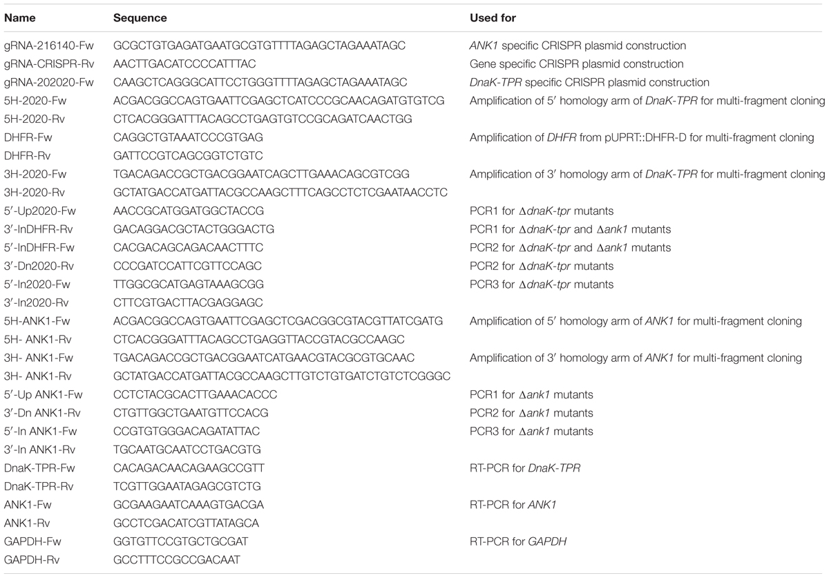
To construct homologous templates for ANK1 or DnaK-TPR knockouts, the 5′- and 3′- homology arms of the corresponding genes were cloned into the pUC19 vector along with the selection marker DHFR, using multi-fragment cloning. The primers used to amplify the homology arms from genomic DNA of ME49 were listed in Table 1. After obtaining each fragment, 5′ homology arm, DHFR and 3′ homology arm were ligated into the linearized pUC19 (achieved through SacI and HindIII digestion) vector by the One Step Cloning Kit (ClonExpress II One Step Cloning Kit, Vazyme, United States) according to manufacturer’s instructions. The gene specific CRISPR plasmids were constructed by site directed mutagenesis of the UPRT targeting CRISPR plasmid, as previously described (Shen et al., 2014, 2017). Primers used for mutagenesis were also listed in Table 1. All constructs were confirmed by Sanger sequencing before use.
To generate gene knockouts in desired strains, corresponding homology construct and gene specific CRISPR plasmid were co-transfected into freshly egressed tachyzoites, as described previously (Shen et al., 2017). Subsequently transfectants were selected with 1 μM pyrimethamine for 3 – 4 passages until the drug resistant pools became stable. Single clones were isolated through limiting dilution in 96-well plates and examined by diagnostic PCRs (PCR1, PCR2 and PCR3; primers are listed in Table 1) to check the disruption of corresponding genes.
Quantitative Real-Time PCR
Approximately 1 × 10⁷ freshly egressed parasites (tachyzoites or bradyzoites) of the ME49 strain were collected and purified through polycarbonate membranes with the pore size of 3 μm. Parasites were washed with PBS and total RNA was extracted from them using the trizol methods, according to manufacturer’s instructions (TransGen Biotech, China). Bradyzoites were obtained by growing parasites in differentiation medium (RPMI 1640 medium supplemented with 1% FBS and 50 mM HEPES, pH 8.2) along with ambient CO2 for 4 days (Walker et al., 2013). After RNA isolation, 2 μg total RNA from each sample was reverse transcribed into cDNA using the PrimeScript RT reagent Kit (Takara Bio, Japan).
Subsequently the cDNA was used as template for real-time PCR (RT-PCR) to estimate the expression levels of target genes. Three pairs of specific primers were designed for this purpose: ANK1-Fw and ANK1-Rv; DnaK-TPR-Fw and DnaK-TPR-Rv; GAPDH-Fw and GAPDH-Rv (Sequences are listed in Table 1). RT-PCR was performed on the ViiA-7 Real-Time system (Life Technologies, Camarillo, CA, United States) using the SYBR Green method, according to the manufacturer’s instructions. Cycling conditions for RT-PCR were set as: initial denaturation at 95°C for 30 s, followed by 40 cycles of 95°C for 15 s, 56°C for 30 s and 72°C for 20 s. Each sample was prepared and examined three times independently. Gene expression levels was estimated by the ΔCT method using the GAPDH gene as internal reference, as described previously (Selseleh et al., 2012).
Whole Genome Sequencing
Freshly egressed tachyzoites (2 × 10⁷) were purified through membrane filtration and washed with PBS. Subsequently genomic DNA was extracted from the purified parasites using the EasyPure Genomic DNA Kit (Transgen Biotech, China). Purified genomic DNA was sheared into short fragments of about 350 bps by sonication and then used for library construction using the TruSeq Library Construction Kit (Illumina, San Diego, CA, United States) according to the manufacturer’s instructions. The libraries were then sequenced by the Illumina HiSeq platform (Illumina, San Diego, CA, United States). The average sequencing depth was around 110X. To detect genetic alterations, clean reads obtained from genome sequencing were mapped to the reference genome of GT1, and SAMTOOLs, BreakDancer and CNVnator were used to detect short indels, structural variations (insertion, deletion, and inversion) and copy number variations, respectively (Chen et al., 2009; Li et al., 2009; Abyzov et al., 2011). The mapping results were visualized by the Integrative Genomics Viewer (Thorvaldsdottir et al., 2013).
Plaque Assay
Human foreskin fibroblasts cells were seeded into six-well plates and grown at 37°C until confluent. Subsequently 100 purified tachyzoites were added to each well and cultured for 10–12 days without disturbance under standard growth conditions. Then the samples were fixed with 4% paraformaldehyde and stained with 0.5% crystal violet to develop the plaques. Finally the plates were scanned by a scanner (FileScan 380, Microtek, China) and the number and sizes of plaques in each well were determined as described previously (Shen and Sibley, 2014). Each strain was tested three times independently, each with triplicates.
Intracellular Replication Assay
Coverslips in 24-well plates seeded with confluent HFF monolayers were infected with freshly harvested tachyzoites (105 parasites per well) for 1 h at 37°C. Extracellular parasites that failed to invade HFF cells were washed away and invaded ones were grown for another 24 h under standard tachyzoite growth conditions. Subsequently the samples were fixed with 4% paraformaldehyde and stained with swine anti-Toxoplasma (gifts from the Zhao lab at Huazhong Agricultural University, to stain extracellular parasites). After extensive washing, samples were permeabilized with 0.1% Triton X-100 for 10 min and stained with rabbit anti-TgALD (gifts from the Sibley lab at Washington University in St. Louis, to stain all parasites). Primary antibodies were detected by Alexa Fluor 594-conjugated goat anti-rabbit IgG and FITC conjugated goat anti-swine IgG secondary antibodies, respectively (Life Technologies, Camarillo, CA, United States). Parasites that were stained red but not green (these were intracellular parasites) were analyzed to determine the number of parasites in each parasitophorous vacuole (PV). Each sample was tested three times independently, each with triplicates.
Bradyzoite Differentiation Assay
Alkaline medium was used to induce bradyzoite differentiation in vitro, as described previously (Walker et al., 2013). Briefly, Purified parasites were used to invade HFF monolayer (104 parasites/well) seeded on coverslips in 24-well plates for 1 h under standard growth conditions. Subsequently non-invaded parasites were washed away with PBS and the rest of the cultures were grown in differentiation medium with ambient CO2 for 4 days. After treatment, samples were fixed with 4% paraformaldehyde, permeablized with 0.1% Triton X-100, and stained with rabbit anti-TgALD and mouse anti-TgBAG1 (gifts from the Zhao lab at Huazhong Agricultural University) antibodies. Alexa Fluor 594-conjugated goat anti-rabbit IgG and Alexa Fluor 488-conjugated goat anti-mouse IgG secondary antibodies (Life Technologies, Camarillo, CA, United States) were used to detect primary antibodies. Hoechst 33342 was used to stain cell nuclei. Parasites that were stained both green and red were counted as bradyzoites, whereas parasites that were stained only red were treated as tachyzoites. Bradyzoite differentiation efficiency was determined by dividing the number of green vacuoles by the number of red vacuoles. At least 100 total vacuoles were counted for each sample in each experiment and each sample was repeated three times independently.
Animal Experiments
All 4–6 weeks old female ICR mice and 10–12 weeks old Kunming mice were purchased from the Hubei Provincial Centers for Disease Control. To test the virulence of parasites in mice, freshly harvested tachyzoites were purified by filtration through 3 μm polycarbonate membranes and then used to infect mice by intraperitoneal injection. Each parasite strain was tested by infection of 10 mice at the dose of 100 tachyzoites per mouse. After infection, mice were monitored daily for 30 days. The cumulative mortality was calculated by dividing the number of mice that died by the number of mice infected. At the conclusion of the virulence tests, mice that survived the infection at day 30 were sacrificed and brain tissues were homogenized to determine the number of Toxoplasma cysts by DBA-FITC staining, as described previously (Buchholz et al., 2013). All animal experiments were approved by The Scientific Ethic Committee of Huazhong Agricultural University (permit #: HZAUMO-2016-041).
Results
Sequence Analysis of ANK1 and DnaK-TPR Proteins
Both ANK1 and DnaK-TPR have been reported previously as proteins whose expression was drastically increased during tachyzoite to bradyzoite conversion. ANK1 was initially reported as 35.m00032 and DnaK-TPR was reported as 20.m00351 (Friesen et al., 2008). According to the current gene model on ToxoDB, ANK1 is a multi-domain protein with 563 amino acids. Sequence analysis using BLAST, Pfam and PROSITE database searching indicated that it has an ankyrin repeat domain in the middle with 5 – 7 ankyrin repeats next to each other, depending on the program used (Figure 1). The TPR prediction program TPRpred (Karpenahalli et al., 2007) predicted that it likely has 3 TPR motifs near the C-terminus. The positions and sequences of these TPR motifs were shown in Figure 1. DnaK-TPR is also a multi-domain protein with 763 amino acids. Sequence analysis suggested that it has a DnaK/HSP70 family domain with molecular chaperone function in the central part (roughly between residues 248 and 396). TPRpred predicted that it may have two TPR motifs near the end of the protein, the one between A711 and S744 is of high probability (Figure 1).
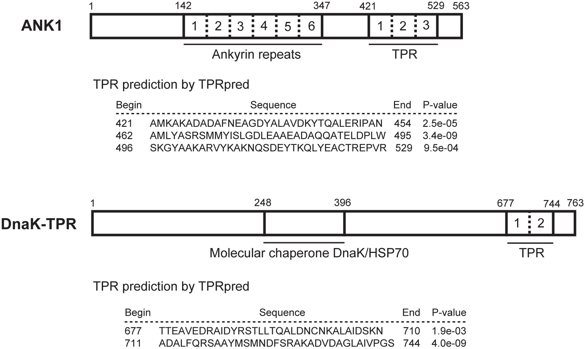
FIGURE 1. Functional domains within ANK1 and DnaK-TPR proteins. The amino acid sequences of ANK1 and DnaK-TPR were subject to BLAST search, as well as motif scanning against Pfam and PROSITE databases. TPR domains were predicted by the online tool TPRpred. Numbers above the boxes indicate the amino acid positions. Different ankyrin repeats or TPR motifs within a protein were separated by dashed lines and individually numbered.
Differential Expression of ANK1 and DnaK-TPR at the Tachyzoite and Bradyzoite Stages
Previous studies (Friesen et al., 2008; Ueno et al., 2011), as well as gene expressing data (Micro-array and RNA-Seq) from ToxoDB, all suggested that the expression of ANK1 and DnaK-TPR was significantly different between the tachyzoite and bradyzoite stages. To quantitatively analyze the expression differences of these two genes between the tachyzoite and bradyzoite stages, real-time PCR (RT-PCR) was performed, using cDNA reverse transcribed from total RNA of tachyzoites or in vitro induced bradyzoites of the ME49 strain. Bradyzoites were obtained by growing parasites in differentiation medium for 4 days with ambient CO2. Using GAPDH expression levels as reference, RT-PCR results showed that expression of ANK1 was barely detectable at the tachyzoite stage, but increased almost 500-fold when cultured under bradyzoite inducing conditions (Figure 2). Similarly, there was a 16-fold increase in the transcript level of DnaK-TPR upon bradyzoite induction (Figure 2). Therefore, expressions of both ANK1 and DnaK-TPR were indeed significantly up-regulated upon bradyzoite induction, consistent with the data from ToxoDB and previous studies (Friesen et al., 2008; Ueno et al., 2011). In addition, using alkaline media or CO2 starvation to treat the parasites, the Roos lab at University of Pennsylvania looked at gene expression changes every 6 h upon bradyzoite induction. Their data deposited in ToxoDB suggest that obvious increase in DnaK-TPR expression was observed 36 h post either treatment. Whereas for ANK1, rise in expression was seen as early as 12 h upon alkaline stress, but did not become apparent until 36 h after CO2 starvation. These results imply that both ANK1 and DnaK-TPR are early induced genes during bradyzoite conversion.
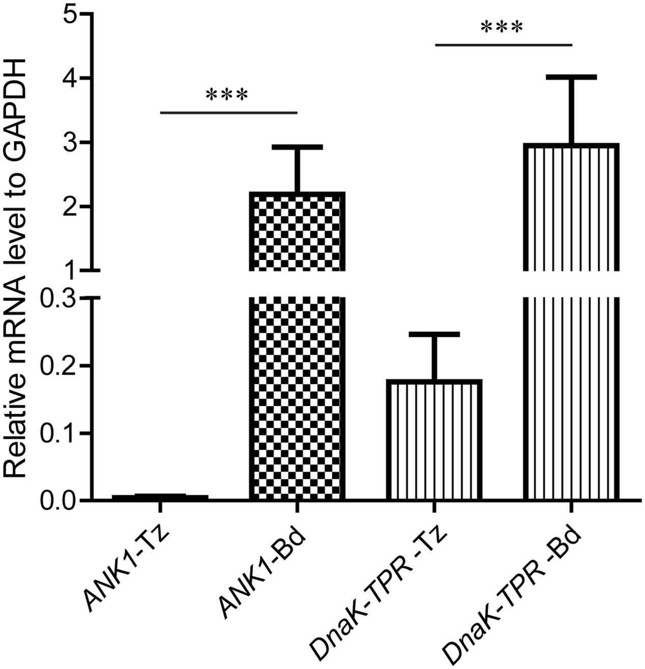
FIGURE 2. Relative expression levels of ANK1 and DnaK-TPR in Toxoplasma tachyzoites and bradyzoites. Total RNA was extracted from 107 tachyzoites or alkaline induced bradyzoites of ME49 and reverse transcribed into cDNA. Transcript levels for ANK1 and DnaK-TPR in each sample were estimated by quantitative real-time PCR, using GAPDH as an internal reference. Means ± SD of three independent experiments. ∗∗∗P < 0.0001, Student’s t-test. Tz, tachyzoites; Bd, bradyzoites.
Generation of ANK1 or DnaK-TPR Knockout Strains Using the CRISPR/CAS9 System
To study the functions of ANK1 and DnaK-TPR during T. gondii growth and development, the CRISPR/CAS9 genome editing technique was used to delete these genes in type 2 strain ME49 and type 1 strain TgHB2. The strategy for deleting target genes by CRISPR/CAS9 mediated homologous recombination was illustrated in Figure 3A. After transfecting the gene specific CRISPR plasmid and homology template into corresponding parental strain (ME49 or TgHB2), transfectants were selected with pyrimethamine and single cloned by limiting dilution. Single clones were then screened by diagnostic PCR to check the correct replacement of target genes by the selection marker DHFR (PCR1/2/3 in Figure 3A). Using this strategy, ANK1 and DnaK-TPR single deletion mutants were generated in both ME49 and TgHB2. Diagnostic PCR results for one clone of ME49 Δank1 and one clone of ME49 ΔdnaK-tpr were shown in Figures 3B,C, respectively. Results for the same mutants produced in TgHB2 were identical to that made in ME49, therefore were not shown here. To further confirm the successful knockout of these genes, we performed whole genome sequencing on the two mutants made in the TgHB2 background. For both TgHB2 Δank1 and TgHB2 ΔdnaK-tpr, complete absence of target gene was observed (Figure 4). Meanwhile, we detected increased sequencing coverage for the exons of the DHFR-TS gene, which corresponds to the increased copy number of DHFR as a consequence of replacing target genes with DHFR (Figure 4). Together, these results confirmed the complete deletion of the ANK1 and DnaK-TPR genes, successful disruption of these genes indicated that neither gene is required for tachyzoite survival, which is consistent with their low expression at the tachyzoite stage.
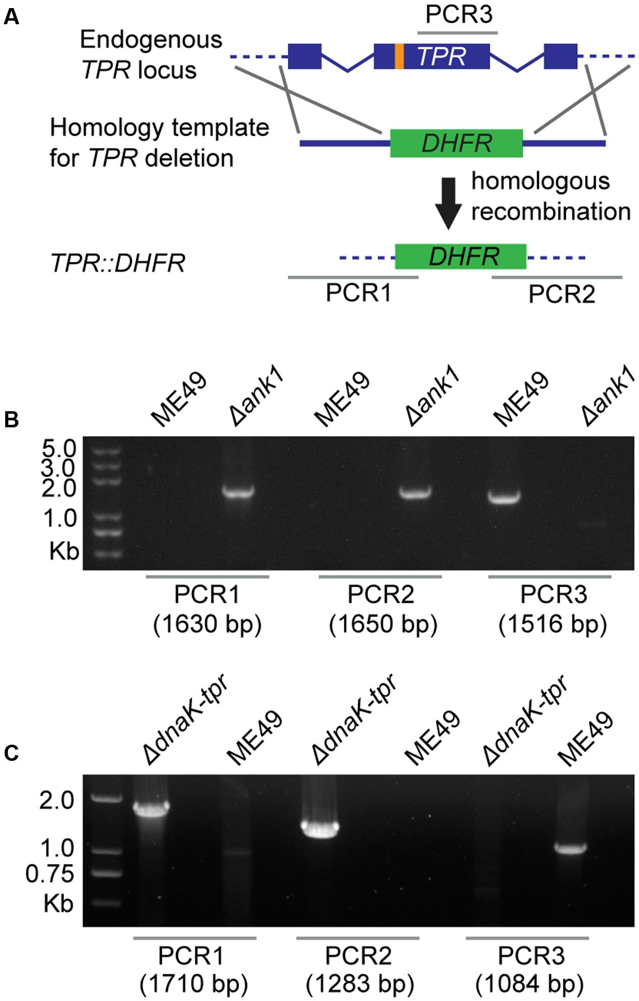
FIGURE 3. Deletion of ANK1 and DnaK-TPR using the CRISPR/CAS9 technology. (A) Schematic illustration of knocking out ANK1 or DnaK-TPR genes (TPR) by CRISPR/CAS9 directed homologous gene replacements. Orange bar indicates the CRISPR targeting site, whereas PCR1/2/3 denote the diagnostic PCRs for knockout strain identification used in (B,C). PCR1 and PCR2 check the 5′ and 3′ integration of the pyrimethamine resistant marker DHFR, whereas PCR3 examines the deletion of target genes. (B) Diagnostic PCRs on a ME49 Δank1 clone. (C) Diagnostic PCRs on a ME49 ΔdnaK-tpr clone.
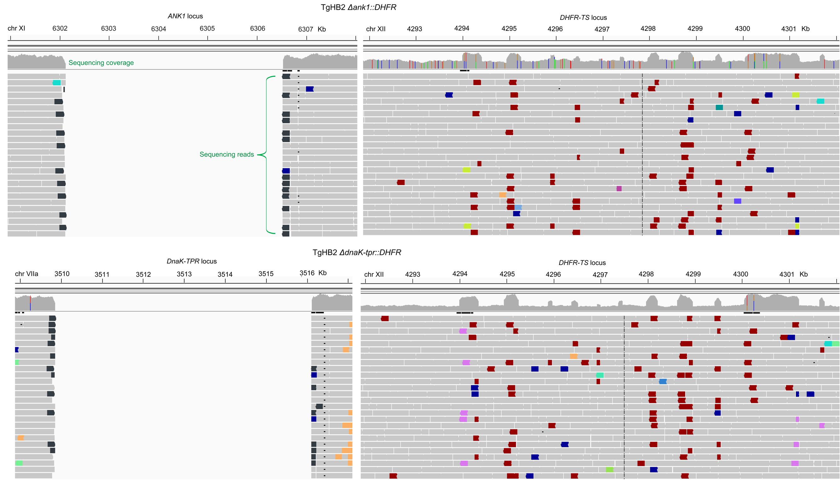
FIGURE 4. Confirm gene deletions by whole genome sequencing. Genomic DNA was extracted from tachyzoites of TgHB2 Δank1 or TgHB2 ΔdnaK-tpr and subject to genome sequencing. Subsequently the clean reads were mapped to the reference genome of the GT1 strain and visualized by the Integrative Genomics Viewer. The numbers above each image indicate the chromosomal position of the corresponding gene in GT1.
Impact of ANK1 and DnaK-TPR on Tachyzoite Growth in Vitro
After obtaining the knockout mutants, we sought to determine the contribution of these genes to the growth of T. gondii tachyzoites in detail. First, plaque assay was used to assess the overall growth rates of these mutants. As shown in Figure 5, after 10 or 12 days’ growth, both ANK1 and DnaK-TPR deletion mutants formed similar number and sizes of plaques on HFF host cell monolayer as the parental strain did. Mutants made in both ME49 and TgHB2 produced plaques with similar efficiencies as their corresponding parental strain. These results suggested that neither ANK1 nor DnaK-TPR had a significant impact on tachyzoite growth in vitro.

FIGURE 5. Growth of ANK1 or DnaK-TPR knockouts as tachyzoites in vitro determined by plaque assay. HFF monolayers infected with 100 tachyzoites of indicated strains were cultured for 10 or 12 days at 37°C, fixed with 70% ethanol, and then stained with crystal violet to develop the plaques.
To further check the parasite growth in vitro, intracellular replication assay was performed to check the proliferation efficiency. Tachyzoites of each strain were used to infect HFF cells for 1 h and successfully invaded parasites were allowed to replicate for 24 h under standard growth conditions. Then the number of parasites in each parasitophorous vacuole (PV) was determined by immunofluorescent staining of Toxoplasma aldolase (Figure 6A). The results showed that both ANK1 or DnaK-TPR deletion mutants displayed very similar replication dynamics as the parental strains (Figures 6B,C), suggesting that ANK1 and DnaK-TPR do not play critical roles for tachyzoite replication in vitro.
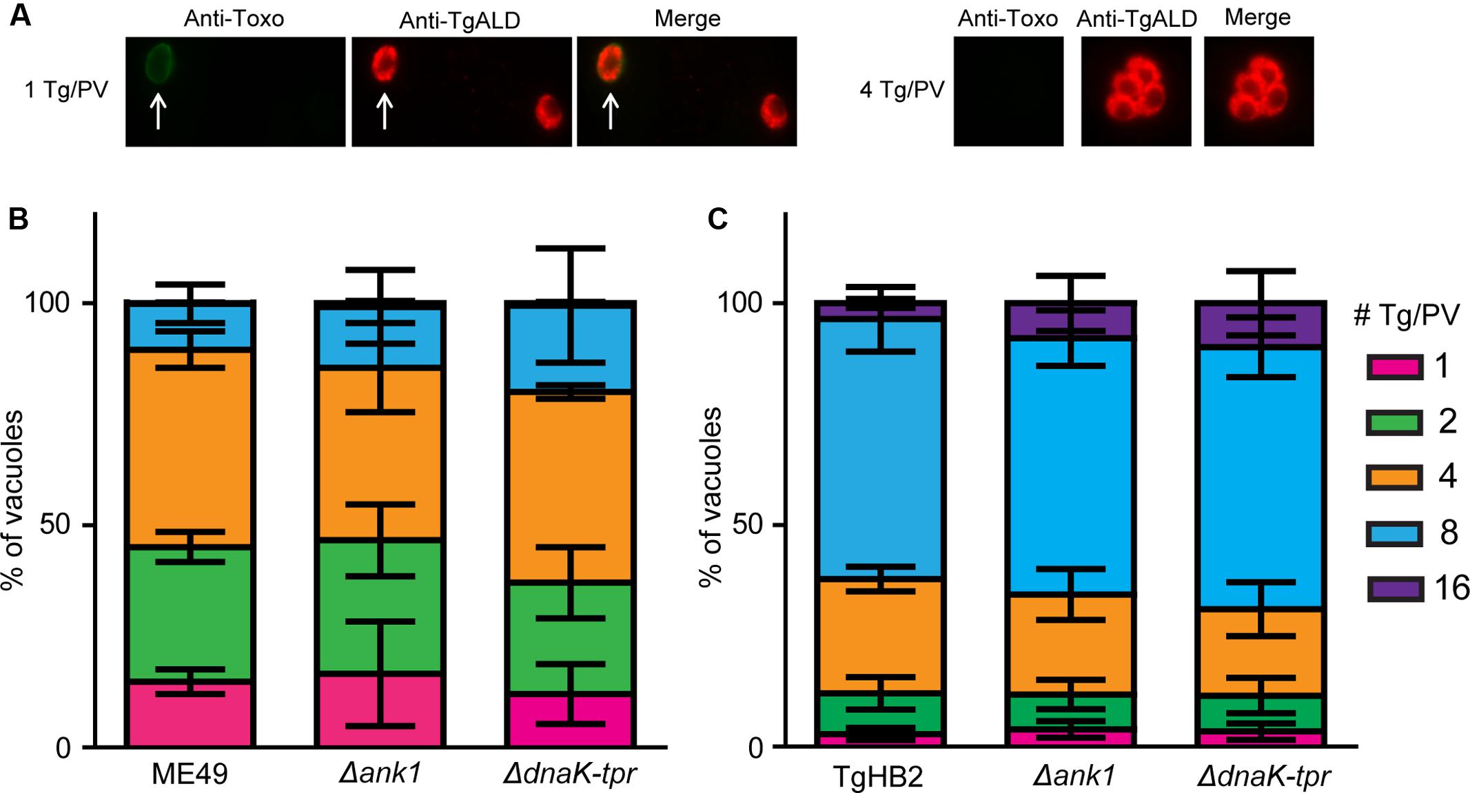
FIGURE 6. Intracellular replication of ANK1 and DnaK-TPR single deletion mutants in vitro. Freshly egressed tachyzoites purified from indicated strains were used to infect HFF monolayer and invaded parasites were cultured for 24 h before fluorescent staining to determine the number of parasites in each parasitophorous vacuole (PV). Swine anti-Toxoplasma (Anti-Toxo) was used to stain extracellular parasites, which were excluded from the intracellular replication analysis. Rabbit anti-TgALD was used to stain total parasites. (A) Examples of PV containing 1 or 4 tachyzoites. The white arrow indicated an extracellular parasite, such parasites were not included in analysis. (B,C) Percentiles of PV containing indicated number of parasites for wild type and mutant strains. Means ± SD of three independent experiments, each with triplicates.
Neither ANK1 nor DnaK-TPR Inactivation Affected Bradyzoite Formation in Vitro
As genes whose expressions were significantly up-regulated at the bradyzoite stage, they were hypothesized to play roles in bradyzoite differentiation or development. To check these possibilities, mutant strains were induced with alkaline conditions to form bradyzoites in vitro, which were detected by positive staining of a bradyzoite marker BAG1 (Figure 7A). After growing parasites under the differentiation condition for 4 days, about 80% of ME49 PV turned BAG1 positive, indicating a high efficiency of bradyzoite transition (Figure 7B). ME49 Δank1 and ME49 ΔdnaK-tpr displayed very similar transition rates as the parental strain ME49. Under this condition, about 70% of the TgHB2 PV became BAG1 positive (Figure 7B), which is slightly lower than that of ME49, consistent with the type 1 nature of this strain. Nonetheless, ANK1 or DnaK-TPR deletion mutants made in this strain also had similar bradyzoite formation efficiencies as the parental strain, suggesting that indeed these two genes did not have significant roles in bradyzoite formation in vitro.
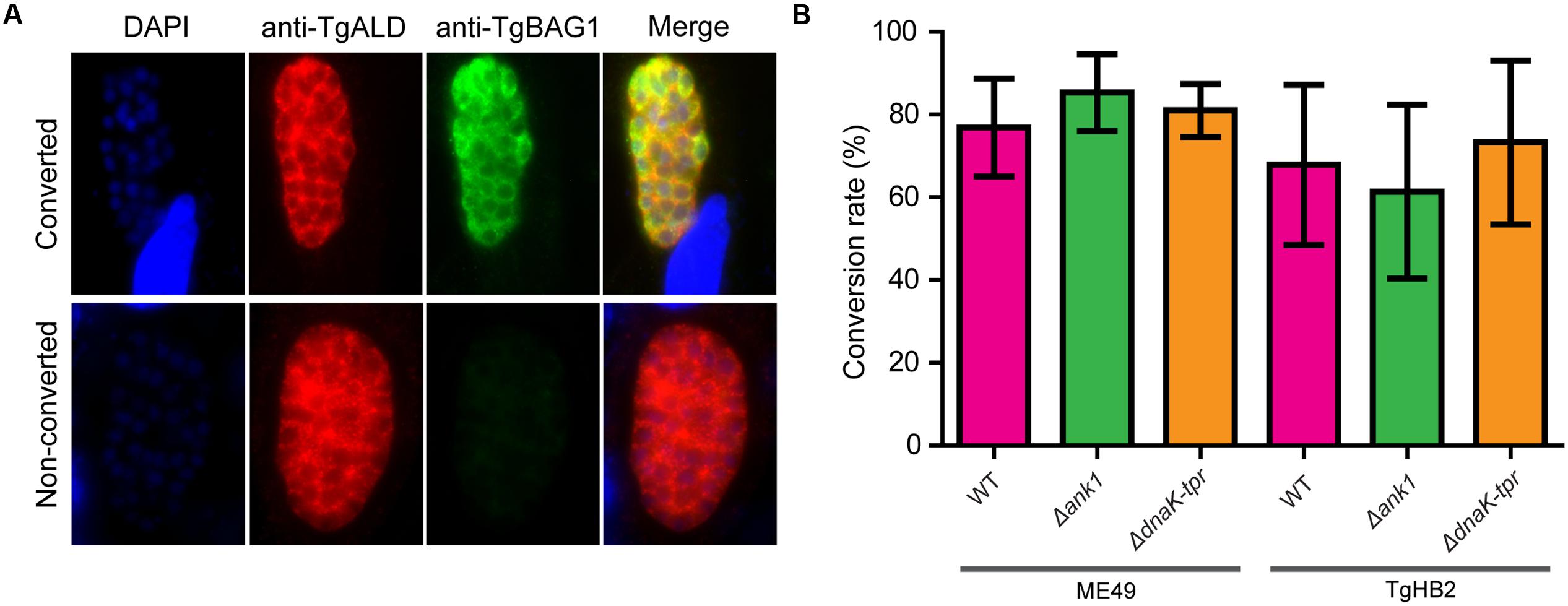
FIGURE 7. Bradyzoite differentiation efficiency of the ANK1 or DnaK-TPR knockout mutants in vitro. HFF monolayers infected with indicated parasite strains were cultured under alkaline conditions (pH 8.2, ambient CO2) for 4 days. Samples were then fixed and stained with mouse anti-BAG1 (a bradyzoite specific marker) and rabbit anti-TgALD (stain of total parasites), to calculate the efficiency of bradyzoite differentiation (# of BAG1+ vacuoles/# of ALD+ vacuoles). (A) Examples of parasites that were converted (BAG1+) or not converted (BAG1-) to bradyzoites. (B) Bradyzoite conversion rates of indicated strains, mean ± SD of three independent experiments.
Both ANK1 and DnaK-TPR Single Deletions Slightly Attenuated Parasite Virulence in Mice
To check the effect of ANK1 or DnaK-TPR disruption on parasite virulence in vivo, tachyzoites of mutant strains were used to infect ICR mice and the survival of mice were monitored. The results for the ME49 serials of strains (ME49, ME49 Δank1, Me49 ΔdnaK-tpr) were shown in Figure 8. At the infection dose of 100 tachyzoites per mouse, the parental strain ME49 killed all the mice, whereas both ANK1 and DnaK-TPR single deletion mutants killed about 70% of infected mice, suggesting that the virulence of both ANK1 and DnaK-TPR mutants was slightly attenuated in laboratory mice.
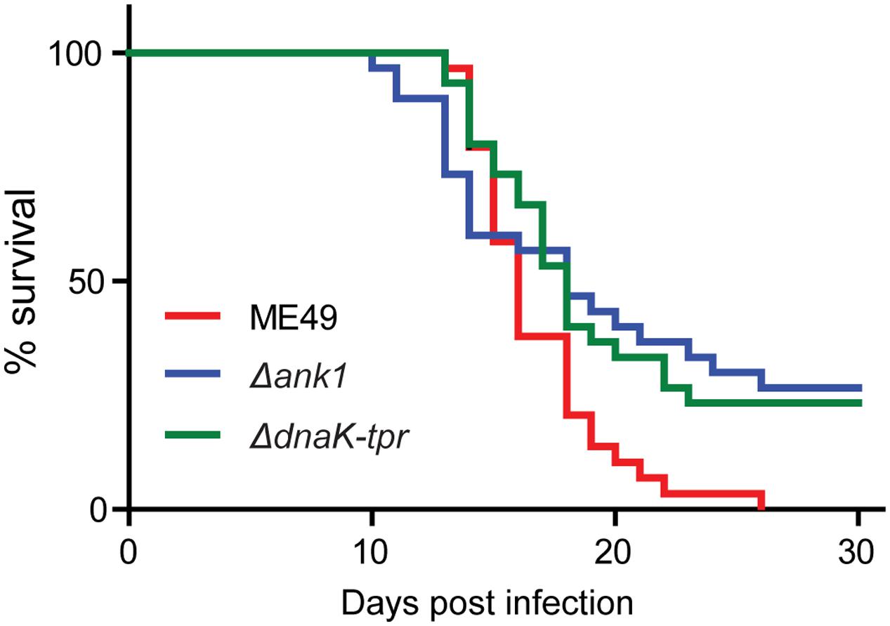
FIGURE 8. Acute virulence of the ANK1 and DnaK-TPR single deletion mutants in mice. ICR mice were infected with freshly egressed tachyzoites (100 parasites/mouse, 40 mice for each strain) of indicated strains by intraperitoneal injection. Subsequently their survival was monitored daily for 30 days.
Since the expression of both ANK1 and DnaK-TPR are dramatically up-regulated in bradyzoites, we sought to determine whether they have any role in chronic infection establishment. To do that, 10–12 weeks old Kunming mice (genetically similar to ICR mice) were infected with 100 tachyzoites of each strain. Survival of mice was monitored daily and the number of cysts in the brain of mice that survived 30 days post infection was estimated. The use of 10–12 weeks old Kunming mice is because typical 4–6 weeks old ICR mice were susceptible to ME49 infection and no survivals could be obtained 30 days post infection. DBA-FITC staining and cyst counting suggested that there was no significant difference in brain cyst formation between the ANK1 or DnaK-TPR knockout mutants and parental strain ME49 (Figure 9), indicating that neither gene directly contributes to chronic infection establishment.
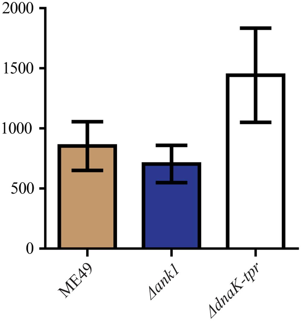
FIGURE 9. Cyst formation of the ANK1 or DnaK-TPR deletion mutants in vivo. Purified tachyzoites from indicated strains were used to infect 10–12 weeks old Kunming mice (100 parasites/mouse) by intraperitoneal injection. Thirty days post infection, mice were sacrificed and the number of Toxoplasma cysts in each brain was determined by DBA-FITC staining and fluorescent microscopy. Mean ± SEM of cyst counting results from 8 mice for each strain.
Discussion
Most of the T. gondii infection cases in animals and humans are chronic infection, where the parasites are encysted as bradyzoites. Under certain circumstances chronic infection can be re-activated and parasites can grow as tachyzoites to cause acute symptoms (Shen et al., 2016; Wohlfert et al., 2017). A large portion of human toxoplasmosis cases are derived from the reactivation of chronic infection, particularly in AIDS and organ transplantation patients (Pereira-Chioccola et al., 2009; Dubey et al., 2012; Meireles et al., 2015). In the wild, chronically infected animals are important sources of T. gondii transmission due to predation. Therefore bradyzoites play key roles in both pathogenesis and transmission of the parasites. However, the biological properties of bradyzoites are poorly understood, very little is known about how they are formed, how they persist and how they get re-activated. Great efforts using different strategies have been paid to compare the gene expression differences between tachyzoites and bradyzoites in vitro (Buchholz et al., 2011), or between acute and chronic infection stages in vivo (Pittman et al., 2014). Studies on these differentially regulated genes provided important insights into the biology of bradyzoite and stage conversion, such as the gene regulation by AP2 factors (White et al., 2014), cyst wall biogenesis (Tomita et al., 2013) and so on. In this study, we focused on two differentially expressed proteins, ANK1 and DnaK-TPR, that have novel TPR motifs involved in protein-protein interactions. Surprisingly, inactivation of either of the two proteins by CRISPR/CAS9 mediated gene knockout did not result in significant bradyzoite formation defect both in vitro and in vivo, although both mutants displayed modest virulence attenuation in mice. These results suggest that not all proteins that are predominantly expressed in bradyzoites are directly involved in bradyzoite formation, but may affect other aspects of the parasites.
One interesting observation from our study is the modest virulence attenuation of the Δank1 and the ΔdnaK-tpr mutants. This is surprising because neither protein is significantly expressed in tachyzoites, therefore their inactivation was not expected to affect the acute virulence of the parasites. The exact mechanisms responsible for the reduced virulence of the mutants are currently unknown but possible explanations include: first, although both ANK1 and DnaK-TPR are drastically up-regulated in bradyzoites, it is possible that they are expressed at very low levels in tachyzoites and the low levels of expression still contribute to the optimal virulence of the parasites. Particularly for DnaK-TPR, its transcript level is about 18% of that of GAPDH at the tachyzoite stage, therefore it is high likely that it is also expressed in tachyzoites, although at relatively low levels. Second, virulence attenuation may be due to indirect effects of gene deletions. Stage specific expression of genes is achieved through complex regulation mechanisms. Loss of the target genes may influence the local chromatin structure and/or the proper function of the regulation mechanisms, which in turn may affect the expression of other genes to alter the virulence properties. Further work is needed to test these possibilities.
Genes that are up or down regulated during bradyzoite conversion serve as good starting points to dissect the complex biology of bradyzoites. However, as the studies accumulate, it has become less clear whether these differentially regulated genes are actively contributing to bradyzoite conversion or their change in expression is just a byproduct of stage conversion. This is particularly true for bradyzoites induced in vitro. For example, the bradyzoite pseudokinase 1 (BPK1) is a component of the cyst wall upregulated in bradyzoites, however, inactivation of this gene did not affect pathogenesis or cyst formation in vitro (Buchholz et al., 2013). Its effect on cyst formation in vivo was also minimal, although it did affect the size and sensitivity of cysts to pepsin-acid (Buchholz et al., 2013). Similarly, deletion of the bradyzoite marker lactate dehydrogenase 2 (LDH2) did not affect bradyzoite transition in vitro (Abdelbaset et al., 2017) or cyst formation in vivo (Xia et al., to be published elsewhere). Disruption of another bradyzoite marker BAG1 also did not affect bradyzoite gene expression in vitro (Bohne et al., 1998; Zhang et al., 1999), nor did it affect cyst formation in vivo as reported in one study (Bohne et al., 1998). In this study when we knocked out either of the two TPR containing proteins (ANK1 and DnaK-TPR), no obvious phenotypes in bradyzoite conversion in vitro or cyst formation in vivo were observed, suggesting that they are not directly involved in chronic infection establishment. Currently it is not clear whether their increased expression in bradyzoites are just byproducts of parasite differentiation, or they actually play roles in other aspects of bradyzoites.
The two proteins we focused in this study were reported before as proteins whose expressions were significantly up-regulated during bradyzoite formation (Friesen et al., 2008; Ueno et al., 2011). But their exact roles were not known at the time of discovery. ANK1 contains an ankyrin repeat domain and a TPR domain. Similar to TPR, ankyrin repeats are also common protein motifs with 30–34 amino acid residues in length and function to mediate protein-protein interactions (Li et al., 2006). Like ANK1, DnaK-TPR also possesses two recognizable domains (DnaK/HSP70 family chaperone domain and TPR domain) that mediate protein-protein interactions. Because of the presence of these novel structural motifs, we initially thought that they might function as scaffolds to mediate protein complex formation during bradyzoite formation. In fact, DnaK-TPR was shown to interact with the p23 co-chaperone in a previous study (Ueno et al., 2011), which confirmed its ability to mediate protein interactions. Nonetheless, individual disruption of these two genes did not cause noticeable changes regarding bradyzoite differentiation and development. Possible reasons include: ANK1 and DnaK-TPR are not directly involved in cyst establishment, but may play roles in the structure integrity or physiological fitness of the cysts, which are not caught in our tests due to limited sensitivity. Alternatively, there are many other TPR containing proteins expressed in the parasites, at both tachyzoite and bradyzoite stages, which may functionally compensate the loss of ANK1 or DnaK-TPR, or ANK1 and DnaK-TPR themselves are functionally redundant. However, although both ANK1 and DnaK-TPR contain TPR motifs, the sequence similarity between them is very limited and TPRs are only small portions of these proteins. Therefore we think the likelihood that they are functionally redundant is low. Nonetheless, the possibilities that they functionally overlap with each other, or with other TPR containing proteins exist. Further work is required to examine these possibilities.
Author Contributions
JY, LZ, and BS conceived and designed the study. JY, LZ, NX, and HD performed the experiments. BS, YZ, and JZ analyzed the data. BS, JY, and LZ wrote the paper.
Funding
This work was supported the National Key R&D Program of China (Project# 2017YFD0500402), National Natural Science Foundation of China (Project# 31572508), the Fundamental Research Funds for the Central Universities in China (Project# 2662015PY104) and The Natural Science Foundation of Hubei Province (Grant# 2017CFA020).
Conflict of Interest Statement
The authors declare that the research was conducted in the absence of any commercial or financial relationships that could be construed as a potential conflict of interest.
Acknowledgments
We thank Drs. Min Hu, Rui Fang, and Lan He from the College of Veterinary Medicine, Huazhong Agricultural University for their thoughtful suggestions during this study. We are also grateful to Mr. Shiwei Zhang from Novogene (Beijing) for his great help on whole genome sequencing analysis.
Footnotes
- ^ https://blast.ncbi.nlm.nih.gov/Blast.cgi
- ^ http://myhits.isb-sib.ch/cgi-bin/motif_scan
- ^ https://toolkit.tuebingen.mpg.de/#/tools/tprpred
References
Abdelbaset, A. E., Fox, B. A., Karram, M. H., Abd Ellah, M. R., Bzik, D. J., and Igarashi, M. (2017). Lactate dehydrogenase in Toxoplasma gondii controls virulence, bradyzoite differentiation, and chronic infection. PLOS ONE 12:e0173745. doi: 10.1371/journal.pone.0173745
Abyzov, A., Urban, A. E., Snyder, M., and Gerstein, M. (2011). CNVnator: an approach to discover, genotype, and characterize typical and atypical CNVs from family and population genome sequencing. Genome Res. 21, 974–984. doi: 10.1101/gr.114876.110
Allan, R. K., and Ratajczak, T. (2011). Versatile TPR domains accommodate different modes of target protein recognition and function. Cell Stress Chaperones 16, 353–367. doi: 10.1007/s12192-010-0248-0
Bohne, W., Heesemann, J., and Gross, U. (1993). Induction of bradyzoite-specific Toxoplasma gondii antigens in gamma interferon-treated mouse macrophages. Infect. Immun. 61, 1141–1145.
Bohne, W., Hunter, C. A., White, M. W., Ferguson, D. J., Gross, U., and Roos, D. S. (1998). Targeted disruption of the bradyzoite-specific gene BAG1 does not prevent tissue cyst formation in Toxoplasma gondii. Mol. Biochem. Parasitol. 92, 291–301. doi: 10.1016/S0166-6851(97)00236-3
Buchholz, K. R., Bowyer, P. W., and Boothroyd, J. C. (2013). Bradyzoite pseudokinase 1 is crucial for efficient oral infectivity of the Toxoplasma gondii tissue cyst. Eukaryot. Cell 12, 399–410. doi: 10.1128/EC.00343-12
Buchholz, K. R., Fritz, H. M., Chen, X., Durbin-Johnson, B., Rocke, D. M., Ferguson, D. J., et al. (2011). Identification of tissue cyst wall components by transcriptome analysis of in vivo and in vitro Toxoplasma gondii bradyzoites. Eukaryot. Cell 10, 1637–1647. doi: 10.1128/EC.05182-11
Cerveny, L., Straskova, A., Dankova, V., Hartlova, A., Ceckova, M., Staud, F., et al. (2013). Tetratricopeptide repeat motifs in the world of bacterial pathogens: role in virulence mechanisms. Infect. Immun. 81, 629–635. doi: 10.1128/IAI.01035-12
Chen, K., Wallis, J. W., McLellan, M. D., Larson, D. E., Kalicki, J. M., Pohl, C. S., et al. (2009). BreakDancer: an algorithm for high-resolution mapping of genomic structural variation. Nat. Methods 6, 677–681. doi: 10.1038/nmeth.1363
Dabritz, H. A., and Conrad, P. A. (2010). Cats and Toxoplasma: implications for public health. Zoonoses Public Health 57, 34–52. doi: 10.1111/j.1863-2378.2009.01273.x
Das, A. K., Cohen, P. W., and Barford, D. (1998). The structure of the tetratricopeptide repeats of protein phosphatase 5: implications for TPR-mediated protein-protein interactions. EMBO J. 17, 1192–1199. doi: 10.1093/emboj/17.5.1192
Dubey, J. P. (1998). Advances in the life cycle of Toxoplasma gondii. Int. J. Parasitol. 28, 1019–1024. doi: 10.1016/S0020-7519(98)00023-X
Dubey, J. P., Lago, E. G., Gennari, S. M., Su, C., and Jones, J. L. (2012). Toxoplasmosis in humans and animals in Brazil: high prevalence, high burden of disease, and epidemiology. Parasitology 139, 1375–1424. doi: 10.1017/S0031182012000765
Dubey, J. P., Lindsay, D. S., and Speer, C. A. (1998). Structures of Toxoplasma gondii tachyzoites, bradyzoites, and sporozoites and biology and development of tissue cysts. Clin. Microbiol. Rev. 11, 267–299.
Elmore, S. A., Jones, J. L., Conrad, P. A., Patton, S., Lindsay, D. S., and Dubey, J. P. (2010). Toxoplasma gondii: epidemiology, feline clinical aspects, and prevention. Trends Parasitol. 26, 190–196. doi: 10.1016/j.pt.2010.01.009
Ferreira da Silva Mda, F., Barbosa, H. S., Gross, U., and Luder, C. G. (2008). Stress-related and spontaneous stage differentiation of Toxoplasma gondii. Mol. Biosyst. 4, 824–834. doi: 10.1039/b800520f
Friesen, J., Fleige, T., Gross, U., and Bohne, W. (2008). Identification of novel bradyzoite-specific Toxoplasma gondii genes with domains for protein-protein interactions by suppression subtractive hybridization. Mol. Biochem. Parasitol. 157, 228–232. doi: 10.1016/j.molbiopara.2007.10.005
Hirano, T., Kinoshita, N., Morikawa, K., and Yanagida, M. (1990). Snap helix with knob and hole: essential repeats in S. pombe nuclear protein nuc2+. Cell 60, 319–328.
Karpenahalli, M. R., Lupas, A. N., and Soding, J. (2007). TPRpred: a tool for prediction of TPR-, PPR- and SEL1-like repeats from protein sequences. BMC Bioinformatics 8:2. doi: 10.1186/1471-2105-8-2
Li, H., Handsaker, B., Wysoker, A., Fennell, T., Ruan, J., Homer, N., et al. (2009). The sequence alignment/map format and SAMtools. Bioinformatics 25, 2078–2079. doi: 10.1093/bioinformatics/btp352
Li, J., Mahajan, A., and Tsai, M. D. (2006). Ankyrin repeat: a unique motif mediating protein-protein interactions. Biochemistry 45, 15168–15178. doi: 10.1021/bi062188q
Manger, I. D., Hehl, A., Parmley, S., Sibley, L. D., Marra, M., Hillier, L., et al. (1998). Expressed sequence tag analysis of the bradyzoite stage of Toxoplasma gondii: identification of developmentally regulated genes. Infect. Immun. 66, 1632–1637.
Meireles, L. R., Ekman, C. C., Andrade, R. H. Jr., and Luna, E. J. (2015). Human toxoplasmosis outbreaks and the agent infecting form. Findings from a systematic review. Rev. Inst. Med. Trop. Sao Paulo 57, 369–376. doi: 10.1590/S0036-46652015000500001
Mouveaux, T., Oria, G., Werkmeister, E., Slomianny, C., Fox, B. A., Bzik, D. J., et al. (2014). Nuclear glycolytic enzyme enolase of Toxoplasma gondii functions as a transcriptional regulator. PLOS ONE 9:e105820. doi: 10.1371/journal.pone.0105820
Pereira-Chioccola, V. L., Vidal, J. E., and Su, C. (2009). Toxoplasma gondii infection and cerebral toxoplasmosis in HIV-infected patients. Future Microbiol. 4, 1363–1379. doi: 10.2217/fmb.09.89
Pittman, K. J., Aliota, M. T., and Knoll, L. J. (2014). Dual transcriptional profiling of mice and Toxoplasma gondii during acute and chronic infection. BMC Genomics 15:806. doi: 10.1186/1471-2164-15-806
Remington, J. S., and Merigan, T. C. (1968). Interferon: protection of cells infected with an intracellular protozoan (Toxoplasma gondii). Science 161, 804–806. doi: 10.1126/science.161.3843.804
Selseleh, M., Modarressi, M. H., Mohebali, M., Shojaee, S., Eshragian, M. R., Selseleh, M., et al. (2012). Real-time RT-PCR on SAG1 and BAG1 gene expression during stage conversion in immunosuppressed mice infected with Toxoplasma gondii Tehran strain. Korean J. Parasitol. 50, 199–205. doi: 10.3347/kjp.2012.50.3.199
Shen, B., Brown, K., Long, S., and Sibley, L. D. (2017). Development of CRISPR/Cas9 for efficient genome editing in Toxoplasma gondii. Methods Mol. Biol. 1498, 79–103. doi: 10.1007/978-1-4939-6472-7_6
Shen, B., Brown, K. M., Lee, T. D., and Sibley, L. D. (2014). Efficient gene disruption in diverse strains of Toxoplasma gondii using CRISPR/CAS9. mBio 5:e01114-14. doi: 10.1128/mBio.01114-14
Shen, B., and Sibley, L. D. (2014). Toxoplasma aldolase is required for metabolism but dispensable for host-cell invasion. Proc. Natl. Acad. Sci. U.S.A. 111, 3567–3572. doi: 10.1073/pnas.1315156111
Shen, B., Yuan, Y., Cheng, J., Pan, M., Xia, N., Zhang, W., et al. (2016). Activation of chronic toxoplasmosis by transportation stress in a mouse model. Oncotarget 7, 87351–87360. doi: 10.18632/oncotarget.13568
Sikorski, R. S., Boguski, M. S., Goebl, M., and Hieter, P. (1990). A repeating amino acid motif in CDC23 defines a family of proteins and a new relationship among genes required for mitosis and RNA synthesis. Cell 60, 307–317. doi: 10.1016/0092-8674(90)90745-Z
Skariah, S., McIntyre, M. K., and Mordue, D. G. (2010). Toxoplasma gondii: determinants of tachyzoite to bradyzoite conversion. Parasitol. Res. 107, 253–260. doi: 10.1007/s00436-010-1899-6
Soete, M., Camus, D., and Dubremetz, J. F. (1994). Experimental induction of bradyzoite-specific antigen expression and cyst formation by the RH strain of Toxoplasma gondii in vitro. Exp. Parasitol. 78, 361–370. doi: 10.1006/expr.1994.1039
Thorvaldsdottir, H., Robinson, J. T., and Mesirov, J. P. (2013). Integrative Genomics Viewer (IGV): high-performance genomics data visualization and exploration. Brief Bioinform. 14, 178–192. doi: 10.1093/bib/bbs017
Tomita, T., Bzik, D. J., Ma, Y. F., Fox, B. A., Markillie, L. M., Taylor, R. C., et al. (2013). The Toxoplasma gondii cyst wall protein CST1 is critical for cyst wall integrity and promotes bradyzoite persistence. PLOS Pathog. 9:e1003823. doi: 10.1371/journal.ppat.1003823
Ueno, A., Dautu, G., Haga, K., Munyaka, B., Carmen, G., Kobayashi, Y., et al. (2011). Toxoplasma gondii: a bradyzoite-specific DnaK-tetratricopeptide repeat (DnaK-TPR) protein interacts with p23 co-chaperone protein. Exp. Parasitol. 127, 795–803. doi: 10.1016/j.exppara.2011.01.015
Walker, R., Gissot, M., Croken, M. M., Huot, L., Hot, D., Kim, K., et al. (2013). The Toxoplasma nuclear factor TgAP2XI-4 controls bradyzoite gene expression and cyst formation. Mol. Microbiol. 87, 641–655. doi: 10.1111/mmi.12121
Weiss, L. M., and Kim, K. (2000). The development and biology of bradyzoites of Toxoplasma gondii. Front. Biosci. 5, D391–D405.
Weiss, L. M., Laplace, D., Takvorian, P. M., Tanowitz, H. B., Cali, A., and Wittner, M. (1995). A cell culture system for study of the development of Toxoplasma gondii bradyzoites. J. Eukaryot. Microbiol. 42, 150–157. doi: 10.1111/j.1550-7408.1995.tb01556.x
White, M. W., Radke, J. R., and Radke, J. B. (2014). Toxoplasma development - turn the switch on or off?. Cell Microbiol. 16, 466–472. doi: 10.1111/cmi.12267
Wohlfert, E. A., Blader, I. J., and Wilson, E. H. (2017). Brains and brawn: toxoplasma infections of the central nervous system and skeletal muscle. Trends Parasitol. 33, 519–531. doi: 10.1016/j.pt.2017.04.001
Keywords: Toxoplasma gondii, bradyzoite, ANK1, DnaK-TPR, stage conversion
Citation: Yang J, Zhang L, Diao H, Xia N, Zhou Y, Zhao J and Shen B (2017) ANK1 and DnaK-TPR, Two Tetratricopeptide Repeat-Containing Proteins Primarily Expressed in Toxoplasma Bradyzoites, Do Not Contribute to Bradyzoite Differentiation. Front. Microbiol. 8:2210. doi: 10.3389/fmicb.2017.02210
Received: 15 June 2017; Accepted: 26 October 2017;
Published: 13 November 2017.
Edited by:
Dongsheng Zhou, Beijing Institute of Microbiology and Epidemiology, ChinaReviewed by:
Shuchao Wang, Academy of Military Medical Sciences, ChinaJozsef Soki, University of Szeged, Hungary
João Marcelo Pereira Alves, University of São Paulo, Brazil
Izabela Marques Dourado Bastos, University of Brasília, Brazil
Daniel Adesse, Fundação Oswaldo Cruz (Fiocruz), Brazil
Copyright © 2017 Yang, Zhang, Diao, Xia, Zhou, Zhao and Shen. This is an open-access article distributed under the terms of the Creative Commons Attribution License (CC BY). The use, distribution or reproduction in other forums is permitted, provided the original author(s) or licensor are credited and that the original publication in this journal is cited, in accordance with accepted academic practice. No use, distribution or reproduction is permitted which does not comply with these terms.
*Correspondence: Bang Shen, c2hlbmJhbmdAbWFpbC5oemF1LmVkdS5jbg==
†These authors have contributed equally to this work.
 Jichao Yang1†
 Yanqin Zhou
 Junlong Zhao
 Bang Shen