- 1Department of Translational Research and New Technologies in Medicine and Surgery, University of Pisa, Pisa, Italy
- 2Department of Biology, University of Pisa, Pisa, Italy
- 3Research Center Nutraceuticals and Food for Health-Nutrafood, University of Pisa, Pisa, Italy
Besides sporulation, Bacillus cereus can undergo a differentiation process in which short swimmer cells become elongated and hyperflagellated swarmer cells that favor migration of the bacterial community on a surface. The functionally enigmatic flagellar protein FlhF, which is the third paralog of the signal recognition particle (SRP) GTPases Ffh and FtsY, is required for swarming in many bacteria. Previous data showed that FlhF is involved in the control of the number and positioning of flagella in B. cereus. In this study, in silico analysis of B. cereus FlhF revealed that this protein presents conserved domains that are typical of SRPs in many organisms and a peculiar N-terminal basic domain. By proteomic analysis, a significant effect of FlhF depletion on the amount of secreted proteins was found with some proteins increased (e.g., B component of the non-hemolytic enterotoxin, cereolysin O, enolase) and others reduced (e.g., flagellin, L2 component of hemolysin BL, bacillolysin, sphingomyelinase, PC-PLC, PI-PLC, cytotoxin K) in the extracellular proteome of a ΔflhF mutant. Deprivation of FlhF also resulted in significant attenuation in the pathogenicity of this strain in an experimental model of infection in Galleria mellonella larvae. Our work highlights the multifunctional role of FlhF in B. cereus, being this protein involved in bacterial flagellation, swarming, protein secretion, and pathogenicity.
Introduction
Bacillus cereus is a Gram-positive, motile, spore-bearing rod, frequently isolated from the soil, where the spore ensures its persistence under adverse conditions. Long known as agent of food-borne diseases, this organism is now recognized to be able to cause local and systemic infections in humans (Bottone, 2010; Logan, 2012; Celandroni et al., 2016). The pathogenic potential of this bacterium is related to the secretion of several virulence proteins, e.g., hemolysins, phospholipases, trimeric toxins (hemolysin BL, HBL; non-hemolytic enterotoxin, NHE), cytotoxin K (CytK), proteases (Senesi and Ghelardi, 2010; Ramarao and Sanchis, 2013; Jeßberger et al., 2015), and to motility modes, such as swimming and swarming (Senesi et al., 2010; Celandroni et al., 2016). Bacterial swarming is a flagellum-driven social form of locomotion in which cells undergo a periodical differentiation process leading to the production of long and hyperflagellated elements, the swarmer cells, which coordinately migrate across surfaces (Kearns, 2010; Partridge and Harshey, 2013). Swarming confers an advantage for the colonization of natural and host surfaces and can contribute to bacterial virulence. Notably, swarming increases HBL secretion by B. cereus (Ghelardi et al., 2007) and enhances the pathogenicity of this bacterium in an experimental endophthalmitis model (Callegan et al., 2006).
In a previous study, we demonstrated that the protein FlhF plays a major role in controlling the arrangement of flagella in B. cereus (Salvetti et al., 2007). The proteins FlhF and FlhG are essential for establishing correct place and quantity of flagella in many but not all bacterial species (Schniederberend et al., 2013). In Bacillus subtilis, FlhF and FlhG behave as two antagonistic proteins regulating the pattern of flagellar basal body formation to control flagella localization, but they do not influence the number of flagella on the cell surface (Guttenplan et al., 2013). In B. cereus, no FlhG homologue was found and the lack of FlhF, other than leading to mislocalization of flagella, causes a reduction in the number of flagella and reduces swimming migration (Salvetti et al., 2007).
FlhF belongs to the GTP-binding signal recognition particle (SRP) family, which also includes Ffh and FtsY (Bange et al., 2007; Schuhmacher et al., 2015). SRP-GTPases are required for the cotranslational targeting of many proteins to the membrane through the recognition and binding of their N-terminus signal peptide during protein synthesis. Nevertheless, FlhF appeared not to be involved in protein secretion mechanisms in B. subtilis (Zanen et al., 2004). Differently, in Pseudomonas putida and B. cereus, the FlhF depletion altered the profile of secreted proteins (Pandza et al., 2000; Salvetti et al., 2007). In particular, a ΔflhF mutant of B. cereus showed an increase in the extracellular levels of NHE and a decrease in HBL and phosphatidyl-choline specific phospholipase C (PC-PLC) (Salvetti et al., 2007). Thus, the aim of the present study was to gain more insight into the function of FlhF in B. cereus by evaluating the effects of FlhF depletion on interconnected cellular functions such as swarming, protein secretion, and virulence, which may all depend from protein targeting to the membrane.
Materials and Methods
Bacterial Strains and Growth Conditions
Bacillus cereus ATCC 14579 wild type (wt), its flhF (GeneBank ID: Bc1670) mutant derivative (ΔflhF, MP06), and complemented strain (ΔflhF-comp, MP07) (Salvetti et al., 2007) stocks from -80° C were streaked on brain heart infusion supplemented with 0.1% (w/v) glucose (BHIG; Becton Dickinson, Cockeysville, NJ, USA) plates and incubated at 37° C. BHIG was also used for liquid cultures. When required, 5 μg/ml erythromycin for strain MP06 or 30 μg/ml kanamycin for strain MP07 were added to the media. MP07 cultures were also supplemented with 4 mM isopropil-β-D-1-tiogalattopiranoside (IPTG; Sigma–Aldrich, St. Louis, MO, USA) in order to induce pspac dependent gene expression.
In silico Analysis
BLAST1 was used for comparative analysis of nucleotide and protein sequences. Protein sequences in the FASTA format were retrieved from the UniProt database2 (The UniProt Consortium, 2015). Functional domain analysis was performed using the ProDom Server3 (Bru et al., 2005). The presumptive secondary and tridimensional structure of proteins were generated using the Phyre2 web portal for protein modeling, prediction and analysis4 (Kelley et al., 2015) and the Raptor X Structure Prediction Server5 (Källberg et al., 2012), respectively.
Swarming Motility
For each experiment, swarm plates (TrA plates; 1% tryptone, 0.5% NaCl, 0.7% granulated agar) were prepared fresh daily and allowed to sit at room temperature overnight before use (Salvetti et al., 2011). Swarming was initiated by spotting 50 μl of a culture containing approximately 2×10⁴ cells/ml onto the center of TrA plates, and incubating cultures at 37° C. Swarming migration was evaluated by measuring colony diameters after 8 h. Since B. cereus flagella are very fragile, bacterial samples were taken by slide overlay of single agar blocks (5 mm ×5 mm) that contained different colony portions. Bacterial cells were stained with tannic acid and silver nitrate (Harshey and Matsuyama, 1994) for microscopy. Several samples were analyzed at 1000× magnification using an optical microscope (BH-2; Olympus, Tokyo, Japan). All experiments were performed in duplicate in three separate days.
Preparation of Culture Supernatants
Protein samples were prepared by growing bacterial cells to the late exponential growth phase in BHIG at 200 rpm for 6 h at 37° C. Culture supernatants were collected by high-speed centrifugation (10000 × g), added with the serine protease inhibitor phenylmethylsulfonyl fluoride 1 mM (PMSF; Sigma–Aldrich), and stored at -80° C until use. Protein concentration in supernatants was determined by the bicinchoninic acid (BCA) assay (Smith et al., 1985), using bovine serum albumin as a standard. The activity of fructose-1,6-bisphosphate aldolase in culture supernatants was spectrophotometrically measured at 30° C by following the rate of NADH oxidation at 340 nm according to the method described by Warth (1980).
Two-Dimensional Electrophoresis (2-DE)
Proteins contained in supernatants were precipitated using the TCA method (trichloroacetic acid, Sigma–Aldrich). Precipitated proteins were washed eight-times with cold 96% (v/v) ethanol, air-dried and suspended in sample rehydration buffer (7 M urea, 2 M thiourea, 4% CHAPS, 2.4% aminosulfobetaine-14; Invitrogen, Carlsbad, CA, USA). ZOOM® IPG strips with a linear pH range of 4–7 (Invitrogen) were rehydrated for 16 h in 400 μl of sample rehydration buffer and 10 μg of protein samples were subsequently added by cup loading. Focusing was carried out at 200 V for 20 min, 450 V for 15 min, 750 V for 15 min, and 2000 V for 30 min using the Xcell SureLockTM Mini-Cell system (Invitrogen). The IPG strips were then equilibrated for 10 min in 5 ml of equilibration solution (LDS 1X; DTT 0.5 mM). For the second dimension, samples were run at 200 V for 35 min on 4–12% gradient SDS-PAGE gel (Bis-Tris ZOOMTM Gel, 1.0 mm IPG-well; Invitrogen). Three independent biological experiments were performed in separate days. Gels were silver or Coomassie Blue (Sigma–Aldrich) stained, according to standard procedures. Gels were scanned at 300 dpi and 8 bits depth on an Image Scanner equipped with a film-scanning unit and analyzed with the Image-Master 2D Platinum v.6 software (GE Healthcare, Little Chalfont, UK). The spots were quantified after normalization and spot volumes (pixel intensity × area) were expressed as percentage of the total volume of the spots on the gel. Presumptive analysis of protein gels was performed by comparison of silver stained 2-DE gels with gels available in the literature (Gohar et al., 2002, 2005) and then protein spots were identified by spot excision from Coomassie-Blue stained gels and subsequent identification. To this purpose, excided spots were digested with 0.1–0.5 μg of trypsin at 37° C for 6 h (Shevchenko et al., 1996). Digested proteins were analyzed by liquid chromatography coupled with tandem mass spectrometry (LC-MS/MS) at Université de Genève, Proteomics Core Facility (Genève, CH). Database searches were performed with the Mascot Server (Matrix Science Ltd; version 04_20146) and results were analyzed and validated using the Scaffold software (Proteome Software Inc.). Searches were performed using trypsin for the fragments cleavage, allowing up to one trypsin miscleavage and without restriction on protein mass or isoelectric point (pI). Fixed modifications included carbamidomethylation of cysteine, while variable modifications comprised oxidation of methionine. Peptide masses ranged from 849.0 m/z to 4001.0 m/z with a charge (z) of 1 +. The mass tolerance was set to ± 25 ppm. Significance was established according to expectancy (e) value transformed into MS score (with significance at a P-value <0.05 at scores over 70), percentage coverage and theoretical pI and molecular weight (Mw) compared to the approximate experimental values observed on 2-DE gels.
Identified proteins were classified based on their biological functions using the Kyoto Encyclopedia of Genes and Genomes (KEGG) database resource7. Protein sequences were analyzed using the SIGNALIP 4.1 Server8, TATP 1.09, SecretomeP 2.0 Server10 and TMpred program11 in order to evaluate the presence of Sec-type signals, Tat-type signals, non-classically secretion signals or transmembrane domains, respectively.
Insects and Infection Experiments
Galleria mellonella larvae, obtained from Mous Live Bait (Balk, Netherland), were selected by weight (0.2–0.3 g) and absence of dark spots on the cuticle. Bacteria were grown to the late exponential growth phase in BHIG at 37° C for 6 h and harvested by centrifugation at 5000 × g for 10 min. Bacterial suspensions were prepared in phosphate buffered saline (PBS: 1 M KH2PO4, 1 M K2HPO4, 5 M NaCl, pH 7.2) and bacteria counted using a hemocytometer. Three infectious doses (about 103, 102, and 101 CFU per larva) were used to infect three groups of 20 larvae. Larva abdomen was accurately cleaned with 70% ethanol and 10 μl of bacterial suspension was injected into the hemocoel through the last right pro-leg, with a sterile Hamilton syringe (Sigma–Aldrich) via a 26-gage needle. A control group of larvae was injected with PBS only. Infectious doses were checked by CFU count after plating appropriate dilutions and incubating 16 h at 37°C. Infected larvae were kept at 37°C and mortality was recorded after 24 h. Each experiment was performed three times in separate days.
To assess bacterial ability to multiply in vivo, groups of 20 animals were infected with 104 CFU/larva of B. cereus wt, ΔflhF, or ΔflhF-comp. Groups of three insects were collected at different times post infection (2, 4, 6, 8, and 24 h) and their surfaces were disinfected with 70% ethanol. Larvae were homogenized in 2 ml of PBS using a Stomacher 400 Circulator (Seward, Worthing, UK). Serial dilutions of the homogenates were plated on LB agar and colonies were counted after incubation at 37°C for 24 h. Non-infected larvae were used as negative control. Three independent biological replicates were performed.
Statistical Analysis
Data were expressed as the mean ± SD. A P-value <0.05 was considered significant. For 2-DE experiments, the One-way Anova analysis and the two-tailored Student’s t-test for equal or unequal variance were applied. For G. mellonella experiments, the 50% lethal dose (LD50) values were estimated by probit analysis (Finney, 1971). Differences in mortality rates obtained for each infectious dose and in the CFU number for each time tested were evaluated by the One-way Anova analysis.
Results
In silico Analysis of B. cereus FlhF
By BLAST alignments, the flhF nucleotide sequence resulted to be highly conserved (from 90 to 100%; Score ≥200) among different B. cereus strains. In a previous work (Salvetti et al., 2007), we showed that B. cereus FlhF possesses a C-terminal G domain, that is strongly conserved among SRP-GTPases (Zanen et al., 2004; Bange et al., 2007; Salvetti et al., 2007; Balaban et al., 2009; Green et al., 2009; Schniederberend et al., 2013; Schuhmacher et al., 2015), and a less conserved N-terminal B domain. Herein, revisited analysis with the ProDom program reveals a nucleoside-triphosphatase (amino acids 176–219), an SRP receptor (amino acids 220–302), an SRP54-type protein GTPase (amino acids 303–353), and an SRP-dependent cotranslational protein-membrane targeting (amino acids 354–439) subdomain inside the NG domain of B. cereus FlhF. Unlike the FlhF proteins of Vibrio cholerae. P. aeruginosa, and B. subtilis in which the N-terminus comprises a GTPase activity subdomain, the N-terminus (B domain) of B. cereus FlhF is unique and functionally unknown.
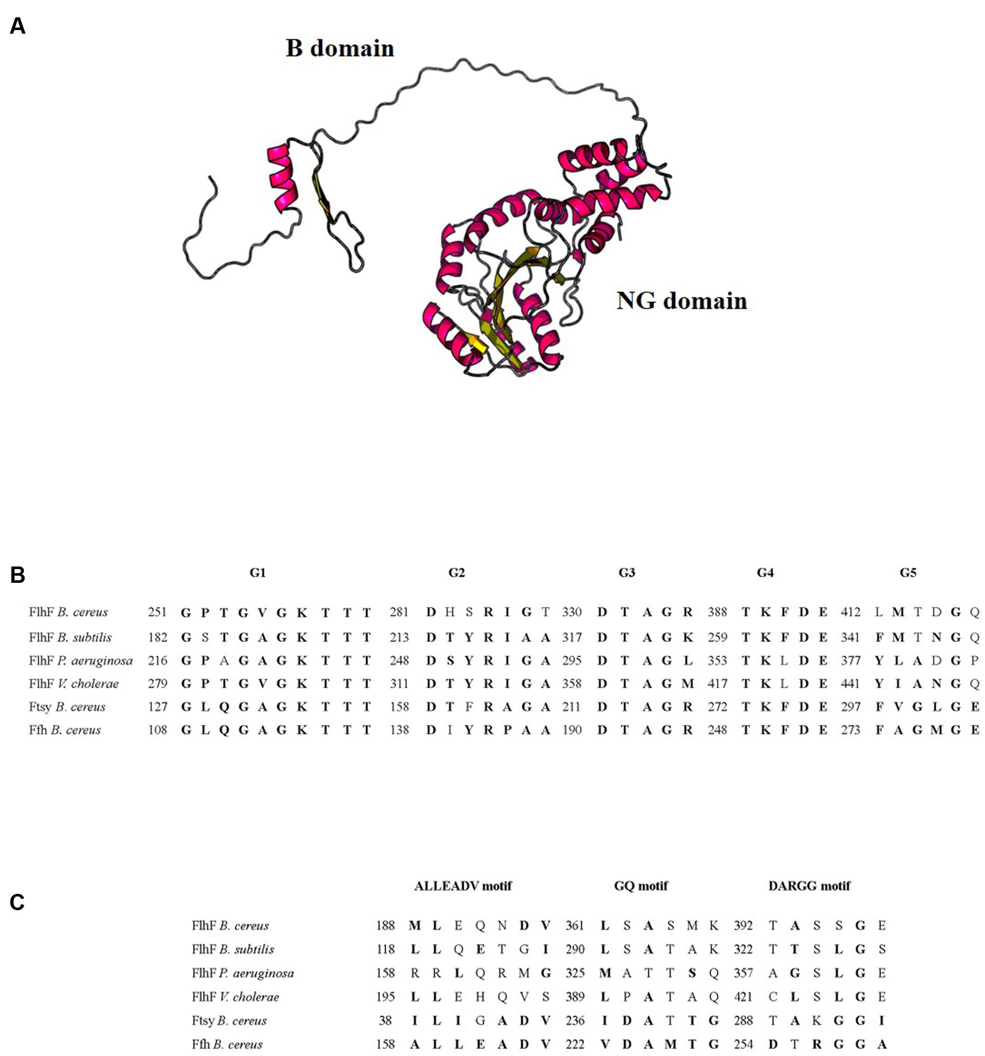
We used the Phyre2 program (Kelley et al., 2015) and the Raptor X Structure Prediction Server (Källberg et al., 2012) in order to define the putative secondary and three-dimensional structure of B. cereus FlhF. The protein appeared to be constituted by 45% α-helices and 30% β-sheets and to consist of two distinct portions connected by an unstructured peptide linker (Figure 1A). The B domain was predicted by the Raptor X Structure Prediction Server only, with a P-value of 2.5 × 10⁻², while the NG domain was predicted by both programs with a good P-value (7.43 × 10⁻⁷ for Raptor X Structure Prediction Server and 100.0% confidence by the single highest scoring template with Phyre2) and it is 100% similar in structure to B. subtilis FlhF.

FIGURE 1. In silico analysis of B. cereus FlhF. (A) Model of the three-dimensional structure of B. cereus FlhF. (B) Alignment of the amino acid sequences of the G1–G5 signatures of the G domain and their respective positions. (C) Alignment of the amino acid sequences of the ALLEADV, GQ and DARGG motifs and their respective positions. Conserved amino acids belonging to the same chemical class are marked in bold.
In all described SRP-GTPases, five signature elements (G1–G5) interacting with the GDP/GTP-Mg2+ complex have been identified in the G domain (Anand et al., 2006; Bange et al., 2007). These elements have never been described in B. cereus SRPs. As shown in Figure 1B, the alignment between the FlhF G1–G5 signatures of B. subtilis, P. aeruginosa, and V. cholerae with the amino acid sequence of B. cereus FlhF, FtsY, and Ffh revealed that all these signatures are also present inside the G domain of B. cereus SRP-GTPases. In particular, G3 and G4 signatures are identical among FlhF, Ffh, and FtsY of B. cereus and G1 is highly conserved in all the analyzed bacterial species. The G2 signature of B. cereus SRP-GTPases are less conserved compared to the same sequence of B. subtilis. P. aeruginosa, and V. cholerae, while G5 is generally less conserved among all the species analyzed.
Ffh and FtsY of many organisms contain additional sequence motifs (ALLEADV in the N domain; GQ and DARGG in the G domain) that are essential for the communication between functional domains (Bange et al., 2007). Since these motifs have never been described in B. cereus SRPs, we first identified them in Ffh and FtsY of B. cereus, through amino acid alignments with the Escherichia coli and B. subtilis orthologs (data not shown). Figure 1C shows the alignment between the ALLEADV, GQ and DARGG motifs in B. cereus SRPs and the putative corresponding regions of FlhF in B. subtilis, P. aeruginosa, and V. cholerae.
FlhF and Swarming Motility
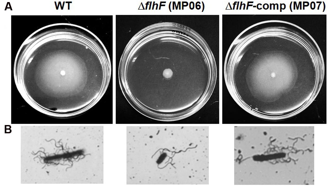
Previous data indicated absence of swarm cells in the ΔflhF mutant of B. cereus grown on swarming-supporting media (Salvetti et al., 2007). In the same experimental conditions, we show that FlhF depletion also impairs cell migration. Indeed, as shown in Figure 2A, the ΔflhF mutant gave rise to colonies (4.2 ± 0.32 mm of diameter) that were significantly smaller than those produced by the swarming proficient wt (18.4 ± 1.28 mm of diameter; P = 0.0039) or ΔflhF-comp strain (14.2 ± 1.8 mm of diameter; P = 0.015). As expected, long and hyperflagellated swarmer cells were visualized in the colonies produced by the wt and the ΔflhF-comp strain only (Figure 2B).

FIGURE 2. Analysis of swarming motility. (A) Colonies produced on swarming TrA plates. (B) Representative cells obtained from swarming TrA plates and stained with tannic acid and silver nitrate.
Effect of FlhF Depletion on Extracellular Proteins
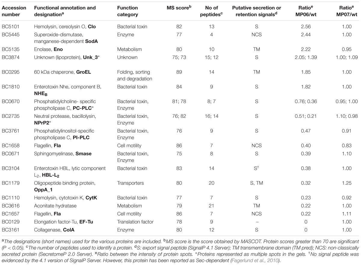
To better characterize the effect of FlhF depletion on protein secretion by B. cereus, we performed comparative proteome analyses of the ΔflhF mutant, the wt, and the ΔflhF-comp strain grown in BHIG. There were no obvious growth differences between these strains in this medium, as already described (Salvetti et al., 2007). Additionally, as previously described (Salvetti et al., 2007), we again evaluated the activity of the cytosolic marker fructose-1,6-bisphosphate aldolase, a cytoplasmic enzyme that is not released by intact cells, in culture supernatants. No enzyme activity was detected. The bacterial supernatants were precipitated with TCA and analyzed using 2-DE. On average, 539 ± 103 spots were detected in the supernatant of the parental strain and 531 ± 99 spots in that derived from the ΔflhF-comp strain. This was not significantly different from the 430 ± 119 spots found in the mutant strain on average (P > 0.05). In the 2-DE gel analysis, 19 protein spots showed a different abundance of >1.5-fold (≥1.5 for upregulated proteins and ≤0.667 for downregulated proteins) in the ΔflhF mutant, compared to the wt (Table 1). These spots were identified by comparison with available gels (Gohar et al., 2002, 2005) and LC-MS/MS analysis. Three proteins were represented as multiple spots in the gel, having either similar mass but different isoelectric points or slight differences in the molecular mass due to putative post-translational modifications. No appreciable difference emerged between the wt and the ΔflhF-comp extracellular proteomes.

TABLE 1. Bacillus cereus ATCC 14579 supernatant proteins appreciably increased, reduced or absent in the supernatant of the ΔflhF (MP06) mutant following growth in BHIG broth.
In the ΔflhF mutant, the amount of six proteins increased and 12 proteins decreased in abundance compared to the parental strain (Table 1). The extracellular protein products significantly more abundant in the ΔflhF mutant included three predicted secretory proteins, cereolysin O (Clo; P = 0.007), the B component of NHE (NHEB; P = 0.041), both contributing to B. cereus pathogenicity (Lund and Granum, 1997; Alouf, 2000), and a protein with unknown function (P = 0.017). Three other proteins that do not exhibit a conventional signal peptide, the SodA superoxide-dismutase (P = 0.048), enolase (Eno; P = 0.007), and the chaperone GroEL (P = 0.01), were also present at higher levels in the supernatant of the ΔflhF mutant. Despite these proteins are cytosolic in nature, their orthologs are commonly detected in an extracellular form in many bacteria, B. cereus and other Bacillus species included (Nouwens et al., 2002; Braunstein et al., 2003; Lenz et al., 2003; Gohar et al., 2005).
Six predicted secretory proteins playing a role in B. cereus pathogenicity, since they are able to degrade phospholipids (PC-PLC; P = 0.017. PI-PLC; P = 0.0034. Smase; P = 0.0072), proteins (bacillolysin, NprP2; P = 0.032 and P = 0.007. Collagenase, ColA; P = 0.0002), and to form pores in planar lipid bilayers (cytotoxin K; P = 0.034), were significantly less abundant in the extracellular proteome of the ΔflhF mutant. The membrane-anchored substrate-binding protein OppA_1 of the oligopeptide permease transporter (Slamti et al., 2015) was also significantly reduced (P = 0.01) in the mutant secretome. In agreement with our previous results (Salvetti et al., 2007), the flagellin proteins BC1657 (P = 0.0091) and BC1658 (P = 0.016), classified as non-conventionally secreted proteins by the SecretomeP 2.0 server, were found significantly less abundant in the ΔflhF strain. In addition, a significant reduction of the L2 component of HBL (HBL-L2; P = 0.023), apparently lacking a signal peptide by the SIGNALIP 4.1 but herein classified as secretory accordingly to Fagerlund et al. (2010), was demonstrated for the mutant strain. The mutant also displayed a reduction in the spots corresponding to aconitate hydratase (BC3616; P = 0.0048) and elongation factor-Tu (EF-Tu; P = 0.002), commonly found in the cytoplasm.
Contribution of FlhF to B. cereus Pathogenicity
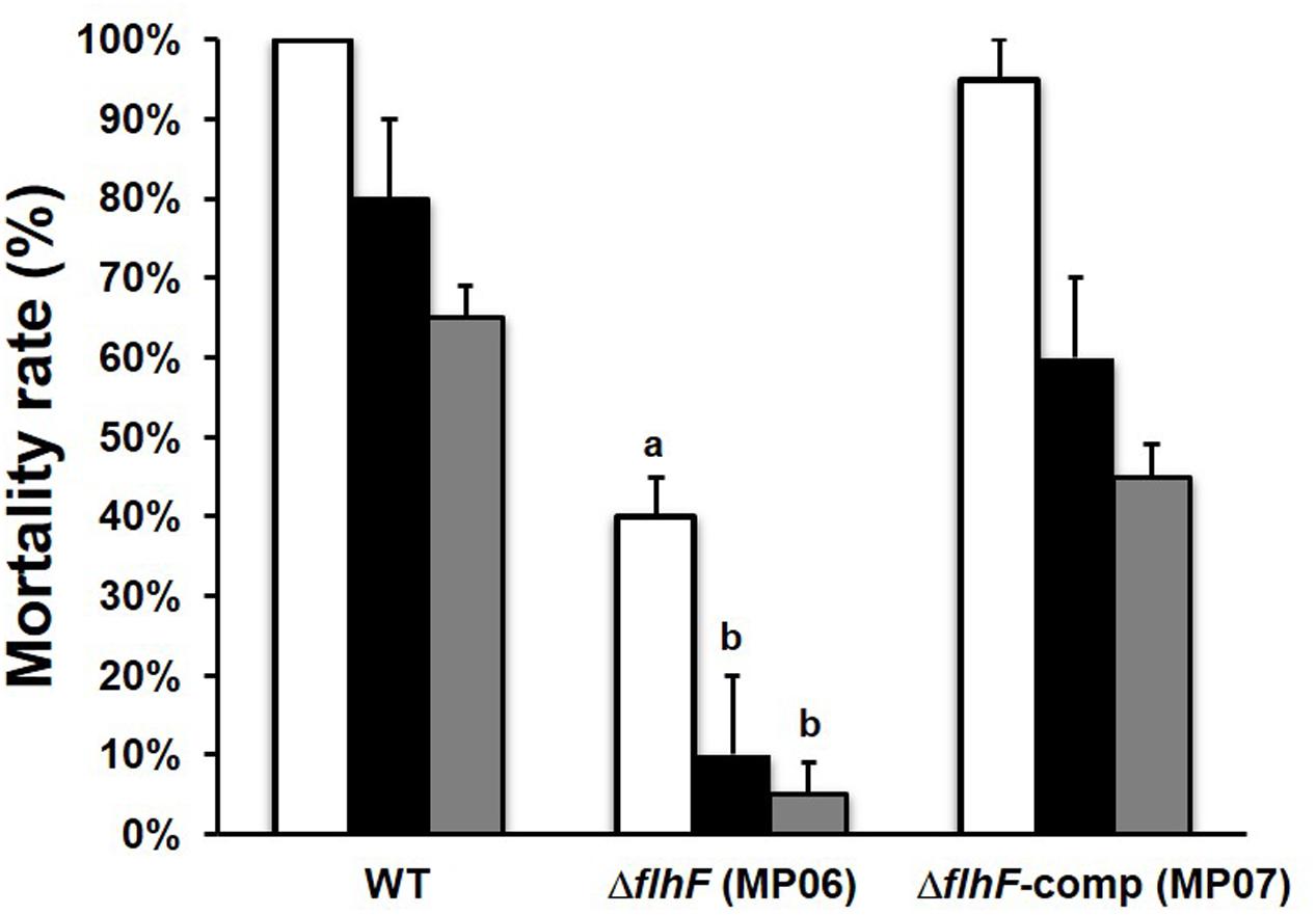
Based on the observation that several virulence proteins were less abundant in the supernatant of the ΔflhF mutant and that this strain was unable to swarm, we wondered whether the deficiency in FlhF would affect B. cereus pathogenicity in an in vivo model of infection. To this aim, larvae of the greater wax moth G. mellonella, which have been shown to provide useful insights into the pathogenesis of a wide range of microbial infections (Jander et al., 2000; Purves et al., 2010), were used to evaluate the pathogenicity of the wt, ΔflhF, and ΔflhF-comp strains. Larvae were intra-hemocoelically injected with three doses of mid-log phase bacteria and the number of dead larvae was recorded. As shown in Figure 3, a significant reduction in larvae mortality compared to the wt was observed when the ΔflhF mutant was inoculated at the three doses (P < 0.01 for the highest dose, and P < 0.05 for the other doses). No significant difference was observed in the mortality rates after injection of the wt or the ΔflhF-comp strain at different doses. As resulted from Probit analysis, the LD50 of the ΔflhF mutant was higher (3.16 ± 0.83 × 10³ CFU per larva) than the value obtained for the wt (9.10 ± 6.73 CFU per larva). The LD50 of the ΔflhF-comp strain was 6.27 ± 4.63 × 101 CFU per larva.

FIGURE 3. Pathogenicity of B. cereus strains in G. mellonella larvae. Mortality rates obtained after infection with 103 (white bars), 102 (black bars) and 101 (gray bars) CFU per larva. aP < 0.01; bP < 0.05.
To test whether differences in larvae mortality could be attributed to a defect in bacterial growth in vivo, the bacterial load in infected larvae was quantified over time. Larvae were infected with 104 CFU of wt, ΔflhF, or ΔflhF-comp. At selected time points, the number of CFU per larva was determined. Infection of G. mellonella with all B. cereus strains resulted in an initial 10-fold reduction compared to the infectious dose in the CFU recovered from infected larvae at 2-h post infection (wt: 1755 ± 214.6 CFU/larva; ΔflhF: 1673 ± 350.4 CFU/larva; ΔflhF-comp: 1780 ± 280.2 CFU/larva). Bacterial recovery progressively increased over time up to 10000-fold the infectious dose at 24- h post infection (wt; 3.45 ± 0.102 × 10⁸ CFU/larva; ΔflhF: 3.48 ± 0.856 × 10⁸ CFU/larva; ΔflhF-comp: 3.46 ± 0.103 × 10⁸ CFU/larva). No significant difference in the CFU recovery of different strains was observed at each time point considered (data not shown). B. cereus was never recovered from non-infected larvae. Our data demonstrate that B. cereus wt, ΔflhF, and ΔflhF-comp are able to replicate at the same rate in G. mellonella.
Discussion
Most of the information on the physiological role of FlhF comes from studies on polarly flagellated, monotrichous bacteria, in which the deletion of flhF results in the absence or mislocalization of flagella leading to swimming/swarming defects (Schuhmacher et al., 2015). In peritrichously flagellates, flhF orthologs are found in Bacillus and Clostridium species and data on FlhF function have been produced for B. subtilis and B. cereus (Carpenter et al., 1992; Zanen et al., 2004; Bange et al., 2007; Salvetti et al., 2007; Guttenplan et al., 2013). In both species, deletion of flhF leads to mislocalization of flagella and, in B. cereus, flagellar number as well as swimming motility are also reduced (Salvetti et al., 2007). FlhF has been suggested to target the first building block to the future flagellar assembly site by an unclear mechanism (Schuhmacher et al., 2015). The B-domain of FlhF seems to play an important role and differences within this domain might be important criteria to define different flagellation patterns in different species.
In this study, in silico analysis shows that, similar to Ffh and FtsY, B. cereus FlhF contains a conserved NG domain that includes the GTPase (G-domain) and the regulatory domain (N-domain). Besides the NG domain, FlhF contains an N-terminal domain of basic character (B-domain) that seems to be natively unfolded (Figure 1A), as already demonstrated for B. subtilis FlhF (Bange et al., 2007), but displays no homology with the same domain of other bacteria. This result is in line with the significant variations in size and conservation found in the B-domain among species, which suggested that this domain might exert species-specific functions (Schuhmacher et al., 2015).
SRP-GTPases are known to form dimers depending on the GTP load (Leipe et al., 2002; Gasper et al., 2006). While Ffh and FtsY aggregate to form a heterodimeric complex, FlhF act as a stable homodimer that bound GTP/GDP-Mg2+ in many organisms (Bange et al., 2007; Green et al., 2009; Schniederberend et al., 2013). SRPs dimerization capacity requires specific amino acid motifs that are typical of eukaryotic and bacterial SRP-GTPases (Anand et al., 2006) and constitute the active site. The presence of typical sequence motifs in B. cereus FlhF (Figures 1B,C) suggests that the protein forms a stable homodimer bound to GTP also in this organism.
By analyzing migration on TrA, an ideal medium for evaluating swarming migration by B. cereus ATCC 14579 (wt; Salvetti et al., 2011), we show that swarming motility is completely abolished in the ΔflhF mutant of B. cereus. This finding together with the previous demonstration that the mutant swimming motility was only reduced (Salvetti et al., 2007) suggests that other factors than flagella may be affected by the FlhF depletion. In fact, while swimming motility of cells primarily depends on flagella, additional factors are required for swarming. Perturbations in the flagellar motor, outer polysaccharides, osmotic agents, surfactants, as well as quorum-sensing signals have been shown to alter swarming in several bacteria (Kearns, 2010; Partridge and Harshey, 2013). Future studies will be required for defining the effects of FlhF deprivation on these factors.
Comparative proteomic analysis of culture supernatants indicated that the ΔflhF mutant of B. cereus displays an alteration in the quantity of certain extracellular proteins (Table 1). The amount of the major part of these proteins was lower in the proteome of the mutant compared to the wt and ΔflhF-comp strain, suggesting that FlhF can be involved in protein secretion, as already reported for P. putida (Pandza et al., 2000). In the SRP system, the SRP (Ffh)/4.5S RNA complex, in its GTP-bound form, attaches to the N-terminal sequence of the newly formed protein as it comes off the ribosome, and then docks with its receptor (FtsY) on the membrane, thus resulting in the insertion of the protein into the membrane (Akopian et al., 2013). B. cereus FlhF is homologous to both the SRP and the membrane receptor of this pathway and, as already suggested for other bacteria (Bange et al., 2007; Balaban et al., 2009), could play a role in either activities. FlhF has been shown to recruit the earliest flagellar component FliF and to insert itself in the cytoplasmic membrane in V. cholerae (Green et al., 2009). B. cereus FlhF could perform a similar function for flagellar proteins, but also for other proteins (PC-PLC, NPrP2, PI-PLC, Smase, HBL-L2, CytK) that need to be directed to Sec translocase to be exported outside the cell. Nevertheless, although less pronouncedly, these proteins are still present in the secretome of the ΔflhF mutant, suggesting that, if FlhF acts as SRP, protein targeting to the membrane can still occur even in the absence of functional FlhF and that other systems may act in the export of these proteins. Although the secretion defects observed in the absence of functional FlhF might be due to its role as SRP-like protein, an effect of FlhF deprivation on regulatory pathways shared between the genes encoding the altered proteins cannot be excluded.
Galleria mellonella was previously shown to be a good alternative to the mouse model for evaluating the virulence of B. cereus (Salamitou et al., 2000). The virulence potential of B. cereus is due to the secretion of a plethora of virulence proteins, but also to the presence of flagella, which play a crucial role in swimming and swarming motility and in bacterial adhesion to surfaces. Infection of G. mellonella with a B. cereus mutant in the Smase gene was previously shown to cause a reduction in larvae mortality, while mutations in nheBC had a little impact on G. mellonella survival (Doll et al., 2013). Infection with a B. cereus mutant in codY, encoding a positive regulator of the PC-PLC gene and many other virulence genes (Frenzel et al., 2012; Slamti et al., 2015), as well as with swimming defective mutant (Frenzel et al., 2015), drastically reduced larvae mortality. G. mellonella infection with a B. thuringiensis strain unable to swarm and to secrete of HBL-L2 resulted in a strong reduction of larvae mortality (Bouillaut et al., 2005). In this study, we demonstrate that lack of FlhF substantially reduces B. cereus pathogenicity in G. mellonella. Since no difference with the wt in the replication of the ΔflhF mutant in G. mellonella was found, we can assume that the reduced pathogenicity of the mutant is due to the different virulence potential this strain shows. Other than being less flagellated and unable to swarm, the mutant strain shows a reduction in the secretion of PC-PLC, Smase, HBL-L2, and many other virulence proteins (PI-PLC, CytK and ColA) (Beecher et al., 2000; Bottone, 2010; Ramarao and Sanchis, 2013). Despite the amount of some virulence proteins (NHEB and Clo) is increased in the secretome of the ΔflhF mutant, we can speculate that these factors have a minor impact on B. cereus pathogenicity in G. mellonella. Therefore, the reduced production of some virulence proteins, of swimming, and the lack of swarming motility appear responsible of the scarce pathogenicity exerted by the ΔflhF mutant in G. mellonella.
Conclusion
The flagellar protein FlhF, which controls the number and localization of flagella in B. cereus (Salvetti et al., 2007), is essential for swarming motility and full virulence. FlhF might act directly or through regulatory mechanisms on protein secretion or synthesis. Protein-protein interaction studies and partial gene deletion mutants will help in clarifying the molecular mechanisms underlying its peculiar activity.
Author Contributions
All authors listed, have made substantial, direct and intellectual contribution to the work, and approved it for publication.
Funding
This research received no specific grant from any founding agency in the public, commercial, or not-for-profit sectors.
Conflict of Interest Statement
The authors declare that the research was conducted in the absence of any commercial or financial relationships that could be construed as a potential conflict of interest.
Acknowledgments
The authors pay tribute to the late Professor Mario Campa for his long-standing inspiration and support.
Footnotes
- ^http://blast.ncbi.nlm.nih.gov/Blast.cgi
- ^http://www.uniprot.org/
- ^http://prodom.prabi.fr/prodom/current/html/home.php
- ^http://www.sbg.bio.ic.ac.uk/phyre2
- ^http://raptorx.uchicago.edu/StructurePrediction/predict/
- ^http://www.matrixscience.com
- ^http://www.genome.jp/kegg/
- ^http://www.cbs.dtu.dk/services/SignalP/
- ^http://www.cbs.dtu.dk/services/TatP/
- ^http://www.cbs.dtu.dk/services/SecretomeP/
- ^http://www.ch.embnet.org/software/TMPRED_form.html
References
Akopian, D., Shen, K., Zhang, X., and Shan, S. O. (2013). Signal recognition particle: an essential protein targeting machine. Annu. Rev. Biochem. 82, 693–721. doi: 10.1146/annurev-biochem-072711-164732
Alouf, J. E. (2000). Cholesterol-binding cytolytic protein toxins. Int. J. Med. Microbiol. 290, 351–356. doi: 10.1016/S1438-4221(00)80039-9
Anand, B., Verma, S. K., and Prakash, B. (2006). Structural stabilization of GTP-binding domains in circularly permuted GTPases: implications for RNA binding. Nucleic Acids Res. 34, 2196–2205. doi: 10.1093/nar/gkl178
Balaban, M., Joslin, S. N., and Hendrixson, D. R. (2009). FlhF and its GTPase activity are required for distinct processes in flagellar gene regulation and biosynthesis in Campylobacter jejuni. J. Bacteriol. 191, 6602–6611. doi: 10.1128/JB.00884-09
Bange, G., Petzold, G., Wild, K., Parltz, R., and Sinning, I. (2007). The crystal structure of the third signal-recognition particle GTPase FlhF reveals a homodimer with bound GTP. Proc. Natl. Acad. Sci. U.S.A. 104, 13621–13625. doi: 10.1073/pnas.0702570104
Beecher, D. J., Olsen, T. W., Somers, E. B., and Wong, A. C. (2000). Evidence for contribution of tripartite hemolysin BL, phosphatidylcholine-preferring phospholipase C, and collagenase to virulence of Bacillus cereus endophthalmitis. Infect. Immun. 68, 5269–5276. doi: 10.1128/IAI.68.9.5269-5276.2000
Bottone, E. J. (2010). Bacillus cereus, a volatile human pathogen. Clin. Microbiol. Rev. 23, 382–398. doi: 10.1128/CMR.00073-09
Bouillaut, L., Ramarao, N., Buisson, C., Gilois, N., Gohar, M., Lereclus, D., et al. (2005). FlhA influences Bacillus thuringiensis PlcR-regulated gene transcription, protein production, and virulence. Appl. Environ. Microbiol. 71, 8903–8910. doi: 10.1128/AEM.71.12.8903-8910.2005
Braunstein, M., Espinosa, B. J., Chan, J., Belisle, J. T., and Jakobs, W. R. Jr. (2003). SecA2 functions in the secretion of the superoxide dismutase a and in the virulence of Mycobacterium tuberculosis. Mol. Microbiol. 48, 453–464. doi: 10.1046/j.1365-2958.2003.03438.x
Bru, C., Courcelle, E., Carrère, S., Beausse, Y., Dalmar, S., and Kahn, D. (2005). The ProDom database of protein domain families: more emphasis on 3D. Nucleic Acids Res. 33, D212–D215. doi: 10.1093/nar/gki034
Callegan, M. C., Novosad, B. D., Ramirez, R., Ghelardi, E., and Senesi, S. (2006). Role of swarming migration in the pathogenesis of Bacillus endophthalmitis. Invest. Ophthalmol. Vis. Sci. 47, 4461–4467. doi: 10.1167/iovs.06-0301
Carpenter, P. B., Hanlon, D. W., and Ordal, G. W. (1992). flhF, a Bacillus subtilis flagellar gene that encodes a putative GTP-binding protein. Mol. Microbiol. 6, 2705–2713. doi: 10.1111/j.1365-2958.1992.tb01447.x
Celandroni, F., Salvetti, S., Gueye, S. A., Mazzantini, D., Lupetti, A., Senesi, S., et al. (2016). Identification and pathogenic potential of clinical Bacillus and Paenibacillus isolates. PLoS ONE 11:e0152831. doi: 10.1371/journal.pone.0152831
Doll, V. M., Ehling-Schulz, M., and Vogelmann, R. (2013). Concerted action of sphingomyelinase and non-hemolytic enterotoxin in pathogenic Bacillus cereus. PLoS ONE 8:e61404. doi: 10.1371/journal.pone.0061404
Fagerlund, A., Lindbäck, T., and Granum, P. E. (2010). Bacillus cereus cytotoxins Hbl, Nhe and CytK are secreted via the Sec translocation pathway. BMC Microbiol. 10:304. doi: 10.1186/1471-2180-10-304
Frenzel, E., Doll, V., Pauthner, M., Lücking, G., Scherer, S., and Ehling-Schulz, M. (2012). CodY orchestrates the expression of virulence determinants in emetic Bacillus cereus by impacting key regulatory circuits. Mol. Microbiol. 85, 67–88. doi: 10.1111/j.1365-2958.2012.08090.x
Frenzel, E., Kranzler, M., Stark, T. D., Hofmann, T., and Ehling-Schulz, M. (2015). The endospore-forming pathogen Bacillus cereus exploits a small colony variant-based diversification strategy in response to aminoglycoside exposure. MBio. 6, e1172–e1115. doi: 10.1128/mBio.01172-15
Gasper, R., Scrima, A., and Wittinghofer, A. (2006). Structural insights into HypB, a GTP-binding protein that regulates metal binding. J. Biol. Chem. 281, 27492–27502. doi: 10.1074/jbc.M600809200
Ghelardi, E., Celandroni, F., Salvetti, S., Ceragioli, M., Beecher, D. J., Senesi, S., et al. (2007). Swarming behavior and hemolysin BL secretion in Bacillus cereus. Appl. Environ. Microbiol. 73, 4089–4093. doi: 10.1128/AEM.02345-06
Gohar, M., Gilois, N., Graveline, R., Garreau, C., Sanchis, V., and Lereclus, D. (2005). A comparative study of Bacillus cereus. Bacillus thuringiensis and Bacillus anthracis extracellular proteomes. Proteomics 5, 3696–3711. doi: 10.1002/pmic.200401225
Gohar, M., Økstad, O. A., Gilois, N., Sanchis, V., Kolstø, A.-B., and Lereclus, D. (2002). Two-dimensional electrophoresis analysis of extracellular proteome of Bacillus cereus reveals the importance of the PlcR regulon. Proteomics 2, 784–791. doi: 10.1002/1615-9861(200206)2:6<784::AID-PROT784>3.0.CO;2-R
Green, J. C. D., Kahramanoglou, C., Rahman, A., Pender, A. M. C., Charbonnel, N., and Fraser, G. (2009). Recruitment of the earliest component of the bacterial flagellum to the old cell division pole by a membrane-associated signal recognition particle family GTP-binding protein. J. Mol. Biol. 391, 679–690. doi: 10.1016/j.jmb.2009.05.075
Guttenplan, S. B., Shaw, S., and Kearns, D. B. (2013). The cell biology of peritrichous flagella in Bacillus subtilis. Mol. Microbiol. 87, 211–229. doi: 10.1111/mmi.12103
Harshey, R. M., and Matsuyama, T. (1994). Dimorphic transition in Escherichia coli and Salmonella typhimurium: surface-induced differentiation into hyperflagellate swarmer cells. Proc. Natl. Acad. Sci. 91, 8631–8635. doi: 10.1073/pnas.91.18.8631
Jander, G., Rahme, L. G., and Ausubel, F. M. (2000). Positive correlation between virulence of Pseudomonas aeruginosa mutants in mice and insects. J. Bacteriol 182, 3843–3845. doi: 10.1128/JB.182.13.3843-3845.2000
Jeßberger, N., Krey, V. M., Rademacher, C., Böhm, M. E., Mohr, A. K., Ehling-Schulz, M., et al. (2015). From genome to toxicity: a combinatory approach highlights the complexity of enterotoxin production in Bacillus cereus. Front. Microbiol. 10:560. doi: 10.3389/fmicb.2015.00560
Källberg, M., Wang, H., Wang, S., Peng, J., Wang, Z., Lu, H., et al. (2012). Template-based protein structure modeling using the raptorX web server. Nat. Protoc. 7, 1511–1522. doi: 10.1038/nprot.2012.085
Kearns, D. B. (2010). A field guide to bacterial swarming motility. Nat. Rev. Microbiol. 8, 634–644. doi: 10.1038/nrmicro2405
Kelley, L. A., Mezulis, S., Yates, C. M., Wass, M. N., and Sternberg, M. J. E. (2015). The Phyre2 web portal for protein modeling, prediction and analysis. Nat. Protoc. 10, 845–858. doi: 10.1038/nprot.2015.053
Leipe, D. D., Wolf, Y. I., Koonin, E. V., and Aravind, L. (2002). Classification and evolution of P-loop GTPases and related ATPases. J. Mol. Biol. 317, 41–72. doi: 10.1006/jmbi.2001.5378
Lenz, L. L., Mohammadi, S., Geissler, A., and Portnoy, D. A. (2003). SecA2-dependent secretion of autolytic enzymes promotes Listeria monocytogenes pathogenesis. Proc. Natl. Acad. Sci. U.S.A. 100, 12432–12437. doi: 10.1073/pnas.2133653100
Logan, N. A. (2012). Bacillus and relatives in foodborne illness. J. Appl. Microbiol. 112, 417–429. doi: 10.1111/j.1365-2672.2011.05204.x
Lund, T., and Granum, P. E. (1997). Comparison of biological effect of the two different enterotoxin complexes isolated from three different strains of Bacillus cereus. Microbiology 143, 3329–3336. doi: 10.1099/00221287-143-10-3329
Nouwens, A. S., Willcox, M. D. P., Walsh, B. J., and Cordwell, S. J. (2002). Proteomic comparison of membrane and extracellular proteins from invasive (PAO1) and cytotoxic (6206) strains of Pseudomonas aeruginosa. Proteomics 2, 1325–1346. doi: 10.1002/1615-9861(200209)2:9<1325::AID-PROT1325>3.0
Pandza, S., Baetens, M., Park, C. H., Au, T., Keyan, M., and Matin, A. (2000). The G-protein FlhF has a role in polar flagellar placement and general stress response induction in Pseudomonas putida. Mol. Microbiol. 36, 414–423. doi: 10.1046/j.1365-2958.2000.01859.x
Partridge, J. D., and Harshey, R. M. (2013). Swarming: flexible roaming plans. J. Bacteriol. 195, 909–918. doi: 10.1128/JB.02063-12
Purves, J., Cockayne, A., Moody, P. C., and Morrissey, J. A. (2010). Comparison of the regulation, metabolic functions, and roles in virulence of the glyceraldehyde-3-phosphate dehydrogenase homologues gapA and gapB in Staphylococcus aureus. Infect. Immun. 78, 5223–5232. doi: 10.1128/IAI.00762-10
Ramarao, N., and Sanchis, V. (2013). The pore-forming haemolysins of Bacillus cereus: a review. Toxins 5, 1119–1139. doi: 10.3390/toxins5061119
Salamitou, S., Ramisse, F., Brehélin, M., Bourguet, D., Gilois, N., Gominet, M., et al. (2000). The plcR regulon is involved in the opportunistic properties of Bacillus thuringiensis and Bacillus cereus in mice and insects. Microbiology 146, 2825–2832. doi: 10.1099/00221287-146-11-2825
Salvetti, S., Faegri, K., Ghelardi, E., Kolstø, A.-B., and Senesi, S. (2011). Global gene expression profile for swarming Bacillus cereus bacteria. Appl. Environ. Microbiol. 7, 5149–5156. doi: 10.1128/AEM.00245-11
Salvetti, S., Ghelardi, E., Celandroni, F., Ceragioli, M., Giannessi, F., and Senesi, S. (2007). FlhF, a signal recognition particle-like GTPase, is involved in the regulation of flagellar arrangement, motility behavior and protein secretion in Bacillus cereus. Microbiology 153, 2541–2552. doi: 10.1099/mic.0.2006/005553-0
Schniederberend, M., Abdurachim, K., Murray, T., and Kazmierczak, B. I. (2013). The GTPase activity of FlhF is dispensable for flagellar localization, but not motility, in Pseudomonas aeruginosa. J. Bacteriol. 195, 1051–1060. doi: 10.1128/JB.02013-12
Schuhmacher, J. S., Thormann, K. M., and Bange, G. (2015). How bacteria maintain location and number of flagella? FEMS Microbiol. Rev. 39, 812–822. doi: 10.1093/femsre/fuv034
Senesi, S., and Ghelardi, E. (2010). Production, secretion and biological activity of Bacillus cereus enterotoxins. Toxins. 2, 1690–1703. doi: 10.3390/toxins2071690
Senesi, S., Salvetti, S., Celandroni, F., and Ghelardi, E. (2010). Features of Bacillus cereus swarm cells. Res. Microbiol. 161, 743–749. doi: 10.1016/j.resmic.2010.10.007
Shevchenko, A., Jensen, O. N., Podtelejnikov, A. V., Sagliocco, F., Wilm, M., Vorm, O., et al. (1996). Linking genome and proteome by mass spectrometry: large-scale identification of yeast proteins from two dimensional gels. Proc. Natl. Acad. Sci. U.S.A. 93, 14440–14445. doi: 10.1073/pnas.93.25.14440
Slamti, L., Lemy, C., Henry, C., Guillot, A., Huillet, E., and Lereclus, D. (2015). CodY regulates the activity of the virulence quorum sensor PlcR by controlling the import of the signaling peptide PapR in Bacillus thuringiensis. Front. Microbiol. 6:1501. doi: 10.3389/fmicb.2015.01501
Smith, P. K., Krohn, R. I., Hermanson, G. T., Mallia, A. K., Gartner, F. H., Provenzano, M. D., et al. (1985). Measurement of protein using bicinchoninic acid. Anal. Biochem. 150, 76–85. doi: 10.1016/0003-2697(85)90442-7
The UniProt Consortium. (2015). UniProt: a hub for protein information. Nucleic Acids Res. 43, D204–D212. doi: 10.1093/nar/gku989
Warth, A. D. (1980). Heat stability of Bacillus cereus enzymes within spores and in extracts. J. Bacteriol. 143, 27–34.
Keywords: FlhF, Bacillus cereus, swarming, protein secretion, virulence
Citation: Mazzantini D, Celandroni F, Salvetti S, Gueye SA, Lupetti A, Senesi S and Ghelardi E (2016) FlhF Is Required for Swarming Motility and Full Pathogenicity of Bacillus cereus. Front. Microbiol. 7:1644. doi: 10.3389/fmicb.2016.01644
Received: 24 June 2016; Accepted: 03 October 2016;
Published: 19 October 2016.
Edited by:
Ivan Mijakovic, Chalmers University of Technology, SwedenReviewed by:
Monika Ehling-Schulz, University of Veterinary Medicine, AustriaMichel Gohar, Institut National de la Recherche Agronomique, France
Copyright © 2016 Mazzantini, Celandroni, Salvetti, Gueye, Lupetti, Senesi and Ghelardi. This is an open-access article distributed under the terms of the Creative Commons Attribution License (CC BY). The use, distribution or reproduction in other forums is permitted, provided the original author(s) or licensor are credited and that the original publication in this journal is cited, in accordance with accepted academic practice. No use, distribution or reproduction is permitted which does not comply with these terms.
*Correspondence: Emilia Ghelardi, ZW1pbGlhLmdoZWxhcmRpQG1lZC51bmlwaS5pdA==
†Present address: Sara Salvetti, San Giuseppe Hospital-USL 11, Empoli, Italy
 Diletta Mazzantini1
 Emilia Ghelardi