- 1Micalis Institute, INRA, AgroParisTech, CNRS, Université Paris-Saclay, Jouy-en-Josas, France
- 2Unité de Recherche Technologies et Valorisation Alimentaire, Laboratoire de Biotechnologie, Université Saint-Joseph, Beirut, Lebanon
- 3UMR UMET: Unité Matériaux et Transformations, Centre National de la Recherche Scientifique, Institut National de la Recherche Agronomique, Université de Lille, Villeneuve d'Ascq, France
Bacillus cereus displays a high diversity of lifestyles and ecological niches and include beneficial as well as pathogenic strains. These strains are widespread in the environment, are found on inert as well as on living surfaces and contaminate persistently the production lines of the food industry. Biofilms are suspected to play a key role in this ubiquitous distribution and in this persistency. Indeed, B. cereus produces a variety of biofilms which differ in their architecture and mechanism of formation, possibly reflecting an adaptation to various environments. Depending on the strain, B. cereus has the ability to grow as immersed or floating biofilms, and to secrete within the biofilm a vast array of metabolites, surfactants, bacteriocins, enzymes, and toxins, all compounds susceptible to act on the biofilm itself and/or on its environment. Within the biofilm, B. cereus exists in different physiological states and is able to generate highly resistant and adhesive spores, which themselves will increase the resistance of the bacterium to antimicrobials or to cleaning procedures. Current researches show that, despite similarities with the regulation processes and effector molecules involved in the initiation and maturation of the extensively studied Bacillus subtilis biofilm, important differences exists between the two species. The present review summarizes the up to date knowledge on biofilms produced by B. cereus and by two closely related pathogens, Bacillus thuringiensis and Bacillus anthracis. Economic issues caused by B. cereus biofilms and management strategies implemented to control these biofilms are included in this review, which also discuss the ecological and functional roles of biofilms in the lifecycle of these bacterial species and explore future developments in this important research area.
Introduction
Bacillus cereus is a large, Gram-positive bacterium which produces spores and displays a peritrichous flagellation. Soil has long been considered to be the natural habitat of this species, although its spores can be isolated from various materials, such as invertebrates, plants, or food (Sneath, 1986). Recently, the ecological niches of B. cereus were suggested to include insects and nematodes guts (Jensen et al., 2003; Ruan et al., 2015), or plant roots (Ehling-Schulz et al., 2015). The high diversity of B. cereus habitats is reflected by the genetic polymorphism of this species (Helgason et al., 2004), and is illustrated by the existence of probiotic (Cutting, 2011) as well as pathogenic strains. B. cereus is indeed one of the most frequent agent of food poisoning outbreaks, which symptoms can be either emetic or diarrheal. Emetic strains of B. cereus can secrete in the food a highly toxic and heat-stable Non-ribosomal cyclic peptide which can withstand cooking temperatures and induce, when ingested, vomitic symptoms (Ehling-Schulz et al., 2015). For diarrheal strains, according to the current model of B. cereus-induced diarrheal gastroenteritis, spores contained in the food are ingested by the host and germinate within the intestine, where vegetative cells can grow and produce enterotoxins. Three enterotoxins (Hbl, Nhe, and CytK) can be secreted by B. cereus (Stenfors Arnesen et al., 2008). In addition to enterotoxins, B. cereus can produce several other toxins (hemolysins HlyI and HlyII) and degradative enzymes (phospholipases and proteases), which are either secreted or directed to the cell-surface, and which are controlled, for most of them, by the PlcR transcriptional activator (Gohar et al., 2008). PlcR is one of the numerous B. cereus quorum-sensing systems, which, together with a great number of chromosomally-encoded sensors and regulators (De Been et al., 2006), make the bacterium highly responsive to environmental changes and give it the ability to adapt to diverse conditions. The adaptative properties of B. cereus is also a consequence of the presence, within the bacterium, of a number of plasmids, which size is in the 2–500 kb range. Bacillus thuringiensis and Bacillus anthracis, for instance, are two species of the B. cereus group sensu lato which differ from B. cereus sensu stricto mainly by the presence of megaplasmids carrying genes encoding toxins specifically active against, respectively, invertebrates or mammals.
B. cereus, B. thuringiensis, and B. anthracis (called hereafter B. cereus sensu lato) are all able to produce biofilms. In most isolates of these species, biofilms are found as floating pellicles, but can also stick on immerged abiotic surfaces or even be present on living tissues. These complex communities are likely to be a key element in the ability of B. cereus to colonize different environments. Together with spores, they confer to the bacterium a high resistance to various stresses and a high adhesive capacity on various substrates, including stainless steel, a material widely used in the food processing lines. In these facilities, B. cereus can persist for long durations and can even withstand sanitization procedures. The exponential increase in the number of articles published on B. cereus biofilms (Figure 1) illustrates the rising interest of the scientific community for this subject. Indeed not only are biofilms a key issue in B. cereus life, they also display interesting specificities. Although some of the molecular mechanisms involved in biofilm formation and in its regulation are shared with Bacillus subtilis—a saprophytic bacterium extensively studied for biofilm formation—striking differences exists between the two species regarding the biofilm structure, the effectors of matrix formation and the regulation pathways controlling them.
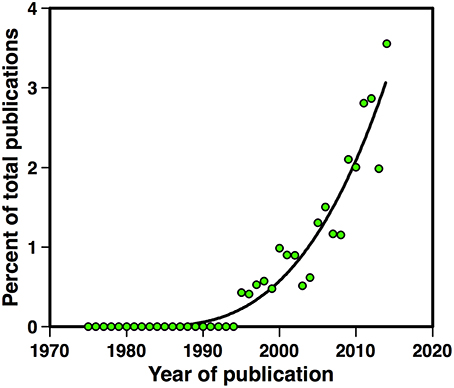
Figure 1. Number of articles published between 1975 and 2015 on B. cereus biofilms. Articles published on B. cereus, B. thuringiensis, or B. anthracis biofilms, in percent of the total number of articles published on the same species.
In the last decade, a considerable knowledge has been accumulated in a wide area of research regarding biofilm formation in B. cereus sensu lato. The aim of this review is to stress a panoramic view of the current knowledge, from the molecular mechanisms involved in biofilms formation in the three species to the functions and roles of these multicellular structures in the bacterium life, including pathogenesis and food industry contamination. From this panoramic view, we expect to draw the most promising incoming research developments and to address some intriguing questions, such as why has B. anthracis, a lethal and capsulated pathogen, kept the ability to produce biofilms. This review will also highlight the variety and prevalence of biofilm formation in the three species and will point, when necessary, to similarities and differences with B. subtilis.
Molecular and Physiological Aspect
The molecular and physiological aspects of biofilm formation discussed here include the various extracellular macromolecules produced by the bacterium and specifically required for the biofilm matrix, cellular elements involved in biofilm formation such as flagella or cell-surface proteins, and the complex regulation network controlling biofilm formation and connecting it to other cellular functions. Also included in this part of the review is phenotypic heterogeneity within the biofilm, a field of growing interest since it is strongly involved in the bacterial survival in changing environments, and the role of mobile genetic elements in biofilm formation.
The Biofilm Matrix
Biofilms are usually embedded in a self-produced matrix whose structural elements are exopolysaccharides, proteins and DNA (Flemming and Wingender, 2010). B. cereus is no exception to this rule and its matrix contains the three components. In B. subtilis, most of the structural exopolysaccharides required for biofilm formation are synthetized by the products of the epsA-O operon (Branda et al., 2001; Kearns et al., 2005). Deletion of epsA-O leads to a Non-structured and fragile biofilm pellicle (Lemon et al., 2008). An eps locus similar to epsA-O is found in bacteria of the cereus group (Ivanova et al., 2003; Gao et al., 2015). This similarity is supported by the presence, within the locus, of an anti-termination RNA element named EAR, found only in epsA-O and in the eps locus of the cereus group (Irnov and Winkler, 2010). However, deletion of the B. cereus eps locus does not affect biofilm formation (Gao et al., 2015), despite the presence of polysaccharides in the B. cereus biofilm matrix (Houry et al., 2012), whose origin therefore remains unknown.
The B. subtilis biofilm matrix also contains the three structural proteins TasA, TapA, and BslA (Vlamakis et al., 2013). BslA (Biofilm surface layer) forms a hydrophobic envelope surrounding the biofilm (Hobley et al., 2013) while TasA assembles into amyloid-like fibers attached to the cell wall by TapA, resulting in a fiber network strengthening the biofilm (Romero et al., 2011). In B. subtilis, tapA, and tasA are included in the tapA-sipW-tasA operon, where sipW codes for a signal peptidase, which releases the two proteins TapA and TasA into the extracellular milieu. There is no paralog of bslA or tapA in the B. cereus genome, but tasA have two paralogs. One is tasA, included in the sipW-tasA operon, and the other is calY, which is located next to sipW-tasA (Caro-Astorga et al., 2015). TasA and CalY are both involved in the production of fibers which can be observed by electron microscopy, and the deletion of their genes or of sipW leads to biofilm defects similar to the ones reported in B. subtilis (Caro-Astorga et al., 2015).
The extracellular DNA (eDNA) contained in the B. cereus biofilm matrix was shown to be produced specifically in biofilms and was reported to be required for adhesion on polystyrene or glass surfaces (Vilain et al., 2009). Its origin remains unknown but might be related to programmed cell death (Abee et al., 2011). However, in planktonic cultures of B. subtilis, the production of eDNA is not a consequence of cell-lysis but requires both competence genes and the Opp oligopeptide permease, and is involved in horizontal gene transfer (Zafra et al., 2012). Other bacterial species, including the Gram-positive bacteria Staphylococcus aureus and Streptococcus pneumonia, also require eDNA for biofilm formation (Whitchurch et al., 2002; Moscoso et al., 2006; Izano et al., 2008). Possible interactions between the eDNA and other consituents of the biofilm matrix have not yet been investigated, neither has the mechanism or the regulation of eDNA production in biofilms.
Role of Flagella
Flagella are cell-surface structures extending far away the bacterial cell. In B. cereus, they are not required for adhesion to glass (Houry et al., 2010), but flagellar motility is involved in biofilm formation through 4 mechanisms. First, motility is a key element of biofilm formation when the bacterium must reach by its own (in static conditions) suitable places for biofilm formation (Houry et al., 2010), at the air-liquid interface. The suppression of motility in a strain which forms biofilms at the air-liquid interface resulted in the formation of submerged biofilms (Hayrapetyan et al., 2015b). Secondly, motile bacteria within the biofilm create channels in the matrix, leading to an increase in nutrients exchange and, conversely, favoring the penetration of toxic substances (Houry et al., 2012). Thirdly, motile planktonic bacteria can enter the biofilm and increase its biomass (Houry et al., 2010, 2012). Fourthly, motile bacteria located at the edge of the growing biofilm extend the surface covered by this structure, resulting in colony spreading (Houry et al., 2010). Although flagellin transcription decreases continuously with biofilm age (Houry et al., 2010), the biofilm bacterial population is heterogeneous and includes a fraction of motile bacteria (Houry et al., 2012) which, in B. subtilis, is located at the edge of the colony (Vlamakis et al., 2008).
Cell-Surface Properties
B. cereus cells in biofilm differ from their planktonic counterparts regarding their cell-surface properties. For example, the structure of the secondary cell wall polymer (SCWP), a polysaccharide linked to the peptidoglycan by phospho-diester linkages, was shown to vary during biofilm aging in B. cereus (Candela et al., 2011). Since SLH (S-layer homology) domain-containing proteins bind to the SCWP, changes in the SCWP structure might result in changes in the proteins displayed on the cell-surface, and possibly involved in the adaptation of the bacterium to its environment. Within these SLH-proteins are autolysins, whose variation during biofilm growth might lead to changes in the bacterial chain length. Similarly, a cell-surface peptidase (CwpFM) involved in autolysis was shown to play a role in biofilm formation, possibly because this autolysin can modulate the length of bacterial chains and consequently act on the motility of the bacterium (Tran et al., 2010).
Regulation Networks
The regulation network controlling B. cereus biofilm formation shows a combination of similarities and differences with B. subtilis. In B. cereus sensu lato, sipW, tasA, and calY transcriptions are repressed by the SinR regulator (Pflughoeft et al., 2011), which controls biofilm formation (Fagerlund et al., 2014) as for B. subtilis. SinR is antagonized by SinI and, in both species, deletion of SinI leads to the absence of biofilm and to hypermotility while the reverse phenotype (biofilm overproduction, no motility) is obtained upon deletion of SinR (Kearns et al., 2005; Fagerlund et al., 2014; Figure 2). Consequently, the SinI/SinR anti-repressor/repressor pair is likely to act as a switch between biofilm formation and swimming motility in B. cereus or B. thuringiensis as it does in B. subtilis. In addition, Spo0A is required for biofilm formation in B. thuringiensis and in B. subtilis, and AbrB represses biofilm formation in both species (Hamon and Lazazzera, 2001; Fagerlund et al., 2014).
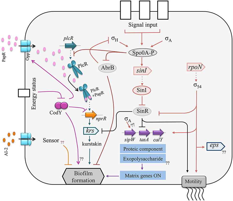
Figure 2. Schematic representation of the regulatory network controlling biofilm formation in B. cereus. Circles symbolize proteins, triangles symbolize open reading frames (ORFs). Arrows indicate activation and blunt lines indicate repression. Dotted arrows represent transcription. The protein component of the matrix is encoded by the sipW-tasA operon and by calY which promoters are activated by σ54 and repressed by SinR. SinR is antagonized by SinI. The transcription of sinI is activated by the master regulator of sporulation Spo0A. Furthermore, Spo0A downregulates the regulator AbrB, resulting in biofilm formation. Several quorum sensing systems are involved in biofilm formation. The regulator PlcR activates the transcription of nprR. NprR promotes kurstakin production, which itself promotes biofilm formation. The autoinducer AI-2 plays an inhibitory effect on biofilm formation.
However, the SinR regulon also displays important differences in the two species: the B. subtilis epsA-O, but not the B. thuringiensis eps, is included in this regulon. Conversely, the production of kurstakin, a lipopeptide biosurfactant, is controlled by SinR in B. thuringiensis while surfactin, a B. subtilis lipopeptide, is not in the SinR regulon. Kurstakin is also included in the NprR necrotrophic regulon required for survival in the insect cadaver (Dubois et al., 2012), and the hemolysin Hbl, controlled by SinR in B. thuringiensis (Fagerlund et al., 2014), is included in the PlcR virulence regulon of this species (Gohar et al., 2008). Other differences, in addition to the SinR regulon, exist between B. subtilis and B. cereus sensu lato for the regulation of biofilm formation. The AI2 autoinducer represses biofilm formation in B. cereus (Auger et al., 2006), but induces biofilm formation in B. subtilis (Duanis-Assaf et al., 2015), and the DegU regulator, which controls biofilm formation in B. subtilis (Kobayashi, 2007b; Cairns et al., 2014), has no homolog in B. cereus.
In B. thuringiensis, there is an interaction between biofilm formation, virulence and necrotrophism in insects (Figure 3), since PlcR promotes NprR transcription (Dubois et al., 2013), which positively controls kurstakin transcription (Dubois et al., 2012), which, in turn, promotes biofilm formation (Gélis-Jeanvoine et al., 2016). In B. cereus strain ATCC14579, PlcR was reported to repress biofilm formation (Hsueh et al., 2006), which is in disagreement with these observations. The disruption of nprR by a transposon in strain ATCC14579, and therefore the shutdown of the necrotrophic regulon, can explain this discrepancy. For the same reason, the regulator CodY was reported, either to repress biofilm formation in the B. cereus ATCC14579 strain (Lindbäck et al., 2012), or to promote biofilm formation in the B. cereus UW101C strain (Hsueh et al., 2008). CodY is a regulator sensing the energy and the nutrient state of the bacterial cell (Sonenshein, 2005). It promotes PlcR transcription in stationary phase (Frenzel et al., 2012; Lindbäck et al., 2012) by inducing the production of a transporter required for the import of the PlcR-activating peptide PapR (Slamti et al., 2015), and represses NprR transcription in exponential phase (Dubois et al., 2013). Therefore, the expected effect of CodY on biofilm formation, if this phenotype is induced in early stationary phase, should rather be positive. The connection between biofilm formation and virulence is mediated by another regulator in B. cereus. In this species, Sigma 54 (RpoN) promotes the transcription of virulence factors, eps genes and flagellins (Hayrapetyan et al., 2015b). These interconnections are an indication that biofilms could be involved in the pathogenic, commensal or necrotrophic lifestyles of B. cereus sensu lato.
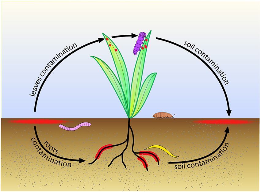
Figure 3. Suggested model for biofilm role in the life cycle of B. cereus and B. thuringiensis in the environment. Biofilms (in red) growing in the topsoil contaminate the roots and leaves of plants. Earthworm (in pink) feeding on soil organic matter, nematodes (in yellow) feeding on plant roots, caterpillar (in purple) feeding on plant leaves, or isopodes (in brown) feeding on plant debris, ingest bacteria, which can then grow as biofilms in their gut. The invertebrates move further in the environment and, upon death, contaminate back the topsoil, giving birth to a new cycle.
Heterogeneity in the Biofilm
The limited diffusion of nutrients and signal molecules within the biofilm matrix creates micro-environments and local quorum-sensing states, resulting in a heterogeneous spatial distribution of bacteria in different physiological states. This heterogeneity has been described in several species, including B. subtilis, where vegetative cells, sporulating cells, and matrix-producing cells co-exist with different spatial localizations (Vlamakis et al., 2008). In B. thuringiensis, motile vegetative cells make from 0.1 to 1% of the total biofilm population and could be beneficial to the whole community by creating channels within the biofilm matrix (Houry et al., 2012). In the same species, in a 48 h-aged biofilm, about 15% of the cells express the enterotoxin Hbl (Fagerlund et al., 2014) which, if it accumulates within the matrix, could make the biofilm a toxic patch-like structure when formed on host tissues. Actually, the biofilm matrix of strains ATCC14579 and ATCC10987 contains the enterotoxins Hbl and Nhe, a collagenase, the phospholipases PI-PLC and sphingomyelinase, and the immune inhibitor protease InhA1, all being virulence factors (Karunakaran and Biggs, 2011). Genes expression heterogeneity within the B. thuringiensis biofilm evolves with time, from 24 to 72 h, and shows a decrease in the proportion of bacteria expressing virulence genes, an increase in the proportion of bacteria expressing necrotrophic genes, and a constant proportion of sporulating cells (about 15%; Verplaetse et al., 2015). Interestingly, necrotrophic bacteria arouse mainly from cells which have previously expressed virulence genes. In a sporulating medium, only necrotrophic and sporulating bacteria were observed in the biofilm (Verplaetse et al., 2016).
Mobile Genetic Elements
Plasmids were shown to be involved in biofilm formation in a variety of Gram-negative and Gram-positive bacterial species (Cook and Dunny, 2014), through conjugative (Ghigo, 2001) as well as Non-conjugative mechanisms, and, conversely, biofilms were reported to favor plasmids transfer, resulting in an increase of genetic exchange between bacteria, including antibiotic resistance genes (Van Meervenne et al., 2014). Plasmids are present in all B. cereus, B. thuringiensis and B. anthracis strains, in number, not including copies, ranging from 1 to 13, and in size ranging from 2 to almost 500 kb (Rasko et al., 2005; Reyes-Ramirez and Ibarra, 2008). Strains of these species also harbor integrated or Non-integrated temperate prophages (Rasko et al., 2005). While mobile genetic elements play a key role in the adaptation of B. cereus and related species to their specific environment, data on their involvement in biofilm formation or on the role of biofilms in their transfer are scarce for this group of bacteria. The role of plasmids in biofilm formation have not been considered until now, although there are indications that large pXO1-like plasmids contained in periodontitis or emetic strains might be involved in the specific behavior of these strains regarding this phenotype. Indeed, addition to the culture medium of cereulide, the product of the ces locus located on the pCER270 emetic strains pXO1-like plasmid, promotes the formation of biofilm (Ekman et al., 2012). Conversely, phages were shown to act on biofilm formation. The GIL01 and GIL16 prophages of the tectiviridae family, present as linear plasmids in B. thuringiensis strains, negatively affect biofilm formation and sporulation, and enhance swarming motility (Gillis and Mahillon, 2014). In B. anthracis, prophages of different families (siphoviridae, myoviridae, or tectiviridae) could either inhibit sporulation (Wip4, Wip5, Frp1), or induce this phenotype (Wip1, Wip2, Frp2) in culture conditions where spore formation does not usually occur—for example absence of aeration (Schuch and Fischetti, 2009). The lysogenic strains containing one of these phages displayed an increased production of cell-surface exopolysaccharides and an enhanced production of biofilms at the air-liquid interface in BHI culture medium (Schuch and Fischetti, 2009). The phages effect on the ability to produce exopolysaccharides or biofilms was the result of a prophage-chromosome dialog mediated by a sigma-factor-like regulator encoded in the prophage sequence (Schuch and Fischetti, 2009).
Structure and Properties
Data related to the biofilm structure are scarcely available in B. cereus. Although the B. cereus biofilm macrostructure has been described, the distribution in the biofilm of the different bacterial subpopulations or its morphogenesis are unknown, even more in the case of multispecies biofilms. Biofilm properties include adhesion to surfaces (which is dealt with in the part 5- Biofilm control in the food environment, of this review) and resistance to stresses. They also include the ability of the biofilm to produce spores, a property which add to the problems induced by the biofilm persistence.
Structure
The B. cereus sensu lato floating pellicle displays differences in its architecture with the one produced by B. subtilis. The B. subtilis floating pellicle exhibits a high number of folds and do not bind to the recipient wall (Kobayashi, 2007a). In contrast, B. cereus biofilm, when formed at the air-liquid interface, includes a ring strongly sticking to the recipient wall, and the pellicle itself which displays protrusions instead of folds (Fagerlund et al., 2014). Wrinkles in the B. subtilis pellicle were shown to be a consequence of biomass extension, confined space, and elasticity of the pellicle, which is dependent from the extracellular matrix (Trejo et al., 2013). In B. subtilis colonies on agar plates, wrinkles forms preferentially where cell death occurs (Asally et al., 2012). The difference in the pellicle architecture between B. cereus and B. subtilis might be a consequence of the strong adhesion of the biofilm to the vessel walls in the former, and of the different polymers present in the matrix produced by the two species.
On immersed surfaces, B. subtilis and some B. cereus strains (see Section Ecological Aspects) are able to produce submerged biofilms. In the B. subtilis immerged biofilm, cells are organized in bundles which can, for some strains, protrude over the biofilm and form aerial structures at heights greater than 100 μm (Bridier et al., 2013). Few data are available on the structure of B. cereus immerged biofilm. The amount of biofilm formed in this condition was variable according to the strain, but a strain isolated from a food processing line produced, on stainless steel coupon, a thick and uneven biofilm with an aerial structure (Faille et al., 2014).
Properties: Sporulation and Resistance to Stresses
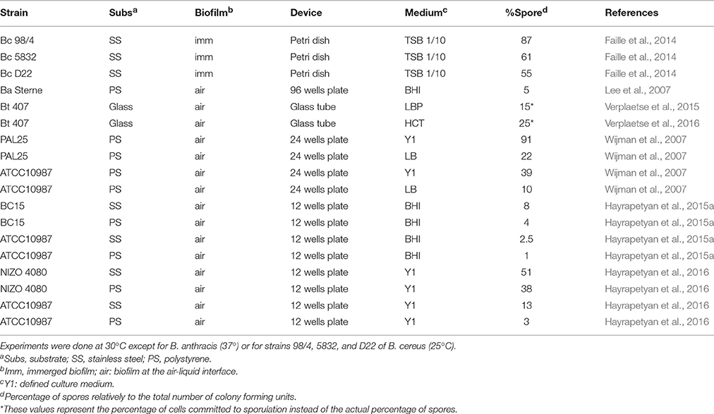
The limited diffusion of nutrients and signal molecules within the matrix creates microenvironments in the biofilm, resulting in a heterogeneity of the bacterial population, which might include cells in the motile, virulent, necrotrophic, or sporulating states, as discussed in the Section Molecular and Physiological Aspects of this review. Sporulation rates in biofilms were highly variable and were dependent from the strain, the culture medium or the device used to form the biofilm (Table 1). Highest rates were obtained with strains isolated from the food environment and grown in poor media, with rates as high as 90%. Sporulation could occur in immerged biofilms although the rate of sporulation was increased when the biofilm was exposed to air or was let to dry (Ryu and Beuchat, 2005; Hayrapetyan et al., 2016), and was greater in the biofilm comparatively to the coexisting planktonic population (Hayrapetyan et al., 2015a). Stainless steel was more favorable to sporulation within the biofilm than polystyrene (Table 1). It was hypothesized that this result could be due to an increased iron availability on stainless steel coupons, as a consequence of corrosion (Hayrapetyan et al., 2015a). In addition to be suitable for sporulation, the biofilm confers to bacteria a protection against stresses. In biofilm, B. anthracis was from 40 (doxycycline) to 150 (ciprofloxacine) times more resistant to antibiotics than planktonic cells (Lee et al., 2007), and a multispecies biofilms containing B. cereus and Pseudomonas fluorescens was more resistant to antimicrobials than the biofilm of each species alone (Simoes et al., 2009).
Ecological Aspects
In nature, bacteria live predominantly in biofilms rather than in a planktonic state (Costerton et al., 1995), and this observation is likely to stand also for B. cereus or B. thuringiensis. Consequently, biofilms are expected to be a key element for the adaptation of these species to their biotopes and to their biocenosis. However, B. cereus and its close relatives are found in a high diversity of biotopes, which questions the role that biofilm formation, in addition to other physiological properties, would play for their fitness to specific environments.
Biofilm Formation among B. Cereus Strains
Although biofilms are suspected to be involved in strains adaptation to their specific environment, there is a considerable variation in the ability to produce biofilms among isolates of B. cereus and B. thuringiensis, and no correlation was found between this ability and the origin (food poisoning, clinical, or environmental) of the strain (Wijman et al., 2007; Auger et al., 2009; Kuroki et al., 2009; Kamar et al., 2013; Hayrapetyan et al., 2015a). However, strains isolated from a specific niche, the oral cavity of periodontitis-diseased patients, were all unable to form biofilms (Auger et al., 2009), although these strains were isolated from dental plaques—which are biofilms. While unexpected, this result looks coherent since periodontal strains of B. cereus, as secondary colonizers of the dental plaque, do not need to initiate biofilms. Another interesting finding from prevalence studies is the observation that about 50% of B. cereus strains isolated from various food preparations produced less biofilms after 48 h than after 24 h of incubation (Hayrapetyan et al., 2015a), a proportion also found in emetic strains (Auger et al., 2009), which are frequent food contaminants (Ehling-Schulz et al., 2015). In contrast, only a minor proportion (less than 15%) of B. cereus strains isolated from blood samples (Kuroki et al., 2009), from the environment, or of B. thuringiensis strains (Auger et al., 2009) showed a drop in the biofilm biomass after 24 h of culture. This decrease can be explained by a massive emigration of biofilm cells. When back to the planktonic state, reverting cells will be able to create new biofilms and to spread the colonized area. Therefore, combined with their resistance to cleaning procedures (see the “Bacillus biofilms and their control in the food environment” section below), this property would confer food isolates the ability to persist and thrive in the food production lines.
Prevalence studies also revealed that the biomass of biofilms produced on stainless steel by B. cereus in LB or in a defined medium (Y1) is greater when they are formed at the air-liquid-solid interface than on submerged surfaces (Wijman et al., 2007). In BHI medium, only one strain, out of 23 isolates from food products, was able to form a submerged biofilm on polystyrene or on stainless steel coupons (Hayrapetyan et al., 2015a). Consequently, the property to form submerged biofilms appear to be rare among B. cereus strains. In the food industry production units, air-liquid interfaces are found in tanks while pipes are mostly in a flooded state. One would expect that the proportion of strains able to produce submerged biofilms would increase in isolates sampled from pipes when compared to isolates from tanks or to other isolates—although we have no data to support this expectation. It would be interesting to proceed to this comparison, since the ability to produce submerged biofilms affect B. cereus persistence within the food processing lines.
B. cereus Role in Multispecies Biofilms
Most biofilms found in natural environments include several bacterial species. B. cereus or B. thuringiensis make no exception to this observation and are found, when in biofilms, in association to other microorganisms. Multispecies biofilms are often described as cooperative consortiums where each partner contributes to the community resilience and development (Davey and O'toole, 2000). For example, periodontitis strains of B. cereus are found in the dental plaque (Rasko et al., 2007), which is one of the best studied multispecies biofilms. The dental plaquee is located at the tooth-gum interface and is a severe illness leading, ultimately, to gum bleeding, ligaments digestion and loosening and loss of teeth. Bacteria build the dental plaquee in a precise sequence, where pioneer species such as Streptococcus mutants bind first to the teeth enamel, followed by secondary colonizer species which bind to pioneer species or to themselves through a co-aggregation process (Kolenbrander et al., 2006). Secondary colonizers benefit from biofilm settlement by primary colonizers and, in turn, might contribute to the biofilm survival and growth. Indeed, B. cereus is able to shift the pH of a Streptococcus mutants biofilm from acidic to neutral values and in this way contributes to the biofilm pH balance (Sissons et al., 1998). It can also strongly participate to host tissues digestion owing to the numerous degradation enzymes which it secretes (Gohar et al., 2002) and which are present in the biofilm matrix (Karunakaran and Biggs, 2011). Likewise, B. cereus strains isolated from multispecies biofilms settled in paper machines were strong producers of exopolysaccharides (Ratto et al., 2005) and could therefore contribute actively to the biofilm development.
The integration of B. cereus vegetative cells can also occur in the depth of a Pre-existing biofilm, thanks to the high motility of these cells, which are able to create channels in the matrix and reach deep areas in the biofilm (Houry et al., 2010). Interestingly, B. cereus and B. thuringiensis secrete a number of bacteriocins (Ahern et al., 2003; Risoen et al., 2004; Oscariz et al., 2006), which, when produced within the integrated biofilm, could lead to drastic changes in the balance of bacterial biofilm populations. For example, a B. thuringiensis strain engineered to produce lysostaphin could invade and replace a Staphylococcus aureus biofilm native population (Houry et al., 2012), which clearly indicate that inter-species competition could occur within biofilms. Another example of competition between bacterial species within a natural biofilm is found in the pretreatment filters of water reclamation systems. These filters contain zeolite stones on which multispecies biofilms can grow. The B. cereus strains found in these biofilms are able to degrade the Gram-negative bacteria quorum sensing signal AHL (acylhomoserine lactone; Hu et al., 2003), interrupting the communication of their cohabitants and thus conferring a competitive advantage to B. cereus.
Biofilms in Soil, Plants, and Invertebrates
The environment is likely to be a major source of food contamination by microorganisms which can live in biofilms on plants or in the soil. B. cereus or B. thuringiensis are often described as saprophytic species whose natural habitat would be the soil (Vilain et al., 2006), from which they can easily be sampled (Vilas-Boas et al., 2002; Anjum and Krakat, 2016) and in which they can persist for long periods (Hendriksen and Carstensen, 2013). Interestingly, a number of B. cereus strains could multiply and form biofilm-like structures when cultivated in a liquid topsoil extract—but not in LB (Vilain et al., 2006), suggesting that some soil components are required to induce the formation of biofilm by B. cereus in the culture conditions used. However, not all soils can support B. cereus or B. thuringiensis growth, since an asporogenic strain of B. thuringiensis could not survive in a sterilized soil (Vilas-Boas et al., 2000), and it was speculated that the invertebrate gut rather than the soil might be the main ecological niche of these species (Jensen et al., 2003). B. cereus and B. thuringiensis were found in the gut of insects (Visotto et al., 2009), earthworms (Hendriksen and Hansen, 2002), nematodes (Schulte et al., 2010; Ruan et al., 2015), and isopods—which are terrestrial crustaceans (Swiecicka and Mahillon, 2006). In the intestine of insects and isopods, B. cereus forms filamentous structures described as “Arthromitus,” which proved to be chains of dividing bacteria (Margulis et al., 1998). Long chains of B. cereus or B. thuringiensis vegetative cells are typically found in biofilms, which suggests that these species can form biofilms in the gut of insects or isopods—and probably in the gut of other invertebrates as well.
In addition to the invertebrates gut, B. cereus is found in the rhizosphere and in the mycorrhiza of plants. When present in these subterranean structures, B. cereus can protect the plant from fungal attacks. For example, B. cereus UW85 produces zwittermicin A and kanosamine, both fungistatic molecules being suspected to contribute to the suppression of damping-off disease of alfalfa caused by Phytophthora medicaginis (Silo-Suh et al., 1994). Another strain of B. cereus (strain 0–9) isolated from roots of wheat cultures, was able to induce a reduction of 31% of the disease caused by the fungal pathogen Rhizoctonia cerealis, the agent of wheat sharp eyespot (Xu et al., 2014). A mutant of this strain obtained by random mutagenesis and selected for defective biofilm formation was unable to colonize wheat roots and to control the fungal disease (Xu et al., 2014). B. cereus is therefore likely to colonize plant roots through biofilm formation. This hypothesis is supported by the finding that, in B. subtilis, tasA, a gene required for biofilm formation which paralog is also required for biofilm formation in B. cereus (Caro-Astorga et al., 2015), is needed for the colonization of Arabidopsis thaliana roots (Lakshmanan et al., 2012). B. cereus can also be associated with plants through the mycorrhiza. It was, for example, isolated from Glomus irregulare spores sampled from the rhizosphere of Agrotis stolonifera growing in a natural stand (Lecomte et al., 2011) and was shown to form biofilms on the hyphae of Glomus sp. (Toljander et al., 2006). The arbuscular myccorhizal fungi are plant roots symbionts which mycelial network can explore soil volumes much larger than the roots themselves (Lecomte et al., 2011).
These data are summarized in the model depicted Figure 3, in which B. cereus and B. thuringiensis growing as biofilms in the topsoil would contaminate germinating plants, leading to biofilms on the rhizosphere and to spores on the phylloplane. Invertebrates feeding on roots (nematodes), soil organic matter (earthworms), vegetal debris (isopods), or leaves (caterpillars) would be infected by these bacteria, which could behave as commensals or as pathogens and settle as biofilms in their host gut. Invertebrates, through their mobility, could disseminate the bacteria in the environment and, upon death, contaminate back the topsoil, thus initiating a new cycle. Biofilms of B. cereus settled in soils and on plants could then contaminate raw food materials.
The Case of B. Anthracis
Formation of biofilms by B. anthracis in the environment is controversial. B. anthracis does not need to produce biofilms for its infective cycle in mammals. Its spore is the infective agent, its toxins are extremely efficient and it is protected against the host immune defenses by a capsule. After the host death, B. anthracis multiply within the host, sporulate, and the spores are finally released into the environment at the host death spot. It is believed that the spores can survive in the soil for a long time, keeping their full infective properties, until their uptake by a new host. Yet, it has been argued that a multiplication step would be required to explain how slow the spore decay in soil is. Indeed, multiplication was observed in soil on plant roots, where B. antthracis formed long chains reminiscent of the bacterial chains found in biofilms (Saile and Koehler, 2006). B. anthracis can also produce biofilms in static and in flow conditions (Lee et al., 2007; Schuch and Fischetti, 2009). It expresses the regulators required for biofilm formation and at least a part the proteic components of the biofilm matrix (Pflughoeft et al., 2011), and can sporulate in biofilms (Lee et al., 2007). In addition, B. anthracis can colonize the earthworm gut for long periods (Schuch and Fischetti, 2009) and is found in flies and mosquitoes (Turell and Knudson, 1987), although only short-term colonization of flies gut was observed (Fasanella et al., 2010). While these data support a multiplication of B. anthracis outside its mammal host, further observations and experiments are required to determine if the model displayed Figure 5 apply to this bacterium.
Biofilms Control in the Food Environment
Bacillus strains, including strains from the B. cereus group, can be isolated from endemic biofilms in various environments such as paperboard production or hospitals (Kolari et al., 2001; Ohsaki et al., 2007; Kuroki et al., 2009), but also food and beverage industries (Evans et al., 2004; Gunduz and Tuncel, 2006; Storgards et al., 2006; Marchand et al., 2012). The presence of biofilms containing B. cereus is a great concern for food industry settings such as fresh products, poultry, dairy, and red meat processing, and they are a potential source of recurrent cross-contamination and Post-processing contamination of finished products, sometimes resulting in food spoilage or foodborne illness (Rajkovic et al., 2008). The contamination of food processing lines by B. cereus biofilms could therefore be a serious public health risk, especially in foods that undergo mild processing such as minimally heat–treated foods (Tauveron et al., 2006). This risk must be given full attention since the total annual cost caused by B. cereus and Staphylococcus aureus in food illness is estimated at $523 million in the United States (Bennett et al., 2013).
B. Cereus, a Food Spoilage Agent
As underlined above, the presence of biofilms in the food industry can result in food spoilage. Indeed, B. cereus strains produce extracellular proteases and lipases resulting in food degradation and spoilage, like sweet curdling and bitterness of milk sour taste, decreasing the shelf life of the product and therefore resulting in significant economic loss to food producers (Fromm and Boor, 2004; Flach et al., 2014). Even if present in raw milk at low concentration, Bacillus sp. become dominant after long periods of storage at a temperature of 10°C (which is often the case in shops), or when produced in improved technological conditions (Samarzija et al., 2012). Consequently, Bacillus spp. are today considered the main microbial causes for the spoilage of milk and milk products, and the main reason for significant economic losses in the dairy industry (Meer et al., 1991; Brown, 2000). It is estimated that the dairy industry has losses of up to 30 % due to spoilage and reduced product quality caused by psychrotrophic bacteria, including Bacillus sp. (Samarzija et al., 2012).
Biofilms in Food Environments
In food environments, Bacillus biofilms are found on every food contact surfaces of open or closed equipment, such as conveyor belts, pasteurizers, evaporators, filling machines, storage tanks, but also on cleaning and handling tools (Christison et al., 2007). Depending on the species or the strain, surfaces of cold rooms and equipment of processes lines where elevated temperatures prevail could be contaminated by Bacillus biofilms (Sharma and Anand, 2002a; Kolari et al., 2003; Evans et al., 2004; Gunduz and Tuncel, 2006; Kumari and Sarkar, 2014). In fact, Bacillus spores or biofilms are capable of contaminating every surface commonly found in food-industry plants, including inert surfaces such as stainless steel surfaces (Faille et al., 2014), plastics or rubber (Mettler and Carpentier, 1997), but also surface of vegetables (Elhariry, 2011). Moreover, Bacillus strains are able to form biofilms both under static and flow conditions, and thick biofilms of B. cereus would particularly develop at the air-liquid interface (Wijman et al., 2007). Along food processing lines, B. cereus is often found in association with other bacterial species to form mixed biofilms (Figure 4) where high levels of Bacillus isolates have sometimes been reported (Mattila et al., 1990). For example, percentages as high as 25% of Bacillus sp. isolates (including B. cereus isolates) have been found in dairy processing industries (Sharma and Anand, 2002c). In addition, sporulation occurs within biofilms (Figure 5) on food contact surfaces (Storgards et al., 2006), sometimes at very high levels (De Vries et al., 2004; Faille et al., 2014), suggesting a potentially significant role for biofilm-derived spores in contamination of food with Bacillus spp. (Scott et al., 2007).
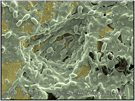
Figure 4. Observation by scanning electron microscopy of a mixed biofilm formed by two strains: B. cereus 98/4 and Comamonas testosteroni CCL24 (Faille et al., 2014).
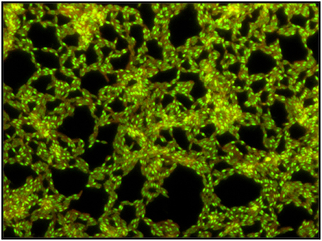
Figure 5. Microscopic images of a B. cereus biofilm grown for 48 h in TSB 1/10. Observation by epifluorescence after staining with the Live/Dead stain (magnification × 400). Endospores produced within the biofilm are stained in green, cells are stained in orange-green.
Biofilms Control
In food plants, disinfection of processing lines (e.g., pipes, heat-exchangers, valves tanks) is preceded by a cleaning step, involving alkali or other cleaning agents. Cleaning and sanitation procedures are set up to guarantee the detachment of organic and inorganic contaminations, disinfection of the cleaned surface and elimination of the residues of the sanitation agents (Vlkova et al., 2008). Unfortunately, the detachment of spores and biofilms but also of food residues in the food processing environment is critical since they often accumulate in areas which are difficult to clean, e.g., crevices, valve, gaskets, and dead ends (Czechowski, 1990; Austin and Bergeron, 1995; Sharma and Anand, 2002b). Of particular concern is the increased resistance of biofilms, compared with bacteria in a free-living environment, to disinfection processes. For example, two widely-used sanitizers, a quaternary ammonium compound and sodium hypochlorite, did not effectively inactivate the adherent single cells and biofilms of B. cereus at concentrations able to induce a reduction in CFU/ml of more than 5.0 log of their planktonic counterparts. Furthermore, the efficacy of both disinfectant was even lower when biofilms were formed on milk Pre-soiled stainless steel (Peng et al., 2002). Adherent Bacillus spores also exhibit a greater resistance to high temperature and disinfectant than spores in suspension (Sagripanti and Bonifacino, 1999; Faille et al., 2001; Kreske et al., 2006a). Indeed, residual Bacillus contamination of equipment surfaces after cleaning and/or sanitizing procedures was detected at different points on milk pasteurization lines and on the surface of the packaging machine (Mattila et al., 1990; Sharma and Anand, 2002b; Salustiano et al., 2009). Hence, considering the difficulty in inactivating adherent Bacillus spores and biofilms, cleaning the biomass from the surfaces is fundamental for controlling biofilm development.
Cleaning-in-Place Protocols
The cleaning-in-place (CIP) protocols used to clean processing lines without dismantling or opening of the equipment, vary according to industries or the food chain and the residues that need to be cleaned, although caustic and acid cleaning has remained the standard method used in many food processing industries. Both chemical (cleaning agents) and mechanical (shear stresses) actions are supposed to play a major role on soil removal. However, the effectiveness of CIP regimes against B. cereus biofilm has not been extensively reported. In the food industries, CIP regimes frequently involve a 60°C cleaning alkali wash (mainly sodium hydroxide), followed by an acid (mainly nitric acid) wash disinfection step (Bremer et al., 2006), but a reduction of viable spores by only 40% has been reported (Andersson et al., 1995). In the case of Bacillus biofilms, relatively low efficiency of the reference CIP regime (1% NaOH at 65°C for 10 min—water rinse—1% HNO3 at 65°C for 10 min—water rinse) has been reported, but the removal would be improved by increasing the concentration of NaOH or the duration of the cleaning procedure (Flint et al., 1997; Bremer et al., 2006; Kumari and Sarkar, 2014).
Mechanical and Chemical Cleaning
In order to better understand the mechanism of spore and biofilm detachment during CIP, the respective role of rinsing vs. cleaning (mechanical and chemical forces) in the detachment of Bacillus biofilms and spores was investigated. When the B. cereus biofilm was formed on milk Pre-soiled stainless chips (Peng et al., 2002) or at different shear stresses (Lemos et al., 2015), a rapid population decrease occurred during the first 5 min whatever the detachment conditions, and no further removal was observed for longer times, either in terms of vegetative cells or spores, even if the amount of detached biofilm was significantly higher in the presence of cleaning agents. Similar observations have been reported when B. cereus biofilm was formed on milk Pre-soiled stainless chips (Peng et al., 2002) or at different shear stresses (Lemos et al., 2015). Further works, performed on spores from the B. cereus group, demonstrated that during a CIP, chemical action plays a major role in the detachment of adherent spores, while mechanical action is poorly effective (less than 90% decrease in the number of adherent spores at wall shear stresses of 500 Pa, whatever the strain; Faille et al., 2013). Spores produced in biofilms showed greater resistance to detachment than the complete biofilms on inert surfaces (Faille et al., 2014) and on vegetables (Elhariry, 2011).
If the contaminated areas are allowed to dry before cleaning, e.g., in half-filled tanks or pipes or on open surfaces, the sporulation level would increase within Bacillus biofilms (Hayrapetyan et al., 2016) and the resistance to shear of attached spores increase concomitantly (Nanasaki et al., 2010). The increase in resistance to detachment is particularly noteworthy for long times and/or high temperature of drying (Faille et al., 2016).
In order to improve the efficiency of cleaning procedures, some industrialists opted to develop enzymatic cocktails effective against biofilms found in food processing plants, which are known to poorly respond to traditional cleaning procedures. The enzymes offer major advantages over traditional cleaning solutions, e.g., low toxicological risk and ecological risk, ease of rinsing external residues and compatibility with different surface material. Many products are nowadays commercially available, essentially for medical use. Some of the commercialized cocktails have proven their efficiency against biofilms produced by B. cereus, B. mycoides or B. flavothermodurans, and also against B. cereus adherent spores (Langsrud et al., 2000; Parkar et al., 2004; Lequette et al., 2010). These enzymatic “detergents” being more expensive than conventional products, their use is proposed as a complementary solution to current cleaning procedures.
Spores and, to a lesser extent, vegetative cells embedded in a B. cereus biofilm are protected against inactivation by the sanitizers commonly used to control foodborne pathogens, such as chlorine and hydrogen peroxide, which are easy to handle, inexpensive, and are soluble in water and relatively stable over a long storage time. For example, hydrogen peroxide or peracetic acid show little activity on adherent B. subtilis and B. cereus spores (Faille et al., 2001; Dequeiroz and Day, 2008). At higher temperatures and longer exposures, a significant reduction in B. cereus viable counts would be observed, but it is not suitable for practical disinfection due to corrosion and toxicity (Langsrud et al., 2000; Dequeiroz and Day, 2008). However, although the peroxygen-based disinfectants are not sporicidal alone, the use of NaOH 1% (typically used at 0.5–2% in the food and beverage industries) or of an enzymatic cocktail would sensitize Bacillus spores to the action of these oxidative disinfectants (Langsrud et al., 2000). The activity of sodium hypochlorite on B. cereus spores on surfaces and in field trials is also limited (Te Giffel et al., 1995). Indeed, although hypochlorite solutions are more stable above pH 9.5, they are only efficient at neutral or acidic pH (Sagripanti and Bonifacino, 1999). However, a marked synergistic effect between both was described on the efficacy to reduce spore counts on contaminated surfaces (Dequeiroz and Day, 2008). The same phenomenon was observed with biofilms produced in immersed conditions or exposed to air (Ryu and Beuchat, 2005). Furthermore, chlorine dioxide was less effective than chlorine in killing Bacillus spores on stainless steel, mainly in the presence of organic soil (Kreske et al., 2006a) and injured B. cereus cells were sometimes seen to recover overnight (Lindsay et al., 2002). Within biofilms, spores were more resistant to chlorine and chlorine dioxide than the vegetative cells (Kreske et al., 2006b).
Control of Multispecies Biofilms Including B. cereus
The control of mixed species biofilms including B. cereus and other Bacillus species has also been investigated. For example, the efficiency of sodium hypochlorite and iodophor, commonly used in the beverage and dairy industries, has been studied in different segments of pasteurization lines (Sharma and Anand, 2002b). Results from this study suggest that sodium iodophors were in some cases more efficient than sodium hypochlorite in inactivating biofilms and that the latter treatment was affected by the constitutive microflora or by spatial heterogeneity of biofilms. However, biofilms were still detected on the different areas even after CIP and iodophor treatment. Since iodophors are much less active against spores than hypochlorite, one can hypothesize that the residual biofilms following treatment with iodophors would be largely composed of Bacillus spores. A laboratory work on dual biofilms (B. cereus and P. fluorescens) showed that dual biofilms are characterized by an increased stability to shear stress and are more resistant to a quaternary ammonium compound (QAC), cetyltrimethylammonium bromide, and glutaraldehyde solutions (sanitizers commonly used in the medical field) than each single species biofilm (Simoes et al., 2009). Once more, a significant proportion of the population of both bacteria remain in a viable state after exposure to antimicrobials. The presence of residual bacterial population after treatment by QACs, also frequently used in food-processing industries, could encourage the development of resistance among food-associated bacteria, as already observed in Gram-negative bacteria and Enterococcus spp. (Sidhu et al., 2002).
Concluding Remarks
In the last decade, a number of studies have shown that although B. cereus sensu lato biofilms looked the same as the B. subtilis ones, there are quite different in several aspects. These studies brought a huge improvement to our understanding of how B. cereus biofilms are built, what is their contribution to the bacterium lifestyle, or how to get rid of them when required. Still, a number of issues stay unresolved or has been brought to light by recent findings. While the role of the TasA-like proteins in the biofilm matrix has been confirmed, the duplication of their genes asks the question of their role in the biofilm formation and in the adaptation of the bacterium to its environment or to its host. Similarly, the genetic determinants required for the building of the polysaccharidic part of the matrix remains a mystery, as well as the regulation of their production and the role of the large epsA-O -like polysaccharidic locus, since this locus does not seems to be involved in biofilm formation. The mechanisms through which eDNA, which was found in high quantities in the B. cereus biofilm matrix, is released remains unknown. The possible involvement of programmed cell death (PCD) in this release as well as in the shaping of the biofilm architecture, and the connection of its regulation to the regulation of biofilm formation represent other exciting issues in the forthcoming work on B. cereus biofilm formation. The impact of plasmids, which are known to play a major role in B. cereus sensu lato pathogenesis, on biofilm formation, and the mechanism through which plasmids act on this phenotype is still to be determined. Regarding pathogenesis, the presence and the evolution of biofilms in vivo has not been yet established, nor has been their exact contribution to the bacterium virulence. Another important issue is relative to the role of biofilms in the B. cereus sensu lato, including B. anthracis, survival and growth in the soil environment. Finally, the traditional hygiene procedures used in the food industry have revealed their limit in the control of surface contamination with Bacillus spores and biofilms. If we consider that B. cereus and other species can act as spoilage organisms and pathogens, these surface contaminations are still of concern in the food industry. This problem is thus far from being resolved and there are many questions that remain to be addressed concerning the different approaches to manage the surface hygiene and limit the risks to consumers.
Author Contributions
All authors listed, have made substantial, direct and intellectual contribution to the work, and approved it for publication.
Funding
Researches were funded by the Agence Nationale pour la Recherche (ANR, France), Campus France, the University St Joseph of Beirut and the Conseil National de la Recherche Scientifique (CNRS-L, Lebanon). These agencies had no role in this work (study design, data analysis, manuscript writing)
Conflict of Interest Statement
The authors declare that the research was conducted in the absence of any commercial or financial relationships that could be construed as a potential conflict of interest.
Acknowledgments
We would like to thank the Lebanese National Council for Scientific Research (CNRS-L), the Grant research program 01-08-15 and the Scholarship Programs 2014-2015, and Campus France for supporting Racha Majed. In addition our gratitude is also extended to the Research Council of Saint-Joseph University: CNRS-FS81 and FS 84. This study was also funded by the French Agence Nationale de la Recherche (Bt-Surf, N°ANR-12-EMMA_0005).
References
Abee, T., Kovács, A. T., Kuipers, O. P., and Van Der Veen, S. (2011). Biofilm formation and dispersal in Gram-positive bacteria. Curr. Opin. Biotechnol. 22, 172–179. doi: 10.1016/j.copbio.2010.10.016
Ahern, M., Verschueren, S., and Van Sinderen, D. (2003). Isolation and characterisation of a novel bacteriocin produced by Bacillus thuringiensis strain B439. FEMS Microbiol. Lett. 220, 127–131. doi: 10.1016/S0378-1097(03)00086-7
Andersson, A., Ronner, U., and Granum, P. E. (1995). What problems does the food industry have with the spore-forming pathogens Bacillus cereus and Clostridium perfringens? Int. J. Food Microbiol. 28, 145–155.
Anjum, R., and Krakat, N. (2016). Detection of multiple resistances, biofilm formation and conjugative transfer of Bacillus cereus from contaminated soils. Curr. Microbiol. 72, 321–328. doi: 10.1007/s00284-015-0952-1
Asally, M., Kittisopikul, M., Rue, P., Du, Y., Hu, Z., çagatay, T., et al. (2012). Localized cell death focuses mechanical forces during 3D patterning in a biofilm. Proc. Natl. Acad. Sci. U.S.A. 109, 18891–18896. doi: 10.1073/pnas.1212429109
Auger, S., Krin, E., Aymerich, S., and Gohar, M. (2006). Autoinducer 2 affects biofilm formation by Bacillus cereus. Appl. Environ. Microbiol. 72, 937–941. doi: 10.1128/AEM.72.1.937-941.2006
Auger, S., Ramarao, N., Faille, C., Fouet, A., Aymerich, S., and Gohar, M. (2009). Biofilm formation and cell surface properties among pathogenic and nonpathogenic strains of the Bacillus cereus group. Appl. Environ. Microbiol. 75, 6616–6618. doi: 10.1128/AEM.00155-09
Austin, J. W., and Bergeron, G. (1995). Development of bacterial biofilms in dairy processing lines. J. Dairy Res. 62, 509–519. doi: 10.1017/S0022029900031204
Bennett, S. D., Walsh, K. A., and Gould, L. H. (2013). Foodborne disease outbreaks caused by Bacillus cereus, Clostridium perfringens, and Staphylococcus aureus–United States, 1998-2008. Clin. Infect. Dis. 57, 425–433. doi: 10.1093/cid/cit244
Branda, S. S., Gonzalez-Pastor, J. E., Ben Yehuda, S., Losick, R., and Kolter, R. (2001). Fruiting body formation by Bacillus subtilis. Proc. Natl. Acad. Sci. U.S.A. 98, 11621–11626. doi: 10.1073/pnas.191384198
Bremer, P. J., Fillery, S., and Mcquillan, A. J. (2006). Laboratory scale Clean-In-Place (CIP) studies on the effectiveness of different caustic and acid wash steps on the removal of dairy biofilms. Int. J. Food Microbiol. 106, 254–262. doi: 10.1016/j.ijfoodmicro.2005.07.004
Bridier, A., Meylheuc, T., and Briandet, R. (2013). Realistic representation of Bacillus subtilis biofilms architecture using combined microscopy (CLSM, ESEM and FESEM). Micron 48, 65–69. doi: 10.1016/j.micron.2013.02.013
Brown, K. L. (2000). Control of bacterial spores. Br. Med. Bull. 56, 158–171. doi: 10.1258/0007142001902860
Cairns, L. S., Hobley, L., and Stanley-Wall, N. R. (2014). Biofilm formation by Bacillus subtilis: new insights into regulatory strategies and assembly mechanisms. Mol. Microbiol. 93, 587–598. doi: 10.1111/mmi.12697
Candela, T., Maes, E., Garénaux, E., Rombouts, Y., Krzewinski, F., Gohar, M., et al. (2011). Environmental and biofilm-dependent changes in a Bacillus cereus secondary cell wall polysaccharide. J. Biol. Chem. 286, 31250–31262. doi: 10.1074/jbc.M111.249821
Caro-Astorga, J., Pérez-García, A., De Vicente, A., and Romero, D. (2015). A genomic region involved in the formation of adhesin fibers in Bacillus cereus biofilms. Front. Microbiol. 5:745. doi: 10.3389/fmicb.2014.00745
Christison, C. A., Lindsay, D., and Von Holy, A. (2007). Cleaning and handling implements as potential reservoirs for bacterial contamination of some ready-to-eat foods in retail delicatessen environments. J. Food Prot. 70, 2878–2883.
Cook, L. C., and Dunny, G. M. (2014). The influence of biofilms in the biology of plasmids. Microbiol. Spectr. 2:0012. doi: 10.1128/microbiolspec.plas-0012-2013
Costerton, J. W., Lewandowski, Z., Caldwell, D. E., Korber, D. R., and Lappin-Scott, H. M. (1995). Microbial biofilms. Annu. Rev. Microbiol. 49, 711–745. doi: 10.1146/annurev.mi.49.100195.003431
Cutting, S. M. (2011). Bacillus probiotics. Food Microbiol. 28, 214–220. doi: 10.1016/j.fm.2010.03.007
Czechowski, M. H. (1990). Bacterial attachment to Buna-n gaskets in milk processing equipment. Aust. J. Dairy Technol. 45, 113–114.
Davey, M. E., and O'toole, G. A. (2000). Microbial biofilms: from ecology to molecular genetics. Microbiol. Mol. Biol. Rev. 64, 847–867. doi: 10.1128/MMBR.64.4.847-867.2000
De Been, M., Francke, C., Moezelaar, R., Abee, T., and Siezen, R. J. (2006). Comparative analysis of two-component signal transduction systems of Bacillus cereus, Bacillus thuringiensis and Bacillus anthracis. Microbiology 152, 3035–3048. doi: 10.1099/mic.0.29137-0
Dequeiroz, G. A., and Day, D. F. (2008). Disinfection of Bacillus subtilis spore-contaminated surface materials with a sodium hypochlorite and a hydrogen peroxide-based sanitizer. Lett. Appl. Microbiol. 46, 176–180. doi: 10.1111/j.1472-765X.2007.02283.x
De Vries, Y. P., Van Der Voort, M., Wijman, J., Van Schaik, W., Hornstra, L. M., De Vos, W. M., et al. (2004). Progress in food-related research focussing on Bacillus cereus. Microbes Environ. 19, 265–269. doi: 10.1264/jsme2.19.265
Duanis-Assaf, D., Steinberg, D., Chai, Y., and Shemesh, M. (2015). The LuxS based quorum sensing governs lactose induced biofilm formation by Bacillus subtilis. Front. Microbiol. 6:1517. doi: 10.3389/fmicb.2015.01517
Dubois, T., Faegri, K., Perchat, S., Lemy, C., Buisson, C., Nielsen-Leroux, C., et al. (2012). Necrotrophism is a quorum-sensing-regulated lifestyle in Bacillus thuringiensis. PLoS Pathog. 8:e1002629. doi: 10.1371/journal.ppat.1002629
Dubois, T., Perchat, S., Verplaetse, E., Gominet, M., Lemy, C., Aumont-Nicaise, M., et al. (2013). Activity of the Bacillus thuringiensis NprR-NprX cell-cell communication system is co-ordinated to the physiological stage through a complex transcriptional regulation. Mol. Microbiol. 88, 48–63. doi: 10.1111/mmi.12168
Ehling-Schulz, M., Frenzel, E., and Gohar, M. (2015). Food-bacteria interplay: pathometabolism of emetic Bacillus cereus. Front. Microbiol. 6:704. doi: 10.3389/fmicb.2015.00704
Ekman, J. V., Kruglov, A., Andersson, M. A., Mikkola, R., Raulio, M., and Salkinoja-Salonen, M. (2012). Cereulide produced by Bacillus cereus increases the fitness of the producer organism in low-potassium environments. Microbiology 158, 1106–1116. doi: 10.1099/mic.0.053520-0
Elhariry, H. M. (2011). Attachment strength and biofilm forming ability of Bacillus cereus on green-leafy vegetables: cabbage and lettuce. Food Microbiol. 28, 1266–1274. doi: 10.1016/j.fm.2011.05.004
Evans, J. A., Russell, S. L., James, C., and Corry, J. E. L. (2004). Microbial contamination of food refrigeration equipment. J. Food Eng. 62, 225–232. doi: 10.1016/S0260-8774(03)00235-8
Fagerlund, A., Dubois, T., Økstad, O. A., Verplaetse, E., Gilois, N., Bennaceur, I., et al. (2014). SinR controls enterotoxin expression in Bacillus thuringiensis biofilms. PLoS ONE 9:e87532. doi: 10.1371/journal.pone.0087532
Faille, C., Benezech, T., Blel, W., Ronse, A., Ronse, G., Clarisse, M., et al. (2013). Role of mechanical vs. chemical action in the removal of adherent Bacillus spores during CIP procedures. Food Microbiol. 33, 149–157. doi: 10.1016/j.fm.2012.09.010
Faille, C., Benezech, T., Midelet-Bourdin, G., Lequette, Y., Clarisse, M., Ronse, G., et al. (2014). Sporulation of Bacillus spp. within biofilms: a potential source of contamination in food processing environments. Food Microbiol. 40, 64–74. doi: 10.1016/j.fm.2013.12.004
Faille, C., Bihi, L., Ronse, G., Baudoin, M., and Zoueshtiagh, F. (2016). Increased resistance to detachment of adherent microspheres and Bacillus spores subjected to a drying step. Colloids Surf. B Biointerfaces 143, 293–300. doi: 10.1016/j.colsurfb.2016.03.041
Faille, C., Fontaine, F., and Benezech, T. (2001). Potential occurrence of adhering living Bacillus spores in milk product processing lines. J. Appl. Microbiol. 90, 892–900. doi: 10.1046/j.1365-2672.2001.01321.x
Fasanella, A., Scasciamacchia, S., Garofolo, G., Giangaspero, A., Tarsitano, E., and Adone, R. (2010). Evaluation of the house fly Musca domestica as a mechanical vector for an anthrax. PLoS ONE 5:e12219. doi: 10.1371/journal.pone.0012219
Flach, J., Grzybowski, V., Toniazzo, G., and Corcao, G. (2014). Adhesion and production of degrading enzymes by bacteria isolated from biofilms in raw milk cooling tanks. Food Sci. Technol. 34, 571–576. doi: 10.1590/1678-457x.6374
Flemming, H. C., and Wingender, J. (2010). The biofilm matrix. Nat. Rev. Microbiol. 8, 623–633. doi: 10.1038/nrmicro2415
Flint, S. H., Bremer, P. J., and Brooks, J. D. (1997). Biofilms in dairy manufacturing plant - Description, current concerns and methods of control. Biofouling 11, 81–97. doi: 10.1080/08927019709378321
Frenzel, E., Doll, V., Pauthner, M., Lucking, G., Scherer, S., and Ehling-Schulz, M. (2012). CodY orchestrates the expression of virulence determinants in emetic Bacillus cereus by impacting key regulatory circuits. Mol. Microbiol. 85, 67–88. doi: 10.1111/j.1365-2958.2012.08090.x
Fromm, H. I., and Boor, K. J. (2004). Characterization of pasteurized fluid milk shelf-life attributes. J. Food Sci. 69, M207–M214. doi: 10.1111/j.1365-2621.2004.tb09889.x
Gao, T., Foulston, L., Chai, Y., Wang, Q., and Losick, R. (2015). Alternative modes of biofilm formation by plant-associated Bacillus cereus. Microbiologyopen 4, 452–464. doi: 10.1002/mbo3.251
Gélis-Jeanvoine, S., Canette, A., Gohar, M., Gominet, M., Slamti, L., and Lereclus, D. (2016). Genetic and functional analyses of kurstakin, a lipopeptide produced by Bacillus thuringiensis. Res. Microbiol. doi: 10.1016/j.resmic.2016.06.002. [Epub ahead of print].
Ghigo, J. M. (2001). Natural conjugative plasmids induce bacterial biofilm development. Nature 412, 442–445. doi: 10.1038/35086581
Gillis, A., and Mahillon, J. (2014). Influence of lysogeny of Tectiviruses GIL01 and GIL16 on Bacillus thuringiensis growth, biofilm formation, and swarming motility. Appl. Environ. Microbiol. 80, 7620–7630. doi: 10.1128/AEM.01869-14
Gohar, M., Faegri, K., Perchat, S., Ravnum, S., Okstad, O. A., Gominet, M., et al. (2008). The PlcR virulence regulon of Bacillus cereus. PLoS ONE 3:e2793. doi: 10.1371/journal.pone.0002793
Gohar, M., Økstad, O. A., Gilois, N., Sanchis, V., Kolsto, A. B., and Lereclus, D. (2002). Two-dimensional electrophoresis analysis of the extracellular proteome of Bacillus cereus reveals the importance of the PlcR regulon. Proteomics 2, 784–791. doi: 10.1002/1615-9861(200206)2:6<784::AID-PROT784>3.0.CO;2-R
Gunduz, G. T., and Tuncel, G. (2006). Biofilm formation in an ice cream plant. Antonie Van Leeuwenhoek 89, 329–336. doi: 10.1007/s10482-005-9035-9
Hamon, M. A., and Lazazzera, B. A. (2001). The sporulation transcription factor Spo0A is required for biofilm development in Bacillus subtilis. Mol. Microbiol. 42, 1199–1210. doi: 10.1046/j.1365-2958.2001.02709.x
Hayrapetyan, H., Abee, T., and Nierop Groot, M. (2016). Sporulation dynamics and spore heat resistance in wet and dry biofilms of Bacillus cereus. Food Control 60, 493–499. doi: 10.1016/j.foodcont.2015.08.027
Hayrapetyan, H., Muller, L., Tempelaars, M., Abee, T., and Nierop Groot, M. (2015a). Comparative analysis of biofilm formation by Bacillus cereus reference strains and undomesticated food isolates and the effect of free iron. Int. J. Food Microbiol. 200, 72–79. doi: 10.1016/j.ijfoodmicro.2015.02.005
Hayrapetyan, H., Tempelaars, M., Nierop Groot, M., and Abee, T. (2015b). Bacillus cereus ATCC 14579 RpoN (Sigma 54) Is a pleiotropic regulator of growth, carbohydrate metabolism, motility, biofilm formation and toxin production. PLoS ONE 10:e0134872. doi: 10.1371/journal.pone.0134872
Helgason, E., Tourasse, N. J., Meisal, R., Caugant, D. A., and Kolsto, A. B. (2004). Multilocus sequence typing scheme for bacteria of the Bacillus cereus group. Appl. Environ. Microbiol 70, 191–201. doi: 10.1128/AEM.70.1.191-201.2004
Hendriksen, N. B., and Carstensen, J. (2013). Long-term survival of Bacillus thuringiensis subsp. kurstaki in a field trial. Can. J. Microbiol. 59, 34–38. doi: 10.1139/cjm-2012-0380
Hendriksen, N. B., and Hansen, B. M. (2002). Long-term survival and germination of Bacillus thuringiensis var. kurstaki in a field trial. Can. J. Microbiol. 48, 256–261. doi: 10.1139/w02-009
Hobley, L., Ostrowski, A., Rao, F. V., Bromley, K. M., Porter, M., Prescott, A. R., et al. (2013). BslA is a self-assembling bacterial hydrophobin that coats the Bacillus subtilis biofilm. Proc. Natl. Acad. Sci. U.S.A. 110, 13600–13605. doi: 10.1073/pnas.1306390110
Houry, A., Briandet, R., Aymerich, S., and Gohar, M. (2010). Involvement of motility and flagella in Bacillus cereus biofilm formation. Microbiology 156, 1009–1018. doi: 10.1099/mic.0.034827-0
Houry, A., Gohar, M., Deschamps, J., Tischenko, E., Aymerich, S., Gruss, A., et al. (2012). Bacterial swimmers that infiltrate and take over the biofilm matrix. Proc. Natl. Acad. Sci. U.S.A. 109, 13088–13093. doi: 10.1073/pnas.1200791109
Hsueh, Y. H., Somers, E. B., Lereclus, D., and Wong, A. C. (2006). Biofilm formation by Bacillus cereus is influenced by PlcR, a pleiotropic regulator. Appl. Environ. Microbiol. 72, 5089–5092. doi: 10.1128/AEM.00573-06
Hsueh, Y. H., Somers, E. B., and Wong, A. C. (2008). Characterization of the codY gene and its influence on biofilm formation in Bacillus cereus. Arch. Microbiol. 189, 557–568. doi: 10.1007/s00203-008-0348-8
Hu, J. Y., Fan, Y., Lin, Y. H., Zhang, H. B., Ong, S. L., Dong, N., et al. (2003). Microbial diversity and prevalence of virulent pathogens in biofilms developed in a water reclamation system. Res. Microbiol 154, 623–629. doi: 10.1016/j.resmic.2003.09.004
Irnov, I., and Winkler, W. C. (2010). A regulatory RNA required for antitermination of biofilm and capsular polysaccharide operons in Bacillales. Mol. Microbiol. 76, 559–575. doi: 10.1111/j.1365-2958.2010.07131.x
Ivanova, N., Sorokin, A., Anderson, I., Galleron, N., Candelon, B., Kapatral, V., et al. (2003). Genome sequence of Bacillus cereus and comparative analysis with Bacillus anthracis. Nature 423, 87–91. doi: 10.1038/nature01582
Izano, E. A., Sadovskaya, I., Wang, H., Vinogradov, E., Ragunath, C., Ramasubbu, N., et al. (2008). Poly-N-acetylglucosamine mediates biofilm formation and detergent resistance in Aggregatibacter actinomycetemcomitans. Microb. Pathog. 44, 52–60. doi: 10.1016/j.micpath.2007.08.004
Jensen, G. B., Hansen, B. M., Eilenberg, J., and Mahillon, J. (2003). The hidden lifestyles of Bacillus cereus and relatives. Environ. Microbiol. 5, 631–640. doi: 10.1046/j.1462-2920.2003.00461.x
Kamar, R., Gohar, M., Jéhanno, I., Rejasse, A., Kallassy, M., Lereclus, D., et al. (2013). Pathogenic potential of Bacillus cereus strains as revealed by phenotypic analysis. J. Clin. Microbiol. 51, 320–323. doi: 10.1128/JCM.02848-12
Karunakaran, E., and Biggs, C. A. (2011). Mechanisms of Bacillus cereus biofilm formation: an investigation of the physicochemical characteristics of cell surfaces and extracellular proteins. Appl. Microbiol. Biotechnol. 89, 1161–1175. doi: 10.1007/s00253-010-2919-2
Kearns, D. B., Chu, F., Branda, S. S., Kolter, R., and Losick, R. (2005). A master regulator for biofilm formation by Bacillus subtilis. Mol. Microbiol. 55, 739–749. doi: 10.1111/j.1365-2958.2004.04440.x
Kobayashi, K. (2007a). Bacillus subtilis pellicle formation proceeds through genetically defined morphological changes. J. Bacteriol. 189, 4920–4931. doi: 10.1128/JB.00157-07
Kobayashi, K. (2007b). Gradual activation of the response regulator DegU controls serial expression of genes for flagellum formation and biofilm formation in Bacillus subtilis molecular Microbiology 66, 395–409. doi: 10.1111/j.1365-2958.2007.05923.x
Kolari, M., Nuutinen, J., Rainey, F. A., and Salkinoja-Salonen, M. S. (2003). Colored moderately thermophilic bacteria in paper-machine biofilms. J. Ind. Microbiol. Biotechnol. 30, 225–238. doi: 10.1007/s10295-003-0047-z
Kolari, M., Nuutinen, J., and Salkinoja-Salonen, M. S. (2001). Mechanisms of biofilm formation in paper machine by Bacillus species: the role of Deinococcus geothermalis. J. Ind. Microbiol. Biotechnol. 27, 343–351. doi: 10.1038/sj.jim.7000201
Kolenbrander, P. E., Palmer, R. J. Jr., Rickard, A. H., Jakubovics, N. S., Chalmers, N. I., and Diaz, P. I. (2006). Bacterial interactions and successions during plaque development. Periodontol. 2000. 42, 47–79. doi: 10.1111/j.1600-0757.2006.00187.x
Kreske, A. C., Ryu, J. H., and Beuchat, L. R. (2006a). Evaluation of chlorine, chlorine dioxide, and a peroxyacetic acid-based sanitizer for effectiveness in killing Bacillus cereus and Bacillus thuringiensis spores in suspensions, on the surface of stainless steel, and on apples. J. Food Prot. 69, 1892–1903.
Kreske, A. C., Ryu, J. H., Pettigrew, C. A., and Beuchat, L. R. (2006b). Lethality of chlorine, chlorine dioxide, and a commercial produce sanitizer to Bacillus cereus and Pseudomonas in a liquid detergent, on stainless steel, and in biofilm. J. Food Prot. 69, 2621–2634.
Kumari, S., and Sarkar, P. K. (2014). In vitro model study for biofilm formation by Bacillus cereus in dairy chilling tanks and optimization of clean-in-place (CIP) regimes using response surface methodology. Food Control 36, 153–158. doi: 10.1016/j.foodcont.2013.08.014
Kuroki, R., Kawakami, K., Qin, L., Kaji, C., Watanabe, K., Kimura, Y., et al. (2009). Nosocomial bacteremia caused by biofilm-forming Bacillus cereus and Bacillus thuringiensis. Intern. Med. 48, 791–796. doi: 10.2169/internalmedicine.48.1885
Lakshmanan, V., Kitto, S. L., Caplan, J. L., Hsueh, Y. H., Kearns, D. B., Wu, Y. S., et al. (2012). Microbe-associated molecular patterns-triggered root responses mediate beneficial rhizobacterial recruitment in Arabidopsis. Plant Physiol. 160, 1642–1661. doi: 10.1104/pp.112.200386
Langsrud, S., Baardsen, B., and Sundheim, G. (2000). Potentiation of the lethal effect of peroxygen on Bacillus cereus spores by alkali and enzyme wash. Int. J. Food Microbiol. 56, 81–86. doi: 10.1016/S0168-1605(00)00221-X
Lecomte, J., St-Arnaud, M., and Hijri, M. (2011). Isolation and identification of soil bacteria growing at the expense of arbuscular mycorrhizal fungi. FEMS Microbiol. Lett. 317, 43–51. doi: 10.1111/j.1574-6968.2011.02209.x
Lee, K., Costerton, J. W., Ravel, J., Auerbach, R. K., Wagner, D. M., Keim, P., et al. (2007). Phenotypic and functional characterization of Bacillus anthracis biofilms. Microbiology 153, 1693–1701. doi: 10.1099/mic.0.2006/003376-0
Lemon, K. P., Earl, A. M., Vlamakis, H. C., Aguilar, C., and Kolter, R. (2008). Biofilm development with an emphasis on Bacillus subtilis Curr. Top. Microbiol. Immunol. 322, 1–16. doi: 10.1007/978-3-540-75418-3_1
Lemos, M., Mergulhão, F., Melo, L., and Simões, M. (2015). The effect of shear stress on the formation and removal of Bacillus cereus biofilms. Food Bioproducts Process. 93, 242–248. doi: 10.1016/j.fbp.2014.09.005
Lequette, Y., Boels, G., Clarisse, M., and Faille, C. (2010). Using enzymes to remove biofilms of bacterial isolates sampled in the food-industry. Biofouling 26, 421–431. doi: 10.1080/08927011003699535
Lindback, T., Mols, M., Basset, C., Granum, P. E., Kuipers, O. P., and Kovacs, A. T. (2012). CodY, a pleiotropic regulator, influences multicellular behaviour and efficient production of virulence factors in Bacillus cereus. Environ. Microbiol. 14, 2233–2246. doi: 10.1111/j.1462-2920.2012.02766.x
Lindsay, D., Brözel, V. S., Mostert, J. F., and Von Holy, A. (2002). Differential efficacy of a chlorine dioxide-containing sanitizer against single species and binary biofilms of a dairy-associated Bacillus cereus and a Pseudomonas fluorescens isolate. J. Appl. Microbiol. 92, 352–361. doi: 10.1046/j.1365-2672.2002.01538.x
Marchand, S., De Block, J., De Jonghe, V., Coorevits, A., Heyndrickx, M., and Herman, L. (2012). Biofilm formation in milk production and processing environments; influence on milk quality and safety. Compr. Rev. Food Sci. Food Saf. 11, 133–147. doi: 10.1111/j.1541-4337.2011.00183.x
Margulis, L., Jorgensen, J. Z., Dolan, S., Kolchinsky, R., Rainey, F. A., and Lo, S. C. (1998). The Arthromitus stage of Bacillus cereus: intestinal symbionts of animals. Proc. Natl. Acad. Sci. U.S.A. 95, 1236–1241. doi: 10.1073/pnas.95.3.1236
Mattila, T., Manninen, M., and Kyläsiurola, A. L. (1990). Effect of cleaning-in-place disinfectants on wild bacterial strains isolated from a milking line. J. Dairy Res. 57, 33–39. doi: 10.1017/S0022029900026583
Meer, R. R., Baker, J., Bodyfelt, F. W., and Griffiths, M. W. (1991). Psychrotrophic Bacillus Spp. in fluid milk-products - a review. J. Food Prot. 54, 969–979.
Mettler, E., and Carpentier, B. (1997). Location, enumeration and identification of the microbial contamination after cleaning of EPDM gaskets introduced into a milk pasteurization line. Lait 77, 489–503. doi: 10.1051/lait:1997435
Moscoso, M., García, E., and López, R. (2006). Biofilm formation by Streptococcus pneumoniae: role of choline, extracellular DNA, and capsular polysaccharide in microbial accretion. J. Bacteriol. 188, 7785–7795. doi: 10.1128/JB.00673-06
Nanasaki, Y., Hagiwara, T., Watanabe, H., and Sakiyama, T. (2010). Removability of bacterial spores made adherent to solid surfaces from suspension with and without drying. Food Control 21, 1472–1477. doi: 10.1016/j.foodcont.2010.04.016
Ohsaki, Y., Koyano, S., Tachibana, M., Shibukawa, K., Kuroki, M., Yoshida, I., et al. (2007). Undetected Bacillus pseudo-outbreak after renovation work in a teaching hospital. J. Infect. 54, 617–622. doi: 10.1016/j.jinf.2006.10.049
Oscáriz, J. C., Cintas, L., Holo, H., Lasa, I., Nes, I. F., and Pisabarro, A. G. (2006). Purification and sequencing of cerein 7B, a novel bacteriocin produced by Bacillus cereus Bc7. FEMS Microbiol. Lett. 254, 108–115. doi: 10.1111/j.1574-6968.2005.00009.x
Parkar, S. G., Flint, S. H., and Brooks, J. D. (2004). Evaluation of the effect of cleaning regimes on biofilms of thermophilic bacilli on stainless steel. J. Appl. Microbiol. 96, 110–116. doi: 10.1046/j.1365-2672.2003.02136.x
Peng, J. S., Tsai, W. C., and Chou, C. C. (2002). Inactivation and removal of Bacillus cereus by sanitizer and detergent. Int. J. Food Microbiol. 77, 11–18. doi: 10.1016/S0168-1605(02)00060-0
Pflughoeft, K. J., Sumby, P., and Koehler, T. M. (2011). Bacillus anthracis sin locus and regulation of secreted proteases. J. Bacteriol. 193, 631–639. doi: 10.1128/JB.01083-10
Rajkovic, A., Uyttendaele, M., Dierick, K., Samapundo, S., Botteldoorn, N., Mahillon, J., et al. (2008). Risk Profile of the Bacillus cereus Group Implicated in Food Poisoning. Report for the Superior Health Council Belgium.
Rasko, D. A., Altherr, M. R., Han, C. S., and Ravel, J. (2005). Genomics of the Bacillus cereus group of organisms. FEMS Microbiol. Rev. 29, 303–329. doi: 10.1016/j.femsre.2004.12.005
Rasko, D. A., Rosovitz, M. J., Okstad, O. A., Fouts, D. E., Jiang, L., Cer, R. Z., et al. (2007). Complete sequence analysis of novel plasmids from emetic and periodontal Bacillus cereus isolates reveals a common evolutionary history among the B. cereus -group plasmids, including Bacillus anthracis pXO1. J. Bacteriol. 189, 52–64. doi: 10.1128/JB.01313-06
Ratto, M., Suihko, M. L., and Siika-Aho, M. (2005). Polysaccharide-producing bacteria isolated from paper machine slime deposits. J. Ind. Microbiol. Biotechnol. 32, 109–114. doi: 10.1007/s10295-005-0210-9
Reyes-Ramirez, A., and Ibarra, J. E. (2008). Plasmid patterns of Bacillus thuringiensis type strains. Appl. Environ. Microbiol. 74, 125–129. doi: 10.1128/AEM.02133-07
Risoen, P. A., Ronning, P., Hegna, I. K., and Kolsto, A. B. (2004). Characterization of a broad range antimicrobial substance from Bacillus cereus J. Appl. Microbiol. 96, 648–655. doi: 10.1046/j.1365-2672.2003.02139.x
Romero, D., Vlamakis, H., Losick, R., and Kolter, R. (2011). An accessory protein required for anchoring and assembly of amyloid fibres in B. subtilis biofilms. Mol. Microbiol. 80, 1155–1168. doi: 10.1111/j.1365-2958.2011.07653.x
Ruan, L., Crickmore, N., Peng, D., and Sun, M. (2015). Are nematodes a missing link in the confounded ecology of the entomopathogen Bacillus thuringiensis? Trends Microbiol. 23, 341–346. doi: 10.1016/j.tim.2015.02.011
Ryu, J. H., and Beuchat, L. R. (2005). Biofilm formation and sporulation by Bacillus cereus on a stainless steel surface and subsequent resistance of vegetative cells and spores to chlorine, chlorine dioxide, and a peroxyacetic acid-based sanitizer. J. Food Prot. 68, 2614–2622.
Sagripanti, J. L., and Bonifacino, A. (1999). Bacterial spores survive treatment with commercial sterilants and disinfectants. Appl. Environ. Microbiol. 65, 4255–4260.
Saile, E., and Koehler, T. M. (2006). Bacillus anthracis multiplication, persistence, and genetic exchange in the rhizosphere of grass plants. Appl. Environ. Microbiol. 72, 3168–3174. doi: 10.1128/AEM.72.5.3168-3174.2006
Salustiano, V. C., Andrade, N. J., Soares, N. F. F., Lima, J. C., Bernardes, P. C., Luiz, L. M. P., et al. (2009). Contamination of milk with Bacillus cereus by post-pasteurization surface exposure as evaluated by automated ribotyping. Food Control 20, 439–442. doi: 10.1016/j.foodcont.2008.07.004
Samarzija, D., Zamberlin, S., and Pogacic, T. (2012). Psychrotrophic bacteria and their negative effects on milk and dairy products quality. Mljekarstvo 62, 77–95.
Schuch, R., and Fischetti, V. A. (2009). The secret life of the anthrax agent Bacillus anthracis: bacteriophage-mediated ecological adaptations. PLoS ONE 4:e6532. doi: 10.1371/journal.pone.0006532
Schulte, R. D., Makus, C., Hasert, B., Michiels, N. K., and Schulenburg, H. (2010). Multiple reciprocal adaptations and rapid genetic change upon experimental coevolution of an animal host and its microbial parasite. Proc. Natl. Acad. Sci. U.S.A. 107, 7359–7364. doi: 10.1073/pnas.1003113107
Scott, S. A., Brooks, J. D., Rakonjac, J., Walker, K. M. R., and Flint, S. H. (2007). The formation of thermophilic spores during the manufacture of whole milk powder. Int. J. Dairy Technol. 60, 109–117. doi: 10.1111/j.1471-0307.2007.00309.x
Sharma, M., and Anand, S. K. (2002a). Bacterial biofilm on food contact surfaces: a review. J Food Sci. Techol. 39, 573–593. doi: 10.2478/v10222-011-0018-4
Sharma, M., and Anand, S. K. (2002b). Biofilms evaluation as an essential component of HACCP for food/dairy processing industry - a case. Food Control 13, 469–477. doi: 10.1016/S0956-7135(01)00068-8
Sharma, M., and Anand, S. K. (2002c). Characterization of constitutive microflora of biofilms in dairy processing lines. Food Microbiol. 19, 627–636. doi: 10.1006/fmic.2002.0472
Sidhu, M. S., Sorum, H., and Holck, A. (2002). Resistance to quaternary ammonium compounds in food-related bacteria. Microb. Drug Resist. 8, 393–399. doi: 10.1089/10766290260469679
Silo-Suh, L. A., Lethbridge, B. J., Raffel, S. J., He, H., Clardy, J., and Handelsman, J. (1994). Biological activities of two fungistatic antibiotics produced by Bacillus cereus UW85. Appl. Environ. Microbiol. 60, 2023–2030.
Simoes, M., Simoes, L. C., and Vieira, M. J. (2009). Species association increases biofilm resistance to chemical and mechanical treatments. Water Res. 43, 229–237. doi: 10.1016/j.watres.2008.10.010
Sissons, C. H., Wong, L., and Shu, M. (1998). Factors affecting the resting pH of in vitro human microcosm dental plaque and Streptococcus mutans biofilms. Arch. Oral Biol. 43, 93–102. doi: 10.1016/S0003-9969(97)00113-1
Slamti, L., Lemy, C., Henry, C., Guillot, A., Huillet, E., and Lereclus, D. (2015). CodY regulates the activity of the virulence quorum sensor plcr by controlling the import of the signaling peptide papr in Bacillus thuringiensis. Front. Microbiol. 6:1501. doi: 10.3389/fmicb.2015.01501
Sneath, P. H. A. (1986). “13 Endospore-forming gram-positive rods and cocci,” in Bergey's Manual of Systematic Bacteriology, Vol. 2., eds J. G. Holt and P. H. A. Sneath (Baltimore, MD: William & Wilkins), 1599.
Sonenshein, A. L. (2005). CodY, a global regulator of stationary phase and virulence in Gram-positive bacteria. Curr. Opin. Microbiol. 8, 203–207. doi: 10.1016/j.mib.2005.01.001
Stenfors Arnesen, L. P., Fagerlund, A., and Granum, P. E. (2008). From soil to gut: Bacillus cereus and its food poisoning toxins. FEMS Microbiol. Rev. 32, 579–606. doi: 10.1111/j.1574-6976.2008.00112.x
Storgards, E., Tapani, K., Hartwall, P., Saleva, R., and Suihko, M. L. (2006). Microbial attachment and biofilm formation in brewery bottling plants. J. Am. Soc. Brewing Chem. 64, 8–15. doi: 10.1094/ASBCJ-64-0008
Swiecicka, I., and Mahillon, J. (2006). Diversity of commensal Bacillus cereus sensu lato isolated from the common sow bug (Porcellio scaber, Isopoda). FEMS Microbiol. Ecol. 56, 132–140. doi: 10.1111/j.1574-6941.2006.00063.x
Tauveron, G., Slomianny, C., Henry, C., and Faille, C. (2006). Variability among Bacillus cereus strains in spore surface properties and influence on their ability to contaminate food surface equipment. Int. J. Food Microbiol. 110, 254–262. doi: 10.1016/j.ijfoodmicro.2006.04.027
Te Giffel, M. C., Beumer, R. R., Van Dam, W. F., Slaghuis, B. A., and Rombouts, F. M. (1995). Sporicidal effect of disinfectants on Bacillus cereus isolated from the milk processing environment. Int. Biodeterior. Biodegradation 36, 421–430. doi: 10.1016/0964-8305(95)00104-2
Toljander, J. F., Artursson, V., Paul, L. R., Jansson, J. K., and Finlay, R. D. (2006). Attachment of different soil bacteria to arbuscular mycorrhizal fungal extraradical hyphae is determined by hyphal vitality and fungal species. FEMS Microbiol. Lett. 254, 34–40. doi: 10.1111/j.1574-6968.2005.00003.x
Tran, S. L., Guillemet, E., Gohar, M., Lereclus, D., and Ramarao, N. (2010). CwpFM (EntFM) is a Bacillus cereus potential cell wall peptidase implicated in adhesion, biofilm formation, and virulence. J. Bacteriol. 192, 2638–2642. doi: 10.1128/JB.01315-09
Trejo, M., Douarche, C., Bailleux, V., Poulard, C., Mariot, S., Regeard, C., et al. (2013). Elasticity and wrinkled morphology of Bacillus subtilis pellicles. Proc. Natl. Acad. Sci. U.S.A. 110, 2011–2016. doi: 10.1073/pnas.1217178110
Turell, M. J., and Knudson, G. B. (1987). Mechanical transmission of Bacillus anthracis by stable flies (Stomoxys calcitrans) and mosquitoes (Aedes aegypti and Aedes taeniorhynchus). Infect. Immun. 55, 1859–1861.
Van Meervenne, E., De Weirdt, R., Van Coillie, E., Devlieghere, F., Herman, L., and Boon, N. (2014). Biofilm models for the food industry: hot spots for plasmid transfer? Pathog. Dis. 70, 332–338. doi: 10.1111/2049-632X.12134
Verplaetse, E., Slamti, L., Gohar, M., and Lereclus, D. (2015). Cell differentiation in a Bacillus thuringiensis population during planktonic growth, biofilm formation, and host infection. MBio 6, e00138–e00115. doi: 10.1128/mBio.00138-15
Verplaetse, E., Slamti, L., Gohar, M., and Lereclus, D. (2016). Two distinct pathways lead Bacillus thuringiensis to commit to sporulation in biofilm. Res. Microbiol. doi: 10.1016/j.resmic.2016.03.006. [Epub ahead of print].
Vilain, S., Luo, Y., Hildreth, M. B., and Brozel, V. S. (2006). Analysis of the life cycle of the soil saprophyte Bacillus cereus in liquid soil extract and in soil. Appl. Environ. Microbiol. 72, 4970–4977. doi: 10.1128/AEM.03076-05
Vilain, S., Pretorius, J. M., Theron, J., and Brozel, V. S. (2009). DNA as an adhesin: Bacillus cereus requires extracellular DNA to form biofilms. Appl. Environ. Microbiol. 75, 2861–2868. doi: 10.1128/AEM.01317-08
Vilas-Bôas, G., Sanchis, V., Lereclus, D., Lemos, M. V., and Bourguet, D. (2002). Genetic differentiation between sympatric populations of Bacillus cereus and Bacillus thuringiensis applied and environmental Microbiology 68, 1414–1424. doi: 10.1128/AEM.68.3.1414-1424.2002
Vilas-Boas, L. A., Vilas-Boas, G. F., Saridakis, H. O., Lemos, M. V., Lereclus, D., and Arantes, O. M. (2000). Survival and conjugation of Bacillus thuringiensis in a soil microcosm. FEMS Microbiol. Ecol. 31, 255–259. doi: 10.1016/S0168-6496(00)00002-7
Visotto, L. E., Oliveira, M. G., Ribon, A. O., Mares-Guia, T. R., and Guedes, R. N. (2009). Characterization and identification of proteolytic bacteria from the gut of the velvetbean caterpillar (Lepidoptera: Noctuidae). Environ. Entomol. 38, 1078–1085. doi: 10.1603/022.038.0415
Vlamakis, H., Aguilar, C., Losick, R., and Kolter, R. (2008). Control of cell fate by the formation of an architecturally complex bacterial community. Genes Dev. 22, 945–953. doi: 10.1101/gad.1645008
Vlamakis, H., Chai, Y., Beauregard, P., Losick, R., and Kolter, R. (2013). Sticking together: building a biofilm the Bacillus subtilis way. Nat. Rev. Microbiol. 11, 157–168. doi: 10.1038/nrmicro2960
Vlkova, H., Babak, V., Seydlova, R., Pavlik, I., and Schlegelova, I. (2008). Biofilms and hygiene on dairy farms and in the dairy industry: sanitation chemical products and their effectiveness on biofilms - a review. Czech. J. Food Sci. 26, 309–323.
Whitchurch, C. B., Tolker-Nielsen, T., Ragas, P. C., and Mattick, J. S. (2002). Extracellular DNA required for bacterial biofilm formation. Science 295:1487. doi: 10.1126/science.295.5559.1487
Wijman, J. G., De Leeuw, P. P., Moezelaar, R., Zwietering, M. H., and Abee, T. (2007). Air-liquid interface biofilms of Bacillus cereus: formation, sporulation, and dispersion. Appl. Environ. Microbiol. 73, 1481–1488. doi: 10.1128/AEM.01781-06
Xu, Y. B., Chen, M., Zhang, Y., Wang, M., Wang, Y., Huang, Q. B., et al. (2014). The phosphotransferase system gene ptsI in the endophytic bacterium Bacillus cereus is required for biofilm formation, colonization, and biocontrol against wheat sharp eyespot. FEMS Microbiol. Lett. 354, 142–152. doi: 10.1111/1574-6968.12438
Keywords: Bacillus, cereus, thuringiensis, anthracis, biofilm, ecology, regulation, food
Citation: Majed R, Faille C, Kallassy M and Gohar M (2016) Bacillus cereus Biofilms—Same, Only Different. Front. Microbiol. 7:1054. doi: 10.3389/fmicb.2016.01054
Received: 07 April 2016; Accepted: 23 June 2016;
Published: 07 July 2016.
Edited by:
Avelino Alvarez-Ordóñez, Teagasc Food Research Centre, IrelandReviewed by:
Francisco Noé Arroyo López, Consejo Superior de Investigaciones Científicas, SpainMonika Ehling-Schulz, University of Veterinary Medicine, Austria
Copyright © 2016 Majed, Faille, Kallassy and Gohar. This is an open-access article distributed under the terms of the Creative Commons Attribution License (CC BY). The use, distribution or reproduction in other forums is permitted, provided the original author(s) or licensor are credited and that the original publication in this journal is cited, in accordance with accepted academic practice. No use, distribution or reproduction is permitted which does not comply with these terms.
*Correspondence: Michel Gohar, bWljaGVsLmdvaGFyQGpvdXkuaW5yYS5mcg==
 Racha Majed
 Christine Faille
 Mireille Kallassy
 Michel Gohar