- 1Department of Animal and Grassland Sciences, Faculty of Agriculture, University of Miyazaki, Miyazaki, Japan
- 2Department of Bacteriology I, National Institute of Infectious Diseases, Tokyo, Japan
- 3Division of Bacteriology, Osaka Prefectural Institute of Public Health, Osaka, Japan
- 4Organization for Promotion of Tenure Track, University of Miyazaki, Miyazaki, Japan
- 5Center for Animal Disease Control, University of Miyazaki, Miyazaki, Japan
- 6Department of Bacteriology, Faculty of Medical Sciences, Kyushu University, Fukuoka, Japan
Serotyping is one of the typing techniques used to classify strains within the same species. O-serogroup diversification shows a strong association with the genetic diversity of O-antigen biosynthesis genes. In a previous study, based on the O-antigen biosynthesis gene cluster (O-AGC) sequences of 184 known Escherichia coli O serogroups (from O1 to O187), we developed a comprehensive and practical molecular O serogrouping (O genotyping) platform using a polymerase chain reaction (PCR) method, named E. coli O-genotyping PCR. Although, the validation assay using the PCR system showed that most of the tested strains were successfully classified into one of the O genotypes, it was impossible to classify 6.1% (35/575) of the strains, suggesting the presence of novel O genotypes. In this study, we conducted sequence analysis of O-AGCs from O-genotype untypeable Shiga toxin-producing E. coli (STEC) strains and identified six novel O genotypes; OgN1, OgN8, OgN9, OgN10, OgN12 and OgN31, with unique wzx and/or wzy O-antigen processing gene sequences. Additionally, to identify these novel O-genotypes, we designed specific PCR primers. A screen of O genotypes using O-genotype untypeable strains showed 13 STEC strains were classified into five novel O genotypes. The O genotyping at the molecular level of the O-AGC would aid in the characterization of E. coli isolates and will assist future studies in STEC epidemiology and phylogeny.
Introduction
Serotyping is a standard method for subtyping of Escherichia coli strains in taxonomical and epidemiological studies (Orskov and Orskov, 1984). In particular, the identification of strains of the same O serogroup is essential in outbreak investigations and surveillance for identifying the diffusion of a pathogenic clone (Frank et al., 2011; Luna-Gierke et al., 2014; Terajima et al., 2014; Heiman et al., 2015). Thus far, the World Health Organization Collaborating Centre for Reference and Research on Escherichia and Klebsiella, which is based at the Statens Serum Institut (SSI) in Denmark1, has recognized 185 E. coli O serogroups. These are designated O1 to O188 (publication of O182 to O188 is pending) and include three pairs of subgroups, O18ab/ac, O28ab/ac, and O112ab/ac; and six missing numbers, O31, O47, O67, O72, O93, and O122 (Orskov and Orskov, 1992; Scheutz et al., 2004).
O-serogroup diversification shows a strong association with the genetic diversity of O-antigen biosynthesis genes. In E. coli, the genes required for O-antigen biosynthesis are clustered at a chromosomal locus flanked by the colanic acid biosynthesis gene cluster (wca genes) and the histidine biosynthesis (his) operon. Sequence comparisons of O-antigen biosynthesis gene clusters (O-AGCs) indicate a variety of genetic structures (DebRoy et al., 2011a). In particular, sequences from O-antigen processing genes (wzx/wzy and wzm/wzt) located on the O-AGCs are highly variable and can be used as gene markers for the identification of O serogroups via molecular approaches. So far, several studies have reported genetic methodologies allowing rapid and low-cost O-typing of isolates (Coimbra et al., 2000; Beutin et al., 2009; Bugarel et al., 2010; Wang et al., 2010, 2014; DebRoy et al., 2011b; Fratamico and Bagi, 2012; Quiñones et al., 2012; Geue et al., 2014). In a previous study (Iguchi et al., 2015a), we analyzed the O-AGC sequences of 184 known E. coli O serogroups (from O1 to O187), and organized 162 DNA-based O serogroups (O-genotypes) on the basis of the wzx/wzy and wzm/wzt sequences. Subsequently we presented a comprehensive molecular O-typing scheme: an E. coli O-genotyping polymerase chain reaction (ECOG-PCR) system using 20 multiplex PCR sets containing 162 O-genotype-specific PCR primers (Iguchi et al., 2015b).
The Shiga toxin-producing E. coli (STEC) constitute one of the most important groups of food-borne pathogens, as they can cause gastroenteritis that may be complicated by hemorrhagic colitis or hemolytic-uremic syndrome (HUS; Tarr et al., 2005). O157 is a leading STEC O serogroup associated with HUS (Terajima et al., 2014; Heiman et al., 2015) and other STEC O serogroups, including O26, O103, O111, O121 and O145, are also recognized as significant food-borne pathogens worldwide (Johnson et al., 2006). Additionally, unexpected STEC O serogroups have sometimes emerged to cause sporadic cases or outbreaks. For example, STEC O104:H4 was responsible for a large food-borne disease outbreak in Europe in Buchholz et al. (2011). For such various O-serogroups, ECOG-PCR is an accurate and reliable approach for subtyping E. coli isolates from patients and contaminated foods (Iguchi et al., 2015b; Ombarak et al., 2016). However, as our previous studies indicated, some of the tested strains were not classified into any of the known O genotypes, suggesting the presence of novel O genotypes (Iguchi et al., 2015b).
Here, we analyzed the O-AGCs from genetically untypeable STEC strains (including strains from patients with diarrhea and hemorrhagic colitis) by the ECOG-PCR. By comparing sequences we revealed six novel O-genotypes and developed specific-PCRs for each novel O-genotype.
Materials and Methods
O Serogrouping/O Genotyping
O serogroup were determined by agglutination tests in microtiter plates using commercially available pooled and single antisera against all recognized E. coli O antigens (O1 to O187; SSI Diagnostica, 156 Hillerød, Denmark). O genotypes were determined by ECOG-PCR as described in our previous study (Iguchi et al., 2015b). Salmonella enterica O42 (SSI Diagnostica) and Shigella boydii type 13 (Denka Seiken Co. Ltd., Japan) single antisera were also used to test for the agglutination reaction.
Source Sequences of Novel O-Genotypes
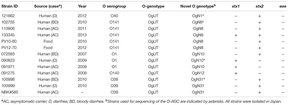
The O-AGC sequences were determined from six O-genotype untypeable (OgUT) STEC strains, of which four were serologically typeable (O1, O39, O40, and O141) and two others were untypeable (OUT) strains (Table 1). All strains were isolated from human feces (including patients with diarrhea and hemorrhagic colitis) in Japan from 2008 to 2012. The O-AGC sequences flanked by wcaM and hisI were extracted from draft genome sequences determined using an Illumina MiSeq sequencer (Illumina, San Diego, CA, USA), as previously described (Ogura et al., 2015). Identification and functional annotation of the coding sequences were performed based on the results of homology searches against the public non-redundant protein database using BLASTP. Six O-AGC sequences reported in this paper have been deposited in the GenBank/EMBL/DDBJ database (accession no. LC125927-LC125932).
Sequence Comparisons
The wzx/wzy sequences from O-serogroup strains (Iguchi et al., 2015a) and OX-groups reference strains (DebRoy et al., 2016) were used. Additionally, O-AGC sequences from O116 (AB812051; Iguchi et al., 2015a), O1 (GU299791; Li et al., 2010), O39 (AB811616; Iguchi et al., 2015a), O141 (DQ868765; Han et al., 2007), O40 (EU296417; Liu et al., 2008), S. enterica O42 (JX975340; Liu et al., 2014), and S. boydii type 13 (AY369140; Feng et al., 2004) were also used. Multiple alignments of DNA and amino acid sequences were constructed by using the CLUSTAL W program (Thompson et al., 1994). Phylogenetic trees were constructed by using the neighbor-joining algorithm using MEGA5 software (Tamura et al., 2007). Homology comparisons of paired sequences were performed by using the In Silico Molecular Cloning Genomics Edition (In Silico Biology, Inc., Yokohama, Japan).
PCR for Identifying Novel O-Genotypes
Polymerase chain reaction primers for specifically identifying novel O genotypes were designed (Table 2) and their specificities were evaluated by using 185 O-serogroup reference strains (O1–O188) from SSI using the following PCR conditions. Genomic DNA from E. coli strains was purified using the Wizard Genomic DNA purification kit (Promega, Madison, WI, USA) or DNeasy Blood & Tissue Kit (QIAGEN, Hilden, Germany) according to the manufacturer’s instructions. PCRs were performed using 10 ng/μl of template DNA. PCR was performed as follows: each 30-μl reaction mixture contained 2 μl of genomic DNA, 6 μl of 5× Kapa Taq buffer, dNTP mix (final concentration, 0.3 mM each), MgCl2 (final concentration, 2.5 mM), primers (final concentration, 0.5 μM each), and 0.8 U of Kapa Taq DNA polymerase (Kapa Biosystems, Woburn, MA, USA). The thermocycling conditions were: 25 cycles of 94°C for 30 s, 58°C for 30 s, and 72°C for 1 min. PCR products (2 μl) were electrophoresed in 1.5% agarose gels in 0.5× TBE (25 mM Tris borate, 0.5 mM EDTA), and photographed under UV light after the gel was stained with ethidium bromide (1 mg/ml).

TABLE 2. Polymerase chain reaction (PCR) primer sequences for identification of six novel O genotypes.
Distributional Survey of Novel O-Genotypes
Thirty five O-serogrouped E. coli strains from our previous study (Iguchi et al., 2015b), whose O genotypes were not identified by ECOG-PCR were used for screening novel O-genotypes by the PCR method designed in this study. The prevalence of stx1, stx2 (Cebula et al., 1995) and eae (Oswald et al., 2000) genes in the tested STEC strains was determined by the PCR.
Results
Novel O-Genotypes
Six types of O-AGC were identified from OgUT STEC strains (Figure 1). Four O-AGCs (named OgN10, OgN31, OgN1, and OgN8 genotypes) were obtained from strains that were serologically classified into O1, O39, O40, and O141, respectively (Table 1). Two others (named OgN9 and OgN12 genotypes) were obtained from strains that were both serologically and genetically unclassified into any groups (Table 1). Actually, the OgN9 strain did not react with any particular antiserum, and OgN12 showed identical agglutination titers with O34 and O140 antisera, which resulted in OUT classsification. OgN8, OgN10, and OgN12 carried rmlBDAC for the synsthesis of deoxythymidine diphosphate (dTDP)-L-rhamnose, and OgN31 carried rmlBA-vioA for dTDP viosamine synthesis (Figure 1). OgN9 carried fnlA-qnlBC for UDP-N-acetyl-L-quinovosamine (UDP-L-QuiNAc) synthesis (Figure 1). All novel O-AGCs carried the wzx/wzy O-antigen processing genes (Figure 1). The wzx/wzy sequences from OgN O-AGCs were compared with those from 171 O-serogroup strains and 11 OX-group reference strains, indicating that their sequences were unique compared to those from known O-AGCs (less than 70% DNA sequence identity of closest pairs), except for wzx of OgN31 (Figure 2). The sequence of OgN31_wzx was 98.7% identical in DNA sequence (99.0% amino acid sequence identity) to that of O116. Sequence comparison of O-AGCs revealed that the left region including wzx and genes for the d-TDP glucose pathway was conserved between OgN31 and Og116, and the right region including the wzy and glycosyltransferase genes was unique (less than 40% DNA sequence identity) in each O-AGC (Figure 3A). O-AGC gene sets from four pairs with members of different genotypes that agglutinated with the same O antisera were compared (Figure 3B). Between OgN10 and Og1 (from O1 strain), and between OgN8 and Og141 (from O141 strain), rmlBDAC genes were highly conserved in both O-AGCs, while other genes including wzx and wzy were diversified (less than 70% DNA sequence identity). Between OgN31 and Og39 (from O39 strain), different types of sugar biosynthesis genes were located on each O-AGC (rmlBA-vioA on OgN31, and rmlBDAC-vioAB and manCB on Og39). There was no genetic similarity between OgN1 and Og40 (from O40 strain). From these results, we were convinced that these six were novel O-AGCs.
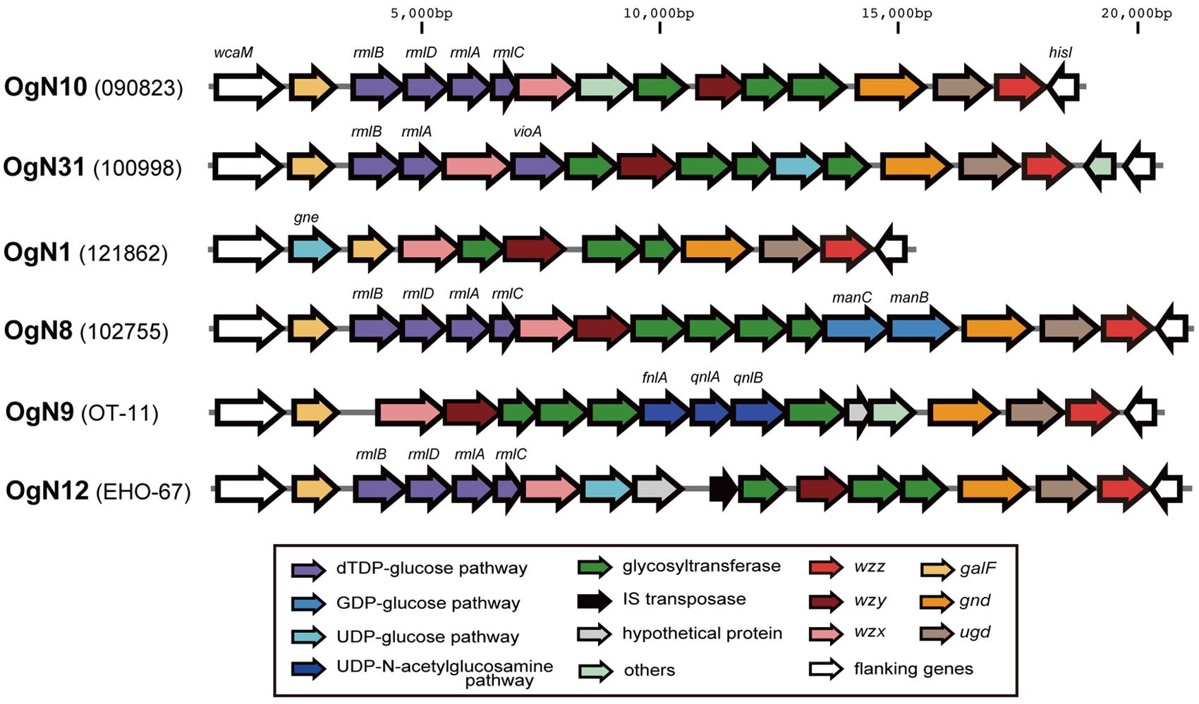
FIGURE 1. Six novel O-AGCs from STEC strains. The O-AGCs flanked by wcaM and hisI were extracted from draft genome sequences.
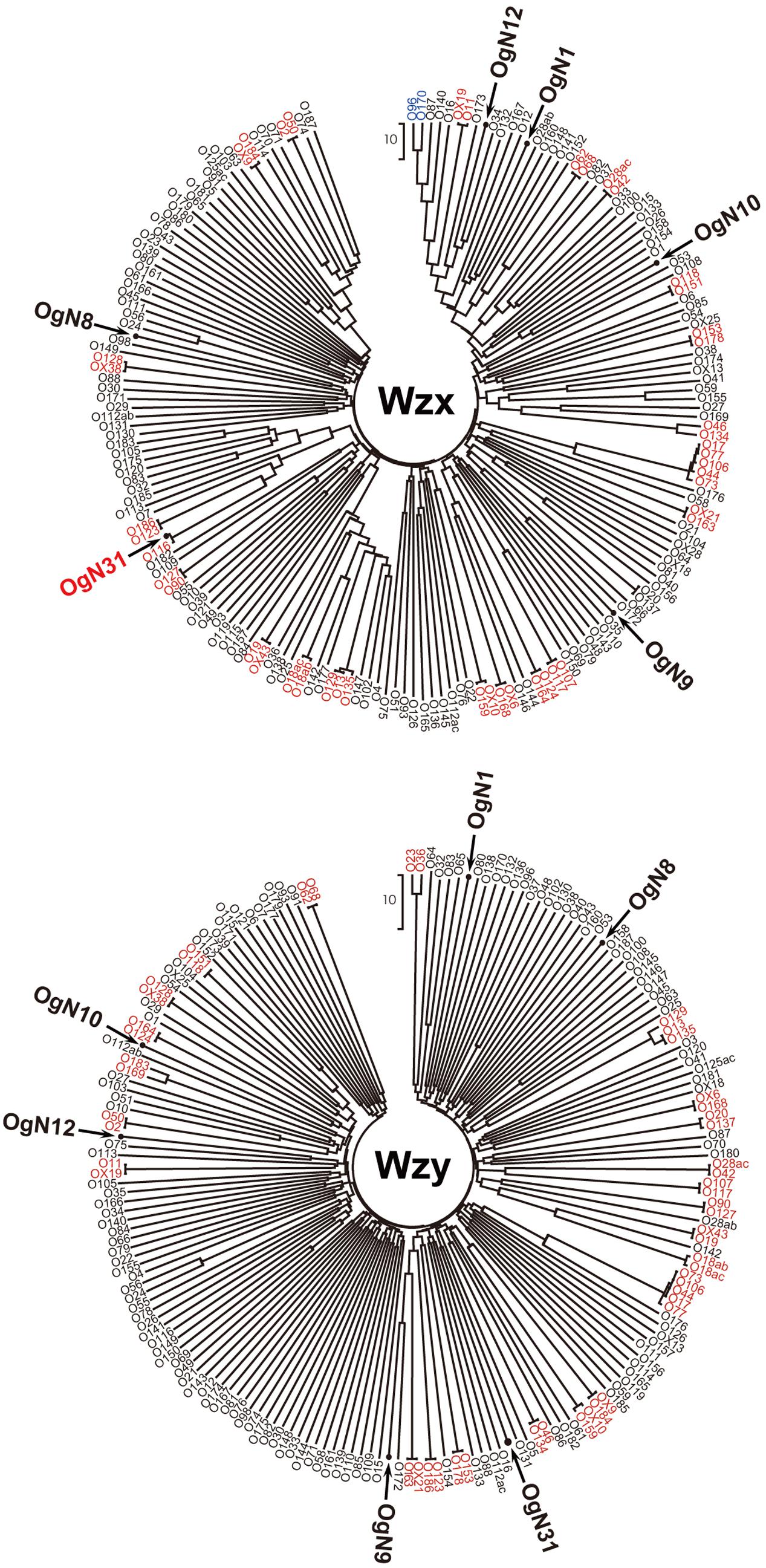
FIGURE 2. Phylogenetic analysis of Wzx and Wzy homologs from six novel O-AGCs, Escherichia coli O-serogroup strains and OX-groups reference strains based on the amino acid sequences (except for wzy of OX25, which is not found in the O-AGC of OX25). Pairs or groups of homologs with ≥95% DNA sequence identity are indicated in red, and ≥70% identity is indicated in blue. Other homologs with low sequence homologies (less than 70%) are indicated in black.
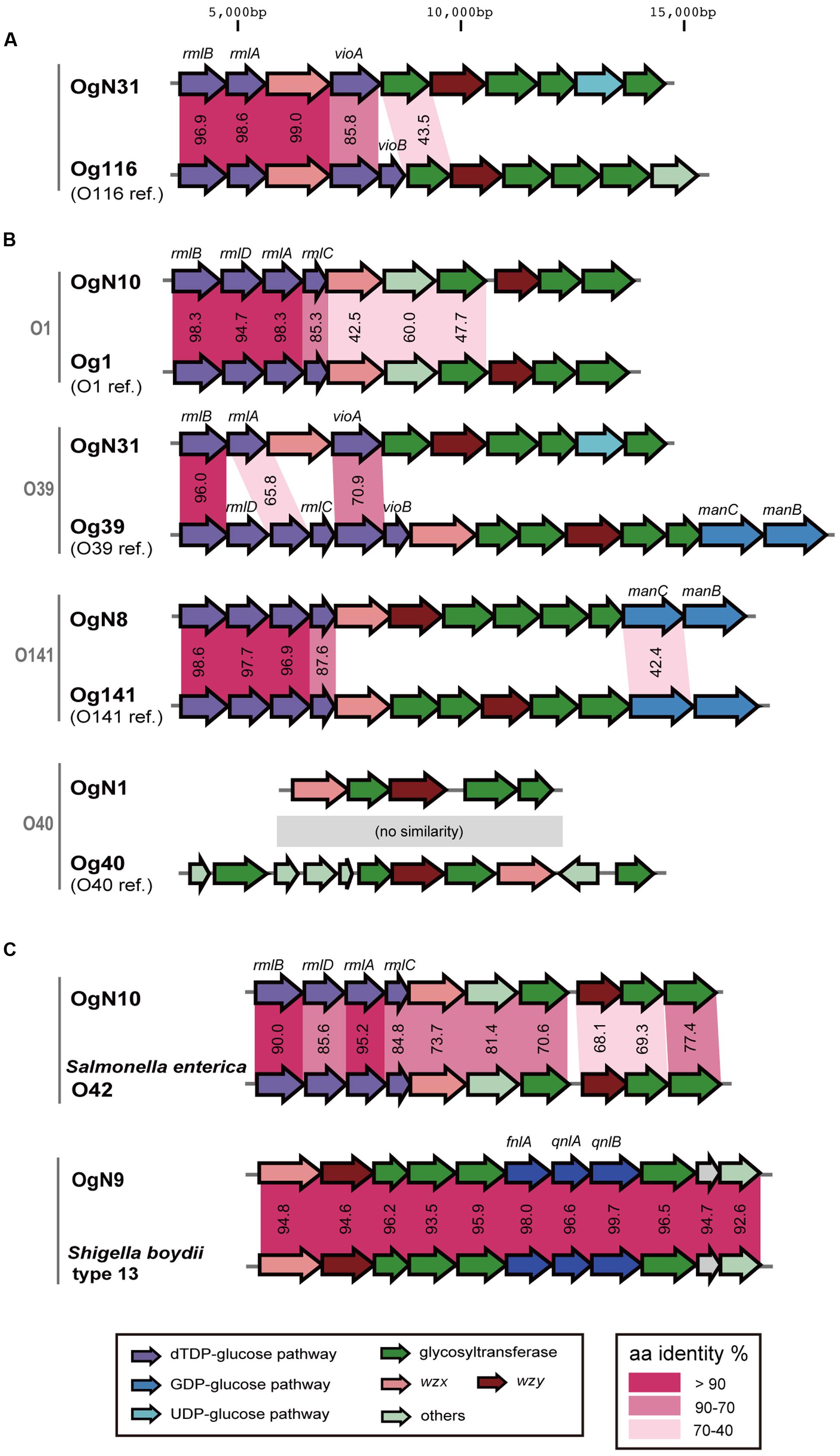
FIGURE 3. Comparison of O-AGCs. (A) The pair of O-AGCs, OgN31 and O116. (B) Four pairs that were serologically agglutinated with the same O antisera carried different types of O-AGCs. Lower genes show the O-AGCs from O-serogroup strains. (C) Similar O-AGCs in strains of other genera. Amino acid sequence identities (%) between homologs are shown in the middle.
A BLAST search of the NCBI database revealed that the OgN10 O-AGC is similar to that of S. enterica O42, and the OgN9 O-AGC was almost identical to that of S. boydii type 13 (Figure 3C). OgN10 and OgN9 strains agglutinated with S. enterica O42 and S. boydii type 13 antisera, respectively (data not shown).
Primers for Identifying Novel O Genotypes
Six PCR primer pairs were designed for identifying the novel O-genotypes (Table 2 and Figure 4). All primer pairs were targeted unique sequences of wzy, except for OgN31 for which primers were targeted to a glycosyltransferase gene. Each PCR was evaluated by using all 185 O-serogroup reference strains from O1 to O188 and six novel O-genotype strains (listed in Table 1). PCR products of the expected sizes on the agarose gel were obtained only with the corresponding strains, and no extra products were observed in the size range between 100 and 1,500 bp (data not shown).
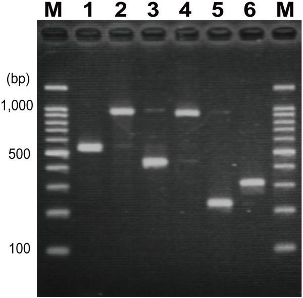
FIGURE 4. Polymerase chain reaction (PCR) products of OgN strains by each specific-primer pair. (1) OgN1 (525 bp), (2) OgN8 (940 bp), (3) OgN9 (423 bp), (4) OgN10 (892 bp), (5) OgN12 (232 bp), (6) OgN31 (311 bp), M; 100-bp DNA ladder markers.
Distribution of Novel O-Genotypes
Among 35 O-serogrouped E. coli strains whose O genotypes were not identified by the ECOG-PCR, five O141, three O1, three O39, one O40, and one O140 strains were classified by using the novel O-genotype PCR into OgN8, OgN10, OgN31, OgN1, and OgN12, respectively (Table 3). All 13 strains classified into five novel O-genotypes were eae-negative STEC, and OgN8, OgN10, and OgN31 had been isolated from patients with bloody diarrhea. OgN8 and OgN12 strains also cross-reacted with O41 and O34 antisera, respectively.
Discussion
In this study, six novel O genotypes were revealed from STEC strains isolated from human patients, and the prevalence of these O genotype strains was confirmed in STECs. The OgN10 strains were serologically classified into the O1 serogroup. The O1-serogroup strain is often seen in extra-intestinal pathogenic E. coli from patients with urinary tract infections (Abe et al., 2008; Mora et al., 2009) and septicemic disease (Mora et al., 2009), and in avian pathogenic E. coli (Mora et al., 2009; Johnson et al., 2012). Our previous study showed that an O1 strain isolated from patient blood was classified into Og1 (Iguchi et al., 2015b) and sequence comparisons showed that both an APEC O1 strain from avian colibacillosis (Johnson et al., 2007) and the G1632 strain from a patient with a urinary tract infection (Li et al., 2010) carried the Og1-type O-AGC, whereas three STEC O1 strains used in this study were all classified into OgN10. Actually, we confirmed that five STEC O1 strains from cattle used in a previous study, described as O1B type (Mekata et al., 2014) were also classified into OgN10 (data not shown). In fact, E. coli O1 strains could be generally subtyped into two genotypes, Og1 and OgN10, which were clearly linked to extra-intestinal/avian pathogenic E. coli and STEC, respectively. Among the O1 serogroup, three types of antigen structures have so far been reported (Baumann et al., 1991; Gupta et al., 1992) and the β-linked side-chain N-acetyl-D-mannosamine residue was suggested to be a common O1-specific epitope (Gupta et al., 1992). A partial kinship of O antigen structure synthesized from different O-AGCs may be serologically recognized as the same O serogroup and may also be represented in OgN1 OgN8, OgN12, and OgN31 strains, related to O40, O141, O140, and O39, respectively. The subdivision within each O-serogroup based on the O-AGC DNA sequences may be useful for obtaining more reliable information for epidemiological studies of pathogenic E. coli. Another advantage for DNA-based typing is that serologically untypeable and ambiguous strains could be clearly classified. At the present time, the PCR-based method reported here is the only way to distinguish OgN9. Although, STEC OgN groups have not emerged as a major public health issue, these groups are believed to be a possible cause of diarrhea and bloody diarrhea. To gain more information about trends in STEC OgNs epidemiology, further studies of global OgN isolates are needed. The PCR method described in this study may help the surveillance and monitoring of the OgN groups. Additionally, published sequences from OgN O-AGCs may be used for other DNA-based methodologies, such as in silico typing using whole genome sequencing data (Joensen et al., 2015).
Author Contributions
Conceived and designed the experiments: AI, MO, and TH. Performed the experiments: AI, SI, KS, and HN. Analyzed the data: AI and YO. Contributed reagents/materials/analysis tools: SI, KS, MO, and HM. Wrote the paper: AI. Critical revision of the paper for important intellectual content: AI, SI, and TH.
Funding
This research was partially supported by the Research Program on Emerging and Re-emerging Infectious Diseases from Japan Agency for Medical Research and development (AMED; 15fk0108008h0001), and by the Grants-in-Aid for Scientific Research (C) from the Ministry of Education, Culture, Sports, Science and Technology (MEXT) to AI (25350180) and SI (15K08486).
Conflict of Interest Statement
The authors declare that the research was conducted in the absence of any commercial or financial relationships that could be construed as a potential conflict of interest.
The reviewer BQ and handling Editor declared their shared affiliation and the handling Editor states that the process nevertheless met the standards of a fair and objective review.
Acknowledgment
We thank Atsuko Akiyoshi and Yuiko Kato for technical assistance.
Footnotes
References
Abe, C. M., Salvador, F. A., Falsetti, I. N., Vieira, M. A., Blanco, J., Blanco, J. E., et al. (2008). Uropathogenic Escherichia coli (UPEC) strains may carry virulence properties of diarrhoeagenic E. coli. FEMS Immunol. Med. Microbiol. 52, 397–406. doi: 10.1111/j.1574-695X.2008.00388.x
Baumann, H., Jansson, P. E., Kenne, L., and Widmalm, G. (1991). Structural studies of the Escherichia coli O1A O-polysaccharide, using the computer program CASPER. Carbohydr. Res. 211, 183–190. doi: 10.1016/0008-6215(91)84159-C
Beutin, L., Jahn, S., and Fach, P. (2009). Evaluation of the ‘GeneDisc’ real-time PCR system for detection of enterohaemorrhagic Escherichia coli (EHEC) O26, O103, O111, O145 and O157 strains according to their virulence markers and their O- and H-antigen-associated genes. J. Appl. Microbiol. 106, 1122–1132. doi: 10.1111/j.1365-2672.2008.04076.x
Buchholz, U., Bernard, H., Werber, D., Böhmer, M. M., Remschmidt, C., Wilking, H., et al. (2011). German outbreak of Escherichia coli O104:H4 associated with sprouts. N. Engl. J. Med. 365, 1763–1770. doi: 10.1056/NEJMoa1106482
Bugarel, M., Beutin, L., Martin, A., Gill, A., and Fach, P. (2010). Micro-array for the identification of Shiga toxin-producing Escherichia coli (STEC) seropathotypes associated with Hemorrhagic Colitis and Hemolytic Uremic Syndrome in humans. Int. J. Food Microbiol. 142, 318–329. doi: 10.1016/j.ijfoodmicro.2010.07.010
Cebula, T. A., Payne, W. L., and Feng, P. (1995). Simultaneous identification of strains of Escherichia coli serotype O157:H7 and their Shiga-like toxin type by mismatch amplification mutation assay-multiplex PCR. J. Clin. Microbiol. 33, 248–250.
Coimbra, R. S., Grimont, F., Lenormand, P., Burguière, P., Beutin, L., and Grimont, P. A. (2000). Identification of Escherichia coli O-serogroups by restriction of the amplified O-antigen gene cluster (rfb-RFLP). Res. Microbiol. 151, 639–654. doi: 10.1016/S0923-2508(00)00134-0
DebRoy, C., Fratamico, P. M., Yan, X., Baranzoni, G., Liu, Y., Needleman, D. S., et al. (2016). Comparison of O-Antigen Gene Clusters of All O-Serogroups of Escherichia coli and Proposal for Adopting a New Nomenclature for O-Typing. PLoS ONE 11:e0147434. doi: 10.1371/journal.pone.0147434
DebRoy, C., Roberts, E., and Fratamico, P. M. (2011a). Detection of O antigens in Escherichia coli. Anim. Health Res. Rev. 12, 169–185. doi: 10.1017/S1466252311000193
DebRoy, C., Roberts, E., Valadez, A. M., Dudley, E. G., and Cutter, C. N. (2011b). Detection of Shiga toxin-producing Escherichia coli O26, O45, O103, O111, O113, O121, O145, and O157 serogroups by multiplex polymerase chain reaction of the wzx gene of the O-antigen gene cluster. Foodborne Pathog. Dis. 8, 651–652. doi: 10.1089/fpd.2010.0769
Feng, L., Senchenkova, S. N., Yang, J., Shashkov, A. S., Tao, J., Guo, H., et al. (2004). Structural and genetic characterization of the Shigella boydii type 13 O antigen. J. Bacteriol. 186, 383–392. doi: 10.1128/JB.186.2.383-392.2004
Frank, C., Werber, D., Cramer, J. P., Askar, M., Faber, M., an der Heiden, M., et al. (2011). Epidemic profile of Shiga-toxin-producing Escherichia coli O104:H4 outbreak in Germany. N. Engl. J. Med. 365, 1771–1780. doi: 10.1056/NEJMoa1106483
Fratamico, P. M., and Bagi, L. K. (2012). Detection of Shiga toxin-producing Escherichia coli in ground beef using the GeneDisc real-time PCR system. Front. Cell Infect. Microbiol. 2:152. doi: 10.3389/fcimb.2012.00152
Geue, L., Monecke, S., Engelmann, I., Braun, S., Slickers, P., and Ehricht, R. (2014). Rapid microarray-based DNA genoserotyping of Escherichia coli. Microbiol. Immunol. 58, 77–86. doi: 10.1111/1348-0421.12120
Gupta, D. S., Shashkov, A. S., Jann, B., and Jann, K. (1992). Structures of the O1B and O1C lipopolysaccharide antigens of Escherichia coli. J. Bacteriol. 174, 7963–7970.
Han, W., Liu, B., Cao, B., Beutin, L., Krüger, U., Liu, H., et al. (2007). DNA microarray-based identification of serogroups and virulence gene patterns of Escherichia coli isolates associated with porcine postweaning diarrhea and edema disease. Appl. Environ. Microbiol. 73, 4082–4088. doi: 10.1128/AEM.01820-06
Heiman, K. E., Mody, R. K., Johnson, S. D., Griffin, P. M., and Gould, L. H. (2015). Escherichia coli O157 Outbreaks in the United States, 2003-2012. Emerg. Infect. Dis. 21, 1293–1301. doi: 10.3201/eid2108.141364
Iguchi, A., Iyoda, S., Kikuchi, T., Ogura, Y., Katsura, K., Ohnishi, M., et al. (2015a). A complete view of the genetic diversity of the Escherichia coli O-antigen biosynthesis gene cluster. DNA Res. 22, 101–107. doi: 10.1093/dnares/dsu043
Iguchi, A., Iyoda, S., Seto, K., Morita-Ishihara, T., Scheutz, F., Ohnishi, M., et al. (2015b). Escherichia coli O-Genotyping PCR: a comprehensive and practical platform for molecular o serogrouping. J. Clin. Microbiol. 53, 2427–2432. doi: 10.1128/JCM.00321-15
Joensen, K. G., Tetzschner, A. M., Iguchi, A., Aarestrup, F. M., and Scheutz, F. (2015). Rapid and easy in silico serotyping of Escherichia coli isolates by use of whole-genome sequencing data. J. Clin. Microbiol. 53, 2410–2426. doi: 10.1128/JCM.00008-15
Johnson, K. E., Thorpe, C. M., and Sears, C. L. (2006). The emerging clinical importance of non-O157 Shiga toxin-producing Escherichia coli. Clin. Infect. Dis. 43, 1587–1595. doi: 10.1086/509573
Johnson, T. J., Kariyawasam, S., Wannemuehler, Y., Mangiamele, P., Johnson, S. J., Doetkott, C., et al. (2007). The genome sequence of avian pathogenic Escherichia coli strain O1:K1:H7 shares strong similarities with human extraintestinal pathogenic E. coli genomes. J. Bacteriol. 189, 3228–3236. doi: 10.1128/JB.01726-06
Johnson, T. J., Wannemuehler, Y., Kariyawasam, S., Johnson, J. R., Logue, C. M., and Nolan, L. K. (2012). Prevalence of avian-pathogenic Escherichia coli strain O1 genomic islands among extraintestinal and commensal E. coli isolates. J. Bacteriol. 194, 2846–2853. doi: 10.1128/JB.06375-11
Li, D., Liu, B., Chen, M., Guo, D., Guo, X., Liu, F., et al. (2010). A multiplex PCR method to detect 14 Escherichia coli serogroups associated with urinary tract infections. J. Microbiol. Methods 82, 71–77. doi: 10.1016/j.mimet.2010.04.008
Liu, B., Knirel, Y. A., Feng, L., Perepelov, A. V., Senchenkova, S. N., Reeves, P. R., et al. (2014). Structural diversity in Salmonella O antigens and its genetic basis. FEMS Microbiol. Rev. 38, 56–89. doi: 10.1111/1574-6976.12034
Liu, B., Knirel, Y. A., Feng, L., Perepelov, A. V., Senchenkova, S. N., Wang, Q., et al. (2008). Structure and genetics of Shigella O antigens. FEMS Microbiol. Rev. 32, 627–653. doi: 10.1111/j.1574-6976.2008.00114.x
Luna-Gierke, R. E., Griffin, P. M., Gould, L. H., Herman, K., Bopp, C. A., Strockbine, N., et al. (2014). Outbreaks of non-O157 Shiga toxin-producing Escherichia coli infection: USA. Epidemiol. Infect. 142, 2270–2280. doi: 10.1017/S0950268813003233
Mekata, H., Iguchi, A., Kawano, K., Kirino, Y., Kobayashi, I., and Misawa, N. (2014). Identification of O serotypes, genotypes, and virulotypes of Shiga toxin-producing Escherichia coli isolates, including non-O157 from beef cattle in Japan. J. Food Prot. 77, 1269–1274. doi: 10.4315/0362-028X.JFP-13-506
Mora, A., López, C., Dabhi, G., Blanco, M., Blanco, J. E., Alonso, M. P., et al. (2009). Extraintestinal pathogenic Escherichia coli O1:K1:H7/NM from human and avian origin: detection of clonal groups B2 ST95 and D ST59 with different host distribution. BMC Microbiol. 9:132. doi: 10.1186/1471-2180-9-132
Ogura, Y., Mondal, S. I., Islam, M. R., Mako, T., Arisawa, K., Katsura, K., et al. (2015). The Shiga toxin 2 production level in enterohemorrhagic Escherichia coli O157:H7 is correlated with the subtypes of toxin-encoding phage. Sci. Rep. 5:16663. doi: 10.1038/srep16663
Ombarak, R. A., Hinenoya, A., Awasthi, S. P., Iguchi, A., Shima, A., Elbagory, A. R., et al. (2016). Prevalence and pathogenic potential of Escherichia coli isolates from raw milk and raw milk cheese in Egypt. Int. J. Food Microbiol. 221, 69–76. doi: 10.1016/j.ijfoodmicro.2016.01.009
Orskov, F., and Orskov, I. (1984). Serotyping of Escherichia coli. Methods Microbiol. 14, 43–112. doi: 10.1016/S0580-9517(08)70447-1
Orskov, F., and Orskov, I. (1992). Escherichia coli serotyping and disease in man and animals. Can. J. Microbiol. 38, 699–704. doi: 10.1139/m92-115
Oswald, E., Schmidt, H., Morabito, S., Karch, H., Marchès, O., and Caprioli, A. (2000). Typing of intimin genes in human and animal enterohemorrhagic and enteropathogenic Escherichia coli: characterization of a new intimin variant. Infect. Immun. 68, 64–71. doi: 10.1128/IAI.68.1.64-71.2000
Quiñones, B., Swimley, M. S., Narm, K. E., Patel, R. N., Cooley, M. B., and Mandrell, R. E. (2012). O-antigen and virulence profiling of shiga toxin-producing Escherichia coli by a rapid and cost-effective DNA microarray colorimetric method. Front. Cell Infect. Microbiol. 2:61. doi: 10.3389/fcimb.2012.00061
Scheutz, F., Cheasty, T., Woodward, D., and Smith, H. R. (2004). Designation of O174 and O175 to temporary O groups OX3 and OX7, and six new E. coli O groups that include Verocytotoxin-producing E. coli (VTEC): O176, O177, O178, O179, O180 and O181. APMIS 112, 569–584. doi: 10.1111/j.1600-0463.2004.apm1120903.x
Tamura, K., Dudley, J., Nei, M., and Kumar, S. (2007). MEGA4: molecular evolutionary genetics analysis (MEGA) software version 4.0. Mol. Biol. Evol. 24, 1596–1599. doi: 10.1093/molbev/msm092
Tarr, P. I., Gordon, C. A., and Chandler, W. L. (2005). Shiga-toxin-producing Escherichia coli and haemolytic uraemic syndrome. Lancet 365, 1073–1086. doi: 10.1016/S0140-6736(05)71144-2
Terajima, J., Iyoda, S., Ohnishi, M., and Watanabe, H. (2014). Shiga Toxin (Verotoxin)-Producing Escherichia coli in Japan. Microbiol. Spectr. 2, 1–9. doi: 10.1128/microbiolspec.EHEC-0011-2013
Thompson, J. D., Higgins, D. G., and Gibson, T. J. (1994). CLUSTAL W: improving the sensitivity of progressive multiple sequence alignment through sequence weighting, position-specific gap penalties and weight matrix choice. Nucleic Acids Res. 22, 4673–4680. doi: 10.1093/nar/22.22.4673
Wang, Q., Ruan, X., Wei, D., Hu, Z., Wu, L., Yu, T., et al. (2010). Development of a serogroup-specific multiplex PCR assay to detect a set of Escherichia coli serogroups based on the identification of their O-antigen gene clusters. Mol. Cell. Probes 24, 286–290. doi: 10.1016/j.mcp.2010.06.002
Keywords: E. coli, O serogroup, genotyping techniques, PCR, STEC
Citation: Iguchi A, Iyoda S, Seto K, Nishii H, Ohnishi M, Mekata H, Ogura Y and Hayashi T (2016) Six Novel O Genotypes from Shiga Toxin-Producing Escherichia coli. Front. Microbiol. 7:765. doi: 10.3389/fmicb.2016.00765
Received: 18 February 2016; Accepted: 06 May 2016;
Published: 20 May 2016.
Edited by:
Pina Fratamico, United States Department of Agriculture – Agricultural Research Service, USAReviewed by:
Beatriz Quiñones, United States Department of Agriculture – Agricultural Research Service, USAEdward G. Dudley, Pennsylvania State University, USA
Copyright © 2016 Iguchi, Iyoda, Seto, Nishii, Ohnishi, Mekata, Ogura and Hayashi. This is an open-access article distributed under the terms of the Creative Commons Attribution License (CC BY). The use, distribution or reproduction in other forums is permitted, provided the original author(s) or licensor are credited and that the original publication in this journal is cited, in accordance with accepted academic practice. No use, distribution or reproduction is permitted which does not comply with these terms.
*Correspondence: Atsushi Iguchi, aWd1Y2hpQG1lZC5taXlhemFraS11LmFjLmpw
 Atsushi Iguchi
 Sunao Iyoda
 Kazuko Seto
 Hironobu Nishii1
