- Department of Microbiology, School of Biology, College of Sciences, University of Tehran, Tehran, Iran
Intracellular life of Helicobacter pylori inside Candida yeast vacuole describes the establishment of H. pylori in yeast as a pre-adaptation to life in human epithelial cells. IgY-Hp conjugated with fluorescein isothiocyanate (FITC) has been previously used for identification and localization of H. pylori inside the yeast vacuole. Here we examined whether FITC-IgY-Hp internalization into yeast follows the endocytosis pathway in yeast. Fluorescent microscopy was used to examine the entry of FITC-IgY-Hp into Candida yeast cells at different time intervals. The effect of low temperature, H2O2 or acetic acid on the internalization of labeled antibody was also examined. FITC-IgY-Hp internalization initiated within 0–5 min in 5–10% of yeast cells, increased to 20–40% after 30 min–1 h and reached >70% before 2 h. FITC-IgY-Hp traversed the pores of Candida yeast cell wall and reached the vacuole where it bound with H. pylori antigens. Internalization of FITC-IgY-Hp was inhibited by low temperature, H2O2 or acetic acid. It was concluded that internalization of FITC-IgY-Hp into yeast cell is a vital phenomenon and follows the endocytosis pathway. Furthermore, it was proposed that FITC-IgY-Hp internalization could be recruited for localization and identification of H. pylori inside the vacuole of Candida yeast.
Introduction
Evolution of eukaryotic cells has been regarded as the most magnificent phenomenon in the history of life. Thousands of DNA mutations in prokaryotic cells caused major changes that led to development of today’s eukaryotes. These changes included formation of endomembrane system and internal cytoskeleton that favored engulfment of smaller prokaryotes through the mechanism of phagotrophy. The phagocytosed cells were digested and used for food or persisted against digestion and became permanent endosymbionts, acting as parasites, mutualists, or slaves (Cavalier-Smith, 1975). Successful endosymbiotic bacteria learned to induce their own internalization, reach the vacuole and persist against destruction. For this purpose, many intracellular bacteria evolved strategies to inhibit phagosomal maturation (Bozue and Johnson, 1996) or prevent oxygen radical production (Haas and Goebel, 1992). The benefits of vacuolar life for the internalized bacteria were being protected against environmental stresses while reaching plenty of nutrients needed for survival and proliferation (Garcia-del Portillo and Finlay, 1995; Finlay and Falkow, 1997; Greub and Raoult, 2004). Establishment of endosymbiotic bacteria within eukaryotic hosts led to maintenance of the vacuole and survival of both partners (Bianciotto et al., 1996). Accordingly, vacuole of eukaryotic cells represents a unique and sophisticated niche in which the endosymbiotic bacteria could survive and promote persistent association with the host cell (Cavalier-Smith, 2002).
In our previous studies we used microscopic and molecular biology methods to present evidence for the existence of non-culturable Helicobacter pylori inside the vacuole of Candida yeast (Siavoshi et al., 1998, 2005, 2013; Salmanian et al., 2008, 2012; Siavoshi and Saniee, 2014). Using anti-H. pylori egg yolk immunoglobulin Y (IgY-Hp) and western blotting, H. pylori-specific proteins; VacA, urease, peroxiredoxin, and thiol peroxidase were detected in the protein pool of Candida yeast, indicating that inside the yeast vacuole H. pylori is alive and expresses proteins (Saniee et al., 2013a). Fluorescent microscopy observations on Candida yeast cells treated with fluorescein isothiocyanate (FITC)-conjugated IgY-Hp, demonstrated the internalization of FITC-IgY-Hp into yeast cells and its specific binding with H. pylori cells, confirming the localization of H. pylori inside the yeast vacuole (Saniee et al., 2013b). Accordingly, yeast vacuole was proposed as a unique and specialized niche for accommodation of H. pylori, providing essential nutrients for its growth and multiplication (Saniee et al., 2013a,b; Siavoshi and Saniee, 2014). Intracellular existence of H. pylori has been reported in epithelial cells (Chu et al., 2010), macrophages and bone marrow-derived dendritic cells (Wang et al., 2009, 2010) and bacterial cells were observed within defined membrane-bound vacuoles (Segal et al., 1996; Dubois and Boren, 2007). It appears that H. pylori has evolved to equip itself for invading the eukaryotic cells and establishing in their vacuole (Dubois and Boren, 2007; Chu et al., 2010).
Reports describe occurrence of endosymbiotic bacteria in many eukaryotes, including protozoa, bivalves and insects (Douglas, 1994), Sponges (Erwin et al., 2012) and fungi (Scannerini and Bonfante, 1991). However, a considerable number of endosymbionts are non-culturable (Ruiz-Lozano and Bonfante, 1999; McFall-Ngai, 2008) and their intracellular localization and identification are possible by recruitment of microscopic and molecular biology methods (Bianciotto et al., 1996). In this regard, fluorescent dyes are ultra-sensitive markers that have been widely used in Live/Dead BacLight staining and fluorescence in situ hybridization (FISH) methods for localization and identification of live but non-culturable bacteria inside eukaryotic cells (Bianciotto et al., 2000). Furthermore, egg yolk antibody (IgY) exhibits high affinity for its target antigen and strongly binds with cell plasma membrane due to positive charge and lipophilic nature (Kovacs-Nolan and Mine, 2012). Results of our previous study showed the internalization of FITC-IgY-Hp and its accumulation in the vacuole of Candida yeast, proposing that IgY when conjugated with a fluorescent dye could serve as a specific probe for localization and identification of intracellular H. pylori (Saniee et al., 2013b).
Endocytosis is a general mechanism by which eukaryotic cells internalize extracellular molecules through the formation of vesicles from the plasma membrane. The endocytosed particles internalize in a free state or while bound to a specific surface receptor. Once internalized by endocytosis, the cargo passes first through early endosome and next late endosome (Prescianotto-Baschong and Riezman, 2002) which fuses with vacuole and releases its contents (Hurley and Emr, 2006). The process of endocytosis is energy- and temperature-dependent and can be impaired by oxidative stress or incubation at low temperature. It is also time-dependent; the half-time for internalization has been estimated as 2–5 min (Pearse and Bretscher, 1981; Steinman et al., 1983). Endocytosis has been widely studied in yeast describing internalization of fluorescent dyes; FM4-64 (Vida and Emr, 1995) and lucifer yellow (Riezman, 1985) and nano gold particles (Prescianotto-Baschong and Riezman, 2002). In this study fluorescent microscopy was used to examine the uptake of FITC-IgY-Hp by Candida yeast cells, at different time intervals and its accumulation in the vacuole. Endocytosis inhibitors; low temperature, H2O2 or acetic acid were recruited to assess whether internalization of FITC-IgY-Hp into yeast cells is a vital phenomenon and follows the endocytosis pathway.
Materials and Methods
Yeast Strains
Two gastric yeasts (G2 and G5) which were isolated from gastric biopsy cultures of two H. pylori-positive patients were used in this study. G2 was isolated from a 54 years old male and G5 from a 68 years old male. Patients were referred to Shariati hospital for endoscopy and diagnosed as having peptic ulcer. Informed consent was signed by the patients and the study was approved by research ethics committee of Tehran University of Medical Sciences. The primary identification of G2 and G5 yeasts as Candida albicans was according to microscopic morphology and production of green colonies on Chromagar (Odds and Bernaerts, 1994). Molecular identification of G2 and G5 yeast was performed by amplification of Candida-specific Topoisomerase II gene, using nested-PCR. Two sets of primers were used; CDF28 (GGTGWMGDAAYGGDTWYGGYGC), CDR148 (CCRTCNTGATCYTGATCBGYCAT) and CABF59 (TTGAACATCTCCAGTTTCAAAGGT), CADBR125 (AGCTA-AATTCATAGCAGAAAGC). PCR was performed according to Kanbe et al. (2002). Size of amplified product was determined as 637 bp by electrophoresis, using 100-bp molecular marker, confirming the identity of yeasts as C. albicans.
Production of FITC-Labeled IgY-Hp
IgY-Hp was produced (Saniee et al., 2013a) and FITC-labeled (Wisdom, 2005; Saniee et al., 2013b) as previously described. Briefly, IgY-Hp was raised in hens by immunization with whole cell lysate of H. pylori and extracted from egg yolk according to Nikbakht Brujeni et al. (2011). FITC-IgY-Hp was prepared by adding FITC solution to the antibody and removing unbound FITC using Sephadex G25 column (Saniee et al., 2013b).
Cultivation of Yeasts
For time and inhibition assays, fresh culture of G2 and G5 yeasts on YGC (yeast extract-glucose-chloramphenicol) agar was inoculated into a home-made broth containing 5 g/L yeast extract (Pronadisa, France), 20 g/L N-acetylglucosamine (Sigma, USA), supplemented with equal volume of fetal bovine serum (Invitrogen, USA). Cultures were grown at 37°C for 24 h, washed twice and re-suspended in phosphate buffered-saline, PBS (0.5X).
Internalization Assays
Time assay
G2 and G5 yeasts were used in this part of study. The intracellular occurrence of H. pylori inside the G2 yeast was determined by light and fluorescent microscopy as well as detection of H. pylori-specific genes and proteins in the whole extract of yeast cells. G5 yeast in which H. pylori-specific genes were not detected by PCR was used as a control (Saniee et al., 2013a,b). A volume of 100 μl of yeast cells in PBS was mixed with 20 μl of 1:5 dilution of FITC-IgY-Hp and 5 μl of evans blue solution (0.01% in PBS). Yeasts were incubated in dark at 37°C while shaking (200 rpm). Internalization of FITC-IgY-Hp into yeast cells was monitored in different time intervals, using fluorescent microscopy. Ten-microliter volumes of treated yeasts were taken after 0–5 min, 15 min, 30 min, 1, 1.5, 2, 3, 4, and 5 h, washed once with distilled water, re-suspended in PBS, spotted on the glass slide and air-dried. Dried spots were covered first with mounting oil (Invitrogen, USA) and next with coverslip. Nail polish was used for fixing the coverslip in place. Immersion oil was used for observing samples by fluorescent microscopy.
Low temperature assay
To examine the impact of low temperature on the endocytosis, a 100-μl volume of G2 yeast cells in PBS was mixed with 20 μl of 1:5 dilution of FITC-IgY-Hp and 5 μl of evans blue solution (0.01% in PBS) and immediately transferred to 4°C where it remained for 1 h while shaking manually every 10 min. Yeast cells were washed with ice-cold water. A 10-μl volume of yeast cells was spotted on pre-cold slide, air-dried on ice and kept cold until observed by fluorescent microscope.
Inhibition assay
To examine the inhibitory effect of H2O2 or acetic acid on internalization of FITC-IgY-Hp into G2 yeast cells, a 200-μl volume of yeast cells in PBS (0.5X) was treated with 10 mM of H2O2 or 140 mM of acetic acid and incubated at 37°C for 1 h with continuous shaking (200 rpm). To examine the possibility of negative effect of H2O2 or acetic acid on FITC-IgY-Hp or G2 yeast cells, at the end of incubation, each sample was divided into two 100-μl aliquots (tubes A and B). In tubes A, inhibitors remained in yeast samples. In tubes B, H2O2 or acetic acid was removed by harvesting and re-suspending the cells in 100 μl of PBS. FITC-conjugated IgY-Hp and evans blue solution were added to tubes A and B as mentioned above. Samples were incubated in dark at 37°C while shaking (200 rpm) and examined at different time intervals (0–5 min, 15 min, 30 min, 1, and 2 h) by fluorescent microscope.
To examine the viability of yeast cells after being treated with H2O2 or acetic acid, a 10-μl volume of yeast cells from tubes A and B was cultured on YGC agar after each time interval and incubated at 37°C.
Results
Effect of Time on Internalization
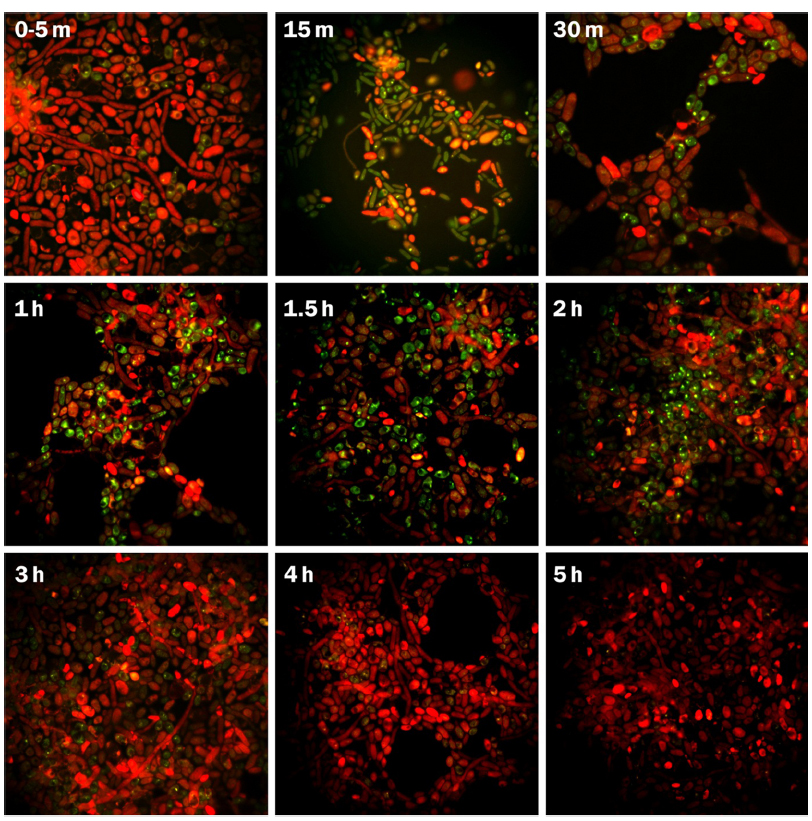
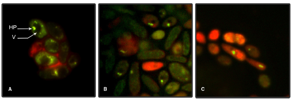
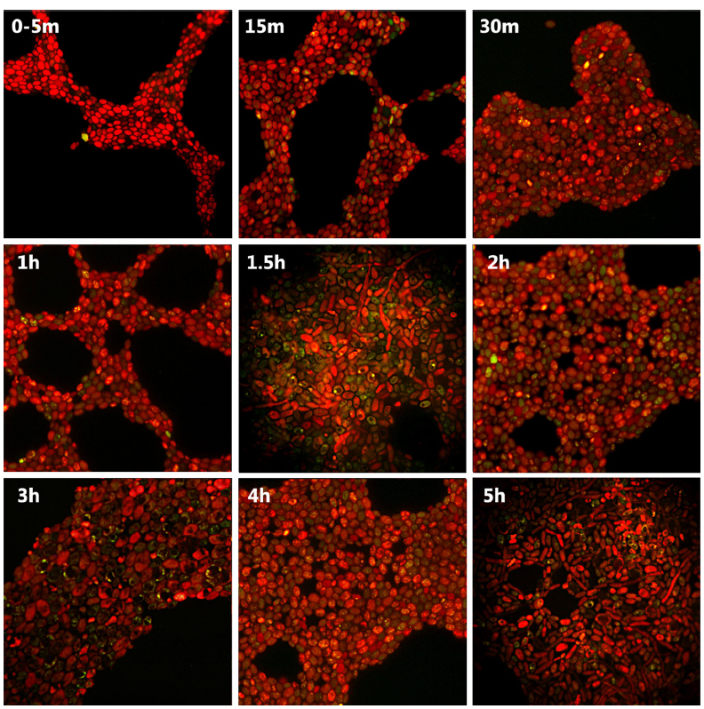
Fluorescent microscopy observations showed the internalization of FITC-IgY-Hp into G2 yeast cells and its accumulation in the vacuoles due to binding with H. pylori antigens. Internalization initiated immediately within 0–5 min and green fluorescent spots were observed in 5–10% of yeast cells. After 30 min–1 h, 20–40% of cells showed green fluorescent spots in their vacuoles. The number of yeast cells with green fluorescent spots reached >70% before 2 h. The green fluorescent-labeled H. pylori cells inside the yeast vacuole remained shining for 3 h and then started fainting. After 4–5 h, green fluorescent spots became invisible and yeast cells appeared completely red, due to evans blue, with dark vacuole (Figure 1). Threefold enlargement of microscopic photographs with original × 1000 magnification showed the specific interaction of FITC-IgY-Hp with H. pylori cells in the yeast vacuole, after 2 h incubation with labeled antibody (Figures 2A–C). Fluorescent microscopy observations of G5 yeast in different time intervals did not show the same pattern as G2 yeast. Green spots were observed in only few yeasts with less fluorescent intensity and persistence (Figure 3).

Figure 1. Fluorescent microscopy of FITC-IgY-Hp-treated G2 yeast cells at different time intervals. Fluorescent microscopy photographs show the internalization of FITC-IgY-Hp in 5–10% of yeast cells within 0–5 min, increased to 20–40% within 30 min to 1 h and reached >70% before 2 h. The green fluorescent-labeled H. pylori cells remained visible up to 3 h and fainted within 4–5 h when yeast cells appeared completely red, due to evans blue, with dark vacuole (magnification × 1000). m, minutes; h, hours.

Figure 2. Enlarged fluorescent microscopy photographs of G2 yeast cells. Threefold enlarged yeast cells with original × 1000 magnification, showing green fluorescent spots in the yeast vacuole after 2 h incubation with FITC-IgY-Hp. (A) H. pylori (HP) in the vacuole (V) of yeast, (B) H. pylori in the vacuole of yeast, (C) H. pylori in the vacuole of a yeast cell and its bud.

Figure 3. Fluorescent microscopy of FITC-IgY-Hp-treated G5 yeast cells at different time intervals. Fluorescent microscopy photographs show no regular pattern of increase in internalized FITC-IgY-Hp by time (up to 3 h) and faint within 4–5 h (magnification × 1000).
Effect of Low Temperature, H2O2 or Acetic Acid on Internalization
Fluorescent microscopy of FITC-IgY-Hp-treated G2 yeast, incubated at 4°C for 1 h showed inhibitory effect of low temperature on internalization of labeled-antibody. Yeast cells did not accumulate green fluorescent antibody and appeared dark (Figure 4A). Fluorescent microscopy showed similar results of the inhibitory effect of H2O2 or acetic acid on the internalization of FITC-IgY-Hp into yeast cells. Fluorescent microscopy photographs showed that in tubes B (inhibitors removed), G2 yeast cells were clearly visible and red with dark vacuoles, without FITC-IgY-Hp accumulation (Figures 4B2,C2). In tubes A (inhibitors present) yeast cells did not accumulate FITC-IgY-Hp and showed altered morphology that could be the result of long exposure to inhibitors (Figures 4B1,C1). These results indicated that 1 h was sufficient for effective inhibition of FITC-IgY-Hp internalization by H2O2 or acetic acid. Inhibitory effect persisted in yeast cells even after 2 h of inhibitors removal. Cultures of G2 yeast on YGC were positive and colonies were observed after 24 h.

Figure 4. Effect of endocytosis inhibitors on internalization of FITC-IgY-Hp into G2 yeast cells. Fluorescent microscopy photographs show that yeast cells inhibited by low temperature (4°C), H2O2 (10 mM) and acetic acid (140 mM) did not accumulate green fluorescent antibody and appeared red. (A) The inhibitory effect of low temperature. (B1) Exposure (2 h) to FITC-IgY-Hp along with H2O2; tube A. (B2) Incubation (2 h) with FITC-IgY-Hp after H2O2 removal; tube B. (C1) Exposure (2 h) to FITC-IgY-Hp along with acetic acid; tube A. (C2) Incubation (2 h) with FITC-IgY-Hp after acetic acid removal; tube B (magnification × 1000).
Discussion
IgY has been recruited as a powerful tool for diagnostic purposes. It is a positively-charged and lipophilic antibody with a molecular mass of ∼180 kDa, slightly larger than mammalian IgG, 150 kDa (Kovacs-Nolan and Mine, 2012). IgY-Hp strongly reacts with H. pylori-specific antigens and can be recruited for effective inhibition or detection of H. pylori (Shin et al., 2002). In our previous studies, strong interaction of IgY-Hp and FITC-IgY-Hp with H. pylori antigens, present in the vacuole of yeast, revealed the occurrence of live H. pylori cells inside the Candida yeast (Saniee et al., 2013a,b).
Results of this study showed that FITC-IgY-Hp could bind with negatively-charged lipid on yeast cell plasma membrane and internalize through endocytotic route in a time-dependent fashion. Internalization of labeled antibody which was determined by visualizing green fluorescent spots in G2 yeast vacuole, initiated immediately (0–5 min) in 5–10% of yeast cells, increased to 20–40% within 30 min–1 h and reached >70% before 2 h. Fainting started before 3 h possibly due to quenching of FITC-IgY-Hp at acidic pH of the yeast vacuole (Clarke et al., 2010). Comparison of photographs of G2 and G5 yeast especially within 1–2 h, showed that the frequency of green fluorescent spots was much higher in G2 yeast than G5 yeast. Furthermore the intensity and persistence of fluorescent spots in G2 yeast was higher than in G5 yeast. This could be due to strong interaction of FITC-IgY-Hp with H. pylori-specific antigens in G2 yeast. In contrast, in G5 yeast without H. pylori few fluorescent spots with less intensity and persistence were observed. These results show that although FITC-IgY-Hp uptake occurred through endocytotic pathway, the fate of labeled antibody was determined by its specific binding with H. pylori antigens, otherwise it would be denatured (Lobo et al., 2004) or quenched in acidic pH of yeast vacuole, in a short time. Internalization of FITC-IgY-Hp was inhibited at cold or upon treatment of yeast cells with endocytosis inhibitors; H2O2 or acetic acid. Inhibition of endocytosis was determined when treated yeast cells did not show accumulation of green fluorescent IgY-Hp and vacuoles appeared dark. Comparison of photographs from yeasts in tubes A and B showed that H2O2 or acetic acid did not have a negative effect on FITC-IgY-Hp. However, long exposure of yeast cells to endocytosis inhibitors led to altered morphology of yeast cells. Endocytosis of FITC-IgY-Hp did not occur in yeasts of tubes B, even 2 h after removal of inhibitors. Moreover, yeast cells in tubes A and B remained viable and produced colonies after 24 h.
Fluorescent microscopy studies on the endocytosis of FM4-64, a lipophilic fluorescent dye, in Saccharomyces cerevisiae revealed that the dye initially stained the plasma membrane and finally reached the vacuolar membrane after 1 h (Vida and Emr, 1995). However, internalization of dye was impaired when yeasts were kept at cold temperature (Huckaba et al., 2004) or exposed to 1–3 mM H2O2 or 80–140 mM acetic acid for 1 h (Pereira et al., 2012). Fluorescence microscopy was also used to study C. albicans cells exposed to a sub-inhibitory concentration (5 μg) of apolipoprotein-derived ApoEdpL-W-Fluo, a labeled antifungal peptide, after different time intervals. Fluorescent peptide was bound to the cell surface and accumulated in the vacuole of 33% of cells within 30 min, 66–87% after 60 min and almost all the cells after 2 h. The ApoEdp-W-Fluo was co-localized with FM4-64, indicating that the target organelle was vacuole (Rossignol et al., 2011). A Fluorescent microscopy study on the internalization of fluorescent dye lucifer yellow into S. cerevisiae vacuole showed that endocytosis of lucifer yellow was dependent on time, temperature and energy (Dulic et al., 1991). Electron microscopy observations on the internalization of positively-charged nano-gold particles into S. cerevisiae endosome revealed that they strongly bound with negatively-charged lipids on the surface, endocytosed and reached the vacuole (Prescianotto-Baschong and Riezman, 1998). Accordingly, results of our study indicate that internalization of FITC-IgY-Hp into yeast cells was a vital phenomenon and followed the principle of endocytosis in yeast.
A number of immunologic and microscopic methods have been recruited for localization and identification of intracellular H. pylori in gastric biopsies (Andersen and Holck, 1990; Ko et al., 1999) and cultured cells (Semino-Mora et al., 2003; Necchi et al., 2007). In these methods, there was no barrier such as cell wall to internalization of antibodies. However, in the present study passage of antibody through the yeast cell wall was the critical step. Internalization of fluorescent-labeled antibodies into yeast cells has been used for recognizing and visualizing intracellular epitopes, especially components of the cytoskeleton (Pringle et al., 1990). The procedure involves fixation of cytoplasmic contents and permeabilization of the yeast cells with cell wall lytic enzyme (Hašek and Streiblová, 1996). Results of this study demonstrated that FITC-IgY-Hp with molecular mass of ∼180 kDa, could traverse the non-permeabilized cell wall of Candida yeast, internalize into yeast cell through endocytosis and accumulate within the vacuole in a short time. The pore size of the yeast Cryptococcus neoformans cell wall was measured by cryoporometry as 1–30 nm (Eisenman et al., 2005) and electron microscopy as 60–300 nm (Rodrigues et al., 2007). Furthermore, atomic force microscopy determined the pore size of S. cerevisiae cell wall as 200 nm that could increase to 400 nm under stressful conditions (de Souza Pereira and Geibel, 1999). Although, controversies exist about the size of molecules that can traverse the pores of yeast cell wall, it has been proposed that fungal cell walls are capable of import or export of vesicles that carry food supply, waste products or digestive enzymes. These vesicles are flexible lipid membranes that can compress and ferry across the cell wall. Accordingly, cell wall plays an important role in vesicular trafficking in yeast and the determining factor is the pore size of the cell wall (Casadevall et al., 2009).
Results of this study indicate that Candida yeast cell wall has pores large enough to let the labeled antibody to pass through, bind with plasma membrane, initiate endocytosis and finally accumulate within the vacuole where it strongly binds with H. pylori antigens. It appears that inside yeast vacuole H. pylori cells maintained their antigenic identity detectable by specific antibodies. Accordingly, it is proposed that endocytotic uptake of FITC-IgY-Hp could be used as an easy and specific tool for demonstrating the intracellular existence of H. pylori inside the yeast vacuole. Furthermore, IgY raised against other intracellular bacteria could be used for their localization and identification inside the yeast vacuole. Recruitment of fluorescent-labeled antibodies against different bacterial species might provide a powerful tool for differentiating and quantifying the members of the possible mixed bacterial population inside the vacuole of yeasts.
Conflict of Interest Statement
The authors declare that the research was conducted in the absence of any commercial or financial relationships that could be construed as a potential conflict of interest.
Acknowledgments
This study was funded by the research council of University of Tehran and Digestive Disease Research Institute, Tehran University of Medical Sciences. Authors wish to thank Kaveh Oskoei for his help in photography and Mehran Ahadi for his support throughout the preparation of the manuscript.
References
Andersen, L. P., and Holck, S. (1990). Possible evidence of invasiveness of Helicobacter (Campylobacter) pylori. Eur. J. Clin. Microbiol. Infect. Dis. 9, 135–138. doi: 10.1007/BF01963640
Bianciotto, V., Bandi, C., Minerdi, D., Sironi, M., Tichy, H. V., and Bonfante, P. (1996). An obligately endosymbiotic mycorrhizal fungus itself harbors obligately intracellular bacteria. Appl. Environ. Microbiol. 62, 3005–3010.
Bianciotto, V., Lumini, E., Lanfranco, L., Minerdi, D., Bonfante, P., and Perotto, S. (2000). Detection and identification of bacterial endosymbionts in arbuscular mycorrhizal fungi belonging to the family Gigasporaceae. Appl. Environ. Microbiol. 66, 4503–4509. doi: 10.1128/AEM.66.10.4503-4509.2000
PubMed Abstract | Full Text | CrossRef Full Text | Google Scholar
Bozue, J. A., and Johnson, W. (1996). Interaction of Legionella pneumophila with Acanthamoeba castellanii: uptake by coiling phagocytosis and inhibition of phagosome-lysosome fusion. Infect. Immun. 64, 668–673.
Casadevall, A., Nosanchuk, J. D., Williamson, P., and Rodrigues, M. L. (2009). Vesicular transport across the fungal cell wall. Trends Microbiol. 17, 158–162. doi: 10.1016/j.tim.2008.12.005
PubMed Abstract | Full Text | CrossRef Full Text | Google Scholar
Cavalier-Smith, T. (1975). The origin of nuclei and of eukaryotic cells. Nature 256, 463–468. doi: 10.1038/256463a0
Cavalier-Smith, T. (2002). The neomuran origin of archaebacteria, the negibacterial root of the universal tree and bacterial megaclassification. Int. J. Syst. Evol. Microbiol. 52, 7–76.
Chu, Y. T., Wang, Y. H., Wu, J. J., and Lei, H. Y. (2010). Invasion and multiplication of Helicobacter pylori in gastric epithelial cells and implications for antibiotic resistance. Infect. Immun. 78, 4157–4165. doi: 10.1128/IAI.00524-10
PubMed Abstract | Full Text | CrossRef Full Text | Google Scholar
Clarke, M., Maddera, L., Engel, U., and Gerisch, G. (2010). Retrieval of the vacuolar H+-ATPase from phagosomes revealed by live cell imaging. PLoS ONE 5:e8585. doi: 10.1371/journal.pone.0008585
PubMed Abstract | Full Text | CrossRef Full Text | Google Scholar
de Souza Pereira, R., and Geibel, J. (1999). Direct observation of oxidative stress on the cell wall of Saccharomyces cerevisiae strains with atomic force microscopy. Mol. Cell. Biochem. 201, 17–24. doi: 10.1023/A:1007007704657
PubMed Abstract | Full Text | CrossRef Full Text | Google Scholar
Dubois, A., and Boren, T. (2007). Helicobacter pylori is invasive and it may be a facultative intracellular organism. Cell. Microbiol. 9, 1108–1116. doi: 10.1111/j.1462-5822.2007.00921.x
PubMed Abstract | Full Text | CrossRef Full Text | Google Scholar
Dulic, V., Egerton, M., Elguindi, I., Raths, S., Singer, B., and Riezman, H. (1991). Yeast endocytosis assays. Methods Enzymol. 194, 697–710. doi: 10.1016/0076-6879(91)94051-D
Eisenman, H. C., Nosanchuk, J. D., Webber, J. B., Emerson, R. J., Camesano, T. A., and Casadevall, A. (2005). Microstructure of cell wall-associated melanin in the human pathogenic fungus Cryptococcus neoformans. Biochemistry 44, 3683–3693. doi: 10.1021/bi047731m
PubMed Abstract | Full Text | CrossRef Full Text | Google Scholar
Erwin, P. M., López-Legentil, S., and Turon, X. (2012). Ultrastructure, molecular phylogenetics, and chlorophyll a content of novel cyanobacterial symbionts in temperate sponges. Microb. Ecol. 64, 771–783. doi: 10.1007/s00248-012-0047-5
PubMed Abstract | Full Text | CrossRef Full Text | Google Scholar
Finlay, B. B., and Falkow, S. (1997). Common themes in microbial pathogenicity revisited. Microbiol. Mol. Biol. Rev. 61, 136–169.
Garcia-del Portillo, F., and Finlay, B. B. (1995). The varied lifestyles of intracellular pathogens within eukaryotic vacuolar compartments. Trends Microbiol. 3, 373–380. doi: 10.1016/S0966-842X(00)88982-9
Greub, G., and Raoult, D. (2004). Microorganisms resistant to free-living amoebae. Clin. Microbiol. Rev. 17, 413–433. doi: 10.1128/CMR.17.2.413-433.2004
PubMed Abstract | Full Text | CrossRef Full Text | Google Scholar
Haas, A., and Goebel, W. (1992). Microbial strategies to prevent oxygen-dependent killing by phagocytes. Free Radic. Res. Commun. 16, 137–157. doi: 10.3109/10715769209049167
PubMed Abstract | Full Text | CrossRef Full Text | Google Scholar
Hašek, J., and Streiblová, E. (1996). “Fluorescence microscopy methods,” in Yeast Protocols: Methods in Cell and Molecular Biology, ed. I. H. Evans (Totowa, NJ: Humana Press), 391–405.
Huckaba, T. M., Gay, A. C., Pantalena, L. F., Yang, H. C., and Pon, L. A. (2004). Live cell imaging of the assembly, disassembly, and actin cable-dependent movement of endosomes and actin patches in the budding yeast, Saccharomyces cerevisiae. J. Cell Biol. 167, 519–530. doi: 10.1083/jcb.200404173
PubMed Abstract | Full Text | CrossRef Full Text | Google Scholar
Hurley, J. H., and Emr, S. D. (2006). The ESCRT complexes: structure and mechanism of a membrane-trafficking network. Annu. Rev. Biophys. Biomol. Struct. 35, 277. doi: 10.1146/annurev.biophys.35.040405.102126
PubMed Abstract | Full Text | CrossRef Full Text | Google Scholar
Kanbe, T., Horii, T., Arishima, T., Ozeki, M., and Kikuchi, A. (2002). PCR-based identification of pathogenic Candida species using primer mixes specific to Candida DNA topoisomerase II genes. Yeast 19, 973–989. doi: 10.1002/yea.892
PubMed Abstract | Full Text | CrossRef Full Text | Google Scholar
Ko, G. H., Kang, S. M., Kim, Y. K., Lee, J. H., Park, C. K., Youn, H. S., et al. (1999). Invasiveness of Helicobacter pylori into human gastric mucosa. Helicobacter 4, 77–81. doi: 10.1046/j.1523-5378.1999.98690.x
PubMed Abstract | Full Text | CrossRef Full Text | Google Scholar
Kovacs-Nolan, J., and Mine, Y. (2012). Egg yolk antibodies for passive immunity. Annu. Rev. Food Sci. Technol. 3, 163–182. doi: 10.1146/annurev-food-022811-101137
PubMed Abstract | Full Text | CrossRef Full Text | Google Scholar
Lobo, E. D., Hansen, R. J., and Balthasar, J. P. (2004). Antibody pharmacokinetics and pharmacodynamics. J. Pharm. Sci. 93, 2645–2668. doi: 10.1002/jps.20178
PubMed Abstract | Full Text | CrossRef Full Text | Google Scholar
McFall-Ngai, M. (2008). Are biologists in ‘future shock’? Symbiosis integrates biology across domains. Nat. Rev. Microbiol. 6, 789–792. doi: 10.1038/nrmicro1982
PubMed Abstract | Full Text | CrossRef Full Text | Google Scholar
Necchi, V., Candusso, M. E., Tava, F., Luinetti, O., Ventura, U., Fiocca, R., et al. (2007). Intracellular, intercellular, and stromal invasion of gastric mucosa, preneoplastic lesions, and cancer by Helicobacter pylori. Gastroenterology 132, 1009–1023. doi: 10.1053/j.gastro.2007.01.049
PubMed Abstract | Full Text | CrossRef Full Text | Google Scholar
Nikbakht Brujeni, G., Jalali, S. A. H., and Koohi, M. K. (2011). Development of DNA-designed avian IgY antibodies for quantitative determination of bovine interferon-gamma. Appl. Biochem. Biotechnol. 163, 338–345. doi: 10.1007/s12010-010-9042-9
PubMed Abstract | Full Text | CrossRef Full Text | Google Scholar
Odds, F. C., and Bernaerts, R. I. A. (1994). CHROMagar Candida, a new differential isolation medium for presumptive identification of clinically important Candida species. J. Clin. Microbiol. 32, 1923–1929.
Pearse, B. M., and Bretscher, M. S. (1981). Membrane recycling by coated vesicles. Annu. Rev. Biochem. 50, 85–101. doi: 10.1146/annurev.bi.50.070181.000505
PubMed Abstract | Full Text | CrossRef Full Text | Google Scholar
Pereira, C., Bessa, C., and Saraiva, L. (2012). Endocytosis inhibition during H2O2-induced apoptosis in yeast. FEMS Yeast Res. 12, 755–760. doi: 10.1111/j.1567-1364.2012.00825.x
PubMed Abstract | Full Text | CrossRef Full Text | Google Scholar
Prescianotto-Baschong, C., and Riezman, H. (1998). Morphology of the yeast endocytic pathway. Mol. Biol. Cell 9, 173–189. doi: 10.1091/mbc.9.1.173
PubMed Abstract | Full Text | CrossRef Full Text | Google Scholar
Prescianotto-Baschong, C., and Riezman, H. (2002). Ordering of compartments in the yeast endocytic pathway. Traffic 3, 37–49. doi: 10.1034/j.1600-0854.2002.30106.x
PubMed Abstract | Full Text | CrossRef Full Text | Google Scholar
Pringle, J. R., Adams, A. E., Drubin, D. G., and Haarer, B. K. (1990). Immunofluorescence methods for yeast. Methods Enzymol. 194, 565–602.
Riezman, H. (1985). Endocytosis in yeast: several of the yeast secretory mutants are defective in endocytosis. Cell 40, 1001–1009. doi: 10.1016/0092-8674(85)90360-5
Rodrigues, M. L., Nimrichter, L., Oliveira, D. L., Frases, S., Miranda, K., Zaragoza, O., et al. (2007). Vesicular polysaccharide export in Cryptococcus neoformans is a eukaryotic solution to the problem of fungal trans-cell wall transport. Eukaryot. Cell 6, 48–59. doi: 10.1128/EC.00318-06
PubMed Abstract | Full Text | CrossRef Full Text | Google Scholar
Rossignol, T., Kelly, B., Dobson, C., and d’Enfert, C. (2011). Endocytosis-mediated vacuolar accumulation of the human ApoE apolipoprotein-derived ApoEdpL-W antimicrobial peptide contributes to its antifungal activity in Candida albicans. Antimicrob. Agents Chemother. 55, 4670–4681. doi: 10.1128/AAC.00319-11
PubMed Abstract | Full Text | CrossRef Full Text | Google Scholar
Ruiz-Lozano, J. M., and Bonfante, P. (1999). Identification of a putative P-transporter operon in the genome of a Burkholderia strain living inside the arbuscular mycorrhizal fungus Gigaspora margarita. J. Bacteriol. 181, 4106–4109.
Salmanian, A. H., Siavoshi, F., Akbari, F., Afshari, A., and Malekzadeh, R. (2008). Yeast of the oral cavity is the reservoir of Helicobacter pylori. J. Oral Pathol. Med. 37, 324–328. doi: 10.1111/j.1600-0714.2007.00632.x
PubMed Abstract | Full Text | CrossRef Full Text | Google Scholar
Salmanian, A. H., Siavoshi, F., Beyrami, Z., Latifi-Navid, S., Tavakolian, A., and Sadjadi, A. (2012). Foodborne yeasts serve as reservoirs of Helicobacter pylori. J. Food Saf. 32, 152–160. doi: 10.1111/j.1745-4565.2011.00362.x
Saniee, P., Siavoshi, F., Nikbakht, B. G., Khormali, M., Sarrafnejad, A., and Malekzadeh, R. (2013a). Immunodetection of Helicobacter pylori-specific proteins in oral and gastric Candida Yeasts. Arch. Iran. Med. 16, 624–630.
Saniee, P., Siavoshi, F., Nikbakht, B. G., Khormali, M., Sarrafnejad, A., and Malekzadeh, R. (2013b). Localization of H. pylori within the vacuole of Candida yeast by direct immunofluorescence technique. Arch. Iran. Med. 16, 705–710.
Scannerini, S., and Bonfante, P. (1991). “Bacteria and bacteria like objects in endomycorrhizal fungi (Glomaceae),” in Symbiosis as Source of Evolutionary Innovation: Speciation and Morphogenesis, eds L. Margulis and R. Fester (Cambridge, MA: The MIT Press), 273–287.
Segal, E. D., Falkow, S., and Tompkins, L. S. (1996). Helicobacter pylori attachment to gastric cells induces cytoskeletal rearrangements and tyrosine phosphorylation of host cell proteins. Proc. Natl. Acad. Sci. U.S.A. 93, 1259–1264. doi: 10.1073/pnas.93.3.1259
PubMed Abstract | Full Text | CrossRef Full Text | Google Scholar
Semino-Mora, C., Doi, S. Q., Marty, A., Simko, V., Carlstedt, I., and Dubois, A. (2003). Intracellular and interstitial expression of Helicobacter pylori virulence genes in gastric precancerous intestinal metaplasia and adenocarcinoma. J. Infect. Dis. 187, 1165–1177. doi: 10.1086/368133
PubMed Abstract | Full Text | CrossRef Full Text | Google Scholar
Shin, J. H., Yang, M., Nam, S. W., Kim, J. T., Myung, N. H., Bang, W. G., et al. (2002). Use of egg yolk-derived immunoglobulin as an alternative to antibiotic treatment for control of Helicobacter pylori infection. Clin. Diagn. Lab. Immunol. 9, 1061–1066. doi: 10.1128/CDLI.9.5.1061-1066.2002
PubMed Abstract | Full Text | CrossRef Full Text | Google Scholar
Siavoshi, F., Nourali-Ahari, F., Zeinali, S., Hashemi-Dogaheh, M. H., Malekzadeh, R., and Massarrat, S. (1998). yeasts protects Helicobacter pylori against the enviromental stress. Arch. Iran. Med. 1, 2–8.
Siavoshi, F., Salmanian, A. H., Kbari, F. A., Malekzadeh, R., and Massarrat, S. (2005). Detection of Helicobacter pylori-specific genes in the oral yeast. Helicobacter 10, 318–322. doi: 10.1111/j.1523-5378.2005.00319.x
PubMed Abstract | Full Text | CrossRef Full Text | Google Scholar
Siavoshi, F., and Saniee, P. (2014). Vacuoles of Candida yeast as a specialized niche for Helicobacter pylori. World J. Gastroenterol. 20, 5263–5273. doi: 10.3748/wjg.v20.i18.5263
PubMed Abstract | Full Text | CrossRef Full Text | Google Scholar
Siavoshi, F., Taghikhani, A., Malekzadeh, R., Sarrafnejad, A., Kashanian, M., Jamal, A. S., et al. (2013). The role of mother’s oral and vaginal yeasts in transmission of Helicobacter pylori to neonates. Arch. Iran. Med. 16, 288–294.
Steinman, R. M., Mellman, I. S., Muller, W. A., and Cohn, Z. A. (1983). Endocytosis and the recycling of plasma membrane. J. Cell Biol. 96, 1–27. doi: 10.1083/jcb.96.1.1
PubMed Abstract | Full Text | CrossRef Full Text | Google Scholar
Vida, T. A., and Emr, S. D. (1995). A new vital stain for visualizing vacuolar membrane dynamics and endocytosis in yeast. J. Cell Biol. 128, 779–792. doi: 10.1083/jcb.128.5.779
PubMed Abstract | Full Text | CrossRef Full Text | Google Scholar
Wang, Y. H., Gorvel, J. P., Chu, Y. T., Wu, J. J., and Lei, H. Y. (2010). Helicobacter pylori impairs murine dendritic cell responses to infection. PLoS ONE 5:e10844. doi: 10.1371/journal.pone.0010844
PubMed Abstract | Full Text | CrossRef Full Text | Google Scholar
Wang, Y. H., Wu, J. J., and Lei, H. Y. (2009). The autophagic induction in Helicobacter pylori-infected macrophage. Exp. Biol. Med. 234, 171–180. doi: 10.3181/0808-RM-252
PubMed Abstract | Full Text | CrossRef Full Text | Google Scholar
Wisdom, G. B. (2005). Conjugation of antibodies to fluorescein or rhodamine. Methods Mol. Biol. 295, 131–134. doi: 10.1385/1-59259-873-0:131
PubMed Abstract | Full Text | CrossRef Full Text | Google Scholar
Keywords: H. pylori, FITC-IgY-Hp, endocytosis, yeast, vacuole
Citation: Saniee P and Siavoshi F (2015) Endocytotic uptake of FITC-labeled anti-H. pylori egg yolk immunoglobulin Y in Candida yeast for detection of intracellular H. pylori. Front. Microbiol. 6:113. doi: 10.3389/fmicb.2015.00113
Received: 22 October 2014; Accepted: 29 January 2015;
Published online: 16 February 2015.
Edited by:
Mohammad H. Derakhshan, University of Glasgow, UKReviewed by:
Ryota Nomura, Osaka University Graduate School of Dentistry, JapanJunko K. Akada, Yamaguchi University, Japan
Copyright © 2015 Saniee and Siavoshi. This is an open-access article distributed under the terms of the Creative Commons Attribution License (CC BY). The use, distribution or reproduction in other forums is permitted, provided the original author(s) or licensor are credited and that the original publication in this journal is cited, in accordance with accepted academic practice. No use, distribution or reproduction is permitted which does not comply with these terms.
*Correspondence: Farideh Siavoshi, Department of Microbiology, School of Biology, College of Sciences, University of Tehran, Enghelab Avenue, Tehran 1417614411, Iran e-mail:c2lhdm9zaGlAa2hheWFtLnV0LmFjLmly
 Parastoo Saniee