- 1Department of Biomedical Engineering, Pennsylvania State University, University Park, PA, United States
- 2Office of Science and Engineering Laboratories, Center for Devices and Radiological Health, U.S. Food and Drug Administration, Silver Spring, MD, United States
- 3Department of Engineering Science and Mechanics, Pennsylvania State University, University Park, PA, United States
- 4Department of Neurosurgery, Penn State Hershey Medical Center, Hershey, PA, United States
- 5Department of Surgery, Penn State Hershey Medical Center, Hershey, PA, United States
A Corrigendum on
Modeling flow in an in vitro anatomical cerebrovascular model with experimental validation
By Bhardwaj S, Craven BA, Sever JE, Costanzo F, Simon SD, Manning KB. (2023). Front. Med. Technol. 5:1130201. doi: 10.3389/fmedt.2023.1130201
Error in Figure/Table
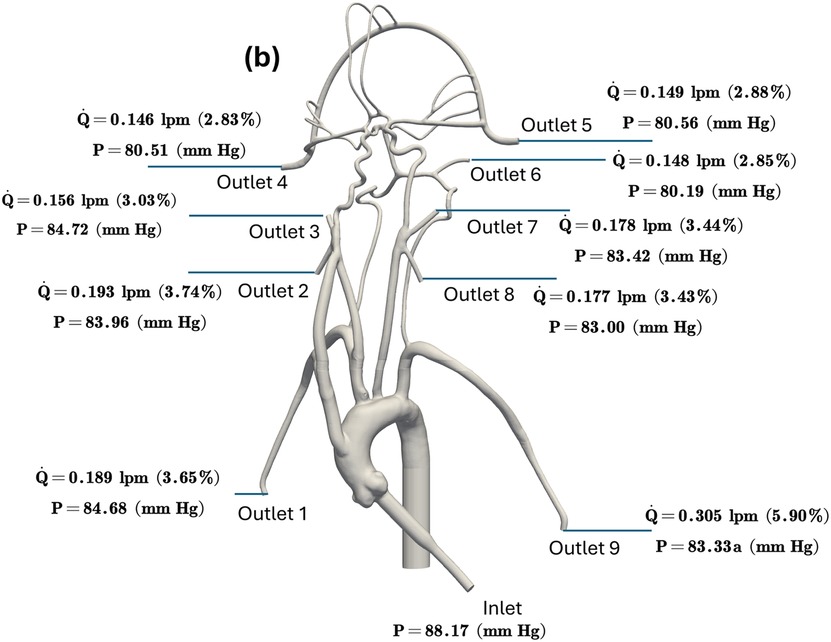
In the published article, there was an error in Figure 3b as published. The simulation was based on an incorrect scale and the values have been updated. The corrected Figure 3b and its caption appear below.

Figure 3. The mean volumetric flow rate and pressure values along with standard deviation at the inlet and all arterial outlets from (a) in vitro experiments and (b) CFD simulation for the normal condition. The percentage values shown in parentheses indicate the fraction of total inlet flow through each arterial outlet.
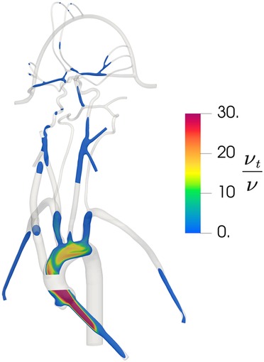
In the published article, there was an error in Figure 5 as published. The simulation was conducted using an incorrect scale and these values have been revised. The corrected Figure 5 and its caption appear below.

Figure 5. Contours of the distribution of the ratio of turbulent viscosity to molecular viscosity in the updated CFD model, illustrating regions of predicted turbulent flow where is large and regions of laminar flow where is small.
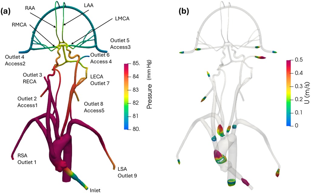
In the published article, there was an error in Figure 6 as published. The simulation was conducted using an incorrect scale and these values have been revised. The corrected Figure 6 and its caption appear below.

Figure 6. (a) Mean pressure contours and (b) profiles of mean velocity at various sections in the anatomical cerebrovascular model from the updated CFD for the normal condition.
In the published article, there was an error in Table 2 as published. The simulation was conducted using an incorrect scale and these values have been revised. The corrected Table 2 and its caption appear below.

Table 2. Mean pressure (mmHg) obtained from in vitro experiments and CFD at various inlet and outlets for the normal condition. The experimental values are reported as mean ± standard deviation (SD) from three experiments.
In the published article, there was an error in Table 3 as published. The simulation was conducted using an incorrect scale and these values have been revised. The corrected Table 3 and its caption appear below.

Table 3. Mean pressure (mmHg) obtained from in vitro experiments and CFD at the inlet and outlets for the stroke condition. The experimental values are reported as mean ± standard deviation (SD) from three experiments.
The authors apologize for this error and state that this does not change the scientific conclusions of the article in any way. The original article has been updated.
Acknowledgments
We would like to thank Boyang Su, Ph.D. for performing the simulations to update the results.
Publisher's note
All claims expressed in this article are solely those of the authors and do not necessarily represent those of their affiliated organizations, or those of the publisher, the editors and the reviewers. Any product that may be evaluated in this article, or claim that may be made by its manufacturer, is not guaranteed or endorsed by the publisher.
Keywords: cerebrovascular model, cerebral blood flow, image based modeling, acute ischemic stroke, fluid dynamics
Citation: Bhardwaj S, Craven BA, Sever JE, Costanzo F, Simon SD and Manning KB (2025) Corrigendum: Modeling flow in an in vitro anatomical cerebrovascular model with experimental validation. Front. Med. Technol. 6:1533412. doi: 10.3389/fmedt.2024.1533412
Received: 23 November 2024; Accepted: 4 December 2024;
Published: 3 January 2025.
Edited and Reviewed by: Claudio Chiastra, Polytechnic University of Turin, Italy
Copyright: © 2025 Bhardwaj, Craven, Sever, Costanzo, Simon and Manning. This is an open-access article distributed under the terms of the Creative Commons Attribution License (CC BY). The use, distribution or reproduction in other forums is permitted, provided the original author(s) and the copyright owner(s) are credited and that the original publication in this journal is cited, in accordance with accepted academic practice. No use, distribution or reproduction is permitted which does not comply with these terms.
*Correspondence: Brent A. Craven, YnJlbnQuY3JhdmVuQGZkYS5oaHMuZ292; Keefe B. Manning, a2JtMTBAcHN1LmVkdQ==
 Saurabh Bhardwaj
 Brent A. Craven
 Jacob E. Sever
 Francesco Costanzo1,3
 Scott D. Simon
 Keefe B. Manning