- 1Department of Gastroenterology, The Second Xiangya Hospital of Central South University, Changsha, China
- 2Research Center of Digestive Disease, Central South University, Changsha, China
Background: The association between grip strength (GS) and non-alcoholic fatty liver disease (NAFLD) has been reported by recent epidemiological studies, however, the results of these studies are inconsistent. This meta-analysis was conducted to collect all available data and estimate the risk of NAFLD among people with low GS, as well as the risk of low GS among patients with NAFLD.
Methods: We systematically searched several literature databases including PubMed, Web of Science, Cochrane Library, and Embase from inception to March 2022. These observational studies reported the risk of NAFLD among people with low GS and/or the risk of low GS among patients with NAFLD. Qualitative and quantitative information was extracted, statistical heterogeneity was assessed using the I2 test, and potential for publication bias was assessed qualitatively by a visual estimate of a funnel plot and quantitatively by calculation of the Begg’s test and the Egger’s test.
Results: Of the citations, 10 eligible studies involving 76,676 participants met inclusion criteria. The meta-analysis of seven cross-section studies (69,757 participants) showed that people with low GS had increased risk of NAFLD than those with normal GS (summary OR = 3.32, 95% CI: 1.91–5.75). In addition, the meta-analysis of four studies (14,920 participants) reported that the risk of low GS patients with NAFLD was higher than those in normal people (summary OR = 3.31, 95% CI: 2.45–4.47).
Conclusion: In this meta-analysis, we demonstrated a strong relationship between low GS and NAFLD. We found an increased risk of NAFLD among people with low GS, and an increased risk of lower GS among NAFLD patients.
Systematic review registration: [www.crd.york.ac.uk/prospero], identifier [CRD42022334687].
Introduction
Currently, non-alcoholic fatty liver disease (NAFLD) has become one of the most common causes of chronic liver disease, it is defined by the presence of steatosis in more than 5% of hepatocytes with little or no alcohol consumption (1). NAFLD is characterized by fatty infiltration of the liver without secondary causes of hepatic steatosis (2).
In the United States, approximately 30% of individuals are diagnosed with NAFLD (3). In addition, over 27% of individuals are affected by NAFLD (4). In China, the prevalence of NAFLD was reported to be between 15 and 36% (5, 6). Additionally, as a result of the aging population and obesity, the prevalence of NAFLD is increasing rapidly. However, to date, there is no effective drug for treatment of NAFLD. As shown by a number of compelling studies, NAFLD is associated with some chronic diseases, such as type 2 mellitus (T2DM), cardiovascular disease (CVD), and chronic kidney disease (CKD) (7, 8). Therefore, understanding the pathobiology and risk factors for development of NAFLD is of great importance.
Grip strength (GS) is a measure of the maximum static force that a hand can apply around a dynamometer. GS is often considered an indicator of muscle mass and muscle strength (9). Researches have suggested that low GS is associated with health damage and higher all-cause mortality (10, 11), such as falls, disability and poor quality of life (12, 13). Indeed, previous studies have also shown association between NAFLD and sarcopenia (14). Low muscle strength is used as a principal determinant of sarcopenia over muscle mass (15), and GS is recommended as a substitute measurement of muscle strength (16). Therefore, in clinical practice, people are increasingly aware of the importance of muscle strength.
Non-alcoholic fatty liver disease is a systemic condition that has a bi-directional relationship with the components of metabolic syndrome (17). According to recent studies, muscular strength is inversely related to insulin sensitivity (18) and excessive body and abdominal fat (19), which are independent risk factors for developing NAFLD. Now, several studies have reported that association between GS and NAFLD, therefore, we collected these studies for meta-analysis as a way to explore the relationship between GS and NAFLD.
Materials and methods
Protocol and guidance
This meta-analysis followed the Preferred Reports Items for Systematic Reviews and Meta-analyses (PRISMA) reporting guideline (20). The protocol for this meta-analysis was registered with PROSPERO (CRD42022334687).
Data sources and searches
Two investigators (LH and SF) independently conducted an electronic literature search using PubMed, Web of Science, Cochrane Library, and Embase, language was restricted to English, from database inception to March 2022. In PubMed, controlled vocabulary terms and the following keywords were used: (“Non-alcoholic Fatty Liver Disease” [Mesh]) OR (Non-alcoholic Fatty Liver Disease) OR (Non-alcoholic Fatty Liver Disease) OR (Fatty Liver, Non-alcoholic) OR (Fatty Livers, Non-alcoholic) OR (Liver, Non-alcoholic Fatty) OR (Livers, Non-alcoholic Fatty) OR (Non-alcoholic Fatty Liver) OR (Non-alcoholic Fatty Livers) OR (Non-alcoholic Steatohepatitis) OR (Non-alcoholic Steatohepatitides) OR (Steatohepatitides, Non-alcoholic) OR (Steatohepatitis, Non-alcoholic) AND (“Hand Strength” [Mesh]) OR (Strength, Hand) OR (Grip Strength) OR (Strength, Grip) OR (Hand Grip Strength) OR (Grip Strength, Hand) OR (Strength, Hand Grip) OR (Grip) OR (Grips) OR (Grasp) OR (Grasps). A similar search strategy was run in other databases. Supplementary Table 1 presents the search strategy.
The database search revealed 224 articles that could have been included in our meta-analysis, and 43 articles were excluded because they were duplicated. After removing duplicates, all titles and abstracts for potential inclusion were screened by two independent researchers (LH and SF). Based on the inclusion and exclusion criteria, 164 articles were excluded after reading the titles and abstracts. Finally, 17 full texts of these records were selected for detailed assessment. The two researchers extracted the related data according to the inclusion criteria. If the studies were potentially eligible for inclusion, the full text was examined. The two reviewers would discuss with each other any disagreements that may have occurred.
Study quality assessment
All studies were assessed for selection and measurement biases according to the Newcastle-Ottawa Scale (NOS) (21). The NOS consists of eight items focused on three domains: selection of study groups, ascertainment of the exposure and outcome, and comparability of groups to assess the quality of observational studies. Ratings were based on a star system and studies with a maximum rating of nine. Studies with one to three stars were categorized as low quality, four to six stars categorized as moderate quality, and seven to nine stars categorized as high quality. Each of included studies was assessed for bias by two independent investigators (LH and SF).
Inclusion criteria
The same two authors evaluated the titles and abstracts of eligible studies and any disagreements were resolved by consensus. The inclusion criteria are as follows: (1) studies on the association between NAFLD and GS; (2) used a standardized index to diagnose and assess NAFLD and GS; (3) reporting odds ratio (OR) and 95% confidence intervals (95% CI) for GS and NAFLD; (4) the full text of the study could be assessed; and (5) the full text of the study could be assessed.
Exclusion criteria
The same two authors evaluated the titles and abstracts of eligible studies and any disagreements were resolved by consensus. The exclusion criteria are as follows: (1) did not use clear diagnostic criteria for NAFLD; (2) the measurement of GS is not accurate; (3) the study did not provide the OR of NAFLD and GS; (4) case reports, case series, reviews, posters, and abstracts were excluded; (5) measured only in vitro parameters or used animal models; and (6) based on the NOS scores, the low-quality studies were excluded.
Data collection process
Data collection process two independent researchers (LH and SF) assessed the full texts of included studies and used a standard data extraction form when extracting data. Any disagreements were resolved by discussion until consensus was reached. The data extracted for the analysis involved: (1) the first author’s name and publication year; (2) the sample size and number of cases; (3) the mean age and sources of participants; (4) the OR with the corresponding 95% CI; and (5) the scores of NOS in the studies.
Statistical analysis
The meta-analysis of comparable data was carried out using Review Manager 5.3. OR and their associated 95% CI were used to assess a comparison between outcomes reported by the studies and a P-value less than 0.05 was considered to be statistically significant. We collected the summary OR of NAFLD and low GS. The heterogeneity of results between studies was determined by the I2 test (22). For I2, values of 25 to <50% were considered low heterogeneity, 50 to <75% moderate, and 75% highly heterogeneous. If significant heterogeneity was not present (I2 < 50%), a fixed-effect model was used to pool outcomes, otherwise a random-effect model was applied for the meta-analysis (I2 > 50%). The publication bias was assessed qualitatively by a visual estimate of the funnel plot and quantitatively by calculation of Begg’s test and Egger’s test (23).
Subgroup analyses and sensitivity analyses
Subgroup analyses were performed according to the method of GS ascertainment [GS and relative grip strength (RGS)], diagnosis of NAFLD [ultrasonography and hepatic steatosis index (HSI)], region (China and Korea), mean age (<60 and >60 years old), several participants (<5,000 and >5,000). One-study-removed sensitivity analyses were performed to determine the relative impact of each study on the overall risk estimate.
Results
Eligible studies and individual characteristics
Ten articles were included in this analysis (Figure 1) (9, 24–32). The selected studies involved 76,676 participants. Among the 10 studies, 7 studies (9, 24–29) reported the odds rate (OR) of NAFLD between low GS group and normal group, 4 studies (26, 30–32) reported the OR of low GS between NAFLD group and normal group and 1 studies (26) involved above the two types of OR. The characteristics of the studies included in our meta-analysis are listed in Table 1.
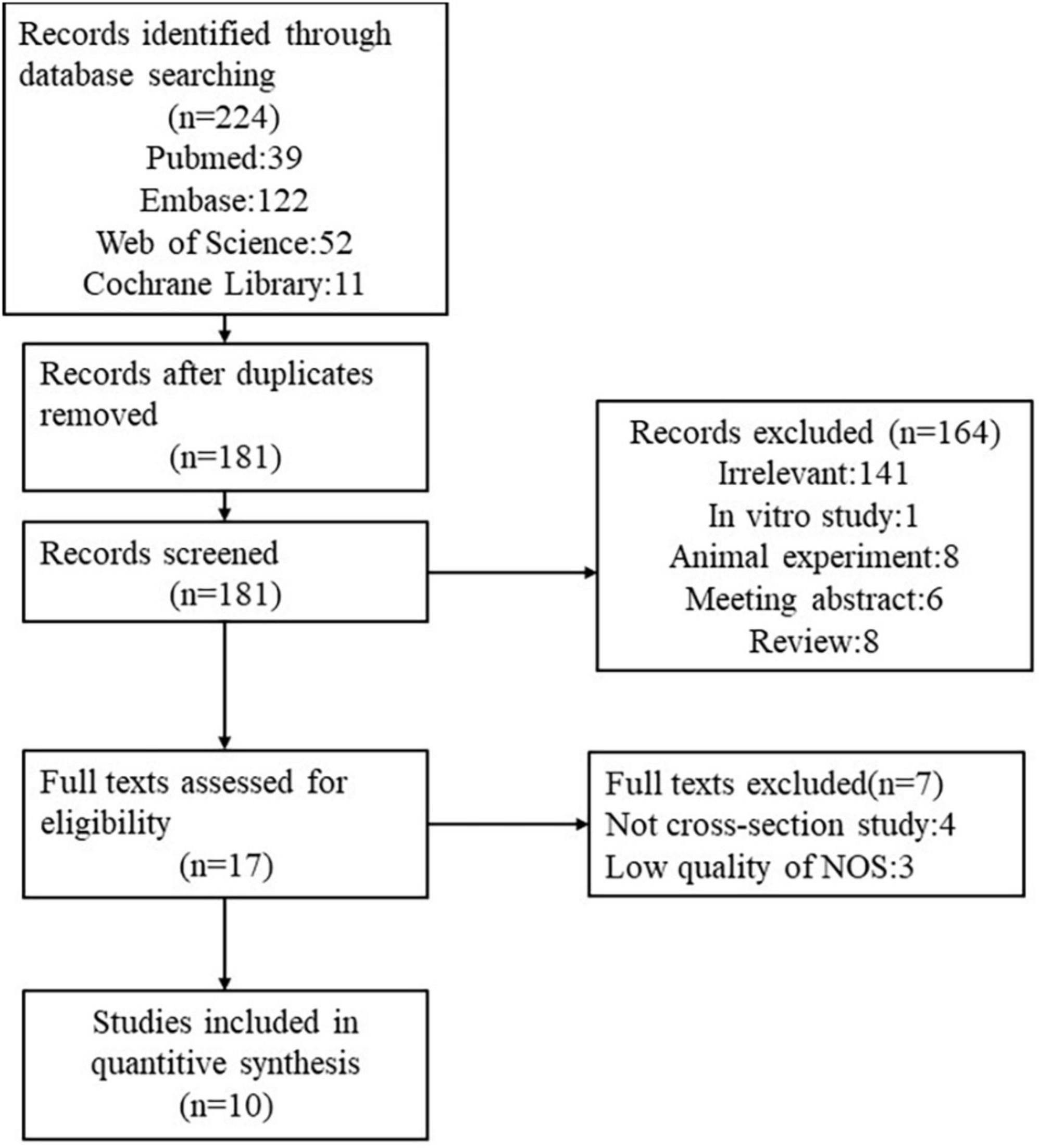
Figure 1. Flow chart of literature search for studies investigating the association between grip strength and NAFLD.
Quality of the individual studies
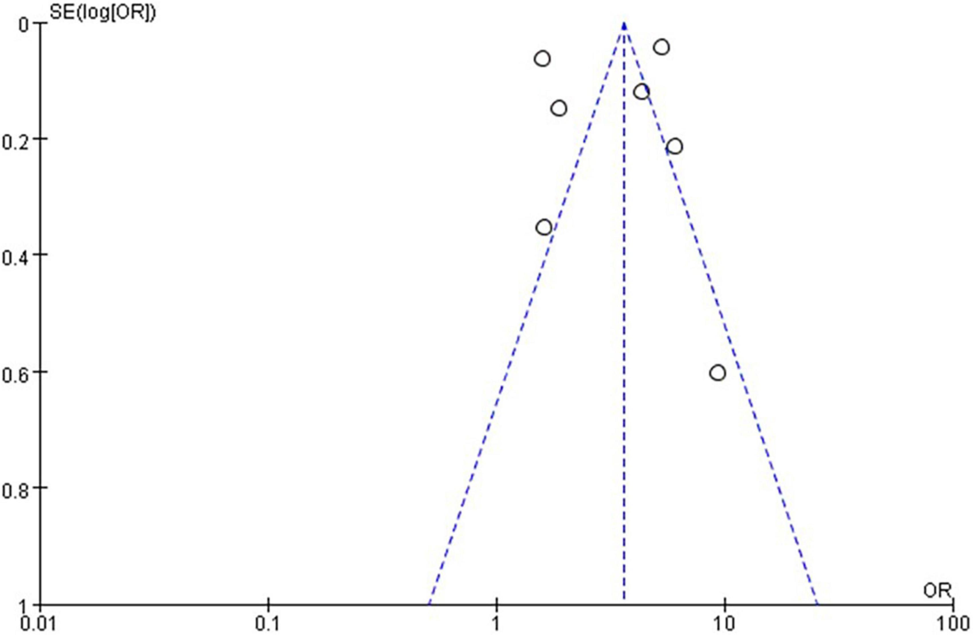
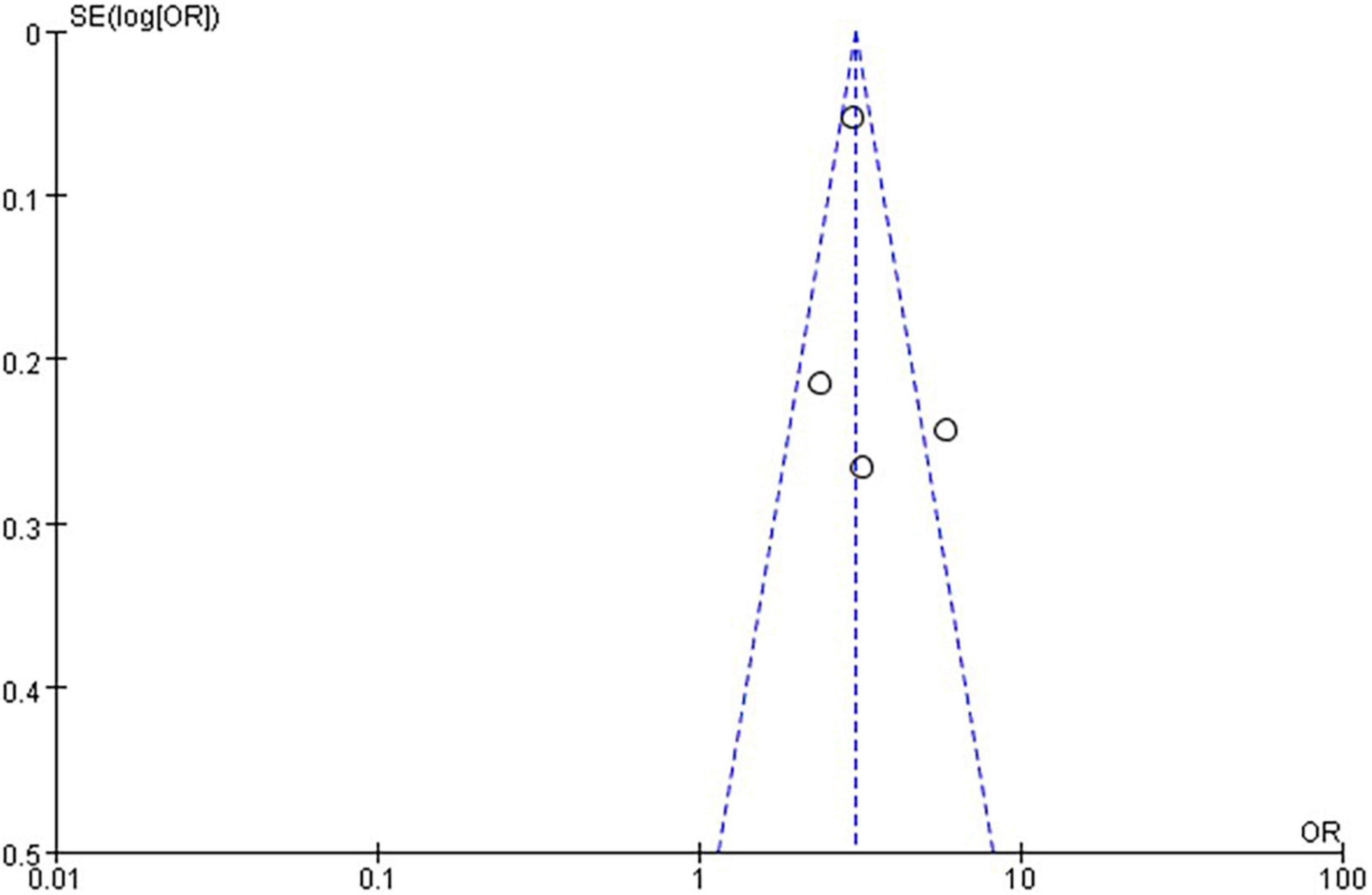
The quality level of each study ranged from 5 to 8 stars (Figure 1). The funnel plot (Figures 2, 3) provided a qualitative estimation of publication bias.
Odds rate of non-alcoholic fatty liver disease between low grip strength group and normal group
In the seven studies of the OR of NAFLD included in this meta-analysis (Table 1), the sample size varied from 538 to 27,531 participants, and the age varied from 18 to 80 years old. As shown in Figure 4, high heterogeneity was present among the seven studies reporting OR (I2=98%), so we chose the random-effects model. Meta-analysis of these studies showed that low GS patients had odds of NAFLD that were 3.32 times as high as normal GS (summary OR = 3.32, 95% CI: 1.91–5.75, Figure 4). The result of Funnel plot analysis is showed in Figure 2, and the result of Begg’s test (P = 1) and Egger’s test (P = 0.785) suggest that there is no significant publication bias.
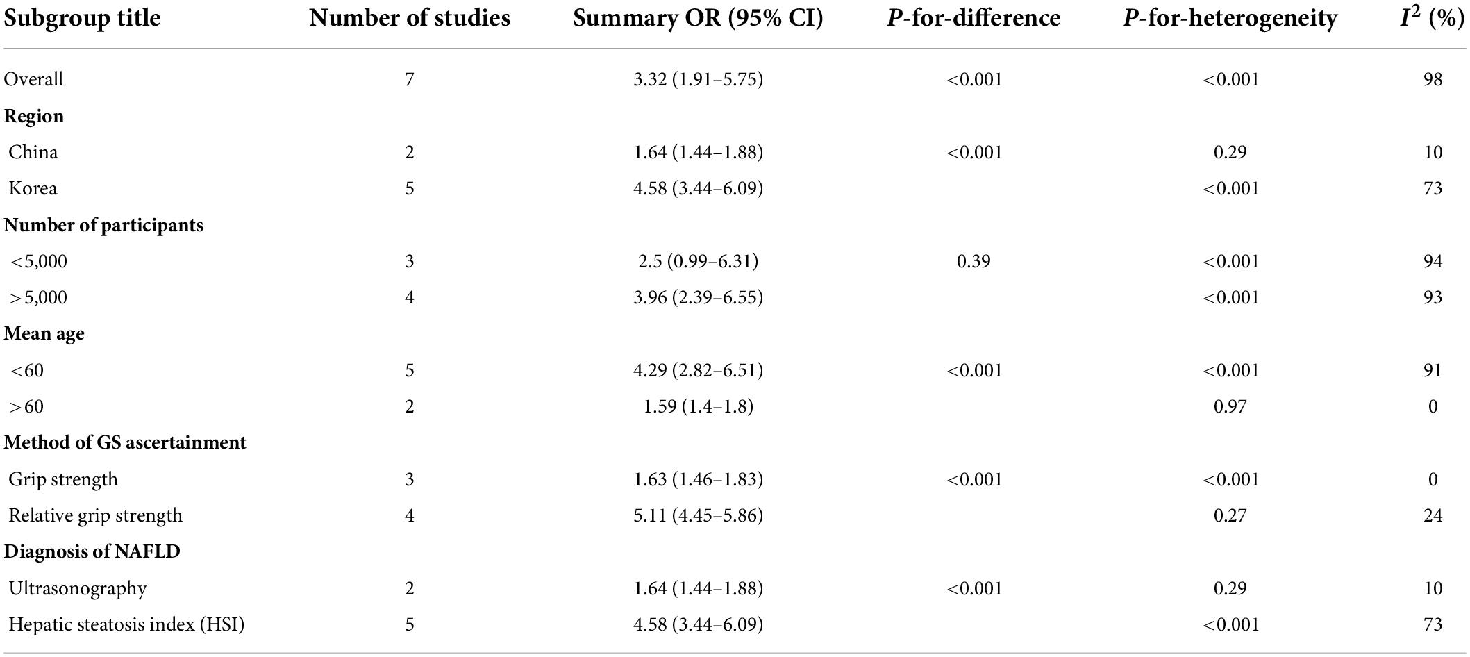
Because of the high heterogeneity, we conducted a series of subgroup analyses to identity the heterogeneity source. The subgroup analysis by several participants revealed no significant difference between numbers (Figure 5), the OR = 2.5, 95% CI: 0.99–6.31 for studies conducted in the number of participants less than 5,000, and OR = 3.96, 95% CI: 2.39–6.55 for studies conducted in several participants more than 5,000. There was a significant association between low GS and risk of NAFLD detected in the studies using RGS (Figure 6, OR = 5.11, 95% CI: 4.45–5.86) compared to the using GS (OR = 1.63, 95% CI: 1.46–1.83). Besides, a significantly greater effect size was observed in the studies using HSI (Figure 7, OR = 4.58, 95% CI: 3.44–6.09) than in the ones applying ultrasonography (OR = 1.64, 95% CI: 1.44–1.88). And the subgroup analysis by region revealed stronger association between GS and risk of NAFLD in the studies in Korea (Figure 8, OR = 4.58, 95% CI: 3.44–6.09) than the studies in China (OR = 1.64, 95% CI: 1.44–1.88). Regarding mean age, compared to the studies with mean age more than 60 years old (Figure 9, OR = 1.59, 95% CI: 1.4–1.8), the studies with mean age of fewer than 60 years old (OR = 4.29, 95% CI: 2.82–6.51) were more strongly associated with the risk of NAFLD. Because of the limited number of original articles, the data are only from China and Korea, therefore, we speculate that the high heterogeneity may be due to the regional distribution of the data and the small number of included articles. And all the subgroup analyses are presented in Table 2.
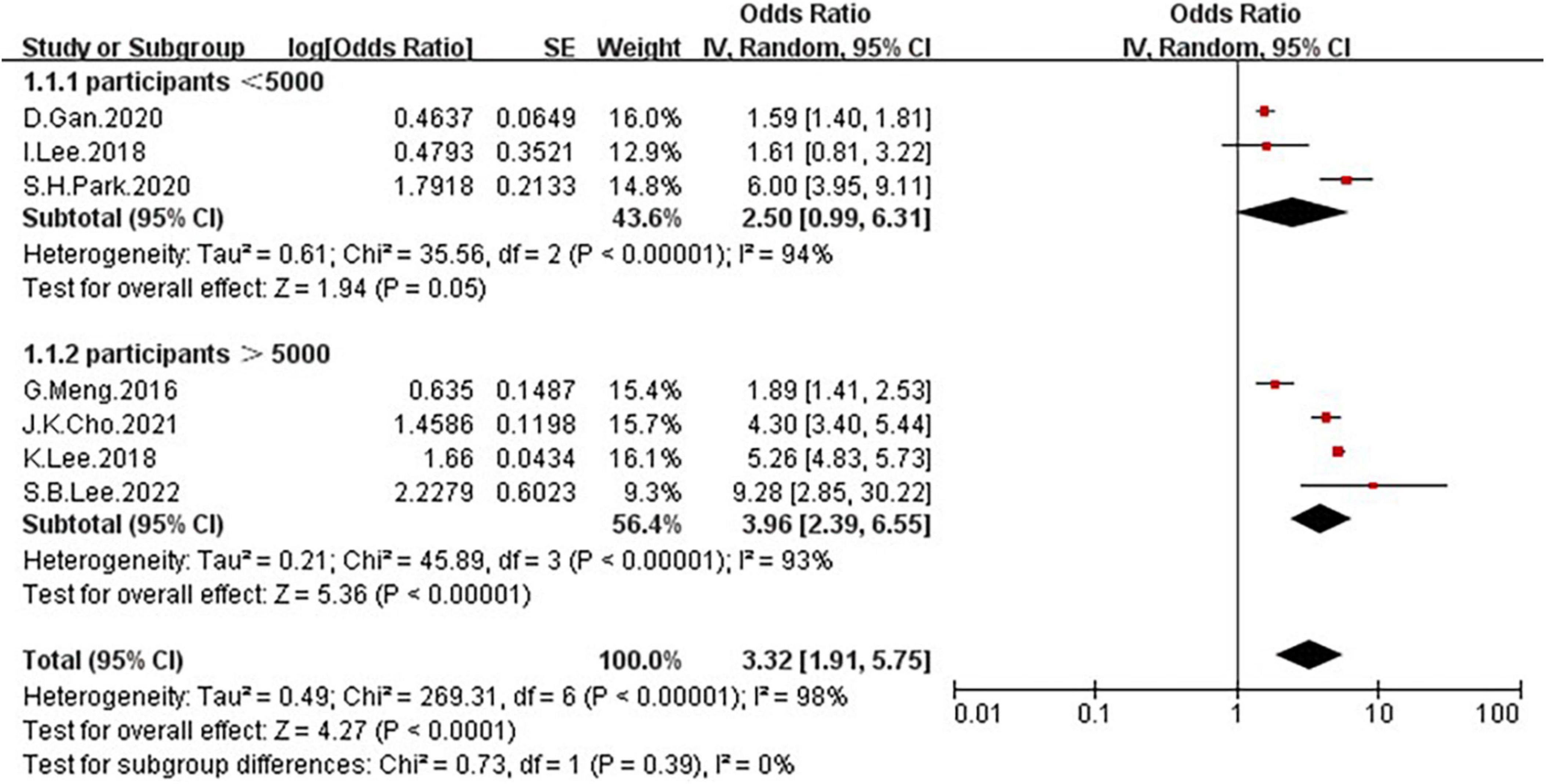
Figure 5. Forest plots depicting the association of low grip strength and the risk of NAFLD were subgrouped by several participants.
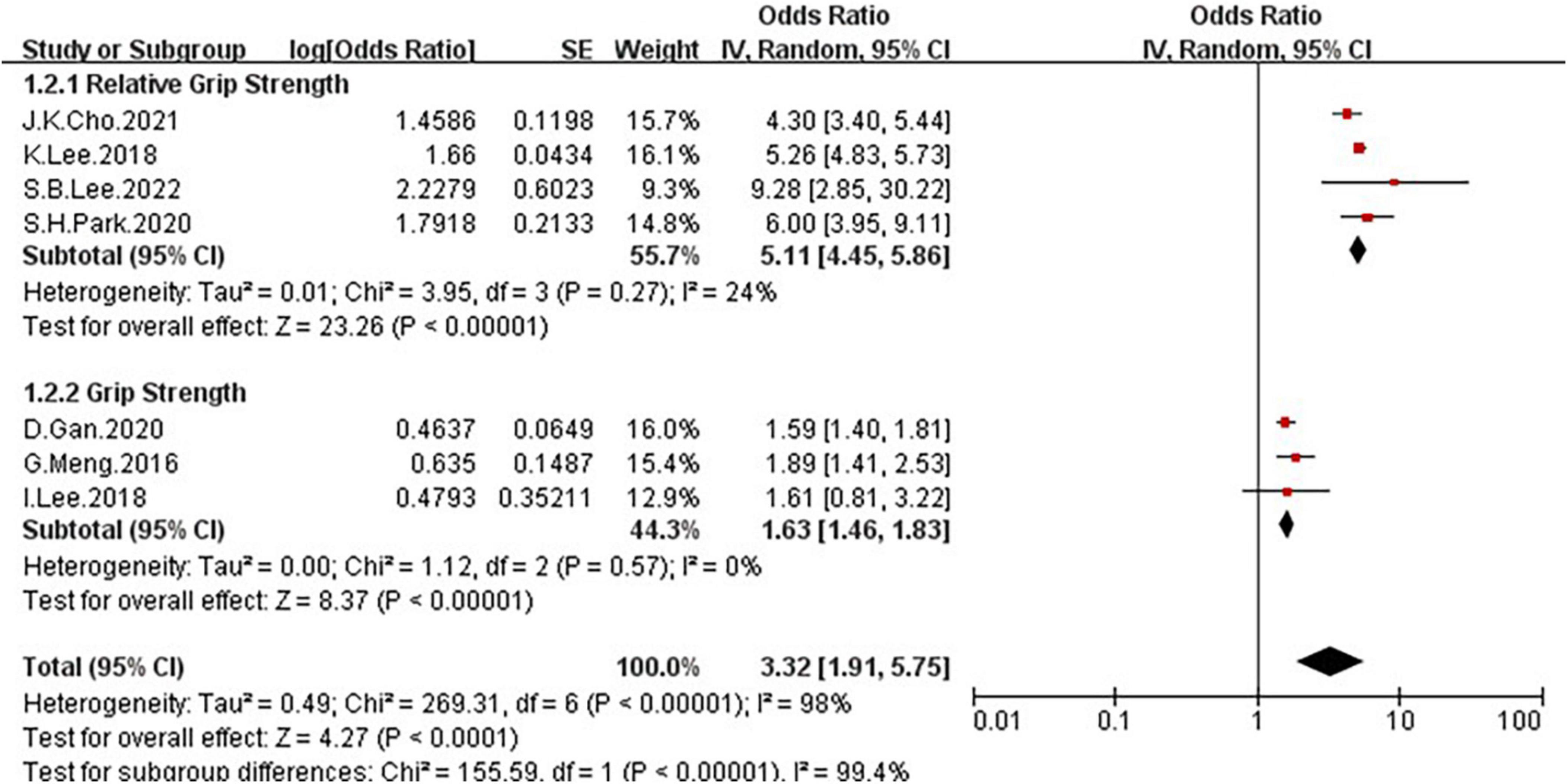
Figure 6. Forest plots depicting the association of low grip strength and the risk of NAFLD were subgrouped by the method of grip strength ascertainment.
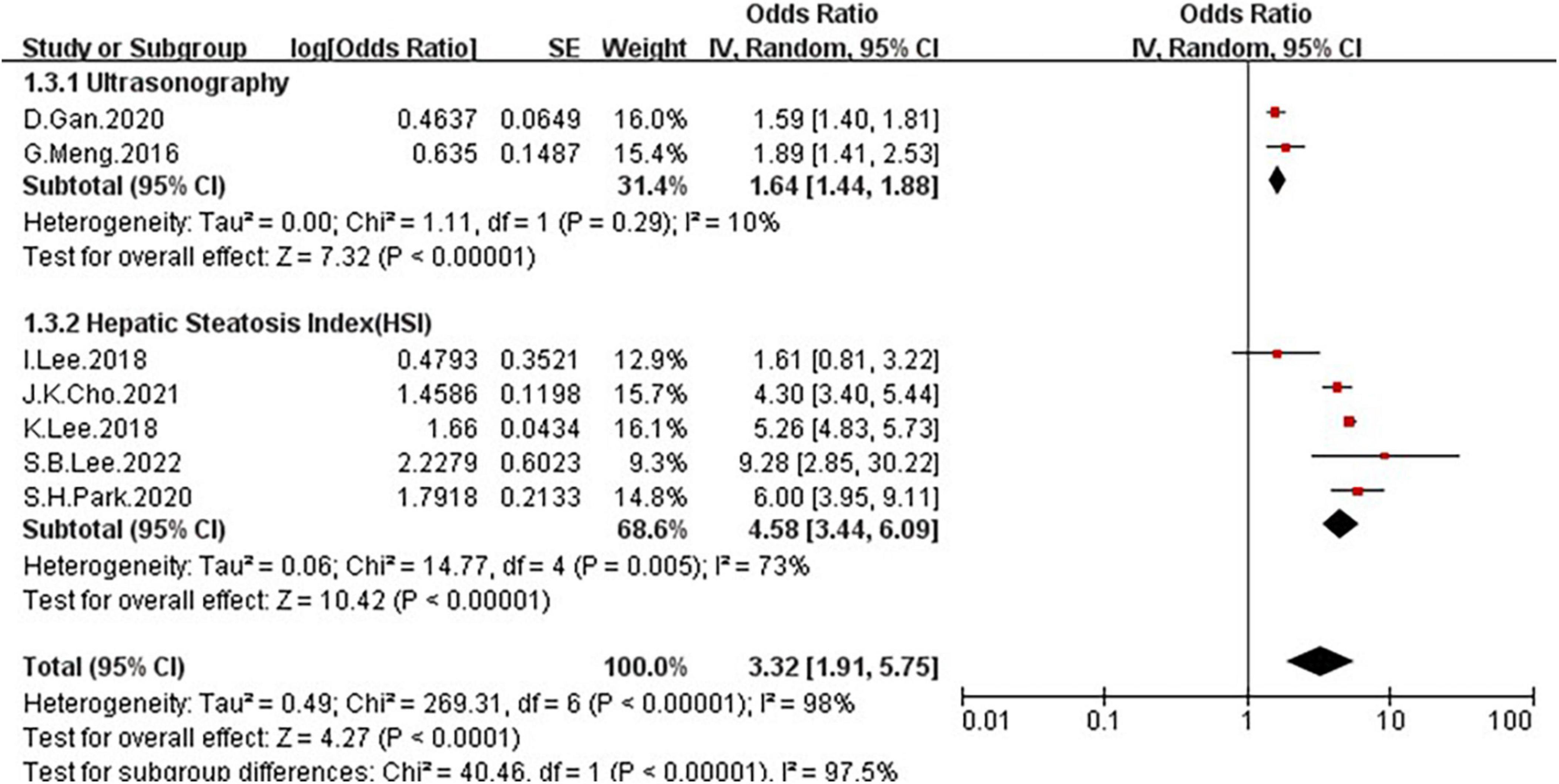
Figure 7. Forest plots depicting the association of low grip strength and the risk of NAFLD were subgrouped by the diagnosis of NAFLD.
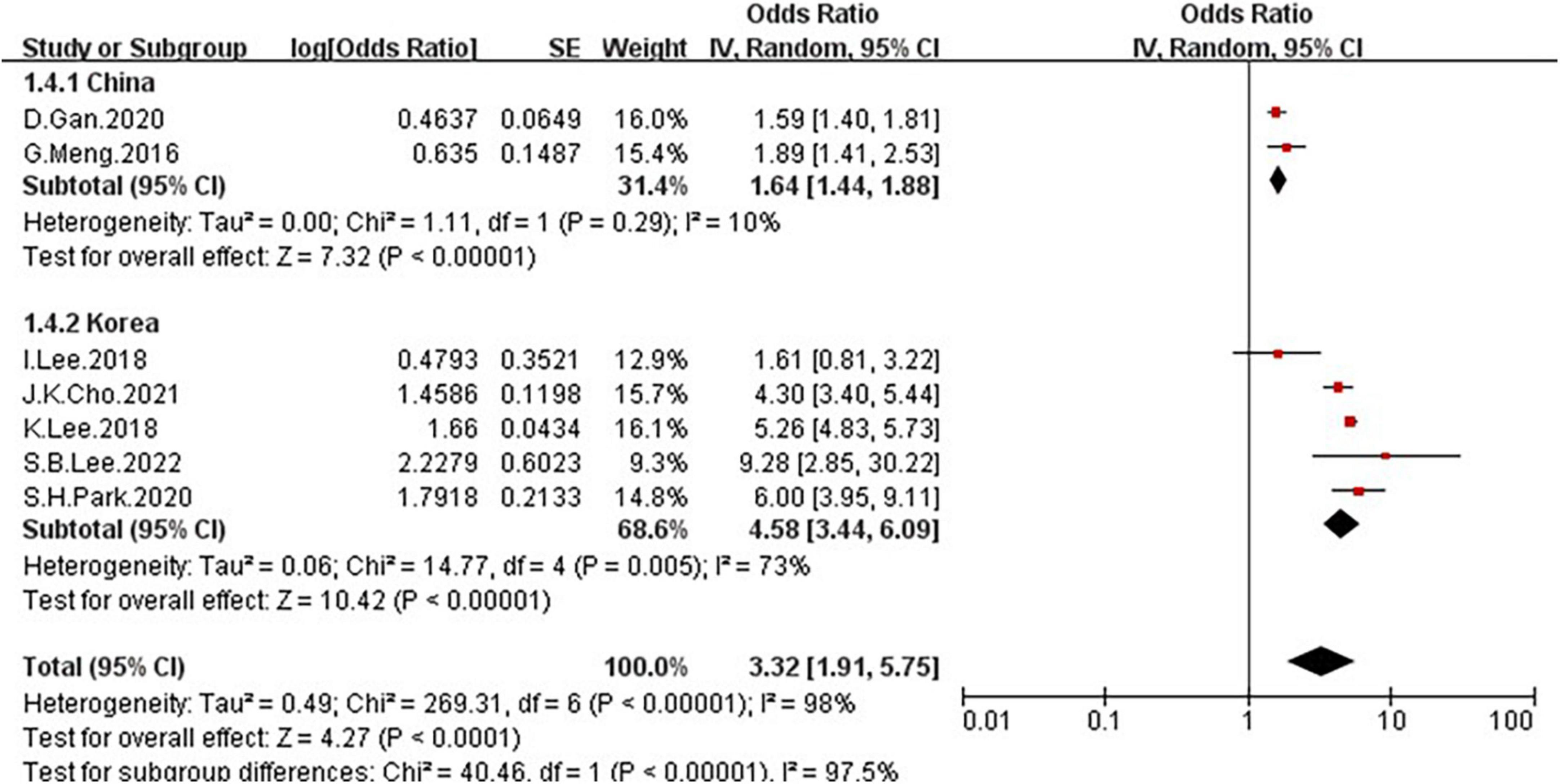
Figure 8. Forest plots depicting the association of low grip strength and the risk of NAFLD were subgrouped by the region.
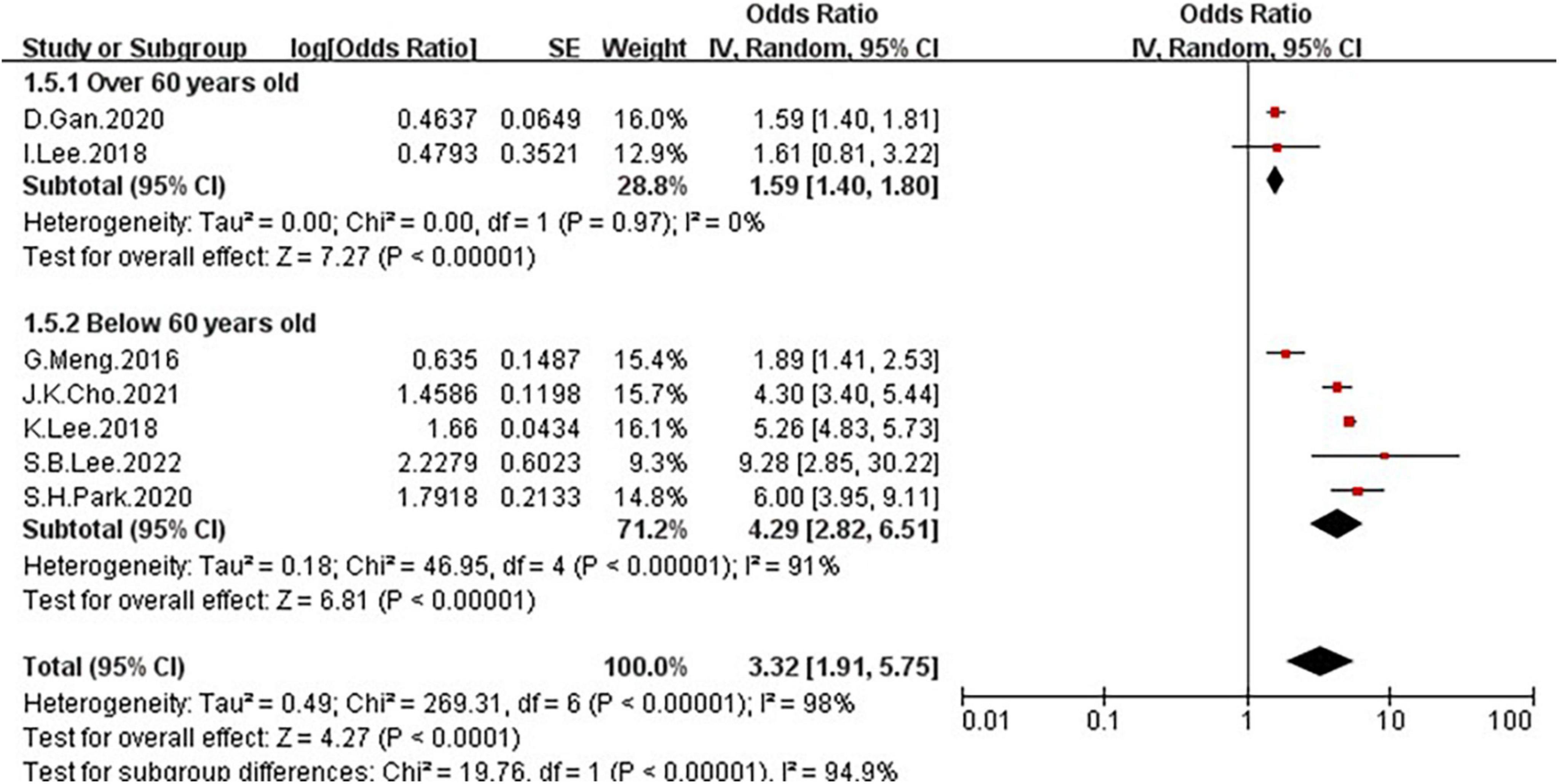
Figure 9. Forest plots depicting the association of low grip strength and the risk of NAFLD were subgrouped by the mean age.
Odds rate of low grip strength between non-alcoholic fatty liver disease group and normal group
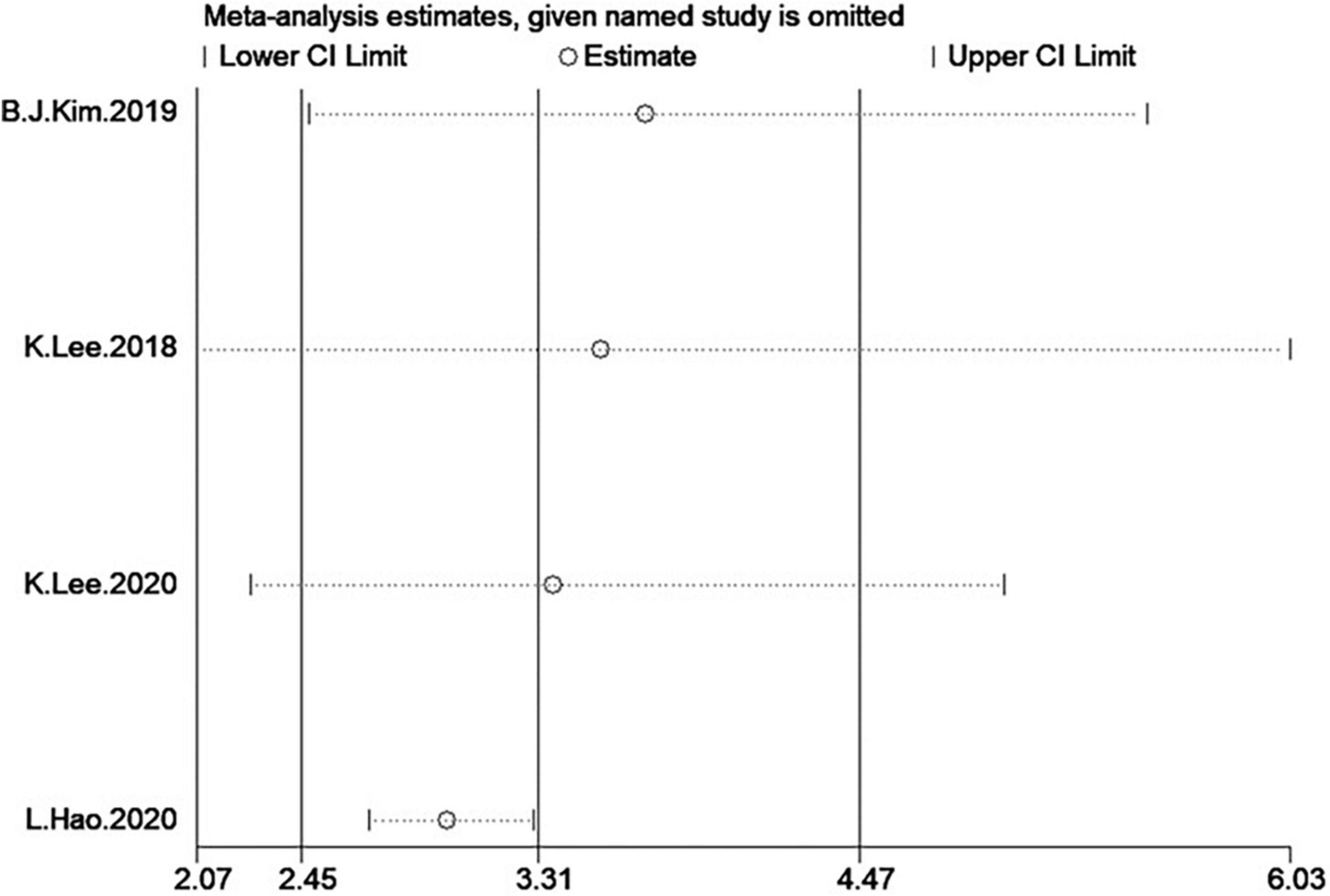
In the four studies of the OR of low GS included in our meta-analysis (Table 1), the sample size varied from 1,126 to 8,001 participants, and the age varied from 10 to 80 years old. As shown in Figure 10, moderate heterogeneity was present among the four studies reporting OR (I2 = 65%). Meta-analysis of these studies showed that patients with NAFLD had odds of low GS that were 3.31 times as high as normal group (summary OR = 3.31, 95% CI: 2.45–4.47, Figure 10). The result of the funnel plot is presented in Figure 3, and the result of Begg’s test (P = 0.734) and Egger’s test (P = 0.630) suggest that there is no significant publication bias.
Because of the moderate heterogeneity, we conduct a subgroup and meta-regression analyses to identify the heterogeneous source. These four studies, all the shown that NAFLD patients have markedly low GS than the non-NAFLD groups. Because meta-regression was performed to examine possible heterogeneous factors for quantitative variables, we used age as a covariate for meta-regression, but the result (P > 0.05) showed that age may not be the cause of high heterogeneity. Additionally, we conducted a subgroup analysis according to the method of GS ascertainment (GS and RGS), there were significant association between NAFLD and low GS was detected in the studies using RGS (Figure 11, OR = 4.02, 95% CI: 2.11–7.65) compared to the studies using GS (OR = 2.69, 95% CI: 1.94–3.73).
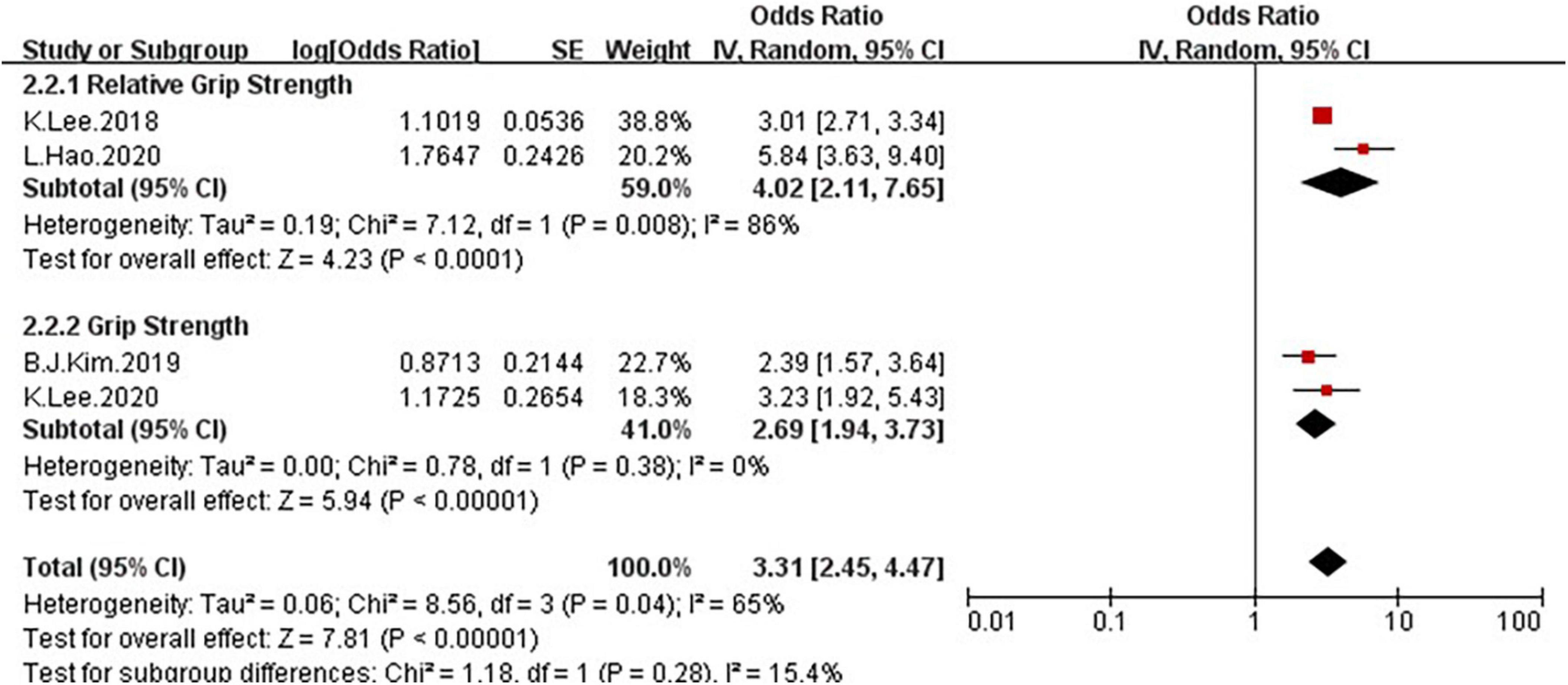
Figure 11. Forest plots depicting the association of NAFLD and the risk of low grip strength were subgrouped by the method of grip strength ascertainment.
Sensitivity analysis
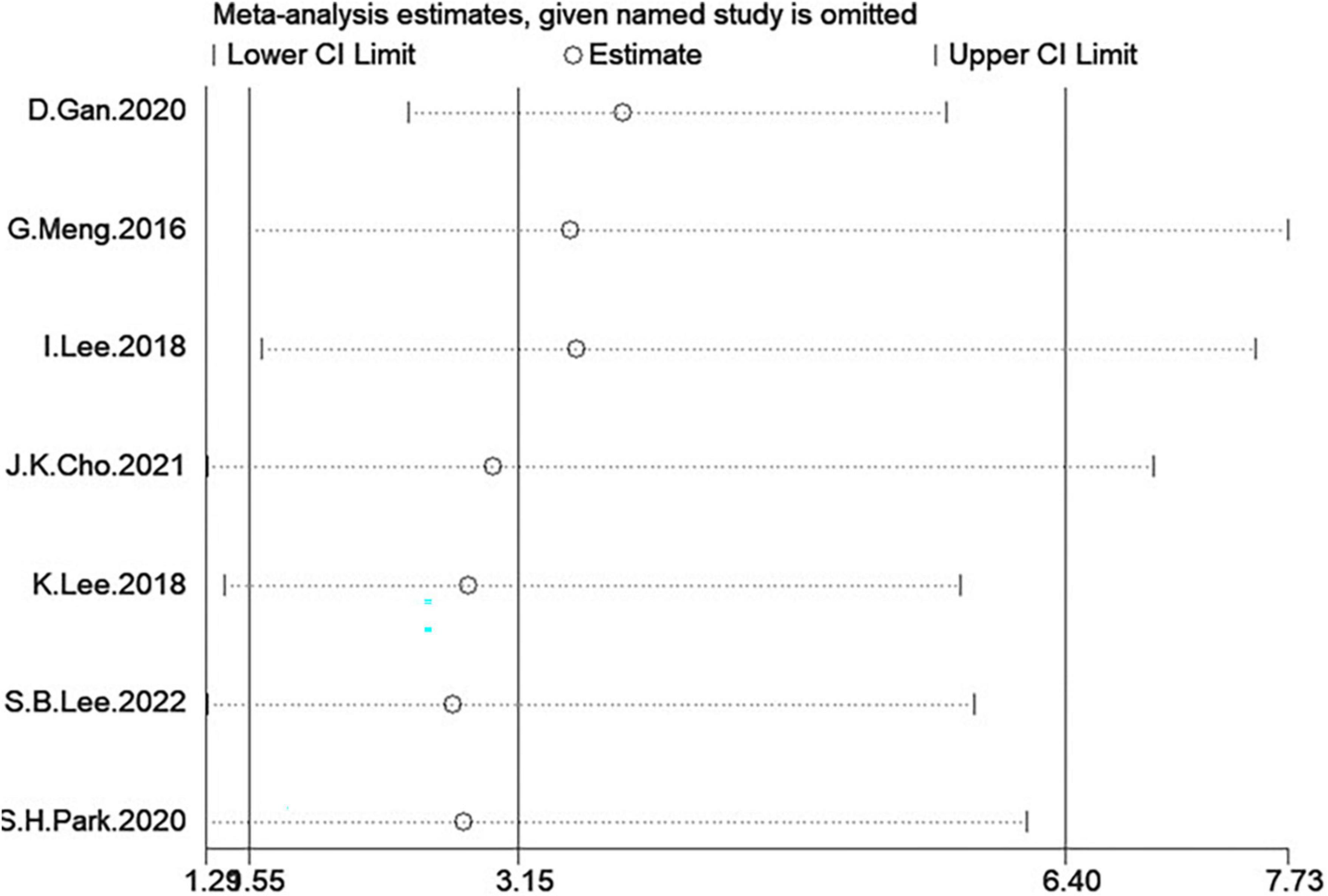
In the two analyses, in one-study-removed sensitivity analyses, we excluded each study and results did not change (Figures 12, 13).
Discussion
To the best of our knowledge, this is the first systematic review and meta-analysis that summarized available studies regarding the association between GS and NAFLD. In this meta-analysis which included 10 studies with a total of 76,676 participants, we performed two types of meta-analysis, and explored the OR of NAFLD in patients with low GS and the OR of low GS among patients with NAFLD, both results suggest a significant association between the NAFLD and GS: the Figure 4 shown that a significantly increased risk of NAFLD among individuals with low GS with the pooled OR of 3.32 (95% CI: 1.91–5.75). And Figure 10 suggested that NAFLD patients had odds of low GS that were 3.31 times as high as normal people (summary OR = 3.31, 95% CI: 2.45–4.47).
Non-alcoholic fatty liver disease encompasses a wide range of diseases from simple steatosis, non-alcoholic steatohepatitis, fibrosis, and even cirrhosis (33). Skeletal muscle is an insulin-responsive and important endocrine organ, because it secretes myokines that influence metabolic processes in liver and muscle (34). Previously, many reliable studies have found that association between skeletal muscle and NAFLD, Guo et al. (35) reported that skeletal muscle index (SMI) is independently associated with the severity of hepatic steatosis and liver fibrosis of related to NAFLD, and they assessed the association of SMI tertiles with NAFLD and liver fibrosis, individuals with low muscle mass were significantly correlated with NAFLD and liver fibrosis. These findings suggest that NAFLD is affected by skeletal muscle even when people do not have sarcopenia. GS is also an important indicator in the assessment of skeletal muscle and sarcopenia. Previously, several studies have shown a link between sarcopenia and NAFLD, mainly due to a common pathological mechanism, insulin resistance and chronic inflammation have been the most frequently proposed mechanisms, and both are hypothetically plausible (14, 36). Firstly, both the liver and muscle are the target organs for insulin action, and insulin resistance is known as a key factor in the pathophysiology of both NAFLD and sarcopenia (37). With aging, the fat mass in muscle cells increases, which becomes a risk factor for insulin resistance (38). Furthermore, ectopic fat accumulation in the liver is closely associated with systemic insulin resistance (37). Hong (39) reported that an increased insulin resistance index in subjects with sarcopenia compared to those without sarcopenia, and insulin resistance and SMI showed a significant negative correlation, and they also found a significant relationship between insulin resistance and liver attenuation index (LAI), which reflects fat accumulation in the liver.
On the other hand, chronic inflammation is the other hypothesis most often cited. There are several studies focus on the mediators that link the muscle-liver-adipose tissue axis (40). For example, myostatin, a transforming growth factor (TGF)-β superfamily member, is a regulator of skeletal muscle mass, and now some animal studies have shown that myostatin has significant hepatic effects by regulating skeletal muscle metabolism, and blocking myostatin not only increases muscle mass but also protects mice from fatty liver and improves insulin resistance (41). It has been demonstrated that oxidative stress and proinflammatory cytokines of chronic inflammation, such as tumor necrosis factor (TNF)-α and Interleukin (IL)-6 can promote fat and muscle metabolism, leading to loss of skeletal muscle (42), various inflammatory factors released from visceral adipocytes can also promote the development of metabolic syndrome (43), and myonectin and irisin have been suggested to contribute to the development of insulin resistance and fatty liver (44, 45), and Hong (39) also found that high-sensitivity C-reactive protein (hsCRP) concentrations were closely correlated with SMI and LAI, which suggests that inflammation may be an important underlying factor associated with both sarcopenia and NAFLD.
Recently, low vitamin D levels have been suggested to be associated with NAFLD and muscle strength. Vitamin D plays an important role in muscle mass and muscle strength. A systematic review revealed that vitamin D supplementation significantly increased muscle strength (46). A separate study also showed that muscle nuclear vitamin D receptor (VDR) was increased by 30% and augmented muscle fiber size by 10% in elderly females taking vitamin D (47). The involvement of vitamin D in mediating several immune-inflammatory (48) and metabolic processes (49) has been demonstrated previously. Roth et al. (50) reported that vitamin D deficiency exacerbates NAFLD through Toll-like receptors (TLR)-activation in a westernized diet rat model, which causes insulin resistance, higher hepatic resistance gene expression, and up-regulation of hepatic inflammatory and oxidative stress. In humans, hepatic VDR expression is inversely correlated with steatosis severity (51). A recent study (52) have shown that liver VDR expression plays an important role in regulating intra-hepatic lipid accumulation.
In addition, gut microbiota is also an important component in the pathological mechanism. The gut microbiota composition is generally shaped in early childhood (53), and by the age of three years old (54), the gut microbiota reaches its mature composition, which is maintained relatively stable over the lifespan, and after age of 65, gut microbiota resilience is generally reduced. In current studies, there is evidence supporting the concept that the gut microbiota composition is moderated by exercise (55), including the animal models (56) and human studies (57). Currently, the most studied putative mediators of the effect of gut microbiota on skeletal muscle function are short-chain fatty acids (SCFA) (58), and the SCFA produced by gut microbiota can enter systemic circulation and be absorbed by skeletal muscle cells, where they act as ligands for free fatty acids receptors 2 and 3 (59), and these receptors have a key role in moderating glucose uptake and metabolism, and in promoting insulin sensitivity (60). In addition, gut microbiota also plays a role in NAFLD. As we all know, increased dietary fat intake, is associated with the development of NAFLD (61), and the high fat diet can alter the gut microbiota, and favoring gut bacteria associated with the development of NAFLD (62).
This study also has several limitations. Firstly, one analysis has moderate heterogeneity, and another analysis has high heterogeneity, which maybe because of the small number of included studies, the restricted regional distribution of the studies, and age differences in each study. Secondly, in the subgroup analyses, some subgroups only have 2 or 3 studies, which may affect the results. Besides, subgroup analyses are observational by nature and may be subject to confounding by study-level characteristics. Finally, the definition of NAFLD is different in included studies, some studies use ultrasound to examine the NAFLD, and some studies use HSI to diagnose NAFLD, which may affect the study findings.
Therefore, GS, as an important parameter of sarcopenia and muscle strength, is associated with NAFLD, not only in pathological mechanisms, such as insulin resistance, chronic inflammation, gut microbiota, and regulation of vitamin D, but also in terms of clinical data that people with low GS have a higher risk of NAFLD, and patient with NAFLD have lower GS than normal people.
Conclusion
In conclusion, there is an association between NAFLD and GS. Compare with the normal group, people with low GS are more likely to develop NAFLD, in addition, GS levels in NAFLD patients are also generally lower than the normal population.
Data availability statement
The original contributions presented in this study are included in the article/Supplementary material, further inquiries can be directed to the corresponding author.
Author contributions
LH: study idea, concept and design, data extraction and interpretation of data, drafting of the manuscript, and review of the final manuscript. SF: data extraction and analysis of data, drafting of the manuscript, and review of the final manuscript. JL: drafting of the manuscript, data analysis, and review of the final manuscript. DL: drafting of the manuscript and review of the final manuscript. YT: study idea, concept and design, drafting of the manuscript, and review of the final manuscript. All authors contributed to the article and approved the submitted version.
Conflict of interest
The authors declare that the research was conducted in the absence of any commercial or financial relationships that could be construed as a potential conflict of interest.
Publisher’s note
All claims expressed in this article are solely those of the authors and do not necessarily represent those of their affiliated organizations, or those of the publisher, the editors and the reviewers. Any product that may be evaluated in this article, or claim that may be made by its manufacturer, is not guaranteed or endorsed by the publisher.
Supplementary material
The Supplementary Material for this article can be found online at: https://www.frontiersin.org/articles/10.3389/fmed.2022.988566/full#supplementary-material
References
1. Cobbina E, Akhlaghi F. Non-alcoholic fatty liver disease (NAFLD) - pathogenesis, classification, and effect on drug metabolizing enzymes and transporters. Drug Metab Rev. (2017) 49:197–211. doi: 10.1080/03602532.2017.1293683
2. Kang SH, Lee HW, Yoo JJ, Cho Y, Kim SU, Lee TH, et al. KASL clinical practice guidelines: Management of nonalcoholic fatty liver disease. Clin Mol Hepatol. (2021) 27:363–401. doi: 10.3350/cmh.2021.0178
3. Cotter TG, Rinella M. Nonalcoholic fatty liver disease 2020: The state of the disease. Gastroenterology. (2020) 158:1851–64. doi: 10.1053/j.gastro.2020.01.052
4. Younossi ZM, Koenig AB, Abdelatif D, Fazel Y, Henry L, Wymer M, et al. Global epidemiology of nonalcoholic fatty liver disease-Meta-analytic assessment of prevalence, incidence, and outcomes. Hepatology. (2016) 64:73–84. doi: 10.1002/hep.28431
5. Fan JG, Zhu J, Li XJ, Chen L, Li L, Dai F, et al. Prevalence of and risk factors for fatty liver in a general population of Shanghai. China. J Hepatol. (2005) 43:508–14. doi: 10.1016/j.jhep.2005.02.042
6. Zhou YJ, Li YY, Nie YQ, Ma JX, Lu LG, Shi SL, et al. Prevalence of fatty liver disease and its risk factors in the population of South China. World J Gastroenterol. (2007) 13:6419–24. doi: 10.3748/wjg.v13.i47.6419
7. Younossi ZM. Non-alcoholic fatty liver disease - A global public health perspective. J Hepatol. (2019) 70:531–44. doi: 10.1016/j.jhep.2018.10.033
8. Adams LA, Anstee QM, Tilg H, Targher G. Non-alcoholic fatty liver disease and its relationship with cardiovascular disease and other extrahepatic diseases. Gut. (2017) 66:1138–53. doi: 10.1136/gutjnl-2017-313884
9. Cho J, Lee I, Park DH, Kwak HB, Min K. Relationships between socioeconomic status, handgrip strength, and non-alcoholic fatty liver disease in middle-aged adults. Int J Environ Res Public Health. (2021) 18:1892. doi: 10.3390/ijerph18041892
10. Schaap LA, Koster A, Visser M. Adiposity, muscle mass, and muscle strength in relation to functional decline in older persons. Epidemiol Rev. (2013) 35:51–65. doi: 10.1093/epirev/mxs006
11. Carson RG. Get a grip: Individual variations in grip strength are a marker of brain health. Neurobiol Aging. (2018) 71:189–222. doi: 10.1016/j.neurobiolaging.2018.07.023
12. Bohannon RW. Hand-grip dynamometry predicts future outcomes in aging adults. J Geriatr Phys Ther. (2008) 31:3–10. doi: 10.1519/00139143-200831010-00002
13. Sayer AA, Syddall HE, Martin HJ, Dennison EM, Anderson FH, Cooper C, et al. Falls, sarcopenia, and growth in early life: Findings from the Hertfordshire cohort study. Am J Epidemiol. (2006) 164:665–71. doi: 10.1093/aje/kwj255
14. Wijarnpreecha K, Panjawatanan P, Thongprayoon C, Jaruvongvanich V, Ungprasert P. Sarcopenia and risk of nonalcoholic fatty liver disease: A meta-analysis. Saudi J Gastroenterol. (2018) 24:12–7. doi: 10.4103/sjg.SJG_237_17
15. Leong DP, Teo KK, Rangarajan S, Lopez-Jaramillo P, Avezum A Jr., Orlandini A, et al. Prognostic value of grip strength: Findings from the Prospective Urban Rural Epidemiology (PURE) study. Lancet. (2015) 386:266–73. doi: 10.1016/S0140-6736(14)62000-6
16. Cruz-Jentoft AJ, Bahat G, Bauer J, Boirie Y, Bruyère O, Cederholm T, et al. Sarcopenia: Revised European consensus on definition and diagnosis. Age Ageing. (2019) 48:601. doi: 10.1093/ageing/afz046
17. Fujii H, Kawada N, Japan Study Group of NAFLD (JSG-NAFLD). The role of insulin resistance and diabetes in nonalcoholic fatty liver disease. Int J Mol Sci. (2020) 21:3863. doi: 10.3390/ijms21113863
18. Cheng YJ, Gregg EW, De Rekeneire N, Williams DE, Imperatore G, Caspersen CJ, et al. Muscle-strengthening activity and its association with insulin sensitivity. Diabetes Care. (2007) 30:2264–70. doi: 10.2337/dc07-0372
19. Jackson AW, Lee DC, Sui X, Morrow JR Jr., Church TS, Maslow AL, et al. Muscular strength is inversely related to prevalence and incidence of obesity in adult men. Obesity (Silver Spring). (2010) 18:1988–95. doi: 10.1038/oby.2009.422
20. Liberati A, Altman DG, Tetzlaff J, Mulrow C, Gøtzsche PC, Ioannidis JP, et al. The PRISMA statement for reporting systematic reviews and meta-analyses of studies that evaluate health care interventions: Explanation and elaboration. PLoS Med. (2009) 6:e1000100. doi: 10.1016/j.jclinepi.2009.06.006
21. Stang A. Critical evaluation of the Newcastle-Ottawa scale for the assessment of the quality of nonrandomized studies in meta-analyses. Eur J Epidemiol. (2010) 25:603–5. doi: 10.1007/s10654-010-9491-z
22. Higgins JP, Thompson SG. Quantifying heterogeneity in a meta-analysis. Stat Med. (2002) 21:1539–58. doi: 10.1002/sim.1186
23. Egger M, Davey Smith G, Schneider M, Minder C. Bias in meta-analysis detected by a simple, graphical test. BMJ. (1997) 315:629–34. doi: 10.1136/bmj.315.7109.629
24. Gan D, Wang L, Jia M, Ru Y, Ma Y, Zheng W, et al. Low muscle mass and low muscle strength associate with nonalcoholic fatty liver disease. Clin Nutr. (2020) 39:1124–30. doi: 10.1016/j.clnu.2019.04.0
25. Lee I, Cho J, Park J, Kang H. Association of hand-grip strength and non-alcoholic fatty liver disease index in older adults. J Exerc Nutrition Biochem. (2018) 22:62–8. doi: 10.20463/jenb.2018.0031
26. Lee K. Relationship between handgrip strength and nonalcoholic fatty liver disease: Nationwide surveys. Metab Syndr Relat Disord. (2018) 16:497–503. doi: 10.1089/met.2018.0077
27. Lee SB, Kwon YJ, Jung DH, Kim JK. Association of muscle strength with non-alcoholic fatty liver disease in Korean adults. Int J Environ Res Public Health. (2022) 19:1675. doi: 10.3390/ijerph19031675
28. Meng G, Wu H, Fang L, Li C, Yu F, Zhang Q, et al. Relationship between grip strength and newly diagnosed nonalcoholic fatty liver disease in a large-scale adult population. Sci Rep. (2016) 6:33255. doi: 10.1038/srep33255
29. Park SH, Kim DJ, Plank LD. Association of grip strength with non-alcoholic fatty liver disease: Investigation of the roles of insulin resistance and inflammation as mediators. Eur J Clin Nutr. (2020) 74:1401–9. doi: 10.1038/s41430-020-0591-x
30. Kim BJ, Ahn SH, Lee SH, Hong S, Hamrick MW, Isales CM, et al. Lower hand grip strength in older adults with non-alcoholic fatty liver disease: A nationwide population-based study. Aging (Albany NY). (2019) 11:4547–60. doi: 10.18632/aging.102068
31. Lee K. Moderation effect of handgrip strength on the associations of obesity and metabolic syndrome with fatty liver in adolescents. J Clin Densitom. (2020) 23:278–85. doi: 10.1016/j.jocd.2019.04.003
32. Hao L, Wang Z, Wang Y, Wang J, Zeng Z. Association between cardiorespiratory fitness, relative grip strength with non-alcoholic fatty liver disease. Med Sci Monit. (2020) 26:e923015. doi: 10.12659/MSM.923015
33. Chalasani N, Younossi Z, Lavine JE, Charlton M, Cusi K, Rinella M, et al. The diagnosis and management of nonalcoholic fatty liver disease: Practice guidance from the American Association for the Study of Liver Diseases. Hepatology. (2018) 67:328–57. doi: 10.1002/hep.29367
34. Argilés JM, Campos N, Lopez-Pedrosa JM, Rueda R, Rodriguez-Mañas L. Skeletal muscle regulates metabolism via interorgan crosstalk: Roles in health and disease. J Am Med Dir Assoc. (2016) 17:789–96. doi: 10.1016/j.jamda.2016.04.019
35. Guo W, Zhao X, Miao M, Liang X, Li X, Qin P, et al. Association between skeletal muscle mass and severity of steatosis and fibrosis in non-alcoholic fatty liver disease. Front Nutr. (2022) 9:883015. doi: 10.3389/fnut.2022.883015
36. De Fré CH, De Fré MA, Kwanten WJ, Op de Beeck BJ, Van Gaal LF, Francque SM, et al. Sarcopenia in patients with non-alcoholic fatty liver disease: Is it a clinically significant entity? Obes Rev. (2019) 20:353–63. doi: 10.1111/obr.12776
37. Takamura T, Misu H, Ota T, Kaneko S. Fatty liver as a consequence and cause of insulin resistance: Lessons from type 2 diabetic liver. Endocr J. (2012) 59:745–63. doi: 10.1507/endocrj.ej12-0228
38. Wang C, Bai L. Sarcopenia in the elderly: Basic and clinical issues. Geriatr Gerontol Int. (2012) 12:388–96. doi: 10.1111/j.1447-0594.2012.00851.x
39. Hong HC, Hwang SY, Choi HY, Yoo HJ, Seo JA, Kim SG, et al. Relationship between sarcopenia and nonalcoholic fatty liver disease: The Korean Sarcopenic Obesity Study. Hepatology. (2014) 59:1772–8. doi: 10.1002/hep.26716
40. Dasarathy S. Is the adiponectin-AMPK-mitochondrial axis involved in progression of nonalcoholic fatty liver disease? Hepatology. (2014) 60:22–5. doi: 10.1002/hep.27134
41. Zhang C, McFarlane C, Lokireddy S, Bonala S, Ge X, Masuda S, et al. Myostatin-deficient mice exhibit reduced insulin resistance through activating the AMP-activated protein kinase signalling pathway. Diabetologia. (2011) 54:1491–501. doi: 10.1007/s00125-011-2079-7
42. Beyer I, Mets T, Bautmans I. Chronic low-grade inflammation and age-related sarcopenia. Curr Opin Clin Nutr Metab Care. (2012) 15:12–22. doi: 10.1097/MCO.0b013e32834dd297
43. Tilg H, Moschen AR. Insulin resistance, inflammation, and non-alcoholic fatty liver disease. Trends Endocrinol Metab. (2008) 19:371–9. doi: 10.1016/j.tem.2008.08.005
44. Polyzos SA, Kountouras J, Anastasilakis AD, Geladari EV, Mantzoros CS. Irisin in patients with nonalcoholic fatty liver disease. Metabolism. (2014) 63:207–17. doi: 10.1016/j.metabol.2013.09.013
45. Merli M, Dasarathy S. Sarcopenia in non-alcoholic fatty liver disease: Targeting the real culprit? J Hepatol. (2015) 63:309–11. doi: 10.1016/j.jhep.2015.05.014
46. Beaudart C, Buckinx F, Rabenda V, Gillain S, Cavalier E, Slomian J, et al. The effects of vitamin D on skeletal muscle strength, muscle mass, and muscle power: A systematic review and meta-analysis of randomized controlled trials. J Clin Endocrinol Metab. (2014) 99:4336–45. doi: 10.1210/jc.2014-1742
47. Ceglia L, Niramitmahapanya S, da Silva Morais M, Rivas DA, Harris SS, Bischoff-Ferrari H, et al. A randomized study on the effect of vitamin D3 supplementation on skeletal muscle morphology and vitamin D receptor concentration in older women. J Clin Endocrinol Metab. (2013) 98:E1927–35. doi: 10.1210/jc.2013-2820
48. Charoenngam N, Holick MF. Immunologic effects of vitamin D on human health and disease. Nutrients. (2020) 12:2097. doi: 10.3390/nu12072097
49. Szymczak-Pajor I, Drzewoski J, Śliwińska A. The molecular mechanisms by which vitamin D prevents insulin resistance and associated disorders. Int J Mol Sci. (2020) 21:6644. doi: 10.3390/ijms21186644
50. Roth CL, Elfers CT, Figlewicz DP, Melhorn SJ, Morton GJ, Hoofnagle A, et al. Vitamin D deficiency in obese rats exacerbates nonalcoholic fatty liver disease and increases hepatic resistin and Toll-like receptor activation. Hepatology. (2012) 55:1103–11. doi: 10.1002/hep.24737
51. Barchetta I, Carotti S, Labbadia G, Gentilucci UV, Muda AO, Angelico F, et al. Liver vitamin D receptor, CYP2R1, and CYP27A1 expression: Relationship with liver histology and vitamin D3 levels in patients with nonalcoholic steatohepatitis or hepatitis C virus. Hepatology. (2012) 56:2180–7. doi: 10.1002/hep.25930
52. Barchetta I, Cimini FA, Chiappetta C, Bertoccini L, Ceccarelli V, Capoccia D, et al. Relationship between hepatic and systemic angiopoietin-like 3, hepatic Vitamin D receptor expression and NAFLD in obesity. Liver Int. (2020) 40:2139–47. doi: 10.1111/liv.14554
53. Dominguez-Bello MG, Costello EK, Contreras M, Magris M, Hidalgo G, Fierer N, et al. Delivery mode shapes the acquisition and structure of the initial microbiota across multiple body habitats in newborns. Proc Natl Acad Sci U S A. (2010) 107:11971–5. doi: 10.1073/pnas.1002601107
54. The Human Microbiome Project Consortium. Structure, function and diversity of the healthy human microbiome. Nature. (2012) 486:207–14. doi: 10.1038/nature11234
55. Cerdá B, Pérez M, Pérez-Santiago JD, Tornero-Aguilera JF, González-Soltero R, Larrosa M, et al. Gut microbiota modification: Another piece in the puzzle of the benefits of physical exercise in health? Front Physiol. (2016) 7:51. doi: 10.3389/fphys.2016.00051
56. Evans CC, LePard KJ, Kwak JW, Stancukas MC, Laskowski S, Dougherty J, et al. Exercise prevents weight gain and alters the gut microbiota in a mouse model of high fat diet-induced obesity. PLoS One. (2014) 9:e92193. doi: 10.1371/journal.pone.0092193
57. Bressa C, Bailén-Andrino M, Pérez-Santiago J, González-Soltero R, Pérez M, Montalvo-Lominchar MG, et al. Differences in gut microbiota profile between women with active lifestyle and sedentary women. PLoS One. (2017) 12:e0171352. doi: 10.1371/journal.pone.0171352
58. Clark A, Mach N. The crosstalk between the gut microbiota and mitochondria during exercise. Front Physiol. (2017) 8:319. doi: 10.3389/fphys.2017.00319
59. den Besten G, Lange K, Havinga R, van Dijk TH, Gerding A, van Eunen K, et al. Gut-derived short-chain fatty acids are vividly assimilated into host carbohydrates and lipids. Am J Physiol Gastrointest Liver Physiol. (2013) 305:G900–10. doi: 10.1152/ajpgi.00265.2013
60. Kimura I, Inoue D, Hirano K, Tsujimoto G. The SCFA receptor GPR43 and energy metabolism. Front Endocrinol (Lausanne). (2014) 5:85. doi: 10.3389/fendo.2014.00085
61. Mollard RC, Sénéchal M, MacIntosh AC, Hay J, Wicklow BA, Wittmeier KD, et al. Dietary determinants of hepatic steatosis and visceral adiposity in overweight and obese youth at risk of type 2 diabetes. Am J Clin Nutr. (2014) 99:804–12.
62. de Wit N, Derrien M, Bosch-Vermeulen H, Oosterink E, Keshtkar S, Duval C, et al. Saturated fat stimulates obesity and hepatic steatosis and affects gut microbiota composition by an enhanced overflow of dietary fat to the distal intestine. Am J Physiol Gastrointest Liver Physiol. (2012) 303:G589–99. doi: 10.1152/ajpgi.00488.2011
Keywords: NAFLD, grip strength, review, meta-analysis, observational study
Citation: Han L, Fu S, Li J, Liu D and Tan Y (2022) Association between grip strength and non-alcoholic fatty liver disease: A systematic review and meta-analysis. Front. Med. 9:988566. doi: 10.3389/fmed.2022.988566
Received: 07 July 2022; Accepted: 03 August 2022;
Published: 26 August 2022.
Edited by:
Huan Tong, Sichuan University, ChinaReviewed by:
Kangsheng Tu, The First Affiliated Hospital of Xi’an Jiaotong University, ChinaNaibin Yang, Ningbo First Hospital, China
Copyright © 2022 Han, Fu, Li, Liu and Tan. This is an open-access article distributed under the terms of the Creative Commons Attribution License (CC BY). The use, distribution or reproduction in other forums is permitted, provided the original author(s) and the copyright owner(s) are credited and that the original publication in this journal is cited, in accordance with accepted academic practice. No use, distribution or reproduction is permitted which does not comply with these terms.
*Correspondence: Yuyong Tan, dGFueXV5b25nQGNzdS5lZHUuY24=
 Liu Han
 Shifeng Fu
 Jianglei Li
 Deliang Liu
 Yuyong Tan
