95% of researchers rate our articles as excellent or good
Learn more about the work of our research integrity team to safeguard the quality of each article we publish.
Find out more
SYSTEMATIC REVIEW article
Front. Med. , 11 November 2022
Sec. Obstetrics and Gynecology
Volume 9 - 2022 | https://doi.org/10.3389/fmed.2022.942666
 Konstantinos S. Kechagias1,2*
 Konstantinos Katsikas-Triantafyllidis2
 Georgios Geropoulos3
 Panagiotis Giannos2,4
 Marina Zafeiri2
 Imran Tariq-Mian2
 Maria Paraskevaidi1
 Anita Mitra1
 Maria Kyrgiou1,4,5
Background: Diverticular disease of the colon represents a common clinical condition in the western world. Its prevalence increases with age and only 5% of cases occur in adults younger than 40 years of age, making it a rare condition during pregnancy. The aim of this review was to provide an overview of the reported cases of diverticulitis during pregnancy.
Methods: We conducted a systematic review of the literature based on preferred reporting items for systematic review and meta-analysis (PRISMA) guidelines. We searched three different electronic databases namely PubMed, Scopus and Web of Science from inception to December 2021. Literature search and data extraction were completed in duplicates.
Results: The initial search yielded 564 articles from which 12 were finally included in our review. Ten articles were case reports and two were observational studies. The mean age of the cases was 34 years. The presenting complain was provided for 11 cases. The majority of the patients (10/11, 91%) presented with abdominal pain located mainly on the left (6/11, 55%) or right (4/11, 36%) iliac fossa. The most common diagnostic modality used for the diagnosis of the condition was ultrasonography in nine cases (9/12, 75%) followed by magnetic resonance imaging (MRI) in two cases (2/12, 17%). In spite of clinical and radiological evaluation, the initial diagnosis was inaccurate in seven cases (7/12, 58%). The therapeutic approach was available for 11 cases and it was based on the administration of intravenous antibiotics in six cases (6/11, 55%) and surgical management in five cases (5/11, 45%). Data for the type of delivery was provided in nine studies with five patients (5/9, 56%) delivering vaginally and four patients (4/9, 44%) delivering with cesarean section.
Conclusion: As advanced maternal age becomes more common, the frequency of diverticulitis in pregnancy may increase. Although available guidelines do not exist, the clinical awareness, early recognition of the disorder, using diagnostic modalities such as ultrasound and MRI, and rapid therapeutic approach with antibiotics, may improve maternal and neonatal outcomes.
Diverticulosis is defined as the anatomic change in the wall of gastrointestinal tract that is characterized by outpouching of the mucosa and submucosa through the muscularis (1). Diverticular disease, which develops at the base of diverticulosis, most commonly affects the colon, with the vast majority of cases located in the sigmoid (2). Its severity can range from asymptomatic, uncomplicated diverticular disease to symptomatic disease with complications such as acute inflammation (diverticulitis) or diverticular haemorrhage (3).
Although diverticular disease is common, its pathogenesis remains poorly understood (4). Epidemiological studies have shown an inverse relation between the incidence of diverticular disease and fiber content of the diet (5). Low dietary fiber decreases the volume of stool and prolongs transit time, leading to higher intraluminal pressures. Theoretically this high intraluminal pressure may cause herniation of colonic mucosa through areas of weakness (6).
Although recent reports suggest that the incidence of diverticulosis as well as diverticulitis has been increased especially in younger patients, the disease remains relatively rare before the age of 30 with its frequency increasing with advancing age (7, 8). Therefore, diverticulitis rarely affects gestation and is rarely considered as a differential diagnosis when managing pregnant women with abdominal symptoms (9). In addition, the decreased peritoneal signs that are encountered during gestation, further obscure the diagnosis of this entity in pregnant patients.
The aim of this systematic review was to provide a holistic overview of the currently available literature on the reported cases of acute diverticulitis during pregnancy and explore the diagnostic and therapeutic approaches used for the management of pregnant patients.
This review was designed and conducted in accordance with the preferred reporting items for systematic review and meta-analysis (PRISMA) guidelines (10).
PubMed, Scopus and Web of science were screened for articles published from inception till December 2021. After removal of duplicates, the remaining titles and abstracts were assessed for inclusion. Two authors (KK and KK-T) searched all databases independently and extracted data in pre-specified forms. Discrepancies in the literature search process and data extraction were discussed and resolved by GG.
There were no language and geographic region restrictions. The terms used for the PubMed search were: (diverticulitis OR diverticular disease OR diverticulosis) AND (pregnancy OR gestation). In Scopus and Web of science, the search was ensued using the aforementioned terms and was further limited based on study type to “articles.” Reference lists of relevant reviews and articles selected for inclusion were additionally manually searched. Two authors (KK and KK-T) extracted data independently on: name of first author, date of publication, country of origin, study design, number of subjects, age of patients, site of diverticulitis, presenting symptoms, diagnostic approach, treatment, gestational age and mode of delivery after the diagnosis of diverticulitis.
The systematic review included all studies which reported cases of diverticular disease or diverticulitis during pregnancy, irrespective of study design. In vitro, animal studies, conference abstracts and other non-peer reviewed sources were excluded from the review. Diagnosis of diverticulitis was established either clinically or using imaging modalities as described in the included studies.
The critical appraisal checklist for case reports provided by the Joanna Briggs Institute (JBI) was employed to evaluate the overall quality of the included studies (11). The assessment was performed based on the reporting of 8 different elements namely, patient demographics, medical history, health status, physical examination and diagnosis, concomitant therapies, post-intervention health status and drug administration reaction interface. The studies were scored either based on “Yes,” “No,” “Unclear or Not/Applicable” depending on the availability of information for every element.
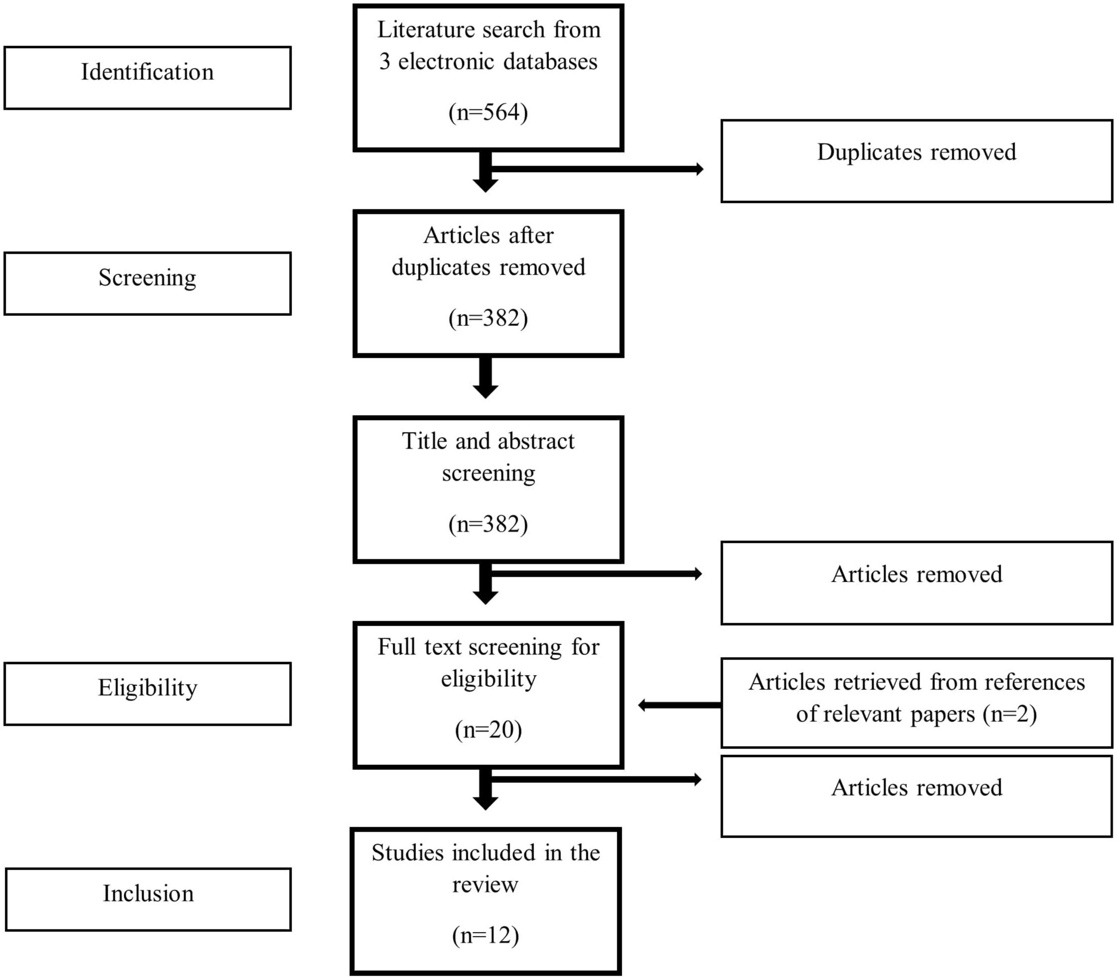
Of the 564 publications retrieved from the literature search, 10 studies were eligible for the systematic review. Two more studies were manually retrieved from references of relevant publications. In total, 12 articles were finally included (12–23). The selection process employed during the systematic literature search is described according to the PRISMA statement in Figure 1. In terms of design, ten studies were case reports and two were observational studies (one cohort and one cross-sectional). Four studies were conducted in North America, four in Europe, three in Asia and one in Africa. The characteristics of the included studies are shown in Table 1.

Figure 1. Preferred reporting items for systematic review and meta-analysis (PRISMA) workflow. Diagram of the selection process during the systematic review of the literature.
A total of 12 cases of diverticulitis during pregnancy were identified. The mean patient age was 34 years, based on available data from 10 studies. The mean gestational age at the time of diagnosis was 25 weeks, based on available data from nine studies.
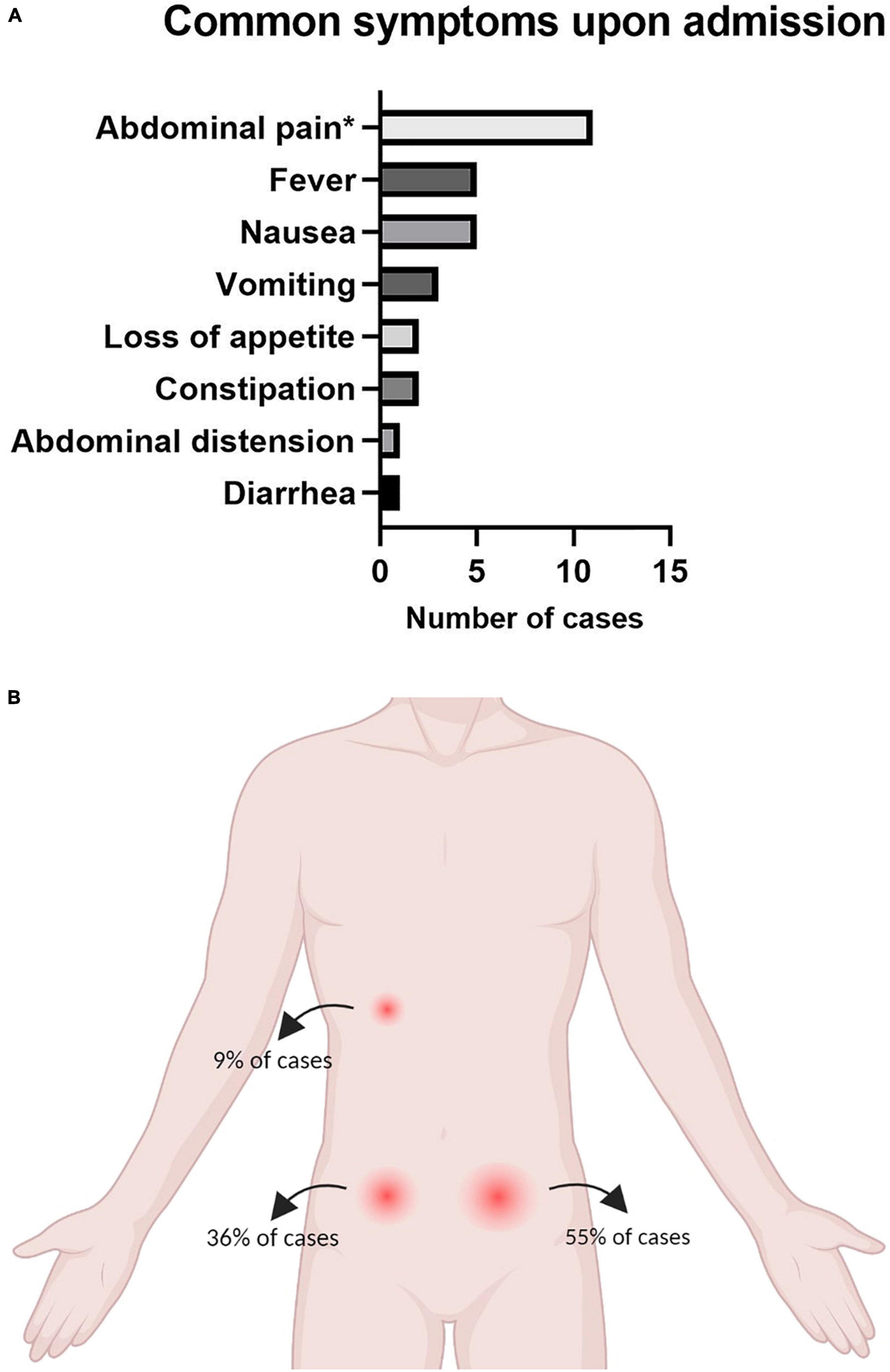
Data relevant to the presenting symptoms of diverticular disease was provided for 11 cases and is demonstrated in Figure 2A. The majority of patients presented with abdominal pain (10/11, 91%). Five (6/11, 55%) and four (4/11, 36%) patients presented with left and right iliac fossa pain, respectively, and one patient (1/11.9%) experienced right upper quadrant pain (Figure 2B). The second most common symptom of presentation was nausea and vomiting, encountered in five cases (5/11, 45%). Less common symptoms included abdominal distension and diarrhea.

Figure 2. (A) Presenting symptoms of the 12 included cases of acute diverticulitis during pregnancy and (B) pain location at the time of presentation.
Regarding the diagnostic approach employed, ultrasound was used as the first diagnostic modality in nine cases (9/12, 75%), and magnetic resonance imaging (MRI) as the initial imaging tool in two cases (2/12, 17%). In four cases (4/12, 33%) an inconclusive ultrasound was followed by an MRI. Despite the clinical and radiological evaluation, the initial diagnosis in seven cases was inaccurate (7/12, 58%), with five initially being diagnosed with acute appendicitis and two with pyelonephritis. In most of the patients, diverticular disease was located in the sigmoid colon (5/12, 42%) followed by the ascending colon (3/12, 25%) and appendix (2/12, 17%). Six women (6/12, 50%) were diagnosed during the third trimester, one (1/12, 8%) during the second trimester and one (1/12, 8%) during the first trimester. The gestational age at the time of diagnosis was not reported for the three remaining cases.
The therapeutic approach was available for 11 cases and it was based on the administration of intravenous antibiotics in six cases (6/11, 55%). Appendicectomy was performed in three cases (3/11, 27%) and simple bowel rest was used in one case (1/11, 9%). Laparotomy and drainage of intra-abdominal abscess with simultaneous cesarean section was performed in one case (1/11, 9%). Additionally, diverticulitis was treated surgically in three cases (3/11, 36%) after delivery due to failed medical management. The operations included right and left hemi-colectomy with anastomosis and Hartmann’s with loop ileostomy.
Data for the type of delivery was provided in nine studies with five patients (5/9, 56%) delivering vaginally and four patients (4/9, 44%) delivering with cesarean section. Two of them were performed as emergency cesarean sections at the time of the diagnostic laparotomy and two as elective procedures. Six of the deliveries (6/9, 67%) were preterm including three spontaneous preterm deliveries (3/9, 33%) and three preterm cesarean sections (3/9, 33%).
Quality assessment of the eligible studies revealed that on average all of the recommended elements were fulfilled and thus, these were considered as low risk of bias. Only two studies did not attain a good score mainly due to inadequate availability of data (Table 2).
Our study constitutes a contemporary systematic review of published cases of diverticular disease during pregnancy and provides an insight into the clinical manifestation and therapeutic management of this rare disorder during gestation. The current review was undertaken and reported using the PRISMA guidelines.
The incidence of diverticulitis increases with age. Only 5% of cases affect patients younger than 40 years (24). However, the number of cases during pregnancy is expected to increase as more women delay their childbearing until later in life (25). A single centre analysis from the USA, which covered a 20-year period, reported an incidence around 1 in 6000 pregnancies (26). Diverticulitis is a well-established infectious cause for intra-abdominal sepsis, which may trigger cytokine release and consequently lead to preterm labour (27). Therefore, the disease should be always considered as part of the differential diagnosis of pregnant patients with abdominal symptoms.
Management of symptomatic diverticular disease during pregnancy should balance both fetal and maternal benefit. Nevertheless, due to the rarity of the disorder in the pregnant population, there are no established clinical protocols to achieve this balance. A recent review on the imaging modalities that can be used during pregnancy proposed that the investigation of pregnant women with abdominal symptoms should include the tools that are used in the diagnosis of appendicitis namely: ultrasonography and MRI (28). While ultrasonography plays a pivotal role in the imaging during pregnancy due to its safety profile, the expertise and experience of the radiologist who performs the scan should always be considered as a general limitation of the test (29, 30). In fact, in recent years there has been a shift toward non-contrast MRI for the evaluation of pregnant women with abdominal pain, either as a secondary test following an inconclusive ultrasound scan or as the primary test for some indications (31). Thus, an approach that initiates with a diagnostic ultrasound and continues with an MRI, seems to offer safety and relatively high sensitivity for intra-abdominal pathologies, including diverticulitis (32).
As far as the treatment is concerned, in the non-pregnant population, most cases of uncomplicated diverticulitis can be treated with antibiotics and dietary restrictions (6, 33). Amoxicillin and clavulanic acid constitute the first line of oral antibiotics followed by trimethoprim and metronidazole or cefalexin and metronidazole. Similarly, intravenous regimes include amoxicillin and clavulanic acid as first line followed by cefuroxime and metronidazole or amoxicillin and gentamicin and metronidazole. It is worth noting that the combination of amoxicillin and clavulanic acid should be avoided in third trimester due to the increased risk of necrotizing enterocolitis of the newborn (34). Similarly, trimethoprim should be avoided in first trimester due to folate antagonism (35). Finally, although quinolones are used in non-pregnant patients with diverticular disease are not routinely used during pregnancy and thus, consultation with a maternal–fetal medicine specialist should be considered (36).
Pregnancy should not be a contraindication to surgical management if this is warranted, for example, due to inadequate response to medical management. In a 2019 statement from the American College of Obstetrics and Gynaecology (ACOG) on non-obstetric surgery during pregnancy it was concluded that medically necessary surgery should not be denied in pregnancy due to the adverse effect that the disease itself may have on the pregnant woman and her fetus (37). Rates of fetal loss in pregnant women with acute appendicitis range from 1.5% with simple appendicitis (38) to 6% with generalized peritonitis and up to 36% with perforation (39), which is likely related to the infective and inflammatory insult from the disease itself. Whilst there is a lack of similar data relating to pregnancy outcomes in diverticulitis, it may well be similar, reinforcing the idea that surgical management in pregnancy should not be disregarded. In addition, ACOG concluded that there is a lack of evidence that in utero exposure to anaesthetic or sedative drugs has any negative impact on the fetal brain (37), which may be reassuring for both clinicians and patients.
Diet is another important aspect related to the management of patients with diverticular disease during pregnancy. During the period of gestation, the need for macro- and micro- nutrients as well as dietary fibre increases sharply to support a healthy pregnancy and childbirth (40). Adequate dietary fibre intake has been particularly linked with a reduced risk of gestational diabetes, preeclampsia, and constipation (41) and conceivably may prevent the reoccurrence of diverticulitis (5, 42). This has been attributed to the reduced contact time between bowel contents and diverticula which as a result reduces mucosal irritation (43, 44). The implementation of different dietary restrictions, from nil by mouth, to clear liquids has been also recommended in the past (45). The rationalization behind this is also associated with the reduction in mucosal irritation and inflammation secondary to decreased bowel motility. However, the aforementioned mechanism is not supported by published evidence and consequently, the dogma of a clear liquid diet has been abandoned by most recent guidelines (46).
The current review provides the only available overview of the clinical presentation, diagnosis, and therapeutic management of diverticular disease during pregnancy. Cases included in this review were identified from comprehensive search of databases using a systematic search approach and the quality of the included studies were assessed with scrutiny.
However, despite having applied stringent inclusion criteria, we were unable to rule out the possibility of missing some important cases aggregated in larger series. The small number of included studies constitutes a major limitation and also reinforces the view that diverticulitis during pregnancy is possibly an underreported condition in the literature. Thus, additional studies are required to reach safer conclusions about the optimal diagnostic and therapeutic management of the disease. Publication bias is another potential weak point as case reports of rare or atypical observations are more likely to be published, potentially excluding more common findings. A wider drawback involves the low-quality nature of case reports and series within our systematic review, which hampers the validity and interpretation of conclusions that can be attained. Therefore, their reported findings although appealing, may not reflect the truth without underlying valid description.
Diverticulitis is a condition obstetricians may expect to see increase as advanced maternal age becomes more common. While available guidelines do not exist, the increased awareness of clinicians and the early recognition of the disorder, using diagnostic modalities, such as ultrasound and MRI, are crucial for the management of these patients. A rapid therapeutic approach with antibiotics may improve overall maternal and fetal outcomes, but surgical management should also be considered irrespective of gestation.
The original contributions presented in the study are included in the article/supplementary material, further inquiries can be directed to the corresponding author.
KK and KK-T: conceptualization, investigation, resources, and writing—original draft preparation. KK, KK-T, and GG: methodology. KK, KK-T, GG, PG, MZ, IT-M, MP, AM, and MK: validation and writing—review and editing. KK: supervision. All authors have read and agreed to the published version of the manuscript.
The authors declare that the research was conducted in the absence of any commercial or financial relationships that could be construed as a potential conflict of interest.
All claims expressed in this article are solely those of the authors and do not necessarily represent those of their affiliated organizations, or those of the publisher, the editors and the reviewers. Any product that may be evaluated in this article, or claim that may be made by its manufacturer, is not guaranteed or endorsed by the publisher.
1. Wan, D, Krisko T. Diverticulosis, diverticulitis, and diverticular bleeding. Clin Geriatr Med. (2021) 37:141–54. doi: 10.1016/j.cger.2020.08.011
2. Tursi A, Scarpignato C, Strate LL, Lanas A, Kruis W, Lahat A, et al. Colonic diverticular disease. Nat Rev Dis Primers. (2020) 6:1–23. doi: 10.1038/s41572-020-0153-5
3. Morris AM, Regenbogen SE, Hardiman KM, Hendren S. Sigmoid diverticulitis: a systematic review. JAMA. (2014) 311:287–97. doi: 10.1001/jama.2013.282025
4. Janes SE, Meagher A, Frizelle FA. Management of diverticulitis. Bmj. (2006) 332:271–5. doi: 10.1136/bmj.332.7536.271
5. Dahl C, Crichton M, Jenkins J, Nucera R, Mahoney S, Marx W, et al. Evidence for dietary fibre modification in the recovery and prevention of reoccurrence of acute, uncomplicated diverticulitis: a systematic literature review. Nutrients. (2018) 10:137. doi: 10.3390/nu10020137
6. Strate LL, Morris AM. Epidemiology, pathophysiology, and treatment of diverticulitis. Gastroenterology. (2019) 156:1282–98.e1. doi: 10.1053/j.gastro.2018.12.033
7. Miulescu MAM. Colonic diverticulosis. Is there a genetic component? Mædica. (2020) 15:105. doi: 10.26574/maedica.2020.15.1.105
8. Bharucha AE, Parthasarathy G, Ditah I, Fletcher J, Ewelukwa O, Pendlimari R, et al. Temporal trends in the incidence and natural history of diverticulitis: a population-based study. Am J Gastroenterol. (2015) 110:1589. doi: 10.1038/ajg.2015.302
9. Masselli G, Derme M, Laghi F, Framarino-dei-Malatesta M, Gualdi G. Evaluating the acute abdomen in the pregnant patient. Radiol Clin. (2015) 53:1309–25. doi: 10.1016/j.rcl.2015.06.013
10. Liberati A, Altman DG, Tetzlaff J, Mulrow C, Gøtzsche PC, Ioannidis JP, et al. The PRISMA statement for reporting systematic reviews and meta-analyses of studies that evaluate health care interventions: explanation and elaboration. J Clin Epidemiol. (2009) 62:e1–34. doi: 10.1016/j.jclinepi.2009.06.006
11. JB Institute. The Joanna Briggs Institute Critical Appraisal Tools for use in JBI Systematic Review: Checklists for Case Reports. Adelaide: The Joanna Briggs Institute (2019).
12. Kasaven LS, Karampitsakos T, Todiwala A. Septiceamia and pre term labour due to severe diverticular abscess in pregnancy. Eur J Obstet Gynecol Reprod Biol X. (2019) 1:100006. doi: 10.1016/j.eurox.2019.100006
13. Kereshi B, Lee KS, Siewert B, Mortele KJ. Clinical utility of magnetic resonance imaging in the evaluation of pregnant females with suspected acute appendicitis. Abdominal Radiol. (2018) 43:1446–55. doi: 10.1007/s00261-017-1300-7
14. Jung JY, Na JU, Han SK, Choi PC, Lee JH, Shin DH. Differential diagnoses of magnetic resonance imaging for suspected acute appendicitis in pregnant patients. World J Emerg Med. (2018) 9:26. doi: 10.5847/wjem.j.1920-8642.2018.01.004
15. Milczarek-Łukowiak M, Pyziak A, Kocemba W, Płusajska J. Complicated colonic diverticulitis at 34 weeks gestation. Ginekol Pol. (2012) 83:943–5.
16. Salah RBH, Mena KB, Bourguiba MB, Moussa MB, Zaouche A. Sigmoïdite diverticulaire compliquée d’une fistule colo-tubaire survenant au cours d’une grossesse. La Tunisie Med. (2011) 89:574–5.
17. Shanbhogue AKP, Kielar A, Nguyen B, Shanbhogue DK, Teo I. Appendiceal diverticulitis in pregnancy. Eur J Radiol Extra. (2009) 71:e29–31. doi: 10.1016/j.ejrex.2009.01.001
18. Bodner J, Windisch J, Bale R, Wetscher G, Mark W. Perforated right colonic diverticulitis complicating pregnancy at 37 weeks’ gestation. Int J Color Dis. (2005) 20:381–2. doi: 10.1007/s00384-004-0643-z
19. Ragu N, Tichoux C, Bouyabrine H, Carabalona J, Taourel P, Bruel J, et al. Diverticulite sigmoïdienne compliquée en cours de grossesse. J Radiol. (2004) 85:1950–2. doi: 10.1016/S0221-0363(04)97766-9
20. Sherer DM, Frager D, Eliakim R. An unusual case of diverticulitis complicating pregnancy at 33 weeks’ gestation. Am J Perinatol. (2001) 18:107–12. doi: 10.1055/s-2001-13639
21. Pelosi M III, Pelosi MA, Villalona E. Right-sided colonic diverticulitis mimicking acute cholecystitis in pregnancy: case report and laparoscopic treatment. Surg Laparosc Endoscopy. (1999) 9:63–7. doi: 10.1097/00019509-199901000-00015
22. Orihata M, Sasaki H, Hata M, Nakagawa H, Kakegawa T, Sagawa F. A case of appendiceal diverticulitis at 15 Weeks’ Gestation. Jpn J Gastroenterol Surg. (1998) 31:1893–6. doi: 10.5833/jjgs.31.1893
23. Schnall M, Phaneuf L, Conway J. Acute diverticulitis of the sigmoid in pregnancy. Am J Obstet Gynecol. (1945) 50:558–9. doi: 10.1016/S0002-9378(16)40153-5
24. Ferzoco LB, Raptopoulos V, Silen W. Acute diverticulitis. N Engl J Med. (1998) 338:1521–6. doi: 10.1056/NEJM199805213382107
25. Adachi T, Endo M, Ohashi K. Uninformed decision-making and regret about delaying childbearing decisions: a cross-sectional study. Nurs Open. (2020) 7:1489–96. doi: 10.1002/nop2.523
26. Longo SA, Moore RC, Canzoneri BJ, Robichaux A. Gastrointestinal conditions during pregnancy. Clin Colon Rectal Surg. (2010) 23:80–9. doi: 10.1055/s-0030-1254294
27. Gilman-Sachs A, Dambaeva S, Salazar Garcia MD, Hussein Y, Kwak-Kim J, Beaman K. Inflammation induced preterm labor and birth. J Reprod Immunol. (2018) 129:53–8.
28. Moreno CC, Mittal PK, Miller FH. Nonfetal imaging during pregnancy: acute abdomen/pelvis. Radiol Clin North Am. (2020) 58:363–80. doi: 10.1016/j.rcl.2019.10.005
29. Schreyer A, Layer G. S2k guidlines for diverticular disease and diverticulitis: diagnosis, classification, and therapy for the radiologist. RöFo. (2015) 187:676–84. doi: 10.1055/s-0034-1399526
30. Cuomo R, Barbara G, Pace F, Annese V, Bassotti G, Binda GA, et al. Italian consensus conference for colonic diverticulosis and diverticular disease. United Eur Gastroenterol J. (2014) 2:413–42. doi: 10.1177/2050640614547068
31. Shur J, Bottomley C, Walton K, Patel JH. Imaging of acute abdominal pain in the third trimester of pregnancy. Bmj. (2018) 361:k2511. doi: 10.1136/bmj.k2511
32. Liljegren G, Chabok A, Wickbom M, Smedh K, Nilsson K. Acute colonic diverticulitis: a systematic review of diagnostic accuracy. Colorectal Dis. (2007) 9:480–8. doi: 10.1111/j.1463-1318.2007.01238.x
33. NICE Guideline. Diverticular Disease: Diagnosis and Management. London: National Institute for Health and Care Excellence (NICE) (2019).
34. Nahum GG, Uhl K, Kennedy DL. Antibiotic use in pregnancy and lactation: what is and is not known about teratogenic and toxic risks. Obstet Gynecol. (2006) 107:1120–38. doi: 10.1097/01.AOG.0000216197.26783.b5
35. Gleckman R, Blagg N, Joubert DW. Trimethoprim: mechanisms of action, antimicrobial activity, bacterial resistance, pharmacokinetics, adverse reactions, and therapeutic indications. Pharmacotherapy. (1981) 1:14–9. doi: 10.1002/j.1875-9114.1981.tb03548.x
36. Yefet E, Schwartz N, Chazan B, Salim R, Romano S, Nachum Z. The safety of quinolones and fluoroquinolones in pregnancy: a meta-analysis. Bjog. (2018) 125:1069–76.
37. Tolcher MC, Fisher WE, Clark SL. Nonobstetric surgery during pregnancy. Obstet Gynecol. (2018) 132:395–403. doi: 10.1097/AOG.0000000000002748
38. Babaknia A, Parsa H, Woodruff JD. Appendicitis during pregnancy. Obstet Gynecol. (1977) 50:40–1. doi: 10.1055/s-0040-1708849
39. Silvestri MT, Pettker CM, Brousseau EC, Dick MA, Ciarleglio MM, Erekson EA. Morbidity of appendectomy and cholecystectomy in pregnant and non-pregnant women. Obstet Gynecol. (2011) 118:1261. doi: 10.1097/AOG.0b013e318234d7bc
40. Potdar RD, Sahariah SA, Gandhi M, Kehoe SH, Brown N, Sane H, et al. Improving women’s diet quality preconceptionally and during gestation: effects on birth weight and prevalence of low birth weight–a randomized controlled efficacy trial in India (Mumbai Maternal Nutrition Project). Am J Clin Nutr. (2014) 100:1257–68. doi: 10.3945/ajcn.114.084921
41. Zerfu TA, Mekuria A. Pregnant women have inadequate fiber intake while consuming fiber-rich diets in low-income rural setting: evidences from Analysis of common “ready-to-eat” stable foods. Food Sci Nutr. (2019) 7:3286–92. doi: 10.1002/fsn3.1188
42. Strate LL, Keeley BR, Cao Y, Wu K, Giovannucci EL, Chan AT. Western dietary pattern increases, and prudent dietary pattern decreases, risk of incident diverticulitis in a prospective cohort study. Gastroenterology. (2017) 152:1023–30.e2. doi: 10.1053/j.gastro.2016.12.038
43. Carabotti M, Annibale B, Severi C, Lahner E. Role of fiber in symptomatic uncomplicated diverticular disease: a systematic review. Nutrients. (2017) 9:161.
44. Slavin J. Fiber and prebiotics: mechanisms and health benefits. Nutrients. (2013) 5:1417–35. doi: 10.3390/nu5041417
45. Quigley EM, Fried M, Gwee KA, Khalif I, Hungin AP, Lindberg G, et al. World gastroenterology organisation global guidelines irritable bowel syndrome: a global perspective update September 2015. J Clin Gastroenterol. (2016) 50:704–13. doi: 10.1097/MCG.0000000000000653
Keywords: pregnancy, diverticulitis, diverticular disease, acute abdomen, delivery
Citation: Kechagias KS, Katsikas-Triantafyllidis K, Geropoulos G, Giannos P, Zafeiri M, Tariq-Mian I, Paraskevaidi M, Mitra A and Kyrgiou M (2022) Diverticulitis during pregnancy: A review of the reported cases. Front. Med. 9:942666. doi: 10.3389/fmed.2022.942666
Received: 12 May 2022; Accepted: 21 October 2022;
Published: 11 November 2022.
Edited by:
Ludovico Abenavoli, Magna Græcia University, ItalyReviewed by:
Nicola Flor, ASST Fatebenefratelli Sacco, ItalyCopyright © 2022 Kechagias, Katsikas-Triantafyllidis, Geropoulos, Giannos, Zafeiri, Tariq-Mian, Paraskevaidi, Mitra and Kyrgiou. This is an open-access article distributed under the terms of the Creative Commons Attribution License (CC BY). The use, distribution or reproduction in other forums is permitted, provided the original author(s) and the copyright owner(s) are credited and that the original publication in this journal is cited, in accordance with accepted academic practice. No use, distribution or reproduction is permitted which does not comply with these terms.
*Correspondence: Konstantinos S. Kechagias, a29uc3RhbnRpbm9zLmtlY2hhZ2lhczE4QGltcGVyaWFsLmFjLnVr
Disclaimer: All claims expressed in this article are solely those of the authors and do not necessarily represent those of their affiliated organizations, or those of the publisher, the editors and the reviewers. Any product that may be evaluated in this article or claim that may be made by its manufacturer is not guaranteed or endorsed by the publisher.
Research integrity at Frontiers

Learn more about the work of our research integrity team to safeguard the quality of each article we publish.