- 1Viro-Immunology Research Unit, Department of Infectious Diseases 8632, Rigshospitalet, University of Copenhagen, Copenhagen, Denmark
- 2Section of Respiratory Medicine, Department of Medicine, Herlev and Gentofte Hospital, University of Copenhagen, Hellerup, Denmark
- 3Department of Clinical Medicine, Faculty of Health and Medical Sciences, University of Copenhagen, Copenhagen, Denmark
Before introducing combination antiretroviral therapy (cART), a higher prevalence of emphysema in people living with HIV (PLWH) than in the background population was reported. This systematic literature review aimed to investigate the prevalence of emphysema in PLWH and to compare the prevalence between PLWH and controls in the current cART era. A systematic literature search was conducted in PubMed, EMBASE, Scopus, and Web of Science (WOS), searching for “human immunodeficiency virus (HIV)” and “emphysema” from January 1, 2000 to March 10, 2021. Eligible studies were published after the introduction of cART, included PLWH, and reported the prevalence of emphysema. A total of 17 studies were included, and nine studies also included controls. The weighted average prevalence of emphysema in PLWH was 23% (95% CI: 16–30). In studies including both PLWH and controls the weighted average prevalence were 22% (95% CI: 10–33) and 9.7% (95% CI: 2.3–17), respectively (p = 0.052). The prevalence of emphysema in never-smoking PLWH and controls was just reported in one study and was 18 and 4%, respectively (p < 0.01). Thirteen of the studies had a moderate risk of bias, mainly due to selection of patients. A tendency to higher prevalence of emphysema was found in PLWH in comparison to controls in the current cART era. However, in the included studies, the definition of emphysema varied largely. Thus, to have a clear overview of the prevalence, further studies with well-designed cohorts of PLWH and controls are warranted.
Introduction
During the early years of the human immunodeficiency virus (HIV) epidemic, acquired immunodeficiency syndrome (AIDS)-related emphysema was reported among people living with HIV (PLWH) (1). Emphysema is a chronic lung disease that is characterized by destruction of alveoli, and smoking, exposure to environmental pollutants, aging, infections, and conditions such as alpha-1-antitrypsin deficiency are among the risk factors (2, 3). The prevalence estimations for emphysema vary in different age groups and by different diagnostic cut-offs, ranging from 0.6 to 24% in the background population (4–7). Before introducing combined antiretroviral therapy (cART), studies reported that both smoking and non-smoking PLWH were more susceptible to emphysema than the background population (8, 9). However, it is uncertain whether there is still an increased prevalence among PLWH after introducing ART. We aimed to investigate the prevalence of emphysema in PLWH and to compare the prevalence between PLWH and controls in the current cART era.
Materials and methods
Search strategy and selection criteria
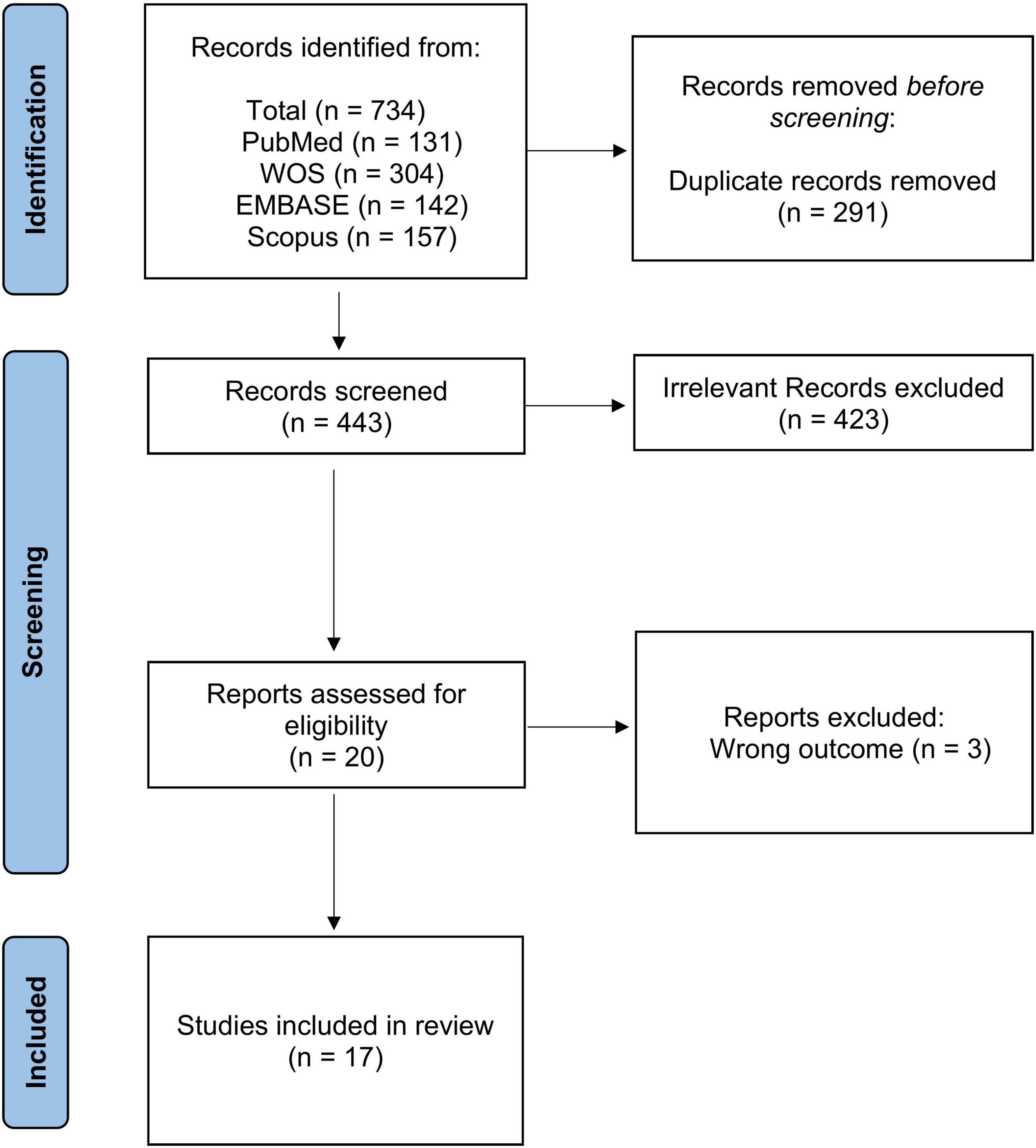
We used the Preferred Reporting Items for Systematic Reviews and Meta-Analyses (PRISMA) statement (10). The clinical question was: What is the prevalence of emphysema in PLWH and non-HIV controls in the current cART era? The clinical question was designed according to the PICOS process (10). The systematic literature review was performed in PubMed, EMBASE, Scopus and Web of Science (WOS) from January 1, 2000 to March 10, 2021. Using the same combination of keywords, we did a complementary search in google. We also read the references list of the retrieved papers for any relevant articles (Figure 1). Two investigators separately performed the searches and screened the retrieved papers by title and abstract. The relevant papers were read in full text and included if they met the inclusion criteria. A third investigator resolved potential conflicts.

Figure 1. Preferred reporting items for systematic reviews and meta-analyses (PRISMA) flow chart. Legend to figure: PRISMA flow diagram for the included studies showing the selection process.
Full electronic search strategy in PubMed
We searched PubMed using MeSH terms from January 1, 2000 to March 10, 2021 and got 15 results. The MeSH terms were: ((“HIV”[Mesh]) OR “AIDS”[Mesh]) AND (“Pulmonary Emphysema”[Mesh] OR “Emphysema”[Mesh]).
In addition, a free-text search was performed using the following terms: (((hiv) OR (aids)) OR (AIDS)) AND ((emphysema) OR (pulmonary emphysema)). This search yielded 116 results. All the search results were imported into Covidence for screening (10).
Eligibility criteria
We did not include studies that were published prior to 2000, to ensure participants were receiving modern cART. All duplicates, as well as all non-English studies and studies on non-human participants were excluded. Further, we excluded studies that diagnosed emphysema clinically or according to spirometry but without doing computed tomography (CT) scans. We included randomized controlled trials, cohort studies, case-control studies, and cross-sectional studies. Systematic reviews and meta-analyses, case reports, and expert opinions/editorials were considered ineligible to ensure stronger level of scientific evidence.
Data extraction
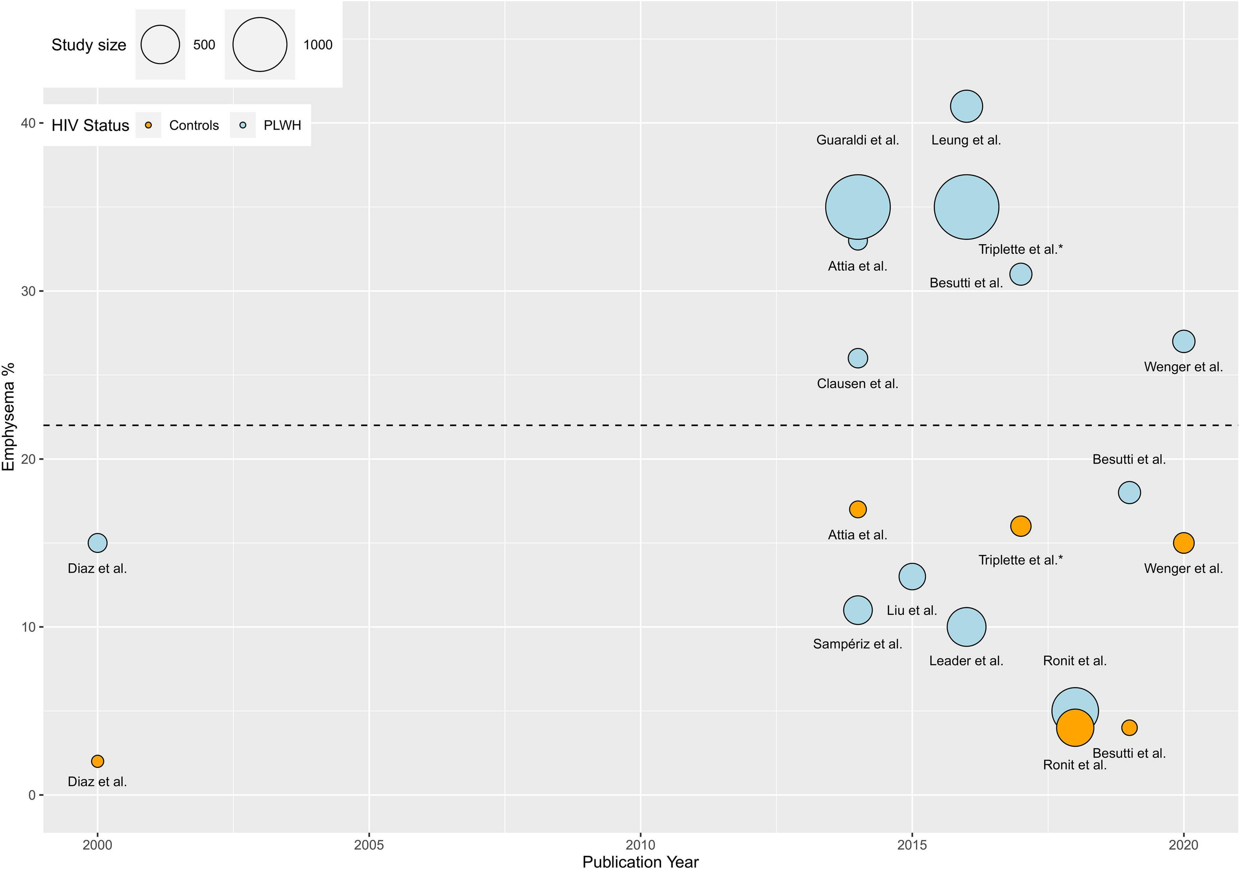
We extracted data using the Extraction 2.0 in Covidence (10). The retrieved studies were thoroughly reviewed, devoting particular attention to their methods and main findings. Specific attention was brought to how each study assessed emphysema as an outcome. The prevalence of emphysema in each study was determined and visually represented with the statistical program R (Figure 2). The weighted average prevalence of emphysema in PLWH and controls was calculated as: prevalence (%) × (N/the sum of all N). The study by Maitre et al. (7) was register-based and included 10,067 hospitalized PLWH and 8,244,682 hospitalized non-HIV controls, and it did not include a definition of emphysema. Therefore, the study by Maitre et al. (7) differed substantially from the others in its study design, population size, and estimations. Lack of emphysema definition prohibits comparison with other studies. Furthermore, including this study in the analyses would cause bias in estimations, and we chose to exclude this study from the calculations. Furthermore, Triplette et al. published four studies with similar prevalences (5, 6, 11, 12). Therefore, average number of PLWH and controls by Triplette et al. was included in the calculations. Unpaired two-samples T-test was used to compare the weighted average prevalence and a p ≤ 0.05 was considered statistically significant. In a sensitivity analysis and to find the effect of new cART drugs on the prevalence of emphysema, we included studies published in 2016 and later (13).

Figure 2. Visual representation of prevalence in PLWH and controls. A visual presentation of the prevalence of emphysema in all the included studies is presented. The weighted average prevalence of emphysema was 23% (95% CI: 16–30) (Horizontal black dotted-line). Triplette et al. published four studies on emphysema prevalence with similar results (prevalence = 31–33% in all studies). Only one of the four studies is shown in the plot for simplicity. The study by Maitre et al. (7) was register-based and included 10,067 hospitalized PLWH and 8,244,682 hospitalized non-HIV controls, and it did not include a definition of emphysema. Therefore, the study by Maitre et al. (7) differed substantially from the others in its study design, population size, and estimations. Lack of emphysema definition prohibits comparison with other studies. Furthermore, including this study in the analyses would cause bias in estimations, and we chose to exclude this study from the calculations.
Risk of bias assessment of included studies
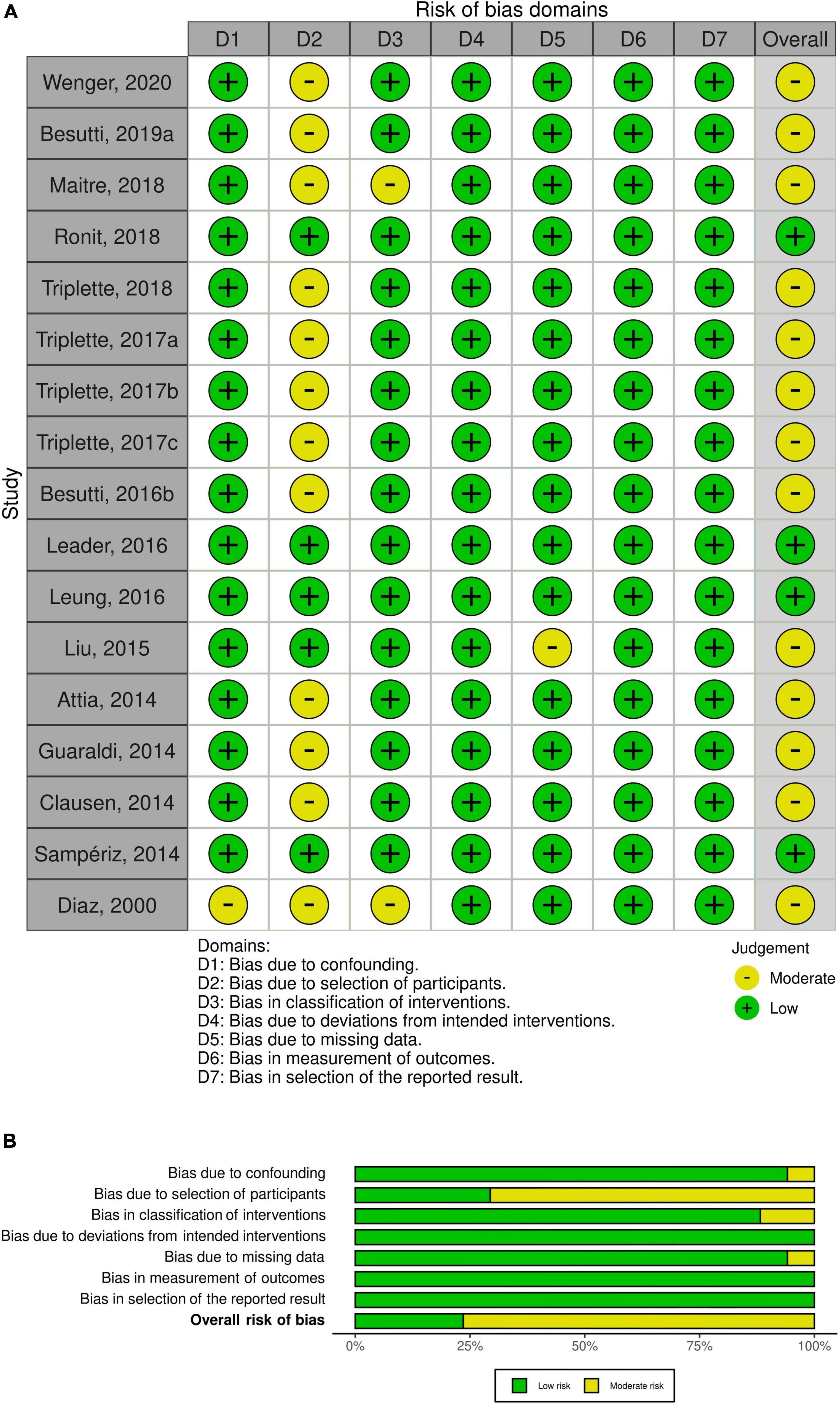
All the included studies were non-randomized, and we used Cochrane risk of bias tool for non-randomized studies (ROBINS-I) (14). We utilized the visualization tool, robvis, to produce weighted bar plots and traffic light plots (15). ROBIN-I contains seven domains with signaling questions that provide a structured approach to the risk of bias (14). The traffic light plots show a low, moderate, or high risk of bias within these important domains and also imply an overall risk of bias for each study. Additionally, the weighted bar plots depict a summary of the judgment within each domain and the studies in general.
Due to the heterogeneous nature of the included studies, it was not possible to perform a meta-analysis.
Results
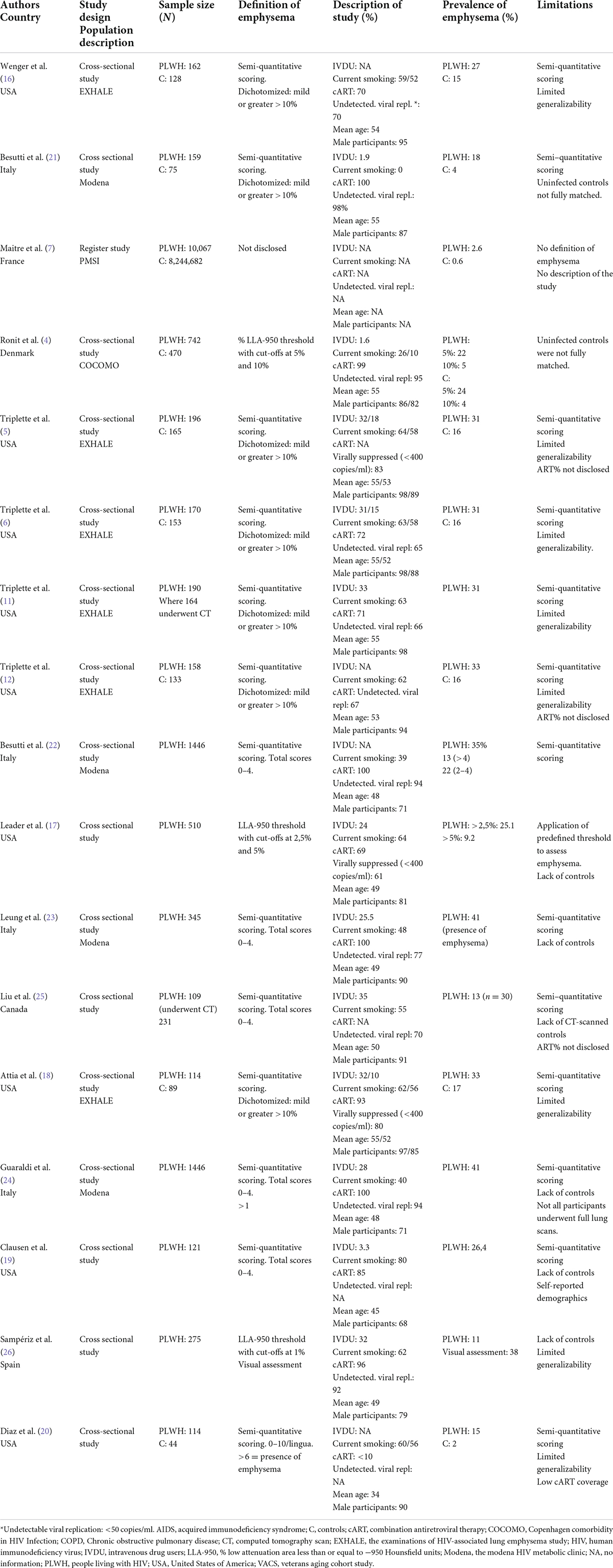
The initial search yielded 443 results. Based on our inclusion criteria, 17 were eligible for data extraction (Figure 1). A summary of the included studies is shown in Table 1. The studies were mainly published in 2014 or later, except for one study published in 2000. Nine studies were conducted in the US (5, 6, 11, 12, 16–20), four in Italy (21–24), one in France (7), one in Denmark (4), one in Canada (25), and one in Spain (26).
Characteristics of study participants
Due to the highly heterogeneous characteristics of the participants in the included studies, a brief presentation of the included cohorts will follow. An overview of the study participants can be found in Table 1. An extended version can be found in Supplementary Table 1.
The examinations of human immunodeficiency virus-associated lung emphysema study
In six studies, the participants were enrolled in the EXHALE study, which was a sub-study of the US Veterans Aging Cohort (VACS) (5, 6, 12, 16, 18, 27). All participants in EXHALE were former soldiers/veterans, and the study included both PLWH and controls. The population was predominantly male, >50 years, and of African-American ethnicity. There was a cART coverage of 70–93%, 59–64% were smokers, and 31–33% had a history of intravenous drug use (IVDU). In four of the studies 62–70% of participants had undetectable viral replication (6, 12, 16, 27). The last two reported viral suppression <400 ml/copies in 80 and 83% of participants, respectively (5, 18).
The Modena human immunodeficiency virus metabolic clinic
Four of the studies were from an outpatient clinic in Italy, the Modena HIV metabolic clinic (21–24). The enrolled participants had >18 months of cART exposure (cART coverage 100%) and were >18 years old. Between 77 and 98% had undetectable viral replication. Among the participants, 1.9–28% had a history of IVDU, and 0–48% had a smoking history. However, one study only included never-smoking PLWH (21).
Copenhagen comorbidity in human immunodeficiency virus infection
One of the included studies was from the COCOMO study which aimed to examine the prevalence, pathogenesis, and incidence of non-AIDS comorbidities in a well-treated cohort of PLWH. Most participants were on cART (99%), and 95% had undetectable viral replication. Of the included PLWH, 26% had a history of smoking, and 1.6% had a history of IVDU.
Programme de médicalisation de systèmes d́Information
Programme de médicalisation de systèmes d́Information (PMSI) is a French nationwide hospital discharge database. Maitre et al. utilized this in a register-based study, where they included all PLWH > 18 who had been hospitalized from 2007 to 2013 for more than 1 day. HIV-negative individuals hospitalized in 2010 were used as controls (7). Smoking status, history of IVDU, cART coverage, and viral replication was not reported.
Others
In five of the studies participants were not part of a larger cohort: Liu et al. (25), Clausen et al. (19), Diaz et al. (20), Leader et al. (17) and Sampériz et al. (26). A description of these studies is presented in Table 1.
Definition of emphysema
Most studies defined emphysema either semi-quantitatively or quantitatively. The PMSI cohort defined emphysema according to the International Statistical Classification of Diseases and Related Health Problems (ICD-10) codes (7).
Semi-quantitative definition
Most studies applied a semi-quantitative definition of emphysema by visual assessment of a CT scan (5, 6, 11, 12, 16, 18–25). In the studies from the EXHALE cohort, the degree of emphysema was characterized by a thoracic radiologist. Participants were scored from 0 (no emphysema) to 5 (>75% emphysema), followed by a dichotomization: Trace or no emphysema (≤10%) and mild or greater emphysema (>10%) (5, 6, 12, 16, 18, 27). In the four studies from the Modena cohort, three radiologists gave scores from 0 to 6 to each lobe and divided emphysematous changes by severity: 0 (absence of emphysema) to >4 (severe emphysema). Finally, they dichotomized the findings and defined presence of emphysema as a score ≥1 (21–24). In Liu et al., emphysematous changes were assessed by two radiologists (25). They gave each lobe and the lingula a score from 0 (absence of emphysema) to 4 (76–100% emphysema). Finally, a total score was given by summation from 0 (absence of emphysema) to >4 (severe emphysema). In the study by Clausen et al., emphysema was visually reviewed by one radiologist and one pulmonologist (19). They determined the extent, distribution and type of emphysema. Scores ranged from 0 (<5%) to 4 (51–75%). They did not mention from which score emphysema was defined, when reporting prevalence. In the study by Diaz et al., the extent of emphysema was given a score from 0 to 10 (20). Two radiologists assessed the CT scans. Emphysema was defined as a total score ≥6.
Quantitative definition
Three studies defined emphysema by densitometry using the low attenuation area −950 Hounsfield Units (% LAA-950 HU) method.% LAA-950 HU was defined as low attenuation area in the lung parenchyma of less than −950 HU. A cut-off in percentage was used to describe the proportion of the lung below this threshold (28). In the COCOMO study, two cut-offs were defined:% LAA-950 >10 and >5% (4). In the Leader et al. study two cut-offs were defined% LAA-950 with cut-offs at >2.5 and >5% (17). Finally, Sampériz et al. defined emphysema as% LAA-950 with a 1% threshold (26).
Prevalence of emphysema in people living with HIV
The reported prevalence of emphysema in PLWH ranged from 2.6 to 41%. The studies including participants from EXHALE found a prevalence of emphysema of 27% (16), 31% (5, 6, 11), and 33% (12, 18). The cohorts from Modena, found a prevalence of emphysema of 18% (21), 35% (22, 24), and 41% (23). The prevalence in the study by Liu et al. was 13% (25). The study by Clausen et al. reported a prevalence of 26% (19). Diaz et al. found the prevalence of emphysema to be 15% (20). The COCOMO study found that 21 and 4.7% had emphysematous changes at the 5 and 10% cut-offs, respectively. The prevalence of emphysema in Leader et al. was 25 and 9.2% at the >2.5 and >5% cut-offs, respectively (17). Sampériz et al. found a prevalence of 11% (26). In PMSI, they found a prevalence of emphysema of 2.6% out of 10067 PLWH without any AIDS-defining events. In Figure 2, visual presentation of the prevalence of emphysema in all the included studies is presented. The weighted average prevalence of emphysema in PLWH was 23% (95% CI: 16–30).
Prevalence of emphysema in people living with HIV and controls
The prevalence of emphysema in both PLWH and controls was reported in nine studies (Figure 2). In eight of the nine studies, the prevalence of emphysema was significantly higher in PLWH than in controls. The COCOMO study found that the prevalence of emphysema in PLWH was not statistically different from that in controls. In COCOMO the prevalence of emphysema was 21 and 24% (p = 0.23) in PLWH and controls at the 5% cut-off, while it was 4.7 and 4% (p = 0.68) at the 10% cut-off (4). Studies from the EXHALE cohort found the prevalence of emphysema to be 27, 31, and 33% in PLWH, whereas the prevalence of emphysema in controls was 15% (16) (p < 0.05), 16% (5, 6) (p = 0.003), and 17% (18) (p = 0.01) respectively. The only study from the Modena cohort, including controls, was the study by Besutti et al., wherein all participants were never smokers. The prevalence of emphysema was determined to be 18% in PLWH and 4% in controls (p < 0.01) (21). Diaz et al. reported the prevalence of emphysema to be 15% in PLWH and 2% in controls (p = 0.025). Finally, the PMSI cohort found a 2.6% prevalence of emphysema in PLWH and 0.6% in controls (7). Further, of the seven studies including controls, only three reported the proportion of males in the control group, and it was comparable in PLWH and in the controls (4–6). In studies including both PLWH and controls the weighted average prevalence were 22% (95% CI: 10–33) and 9.7% (95% CI: 2.3–17), respectively (p = 0.052).
Sensitivity analysis
In four studies that were published after 2016, the weighted average prevalence was 20% (95% CI: 3.2–38) and 9.8% (95% CI: 0.0–20) in PLWH and controls, respectively (p = 0.18).
Risk of bias
The overall risk of bias was low for four out of seventeen studies. Most of the studies had moderate overall risk of bias mainly due to bias in selection of the participants. Figures 3A,B show a visual representation of bias assessment.

Figure 3. Risk of bias for the included studies. (A) Traffic light plots; (B) weighted bar plots for non–randomized clinical trial. The overall risk of bias was low in 4 of 17 studies and moderate in 13 studies.
Discussion
This systematic review aimed to investigate the prevalence of emphysema in PLWH and to compare the prevalence between PLWH and controls in the current cART era. The majority of the participants in the included studies were middle-aged smoking men receiving ART, and a considerable proportion had a history of intravenous or inhalational drug use. The weighted average prevalence of emphysema in PLWH was 23%.
The prevalence of emphysema was more than 10% in studies that determined emphysema semi-quantitatively using visual assessment (5, 6, 11, 12, 16, 18–25), while the reported prevalence was lower in studies that used a quantitative assessment method (4, 17, 26). Previous studies reported that there is merely a moderate agreement between semi-quantitative and quantitative assessments and the presence of emphysema (29). The semi-qualitative assessment, which radiologists often use, is experience-dependent and has inter-individual variations with risk of overestimation (30). The quantitative assessment is highly reproducible, can report a lower% LAA-950, and have a better correlation with microscopic and macroscopic emphysema than semi-quantitative methods (31–33). This might explain the differences in the prevalence of emphysema between the included studies, which use different methods.
Approximately half of the studies included controls, and a tendency toward a higher prevalence of emphysema was found in PLWH than in controls. However, although the same tendency was observed, the difference was not statistically significant when we only included four published studies after 2016. The prevalence of emphysema in never smoker PLWH and controls was only reported in one study and was almost fivefold higher in PLWH (21).
Smoking is a well-known risk factor for emphysema, and in studies that reported data on smoking, the prevalence of smoking was higher in PLWH than in controls (4–6, 16, 20). Although, other theories beyond tobacco smoking have been suggested to explain why the prevalence of emphysema might be higher in PLWH. Some of those are; a higher proportion of previous or recurrent respiratory infections (34, 35), presence of chronic inflammation (36), and also long-term exposure to cART (37). In the EXHALE cohort, the authors proposed that a CD4 nadir <200 cells/μl was an independent risk factor for emphysema, indicating that an immunocompromised state can predispose PLWH to develop emphysema (18). Moreover, the EXHALE proposed that a low CD4/CD8 ratio can be a risk factor for emphysema (27). Furthermore, it has been shown in a study from Denmark that a low nadir CD4 is associated with dynamic measures of pulmonary function (38). It is worth mentioning that a low CD4/CD8 ratio is a marker of the enduring immune activation in PLWH and residual inflammation. Even though, there are studies that did not find an association between emphysema and HIV-related factors, such as viral load and CD4-cell count (19, 23).
Our review has potential limitations; we only included English literature, while potentially relevant material may have been overlooked in other languages. Different standards for defining emphysema were used since no consensus gold standard exists, which could have led to difficulties in comparing results and both under- and overestimating emphysema. Most of the included studies had a risk of bias due to the selection of patients. Therefore, it may imply that the relatively high prevalence found in PLWH is only applicable to certain groups such as IVDUs. Finally, only one of the studies reported that the prevalence of emphysema did not differ between PLWH and controls; hence there is a risk of publication bias because of unpublished negative results.
Conclusion
To conclude, the weighted average prevalence of emphysema in PLWH was 23%, and a tendency to higher prevalence of emphysema was found in PLWH than in controls in the current cART era. However, the definition of emphysema varied largely. Thus, to gain a clear overview, further studies with well-designed cohorts of PLWH and compared with controls are warranted.
Data availability statement
The original contributions presented in this study are included in the article/Supplementary material, further inquiries can be directed to the corresponding author.
Author contributions
HR, RT, and SN designed the study. HR and OR did the search. HR, RT, and OR screened manuscripts, determined risk of bias and extracted data, and wrote the first draft of the manuscript. SN and J-UJ commented and revised the manuscript. All authors read and approved the final version of the manuscript.
Funding
The Research Foundation of Rigshospitalet, Novo Nordisk Foundation, and the Independent Research Fund (FSS).
Conflict of interest
Author OR received a grant from The Research Foundation of Rigshospitalet related, and a grant from AP Møller Fonden not related to this work; Author SN received a grant from Novo Nordisk Foundation and the Independent Research Fund (FSS).
The remaining authors declare that the research was conducted in the absence of any commercial or financial relationships that could be construed as a potential conflict of interest.
Publisher’s note
All claims expressed in this article are solely those of the authors and do not necessarily represent those of their affiliated organizations, or those of the publisher, the editors and the reviewers. Any product that may be evaluated in this article, or claim that may be made by its manufacturer, is not guaranteed or endorsed by the publisher.
Supplementary material
The Supplementary Material for this article can be found online at: https://www.frontiersin.org/articles/10.3389/fmed.2022.897773/full#supplementary-material
References
1. Kuhlman JE, Knowles MC, Fishman EK, Siegelman SS. Premature bullous pulmonary damage in AIDS: CT diagnosis. Radiology. (1989) 173:23–6. doi: 10.1148/radiology.173.1.2781013
2. Janssen R, Piscaer I, Franssen FME, Wouters EFM. Emphysema: looking beyond alpha-1 antitrypsin deficiency. Expert Rev Respir Med. (2019) 13:381–97. doi: 10.1080/17476348.2019.1580575
3. Agustí A, Hogg JC. Update on the pathogenesis of chronic obstructive pulmonary disease. N Engl J Med. (2019) 381:1248–56. doi: 10.1056/NEJMra1900475
4. Ronit A, Kristensen T, Hoseth VS, Abou-Kassem D, Kühl JT, Benfield T, et al. Computed tomography quantification of emphysema in people living with HIV and uninfected controls. Eur Respir J. (2018) 52:2018. doi: 10.1183/13993003.00296-2018
5. Triplette M, Justice A, Attia EF, Tate J, Brown ST, Goetz MB, et al. Markers of chronic obstructive pulmonary disease are associated with mortality in people living with HIV. Aids. (2018) 32:487–93. doi: 10.1097/QAD.0000000000001701
6. Triplette M, Attia E, Akgün K, Campo M, Rodriguez-Barradas M, Pipavath S, et al. The differential impact of emphysema on respiratory symptoms and 6-minute walk distance in HIV infection. JAIDS J Acquir Immune Defic Syndr. (2017) 74:e23–9. doi: 10.1097/QAI.0000000000001133
7. Maitre T, Cottenet J, Beltramo G, Georges M, Blot M, Piroth L, et al. Increasing burden of noninfectious lung disease in persons living with HIV: a 7-year study using the French nationwide hospital administrative database. Eur Respir J. (2018) 52:2018. doi: 10.1183/13993003.00359-2018
8. Diaz PT, Clanton TL, Pacht ER. Emphysema-like pulmonary disease associated with human immunodeficiency virus infection. Ann Intern Med. (1992) 116:124–8. doi: 10.7326/0003-4819-116-2-124
9. Huang L, Stansell JD. AIDS and the lung. Med Clin North Am. (1996) 80:775–801. doi: 10.1016/S0025-7125(05)70467-3
10. Preferred Reporting Items for Systematic Reviews and Meta-Analyses [PRISMA]. Covidence Systematic Review Software. Melbourne, Australia: Veritas Health Innovation (2020).
11. Triplette M, Attia EF, Akgün KM, Hoo GWS, Freiberg MS, Butt AA, et al. A low peripheral blood CD4/CD8 ratio is associated with pulmonary emphysema in HIV. PLoS One. (2017) 2017:170857. doi: 10.1371/journal.pone.0170857
12. Triplette M, Sigel KM, Morris A, Shahrir S, Wisnivesky JP, Kong CY, et al. Emphysema and soluble CD14 are associated with pulmonary nodules in HIV-infected patients: implications for lung cancer screening. Aids. (2017) 31:1715–20. doi: 10.1097/QAD.0000000000001529
13. Ghosn J, Taiwo B, Seedat S, Autran B, Katlama C. HIV. Lancet. (2018) 392:685–97. doi: 10.1016/S0140-6736(18)31311-4
14. Sterne JAC, Hernán MA, Reeves BC, Savoviæ J, Berkman ND, Viswanathan M, et al. ROBINS-I: a tool for assessing risk of bias in non-randomised studies of interventions. BMJ. (2016) 355:4919. doi: 10.1136/bmj.i4919
15. McGuinness LA, Higgins JPT. Risk-of-bias visualization (robvis): an R package and shiny web app for visualizing risk-of-bias assessments. Res Synth Methods. (2021) 12:55–61. doi: 10.1002/jrsm.1411
16. Wenger DS, Triplette M, Shahrir S, Akgun KM, Wongtrakool C, Brown ST, et al. Associations of marijuana with markers of chronic lung disease in people living with HIV. HIV Med. (2020) 22:92–101. doi: 10.1111/hiv.12966
17. Leader JK, Crothers K, Huang L, King MA, Morris A, Thompson BW, et al. Risk factors associated with quantitative evidence of lung emphysema and fibrosis in an HIV-infected cohort. J Acquir Immune Defic Syndr. (2016) 71:420–7. doi: 10.1097/QAI.0000000000000894
18. Attia EF, Akgün KM, Wongtrakool C, Goetz MB, Rodriguez-Barradas MC, Rimland D, et al. Increased risk of radiographic emphysema in HIV is associated with elevated soluble CD14 and nadir CD4. Chest. (2014) 146:1543–53. doi: 10.1378/chest.14-0543
19. Clausen E, Wittman C, Gingo M, Fernainy K, Fuhrman C, Kessinger C, et al. Chest computed tomography findings in HIV-infected individuals in the era of antiretroviral therapy. PLoS One. (2014) 9:e112237. doi: 10.1371/journal.pone.0112237
20. Diaz PT, King MA, Pacht ER, Wewers MD, Gadek JE, Nagaraja HN, et al. Increased susceptibility to pulmonary emphysema among HIV-seropositive smokers. Ann Intern Med. (2000) 132:369–72. doi: 10.7326/0003-4819-132-5-200003070-00006
21. Besutti G, Santoro A, Scaglioni R, Neri S, Zona S, Malagoli A, et al. Significant chronic airway abnormalities in never-smoking HIV-infected patients. HIV Med. (2019) 20:657–67. doi: 10.1111/hiv.12785
22. Besutti G, Raggi P, Zona S, Scaglioni R, Santoro A, Orlando G, et al. Independent association of subclinical coronary artery disease and emphysema in HIV-infected patients. HIV Med. (2016) 17:178–87. doi: 10.1111/hiv.12289
23. Leung JM, Malagoli A, Santoro A, Besutti G, Ligabue G, Scaglioni R, et al. Emphysema distribution and diffusion capacity predict emphysema progression in human immunodeficiency virus infection. PLoS One. (2016) 11:e0167247. doi: 10.1371/journal.pone.0167247
24. Guaraldi G, Besutti G, Scaglioni R, Santoro A, Zona S, Guido L, et al. The burden of image based emphysema and bronchiolitis in HIV-infected individuals on antiretroviral therapy. PLoS One. (2014) 9:e109027. doi: 10.1371/journal.pone.0109027
25. Liu JCY, Leung JM, Ngan DA, Nashta NF, Guillemi S, Harris M, et al. Absolute leukocyte telomere length in HIV-infected and uninfected individuals: evidence of accelerated cell senescence in HIV-associated chronic obstructive pulmonary disease. PLoS One. (2015) 10:e0124426. doi: 10.1371/journal.pone.0124426
26. Sampériz G, Guerrero D, López M, Valera JL, Iglesias A, Ríos Á, et al. Prevalence of and risk factors for pulmonary abnormalities in HIV-infected patients treated with antiretroviral therapy. HIV Med. (2014) 15:321–9. doi: 10.1111/hiv.12117
27. Triplette M, Attia EF, Akgü NKM, Soo Hoo GW, Freiberg MS, Butt AA, et al. A low peripheral blood CD4/CD8 ratio is associated with pulmonary emphysema in HIV. PLoS One. (2017) 12:e0170857. doi: 10.1371/journal.pone.0170857
28. Tenda ED, Ridge CA, Shen M, Yang G-Z, Shah PL. Role of quantitative computed tomographic scan analysis in lung volume reduction for emphysema. Respiration. (2019) 98:86–94. doi: 10.1159/000498949
29. Barr RG, Berkowitz EA, Bigazzi F, Bode F, Bon J, Bowler RP, et al. A combined pulmonary-radiology workshop for visual evaluation of COPD: study design, chest CT findings and concordance with quantitative evaluation. COPD J Chronic Obstr Pulm Dis. (2012) 9:151–9. doi: 10.3109/15412555.2012.654923
30. Gietema HA, Müller NL, Nasute Fauerbach PV, Sharma S, Edwards LD, Camp PG, et al. Quantifying the extent of emphysema: factors associated with radiologists’ estimations and quantitative indices of emphysema severity using the ECLIPSE cohort. Acad Radiol. (2011) 18:661–71. doi: 10.1016/j.acra.2011.01.011
31. Bankier AA, De Maertelaer V, Keyzer C, Gevenois PA. Pulmonary emphysema: Subjective visual grading versus objective quantification with macroscopic morphometry and thin-section CT densitometry. Radiology. (1999) 211:851–8. doi: 10.1148/radiology.211.3.r99jn05851
32. Cavigli E, Camiciottoli G, Diciotti S, Orlandi I, Spinelli C, Meoni E, et al. Whole-lung densitometry versus visual assessment of emphysema. Eur Radiol. (2009) 19:1686–92. doi: 10.1007/s00330-009-1320-y
33. Ashraf H, Lo P, Shaker SB, De Bruijne M, Dirksen A, Tønnesen P, et al. Short-term effect of changes in smoking behaviour on emphysema quantification by CT. Thorax. (2011) 66:55–60. doi: 10.1136/thx.2009.132688
34. Norris KA, Morris A, Patil S, Fernandes E. Pneumocystis colonization, airway inflammation, and pulmonary function decline in acquired immunodeficiency syndrome. Immunol Res. (2006) 36:175–87. doi: 10.1385/IR:36:1:175
35. Morris A, Sciurba FC, Norris KA. Pneumocystis: a novel pathogen in chronic obstructive pulmonary disease? COPD. (2008) 5:43–51. doi: 10.1080/1541255070181756
36. Thudium RF, Knudsen AD, Von Stemann JH, Hove-Skovsgaard M, Hoel H, Mocroft A, et al. Independent association of interleukin 6 with low dynamic lung function and airflow limitation in well-treated people with human immunodeficiency virus. J Infect Dis. (2021) 223:1690–8. doi: 10.1093/infdis/jiaa600
37. George MP, Kannass M, Huang L, Sciurba FC, Morris A. Respiratory symptoms and airway obstruction in HIV-infected subjects in the HAART era. PLoS One. (2009) 4:e6328. doi: 10.1371/journal.pone.0006328
Keywords: HIV, emphysema, comorbidity, systematic review, antiretroviral therapy
Citation: Ringheim H, Thudium RF, Jensen J-US, Rezahosseini O and Nielsen SD (2022) Prevalence of emphysema in people living with human immunodeficiency virus in the current combined antiretroviral therapy era: A systematic review. Front. Med. 9:897773. doi: 10.3389/fmed.2022.897773
Received: 16 March 2022; Accepted: 01 September 2022;
Published: 21 September 2022.
Edited by:
Leland Shapiro, University of Colorado Anschutz Medical Campus, United StatesReviewed by:
Patrick Geraghty, Downstate Health Sciences University, United StatesVicente Benavides-Cordoba, Pontificia Universidad Javeriana Cali, Colombia
Copyright © 2022 Ringheim, Thudium, Jensen, Rezahosseini and Nielsen. This is an open-access article distributed under the terms of the Creative Commons Attribution License (CC BY). The use, distribution or reproduction in other forums is permitted, provided the original author(s) and the copyright owner(s) are credited and that the original publication in this journal is cited, in accordance with accepted academic practice. No use, distribution or reproduction is permitted which does not comply with these terms.
*Correspondence: Susanne D. Nielsen, c2RuQGRhZGxuZXQuZGs=
 Hedda Ringheim
 Rebekka F. Thudium
 Jens-Ulrik S. Jensen2,3
 Omid Rezahosseini
 Susanne D. Nielsen