
94% of researchers rate our articles as excellent or good
Learn more about the work of our research integrity team to safeguard the quality of each article we publish.
Find out more
CASE REPORT article
Front. Med., 29 September 2022
Sec. Infectious Diseases – Surveillance, Prevention and Treatment
Volume 9 - 2022 | https://doi.org/10.3389/fmed.2022.1008585
This article is part of the Research TopicCOVID-19: From Bedside to Follow-upView all 11 articles
 Mateus V. de Castro1†
 Monize V. R. Silva1†
 Flávia B. Soares1,2†
 Vivian R. Cória1
 Michel S. Naslavsky1,2Marilia O. Scliar1,2
 Erick C. Castelli3
 Jamile R. de Oliveira4,5Giuliana X. de Medeiros4,5Greyce L. Sasahara5
 Keity S. Santos4,5,6Edecio Cunha-Neto4,5,6Jorge Kalil4,5,6
 Mayana Zatz1,2*
Background: The influence of the host genome on coronavirus disease 2019 (COVID-19) susceptibility and severity is supported by reports on monozygotic (MZ) twins where both were infected simultaneously with similar disease outcomes, including several who died due to the SARS-CoV-2 infection within days apart. However, successive exposures to pathogens throughout life along with other environmental factors make the immune response unique for each individual, even among MZ twins.
Case presentation and methods: Here we report a case of a young adult monozygotic twin pair, who caught attention since both presented simultaneously severe COVID-19 with the need for oxygen support despite age and good health conditions. One of the twins, who spent more time hospitalized, reported symptoms of long-COVID even 7 months after infection. Immune cell profile and specific responses to SARS-CoV-2 were evaluated as well as whole exome sequencing.
Conclusion: Although the MZ twin brothers shared the same genetic mutations which may be associated with their increased risk of developing severe COVID-19, their clinical progression was different, reinforcing the role of both immune response and genetics in the COVID-19 presentation and course. Besides, post-COVID syndrome was observed in one of them, corroborating an association between the duration of hospitalization and the occurrence of long-COVID symptoms.
The ongoing global pandemic of coronavirus disease 2019 (COVID-19) caused by the SARS-CoV-2 virus has already affected the health of millions of people worldwide, with a significant number of deaths (1). Although older patients and those with comorbidities who are infected are more subject to unfavorable outcomes, reports of young people without chronic diseases who died from COVID-19 support the existence of genetic and immunological risk factors (2, 3). Also, several reports of identical twins deceased due to COVID-19 within days apart give further support to the influence of the host genome on COVID-19.
The first worldwide case of death from COVID-19, within 3 days apart, in one pair of adult unvaccinated MZ was reported in April 2020 in the United Kingdom. Aged 37, both twin sisters worked as nurses and had the same underlying health condition. Recently (2022), France’s famous twin Bogdanoff’s brothers died of COVID-19 6 days apart. Aged 72, the brothers had not been vaccinated against COVID-19 either.
The identification of genetic variants related to immune response, associated with higher susceptibility to the infection or severe COVID-19 has been the focus of numerous studies around the world (2, 4–8). Currently, genome-wide association studies (GWAS) have identified some genetic variants, including rare loss-of-function variants in genes involved in type I interferon (IFN) pathways (3, 9, 10) or missense variants that affect the activity of transmembrane serine protease 2 (3, 11, 12), that contribute to susceptibility or severe COVID-19, respectively. Here, we investigated a case of simultaneous critical COVID-19 in young adult MZ brothers in 2021, before being vaccinated. We present a comprehensive assessment of their innate and adaptive immunity, genetic profiling, and systemic biomarkers.
In June 2021, a 31-year-old Brazilian monozygotic twin brother pair (ID 01 and ID 02) started with cough and fever on the 26th. Both tested positive for SARS-CoV-2 infection on June 29th. During the following days symptoms worsened with dyspnea. From June 30th to July 4th, azithromycin was used. Blood oxygen saturation reached a critical level of 80% and the twin brothers were admitted directly to the intensive care unit on July 8th. They were intubated due to pulmonary involvement and were extubated after 5 days of mechanical ventilation (July 13th). The Gamma variant was the only SARS-CoV-2 variant identified in the region at the end of June 2021 (13) and it is known that this variant was associated with increased mortality risk and severity of COVID-19 cases in younger age groups, particularly in the unvaccinated population at the time (14). The twins received the same supportive ICU measures (sedation and proning). Also, at the hospital, both twins received the same treatment: dexamethasone (from July 9th to July 19th), to inhibit inflammation in the lungs. Due to detected resistant bacterial infections in both twins after extubation, they were treated with meropenem (from July 13th to July 20th). ID 01 was discharged on the 22nd of July and ID 02 7 days later. Both required respiratory physiotherapy for at least 3 months after hospital discharge. Seven months after the COVID-19 episode, ID 02 complained of persistent muscle fatigue, commonly associated with the post-COVID syndrome. The twins lived apart but worked at the same company as realtors. They did not have any known health conditions or comorbidities. The entire timeline of main events is presented in Figure 1.
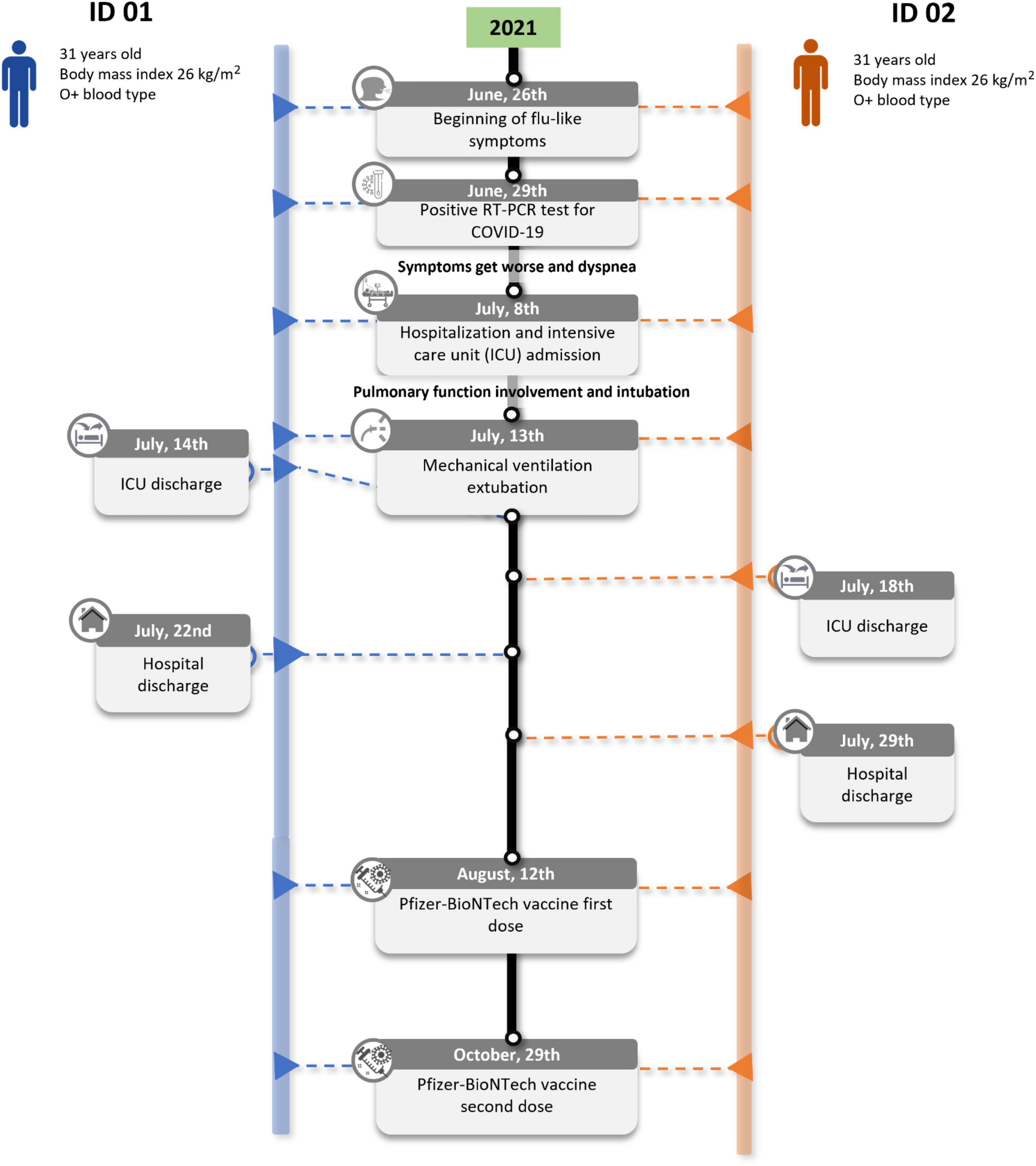
Figure 1. Clinical timeline of the major events of the twins’ case.
Blood samples from the twins were collected in February 2022 (7 months after COVID-19 diagnosis and 4 months after getting a second dose of Pfizer-BioNTech COVID-19 vaccine) at our Research Center (HUG-CELL) for global immune profiling and genetic investigation. Peripheral blood mononuclear cells (PBMCs), plasma, and serum were obtained to perform the immunological assays and DNA for whole-exome sequencing (WES). Complementary clinical laboratory analyses were also performed in whole blood samples.
Surface immunophenotyping of PBMC was performed by flow cytometry (Table 1). The twins displayed normal frequencies of CD3+, CD4+, and CD8+ T-cells, monocytes, NK cells, and lymphocytes B as expected in healthy donors (15). The type I/III IFN production by PBMCs after toll-like receptor (TLR) stimulus (double-stranded RNA Poly I:C), was evaluated for 1, 4, and 8 h. Although there was heterogeneity in IFN or IFN-induced gene expression, the twins presented an early and strong (FC = 20 or higher) mRNA expression of at least two of the five types of I/III IFN analyzed. Thus, no failure in the innate IFN response was observed. The production of interferon-gamma (IFN-γ) and interleukin-2 (IL-2) by PBMC, after stimulation by SARS-CoV-2 peptides, was also evaluated. Similar results were observed in both twins, for CD4 + T lymphocyte responses. ELISA serological assays were performed for SARS-CoV-2 IgA, IgG, and IgM for the receptor-binding domain (RBD) and nucleocapsid protein (NP) to assess their humoral immune response. The antibody profiles of SARS-CoV-2 IgA, IgM, and IgG were virtually identical between the MZ twin brothers. The global immune profiling of the twins is presented in Figure 2.
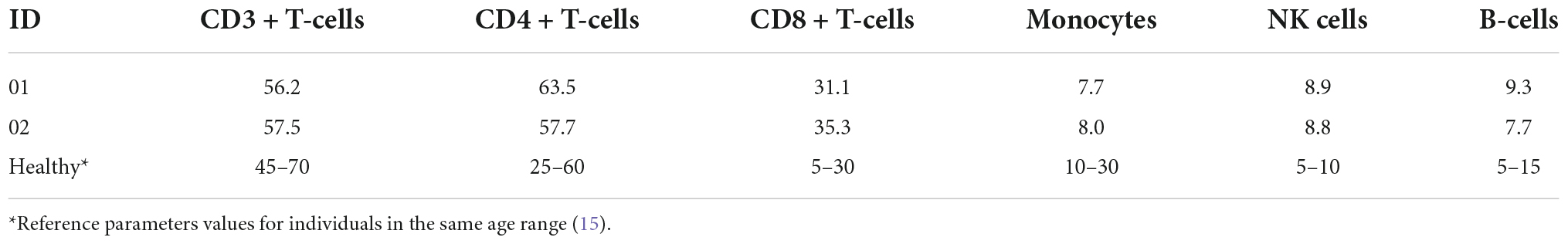
Table 1. PBMCs immunophenotyping of the twin volunteers, 7 months after COVID-19 episode.
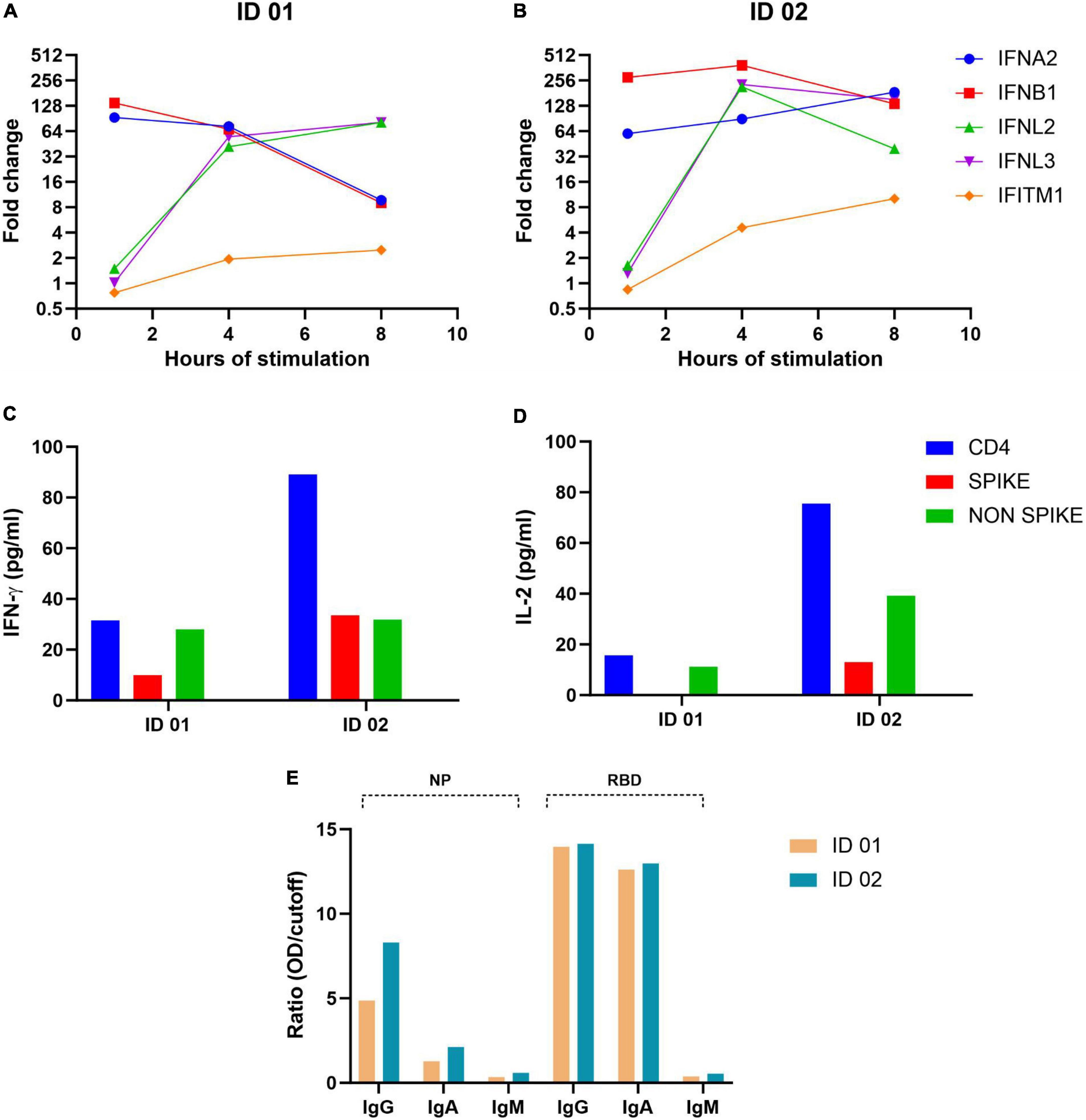
Figure 2. Global immune profiling of the twin volunteers, 7 months after the COVID-19 episode. (A,B) Type I/III IFN production by PBMCs after toll-like receptor (TLR) stimulus. (C,D) IFN-γ and IL-2 production by PBMC when stimulated by SARS-CoV-2 peptides. (E) Serological assays for SARS-CoV-2 IgA, IgG, and IgM through ELISA for the receptor-binding domain (RBD) and nucleocapsid protein (NP).
Hematologic and systemic parameters of the post-COVID phase (Table 2) revealed great similarity between the MZ twins, except for a very slight increase in creatine phosphokinase (an enzyme specific to muscle tissues, which may increase after muscle injuries) and ferritin (an acute phase reactant that can increase its serum concentration during inflammation), presented by ID 02. These findings might be related to the fatigue reported only by this twin. Likewise, both presented mild changes in erythrocyte sedimentation rate, a parameter that may be increased in different inflammatory conditions.
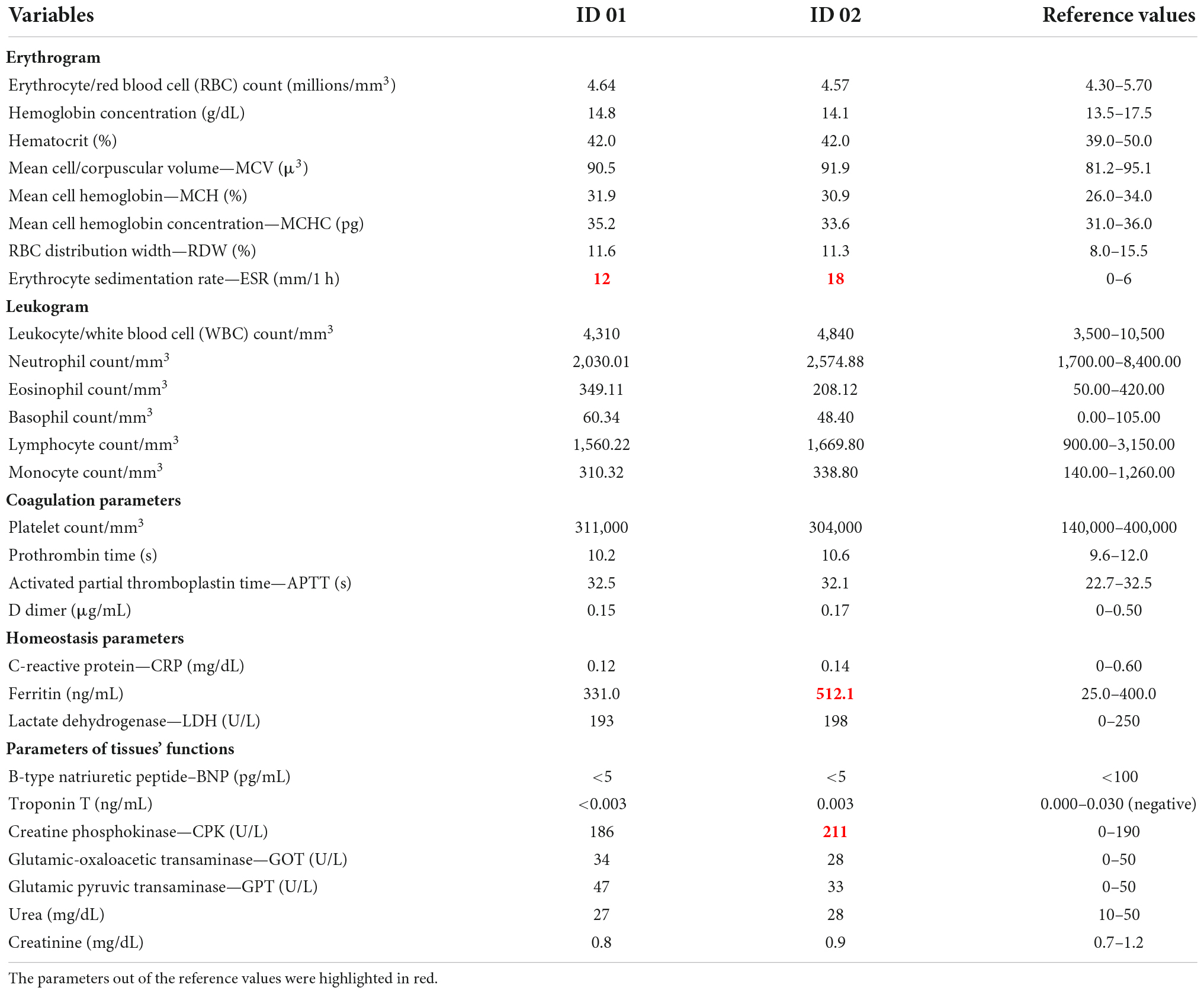
Table 2. Blood test parameters of the volunteers, 7 months after the COVID-19 episode.
WES was performed in peripheral blood DNA with the Illumina NovaSeq platform at HUG-CELL facilities. The identical twins do not carry any rare variants in genes associated with inborn errors (IE) of Toll-like receptor 3 (TLR3) and IFN regulatory factor 7 (IRF7) dependent type I IFN immunity, which underlies life-threatening COVID-19 pneumonia (5, 16). Also, we did not detect any copy number variation (CNV) in IE genes. The Neanderthal-derived genetic variant rs35044562 (17) was not detected in the twins. However, we detected two rare missense variants (with a mean CADD score > 20), one in the BTK gene (NM_000061:exon8:c.G684A:p.M228I) carried in homozygosity and one in the NFKB2 gene (NM_002502:exon22:c.T2531C:p.V844A) carried in the heterozygous state, which may be linked to their increased risk of developing severe COVID-19. In addition, we analyzed the genotypes and haplotypes (Supplementary Table 1) of the HLA cluster in the MHC region by using a hla-mapper (version 4) (18) to optimize read alignment along the MHC region. Interestingly, the twins present the alleles HLA-A*02:01 and HLA-E*01:01 (both carried in the heterozygous state), which were associated with the high severity of COVID-19.
Twin studies are important to investigate the contribution of genetics vs. the environment, in the susceptibility or resistance to infectious diseases as well as their pathomechanisms. Moreover, the study of the monozygotic ones may represent a powerful approach to further explore the immunological factors that contributed to the host defense. Beyond the host genotype, the individual immune response plays a determining role in the success or failure against SARS-CoV-2 (15). The immune repertoire which is also somatically defined by mutations occurring at later stages of development could justify different disease courses, and/or outcomes even in monozygotic twins (19, 20).
The genetic causes responsible for the clinical variability associated with COVID-19 remain the subject of investigation. Worldwide genomic studies of large cohorts of individuals with different clinical manifestations have been published, suggesting the involvement of different genetic variants responsible for greater susceptibility or resistance to SARS-CoV-2. GWAS identified a potential effect of variants in the SLC6A20, LZTFL1, CCR9, FYCO1, CXCR6, and XCR1 genes as responsible for greater susceptibility to SARS-CoV-2 (2, 19) in addition to variants in the genes REXO2, C11orf71, NNMT, and CADM1, involved in the immune response (21). Additionally, variants in many genes involved with the innate immune response seem to be involved in the susceptibility/predisposition to severe cases of COVID-19 such as those involved in the IFN and TLRs pathways, as well as the ACE2 and TMPRSS2 genes involved in virus entry into the cell (3, 7, 22). Variants in the genes IL1B, IL1R1, IL1RN, IL6, IL17A, FCGR2A, and TNF which encode cytokines would also have a possible relation with disease susceptibility and cytokine storm development. The Human Leukocyte Antigen (HLA) gene cluster and genes associated with the Major Histocompatibility Complex (MHC) are important candidates for the mechanisms of innate and adaptative immunity and susceptibility to COVID-19 infection and manifestation (23).
Interestingly the identified heterozygous NFKB1 missense variant (NM_002502:exon22:c.T2531C:p.V844AI) and the hemizygous missense variant in the BTK gene (NM_000061:exon8:c.G684A:p.M228I) are the central hubs that connect proinflammatory signaling pathways for survival, proliferation, cytokine production, and lymphocyte development. Interestingly, variants in both genes have been reported in primary antibody immunodeficiencies (24, 25). However, since these variants were not studied at the protein or functional level, their pathogenicity is yet to be determined. Genetic variant in chromosome 3, previously associated with high severity cases of COVID-19 and inherited from Neanderthals (rs35044563), was not detected in both volunteers (17). Regarding the HLA complex, the twins present the alleles HLA-A*02:01 and HLA-E*01:01 (both carried in the heterozygous state), which were associated with high severity of COVID-19 (13).
The global immune profiling assays, after 7 months of the COVID-19 episode, revealed great similarities between the MZ twins. It is known that the failure to elicit a strong type I IFN response contributes to severe COVID-19 (14). Infections trigger massive T cell expansion, leading to the skewing of the TCR repertoire due to antigen−specific T cell expansion. A low clonotype sharing between MZ twins with rheumatoid arthritis that were mismatched for SARS-CoV-2 infection suggests an immune repertoire reshaping might be induced after COVID-19 (26). However, clonality and alterations of T-cell receptors’ repertoires were partly associated with immune activation mediated by IFN type I and III (27) and here both twins displayed early and strong I/III IFN responses. The production of cytokines IFN-γ and IL-2 by T lymphocytes, when stimulated by SARS-CoV-2 peptides, was expressive. IL-2 and IFN-γ, which play a critical role in the activation of macrophages and other immune cells related to viral clearance, were found to be specific biomarkers of SARS-CoV-2 cellular response (28). The virus-specific antibody responses showed a vigorous IgA and IgG similar response in both twins. Since these analyses were done post-vaccination, it is not clear whether it was the viral infection or the vaccines that stimulated the production of these antibodies but it is clear there is no deficient humoral response. Taken together, regarding the immune response, all parameters analyzed were practically identical among the MZ twins.
Although both twins required intensive care and mechanical ventilation for 5 days, ID 02 required longer hospitalization and presented long-term symptoms consistent with long COVID. After 7 months of follow-up, twin ID 02 reported persistent muscle weakness and fatigue, while twin ID 01 referred to a return to his usual state of health. Muscle dysfunction (intense fatigue) is among the most reported symptoms of the post-COVID syndrome (17). The laboratory values obtained at 7 months demonstrated that twin ID 02 had an elevation in ferritin and CPK but otherwise had similar hematological and functional parameters relative to twin ID 01. Importantly, the twins were not on any medications or supplements. The CK levels at admission are reported to be higher in those subjects who later experience more severe outcomes and were associated with a worse prognosis (20) while severe to critical COVID-19 patients showed higher ferritin levels compared to mild to moderate COVID-19 patients (29). Thus, the slightly abnormal ferritin and CPK from ID 02 even after 7 months post-hospitalization might play a role in the pathogenesis of post-acute sequelae of COVID-19.
This case study on two young-adult monozygotic twins simultaneously infected with SARS-CoV-2, both requiring ICU care but with different periods of clinical progression suggests the contribution of both immune response and the genetics in the COVID-19 presentation and course. Besides, the post-COVID syndrome was observed in one of them, corroborating an association between the duration of hospitalization and the occurrence of long-COVID symptoms.
The original contributions presented in this study are included in the article/Supplementary material, further inquiries can be directed to the corresponding author.
The studies involving human participants were reviewed and approved by CAAE 34786620.2.0000.5464. The patients/participants provided their written informed consent to participate in this study. Written informed consent was obtained from the individual(s) for the publication of any potentially identifiable images or data included in this article.
MC and MVRS: data curation, investigation, formal analysis, visualization, and writing—original draft. FS: data curation and investigation. VC and EC-N: writing—review and editing. MN: conceptualization, formal analysis, investigation, methodology, software, and writing—review and editing. MOS and EC: formal analysis, investigation, methodology, software, and writing—review and editing. JO and GS: methodology and writing—review and editing. KS: investigation, visualization, and writing—review and editing. JK: funding acquisition, resources, and writing—review and editing. MZ: conceptualization, funding acquisition, project administration, writing—original draft, and writing—review and editing. All authors contributed to the article and approved the submitted version.
This work was supported by the São Paulo Research Foundation (FAPESP) (grant nos. 2013/08028-1, 2014/50931-3, 2014/50890-5, and 2020/09702-1), the National Council for Scientific and Technological Development (CNPq) (grant nos. 465434/2014-2, 465355/2014-5, and 404134/2020-3), and the Coordenação de Aperfeiçoamento de Pessoal de Nível Superior-Brasil (CAPES)—Finance Code 001 and JBS S.A (grant no. 69004). The funders were not involved in the study design, collection, analysis, interpretation of data, the writing of this case report, or the decision to submit it for publication. All the cited funders supported the conduction of the experiments equally.
We are extremely grateful for the participation and collaboration of the twin brothers ID 01 and ID 02, the nurses for sample collection, the clinic that performed the blood tests, and the HUG-CELL technical team. We also thank Brazilian Senator Mara Gabrilli for financial support.
The authors declare that the research was conducted in the absence of any commercial or financial relationships that could be construed as a potential conflict of interest.
All claims expressed in this article are solely those of the authors and do not necessarily represent those of their affiliated organizations, or those of the publisher, the editors and the reviewers. Any product that may be evaluated in this article, or claim that may be made by its manufacturer, is not guaranteed or endorsed by the publisher.
The Supplementary Material for this article can be found online at: https://www.frontiersin.org/articles/10.3389/fmed.2022.1008585/full#supplementary-material
1. World Health Organization. WHO coronavirus (COVID-19) dashboard. Geneva: World Health Organization (2022).
2. Fricke-Galindo I, Falfán-Valencia R. Genetics insight for COVID-19 susceptibility and severity: A review. Front Immunol. (2021) 12:622176. doi: 10.3389/fimmu.2021.622176
3. Velavan TP, Pallerla SR, Rüter J, Augustin Y, Kremsner PG, Krishna S, et al. Host genetic factors determining COVID-19 susceptibility and severity. EBioMedicine. (2021) 72:103629. doi: 10.1016/j.ebiom.2021.103629
4. Bastard P, Rosen LB, Zhang Q, Michailidis E, Hoffmann HH, Zhang Y, et al. Autoantibodies against type I IFNs in patients with life-threatening COVID-19. Science. (2020) 370:eabd4585.
5. Zhang Q, Bastard P, Liu Z, Le Pen J, Moncada-Velez M, Chen J, et al. Inborn errors of type I IFN immunity in patients with life-threatening COVID-19. Science. (2020) 370:eabd4570.
6. Asgari S, Pousaz LA. Human genetic variants identified that affect COVID susceptibility and severity. Nature. (2021) 600:390–1. doi: 10.1038/d41586-021-01773-7
7. SeyedAlinaghi S, Mehrtak M, MohsseniPour M, Mirzapour P, Barzegary A, Habibi P, et al. Genetic susceptibility of COVID-19: A systematic review of current evidence. Eur J Med Res. (2021) 26:46. doi: 10.1186/s40001-021-00516-8
8. Casanova JL, Su HC. COVID human genetic effort. A global effort to define the human genetics of protective immunity to SARS-CoV-2 infection. Cell. (2020) 181:1194–9.
9. Povysil G, Butler-Laporte G, Shang N, Wang C, Khan A, Alaamery M, et al. Rare loss-of-function variants in type I IFN immunity genes are not associated with severe COVID-19. J Clin Invest. (2021) 131:147834. doi: 10.1172/JCI152475
10. van der Made CI, Simons A, Schuurs-Hoeijmakers J, van den Heuvel G, Mantere T, Kersten S, et al. Presence of genetic variants among young men with severe COVID-19. JAMA. (2020) 324:663–73. doi: 10.1001/jama.2020.13719
11. Jahanafrooz Z, Chen Z, Bao J, Li H, Lipworth L, Guo X. An overview of human proteins and genes involved in SARS-CoV-2 infection. Gene. (2022) 808:145963. doi: 10.1016/j.gene.2021.145963
12. Monticelli M, Hay Mele B, Benetti E, Fallerini C, Baldassarri M, Furini S, et al. Protective role of a TMPRSS2 variant on severe COVID-19 outcome in young males and elderly women. Genes. (2021) 12:596. doi: 10.3390/genes12040596
13. Saulle I, Vicentini C, Clerici M, Biasin M. Antigen presentation in SARS-CoV-2 infection: The role of class I HLA and ERAP polymorphisms. Hum Immunol. (2021) 82:551–60. doi: 10.1016/j.humimm.2021.05.003
14. Setaro AC, Gaglia MM. All hands on deck: SARS-CoV-2 proteins that block early anti-viral interferon responses. Curr Res Virol Sci. (2021) 2:100015. doi: 10.1016/j.crviro.2021.100015
15. Kokuina E, Breff-Fonseca MC, Villegas-Valverde CA, Mora-Díaz I. Normal values of T, B and NK lymphocyte subpopulations in peripheral blood of healthy cuban adults. MEDICC Rev. (2019) 21:16–21. doi: 10.37757/MR2019.V21.N2-3.5
16. Zhang Q, Bastard P, Cobat A, Casanova JL. Human genetic and immunological determinants of critical COVID-19 pneumonia. Nature. (2022) 603:587–98.
17. Zeberg H, Pääbo S. The major genetic risk factor for severe COVID-19 is inherited from Neanderthals. Nature. (2020) 587:610–2. doi: 10.1038/s41586-020-2818-3
18. Castelli EC, Paz MA, Souza AS, Ramalho J, Mendes-Junior CT. Hla-mapper: An application to optimize the mapping of HLA sequences produced by massively parallel sequencing procedures. Hum Immunol. (2018) 1:678. doi: 10.1016/j.humimm.2018.06.010
19. Severe Covid-19 Gwas Group, Ellinghaus D, Degenhardt F, Bujanda L, Buti M, Albillos A, et al. Genomewide association study of severe covid-19 with respiratory failure. N Engl J Med. (2020) 383:1522–34. doi: 10.1056/NEJMoa2020283
20. Orsucci D, Trezzi M, Anichini R, Blanc P, Barontini L, Biagini C, et al. Increased creatine kinase may predict a worse COVID-19 outcome. J Clin Med. (2021) 10:1734. doi: 10.3390/jcm10081734
21. Li Y, Ke Y, Xia X, Wang Y, Cheng F, Liu X, et al. Genome-wide association study of COVID-19 severity among the Chinese population. Cell Discov. (2021) 7:1–16. doi: 10.1038/s41421-021-00318-6
22. Hou Y, Zhao J, Martin W, Kallianpur A, Chung MK, Jehi L, et al. New insights into genetic susceptibility of COVID-19: An ACE2 and TMPRSS2 polymorphism analysis. BMC Med. (2020) 18:216. doi: 10.1186/s12916-020-01673-z
23. Castelli EC, de Castro MV, Naslavsky MS, Scliar MO, Silva NSB, Andrade HS, et al. MHC variants associated with symptomatic versus asymptomatic SARS-CoV-2 infection in highly exposed individuals. Front Immunol. (2021) 12:3898. doi: 10.3389/fimmu.2021.742881
24. Shi C, Wang F, Tong A, Zhang XQ, Song HM, Liu ZY, et al. NFKB2 mutation in common variable immunodeficiency and isolated adrenocorticotropic hormone deficiency: A case report and review of literature. Medicine. (2016) 95:e5081. doi: 10.1097/MD.0000000000005081
25. Tsukada S, Rawlings DJ, Witte ON. Role of Bruton’s tyrosine kinase in immunodeficiency. Curr Opin Immunol. (1994) 6:623–30. doi: 10.1016/0952-7915(94)90151-1
26. Arruda LCM, Gaballa A, Da Silva Rodrigues R, Makower B, Uhlin M. SARS-CoV-2 (COVID-19)-specific T cell and B cell responses in convalescent rheumatoid arthritis: Monozygotic twins pair case observation. Scand J Immunol. (2022) 95:e13151. doi: 10.1111/sji.13151
27. Schultheiß C, Paschold L, Simnica D, Mohme M, Willscher E, von Wenserski L, et al. Next-generation sequencing of T and B cell receptor repertoires from COVID-19 patients showed signatures associated with severity of disease. Immunity. (2020) 53:442.e–55.e. doi: 10.1016/j.immuni.2020.06.024
28. Pérez-Cabezas B, Ribeiro R, Costa I, Esteves S, Teixeira AR, Reis T, et al. IL-2 and IFN-γ are biomarkers of SARS-CoV-2 specific cellular response in whole blood stimulation assays. medRxiv [Preprint] (2021). doi: 10.1101/2021.01.04.20248897
Keywords: COVID-19, monozygotic twins, SARS-CoV-2, immunity, genetic variants
Citation: de Castro MV, Silva MVR, Soares FB, Cória VR, Naslavsky MS, Scliar MO, Castelli EC, de Oliveira JR, de Medeiros GX, Sasahara GL, Santos KS, Cunha-Neto E, Kalil J and Zatz M (2022) Follow-up of young adult monozygotic twins after simultaneous critical coronavirus disease 2019: a case report. Front. Med. 9:1008585. doi: 10.3389/fmed.2022.1008585
Received: 01 August 2022; Accepted: 15 September 2022;
Published: 29 September 2022.
Edited by:
Sara Cajander, Örebro University, SwedenReviewed by:
Lucas Coelho Marlière Arruda, Karolinska Institutet (KI), SwedenCopyright © 2022 de Castro, Silva, Soares, Cória, Naslavsky, Scliar, Castelli, de Oliveira, de Medeiros, Sasahara, Santos, Cunha-Neto, Kalil and Zatz. This is an open-access article distributed under the terms of the Creative Commons Attribution License (CC BY). The use, distribution or reproduction in other forums is permitted, provided the original author(s) and the copyright owner(s) are credited and that the original publication in this journal is cited, in accordance with accepted academic practice. No use, distribution or reproduction is permitted which does not comply with these terms.
*Correspondence: Mayana Zatz, bWF5YXphdHpAdXNwLmJy
†These authors have contributed equally to this work
Disclaimer: All claims expressed in this article are solely those of the authors and do not necessarily represent those of their affiliated organizations, or those of the publisher, the editors and the reviewers. Any product that may be evaluated in this article or claim that may be made by its manufacturer is not guaranteed or endorsed by the publisher.
Research integrity at Frontiers
Learn more about the work of our research integrity team to safeguard the quality of each article we publish.