- Department of Dermatology, Rheumatology, and Immunology, Xinqiao Hospital of Army Medical University, Chongqing, China
Purpose: The advantage of pulsed dye laser (PDL) for the treatment of rosacea is not yet clear. This meta-analysis compared the curative effect of PDL to neodymium:yttrium-aluminum-garnet (Nd:YAG) laser for the treatment of rosacea.
Methods: The PubMed, Embase, and Cochrane Library databases were searched for clinical studies on the efficacy of PDL for the treatment of rosacea through October 13, 2021, and heterogeneity tests among studies were evaluated. Meta-analysis was conducted to combine the effects of physicians' clinical assessments, patient global assessment, erythema index, and visual analog scale.
Results: A total of 326 articles were obtained from three databases and ten articles were finally included. The clinical improvements of >50% clearance of up to 68.6% in the PDL group and 71.4% in the control group, and the subjective satisfaction rate of patients in the PDL group of 88.6% compared to 91.4% in the Nd:YAG group, but there were no significant differences in the rates of patients with rosacea with clinical improvement (>50% clearance) (relative risk [RR] = 0.94, 95% confidence interval [CI]: 0.75–1.17, P = 0.578) or patient subjective satisfaction rate (RR = 0.96, 95% CI: 0.70–1.33, P = 0.808) between PDL and Nd:YAG groups for rosacea treatment. Also, the pain score for PDL and Nd:YAG were not significant (mean = 3.07, 95% CI: 1.82–4.32, P = 0.115).
Conclusion: Two treatments all showed clinical efficacy and patient satisfaction for the treatment of rosacea, with no significant differences observed between treatments. The pain scores for PDL and Nd:YAG were not significant.
Highlights
- Two methods all showed clinical efficacy and satisfaction for the therapy of rosacea.
- Clinical efficacy and satisfaction were similar between the two treatments.
- The pain scores for PDL and Nd:YAG were not significant.
Introduction
Rosacea, also known as rose acne, is a facial inflammatory disease with clinical manifestations of transient or persistent erythema, papules, pustules, burning sensation, pruritus, or telangiectasia in the middle of the face (1). It is well known that rosacea is related to genetic factors, congenital, and adaptive immune system disorders, vascular, and neurological dysfunction, Demodex folliculorum, inflammation, and microorganism factors (2, 3), while the specific pathogenesis of rosacea is not fully understood. A study conducted in a genome-wide association identified two single nucleotide polymorphisms in European individuals with rosacea which might predispose them to the development of rosacea (4). Immune dysregulation is also an important cause of rosacea. In patients with rosacea, the innate immune system was activated and thus increased cytokines and antimicrobial peptides (5). Increased human cationic antibacterial protein 18kDa (hCAP18) is cleaved into LL-37, which promotes angiogenesis, induces leukocyte chemotaxis, and is involved in the production of proinflammatory cytokines (6). Activation of transient receptor potential (TRPs) in patients with rosacea leads to the release of mediators of neurogenic inflammation and pain, such as substance P and calcitonin gene-related peptide. These vasoregulatory neuropeptides are critical mediators that induce sustained flushing that is a characteristic of rosacea (5). The reported aggravating factors for rosacea include high temperature, alcohol, sunlight, stress, menstruation, drugs, and certain foods (7). The onset age of rosacea is usually older than that for acne, and rosacea mainly occurs in middle-aged people.
Clinically, the overall incidence of rosacea is increasing (8). Rosacea not only affects the normal life of patients, but may also progress to capacitive skin diseases and blindness (6, 9). The diversity of clinical manifestations of rosacea requires a variety of appropriate and targeted treatment methods, including skincare, local or systemic drug treatment, or physical and surgery therapies (10, 11). However, telangiectatic (ET) and rhinophyma (RP) changes have limited therapeutic effects on drugs, and traditional methods (12).
Recently, pulsed dye laser (PDL) has been widely used to treat rosacea-related erythema and telangiectasia (13, 14). PDL emits pulsed laser energy at wavelengths of 585 or 595 nm, which is absorbed by oxyhemoglobin to inhibit endothelial cell formation, reduce angiogenesis, and destruct the existing vessels (15). Many studies have also demonstrated the efficacy of other photoelectric treatments including long-pulsed neodymium: yttrium-aluminum-garnet laser (Nd:YAG) on rosacea (16). Heterogeneous therapeutic effect for rosacea occurred in different studies in which PDL reduced more facial redness than Nd:YAG in the study of Alam (17) while exhibited no significant differential treatment outcome in the study of Hyun-Min (18). The various results might stem from the sample size, study method like fluence, and so on. Meta-analysis provides a well-done understanding for combining the different studies with differential features. A systematical meta-analysis compared the differences between the two treatments and showed that the PDL leads to worse improvement for erythema (19), providing a general understanding for researchers. However, the study was conducted in March 2020, with only 5 papers included, and did not perform the methodological quality and heterogeneity test. The meta-analysis which evaluated the differences between PDL and Nd: YAG for the treatment of rosacea in terms of curative effect and patient satisfaction needed to be further conducted to provide evidence for clinical treatment.
Methods
Literature Retrieval Strategy
Systematic literature retrieval was performed from the PubMed, Embase, Cochrane Library databases according to the pre-defined retrieval strategy. The search keywords were Rosacea AND [PDL OR (Pulsed Dye Laser)] OR [Nd:YAG OR (neodymium: yttrium aluminum-garnet laser)]. Subject and free words were performed in this search, and the search format changed according to the characteristics of the database (for the specific different retrieval steps of each database, see Supplementary Tables 1–3). The retrieval time was up to October 13, 2021, and there was no language restriction. In addition, we also manually retrieved the paper articles, and screened the bibliographies of relevant systematic reviews for more sources that could be included in the meta-analysis.
Inclusion and Exclusion Criteria
The inclusion criteria were (1) subjects with erythemato-telangiectatic rosacea or papulopustular rosacea patients; (2) the studies reported that the efficacy or safety of PDL vs. Nd:YAG treated rosacea or the efficacy or safety of PDL/Nd:YAG vs. other treatment treated rosacea, or the single-arm study of PDL/Nd:YAG treated rosacea; (3) study outcomes, included clinical efficacy, subjective improvement, erythema index, or pain score (visual analog scale), after the intervention; and (4) study types were randomized controlled trial (RCT) or prospective cohort studies.
The exclusion criteria were (1) studies using PDL in combination with other drugs or interventions; (2) studies with fewer than 10 patients to reduce the risk of publication bias; (3) studies with unclear rosacea type; (4) reviews, letters, comments, etc.; and (5) repeated publications or use of the same data for multiple articles (only the study with the most complete information was included, and the rest were excluded).
Data Extraction and Quality Evaluation
Two investigators independently completed the literature screening according to the inclusion and exclusion criteria and independently carried out data extraction according to the standardized table designed in advance. The extracted information included the first author name and article publication year, the area where the study was carried out, participant information (age, sample size, and sex), intervention plans, and outcome indicators. After data extraction, two investigators exchanged audit extraction forms. Any inconsistencies were resolved by discussion.
The Cochrane Collaboration's tool for evaluating risk was used to evaluate the quality of the included RCT studies (20). In addition, the Newcastle-Ottawa scale (NOS) was conducted to assess the methodological quality of the cohort studies (21).
Differences in the quality evaluations were resolved by reaching a consensus after a group discussion with the third author.
Statistic Analysis
There were two comparisons in this meta-analysis, including (1) the efficacy or safety of PDL, the Nd:YAG treated rosacea were respectively obtained, and the Mean [95% confidence intervals (CIs)] was used to merge continuous variables, and the Incidence Rate (IR) and 95%CI were utilized to merge classified variable. The combined results between two groups were performed the indirect comparison, and the Altman, DG (24), and Delong, ER (25) methods were used to conduct the difference significance test; (2) direct comparison: the efficacy or safety of PDL vs. Nd:YAG treated rosacea were obtained, weighted mean differences (WMDs), and their 95% confidence intervals (CIs) were used to merge continuous variables, while relative risks (RRs) with 95% CIs were used to the merge effect values of classified variables. Cochran's Q and I2 tests were applied for heterogeneity assessment (26). If significant heterogeneity among studies was evaluated, defined as Q Statistic P ≤ 0.05 or I2 > 50%. The random-effects model was conducted on the meta-analysis because there was a large clinical and methodological heterogeneity between the included studies. All statistical analyses in this study were performed using RevMan5.3 (Copenhagen: The Nordic Cochrane Center and The Cochrane Collaboration) and Stata15.0 (Stata Corp., TX, USA) software.
Results
Literature Retrieval
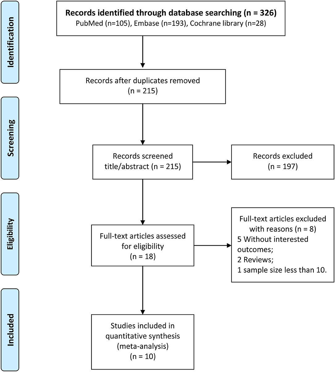
The process of literature screening is shown in Figure 1. A total of 105, 193, and 28 articles were initially identified in the PubMed, Embase, and Cochrane Library databases, respectively. After excluding repeated articles, 215 articles remained,197 of which did not meet the inclusion criteria after browsing the titles and abstracts. Eight of the remaining 18 articles were excluded after reading the full texts. No additional eligible articles were found through manual searching; finally, ten articles (16–18, 22, 23, 27–31) were included in this study.
Characteristics of the Included Studies
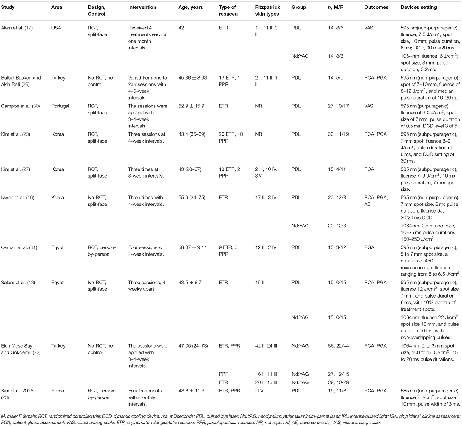
The included papers were published in 2011–2020, and distributed in the United States, Egypt, and South Korea. Among the ten included studies, six studies were RCTs (17, 23, 27, 28, 30, 31) and four studies were prospective cohorts (17, 25, 15, 28). The total sample size of the six studies was 235 cases. There was no significant difference in age between the PDL and control groups in each study; however, differences in Fitzpatrick skin types were observed among participants of each study. Other baseline information is detailed in Table 1.
Quality of the Included Studies
The methodology quality of each included study was evaluated. Although six RCT studies implemented the blinding of subjects, the clinicians were not blinded; thus the evaluations of the “Blinding of participants and personnel” for all three included studies were “unclear risk”. Only Alam et al. (17), Campos et al. (30), and Kim et al. (27) reported specific randomization, while the studies by Alam et al. (17) and Seo et al. (23)reported allocation concealment. All three studies showed low risks in terms of “detection bias”, “attrition bias”, “reporting bias”, and “other bias”. Overall, the methodology quality of the included RCTs was moderate (Figure 2). The results of the NOS evaluation of the four prospective cohort studies showed moderate-high quality (Table 2).
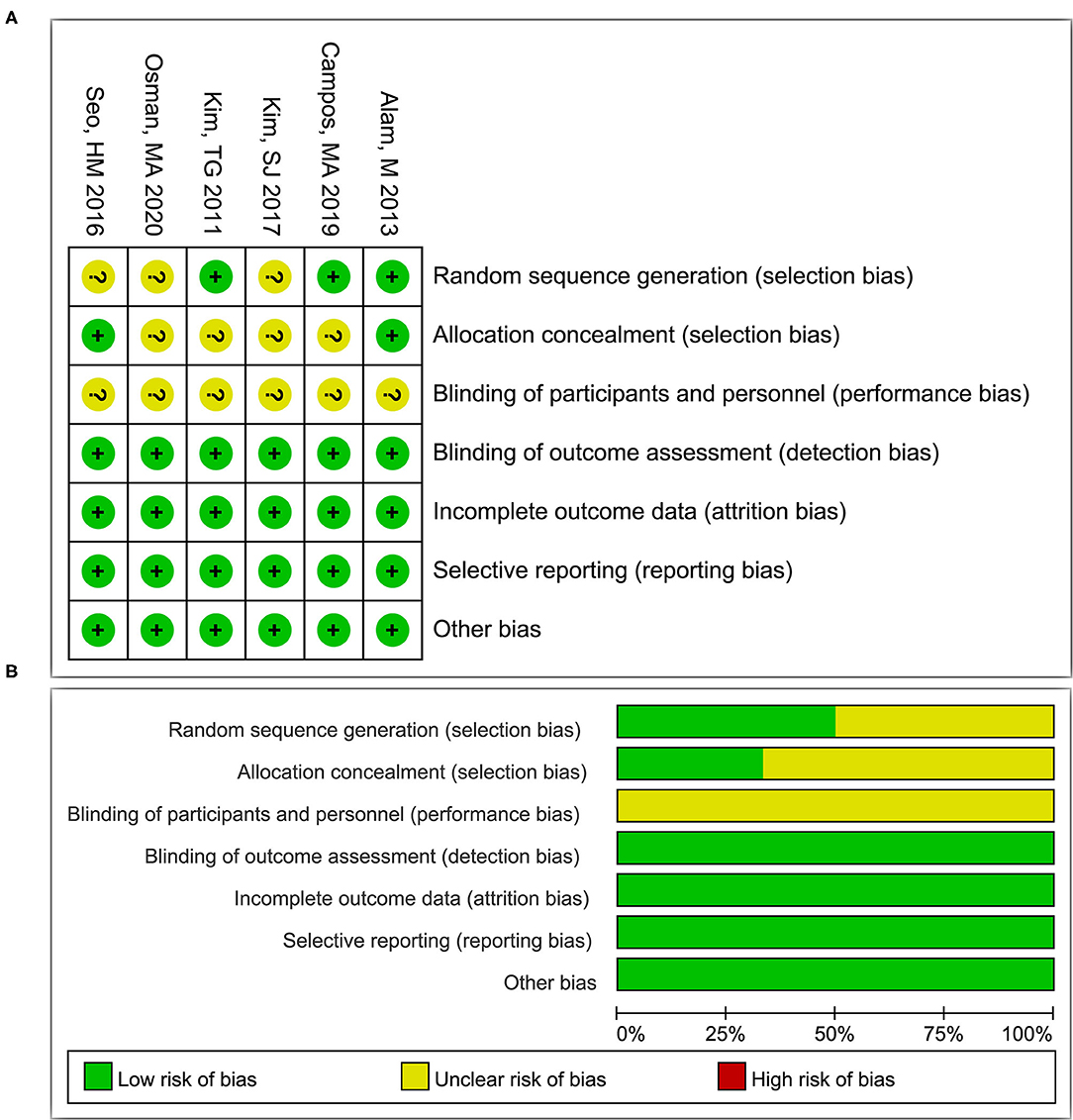
Figure 2. The risk of bias assessments in each item for studies (A) and summary of the risk of bias in each item by all studies (B). +, low risk of bias; -, high risk of bias; ?, unclear risk of bias.

Table 2. Quality assessment of the prospective cohort studies with Newcastle-Ottawa quality assessment scale.
Results of Meta-Analysis
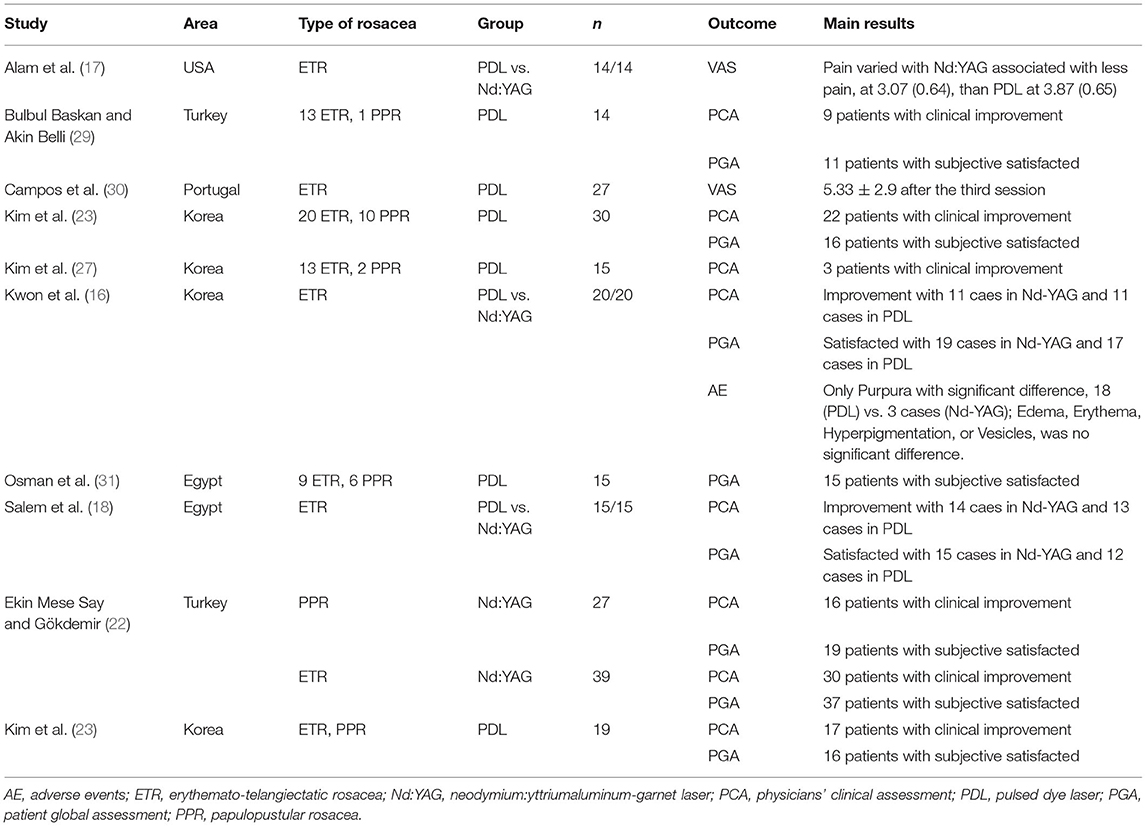
The main outcomes and results of each of the ten included articles were listed in Table 3. A total of six studies (16, 18, 23, 27–29) reported the physicians' clinical assessment (PCA) of the PDL group, and the combined results showed IR (95%CI) = 0.663 (0.456–0.844), and significant heterogeneity was found between these studies (I2 = 78.6%, P < 0.001) (Figure 3A). Three studies (17, 25, 15) reported the PCA of Nd:YAG and the combined results showed IR (95%CI) = 0.718 (0.547–0.864), and significant heterogeneity was found between these studies (I2 = 66.3%, P = 0.031) (Figure 3B). No significant difference between these two groups was detected in the combined results (P = 0.667).
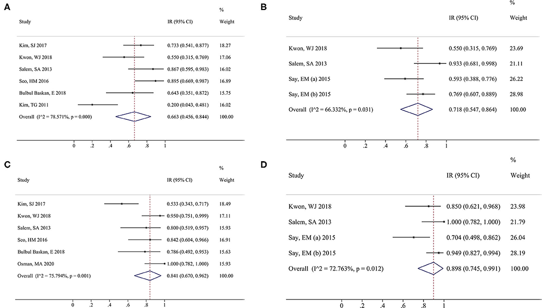
Figure 3. Forest plot of overall risk ratios of physicians' clinical assessment (PCA) and patient global assessment (PGA) in the pulsed dye laser (PDL) group vs. neodymium:yttrium-aluminum-garnet (Nd:YAG) groups. PCA of PDL group (A) and Nd:YAG group (B); PGA of PDL group (C) and Nd:YAG group (D). Squares indicate the estimates for the corresponding study; the size of the square is proportional to the weight of the study to the overall estimate. Diamonds indicate an overall pooled estimate while horizontal lines represent the 95% CI. PDL, pulsed dye laser; Nd:YAG, neodymium:yttrium-aluminum-garnet.
In addition, a total of six studies (16, 18, 23, 28, 29, 31) reported the patient global assessment (PGA) of the PDL group, and the combined results showed IR (95%CI) = 0.841 (0.670–0.962), and significant heterogeneity was found between these studies (I2 = 75.8%, P = 0.001) (Figure 3C). Three studies (17, 25, 15) reported the PGA of Nd:YAG and the combined results showed IR (95%CI) = 0.898 (0.745–0.991), and significant heterogeneity was found between these studies (I2 = 72.8%, P = 0.012) (Figure 3D). There was no significant difference between the two groups (P = 0.559).
The clinical efficacies of PDL and Nd:YAG were compared based on PCA to evaluate clinical improvement (defined as of >50% clearance, a subjective method for blinded dermatologists to assess the improvement in the severity of erythema). A total of two articles (17, 15) reported this outcome, and no significant heterogeneity was found between the two studies (I2 = 0.0%, P = 0.759); thus, a fixed-effect model was used to combine data. The result showed clinical improvements of >50% clearance of up to 68.6% in the PDL group and 71.4% in the Nd:YAG group, but no significant differences in the rates of patients with rosacea with clinical improvement between the two groups (RR = 0.94, 95%CI: 0.75–1.17, P = 0.578) (Figure 4A).

Figure 4. Forest plot of the overall risk ratios of clinical efficacies of rosacea treated compared with PDL than Nd:YAG. PCA (A) and PGA (B). PDL, pulsed dye laser; Nd:YAG, neodymium:yttrium-aluminum-garnet.
Two studies (17, 15) reported the results of PGA in the PDL and Nd:YAG groups. Significant heterogeneity was found between the two studies (I2 = 71.5%, P = 0.061). The merged result showed a subjective satisfaction rate of patients in the PDL group of 88.6% compared to 91.4% in the control group, a difference between the two groups that was not statistically significant (RR = 0.96, 95%CI: 0.70–1.33, P = 0.808) (Figure 4B).
Two studies (17, 30) reported the pain score of the PDL group, and one study reported the pain score of the Nd:YAG group. Based on the visual analog scale (VAS) as the pain outcome index, the combined result of the PDL group showed a mean (95%CI) = 4.60 (3.17–6.04), indicating not significantly higher (P = 0.115) pain sensation of patients treated with PDL than that in patients treated with Nd:YAG, mean (95%CI) = 3.07 (1.82–4.32) (Figure 5).
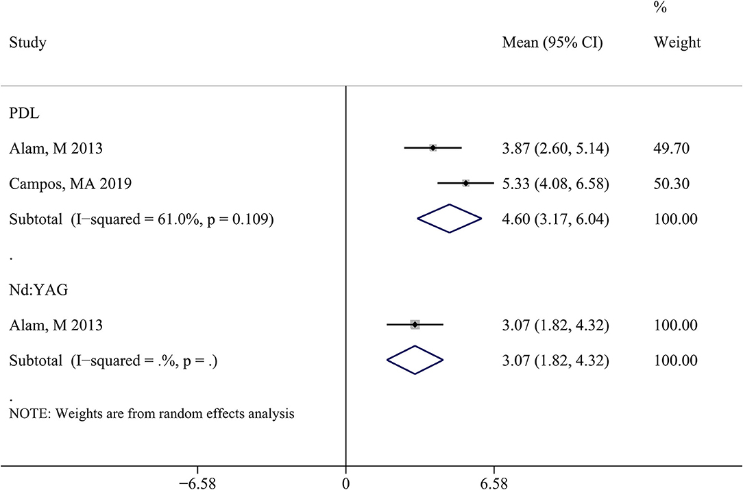
Figure 5. Forest plot of standard mean difference to compare pain score between PDL and Nd:YAG groups. PDL, pulsed dye laser; Nd:YAG, neodymium:yttrium-aluminum-garnet.
Discussion
To explore the different clinical effects of PDL and Nd:YAG on rosacea, PCA, PGA, VAS, and adverse events were defined as the outcome indexes. The results of our analyses showed no significant differences in the percentage of cases with clinical improvement (>50% clearance) and pain score.
Hemangiectasis in rosacea is located in deep subdermal blood vessels located approximately 3 mm deep and mainly requires long-pulse Nd:YAG laser (1064 nm) for treatment (32). Nd:YAG has a good clinical effect for the treatment of the vascular and inflammatory lesions of rosacea (22). In a double-blind RCT study comparing Nd:YAG to PDL for treating diffuse facial erythema, the efficacy of PDL was better than that of Nd:YAG, but the pain of Nd:YAG was less than that of PDL (17). Our study revealed that PDL and Nd:YAG were both effective for treating rosacea with comparable physician assessments and patient satisfaction; however, there was no statistical significance between the rosacea improvement effects of the two groups. Consistent with our findings, two experiments also point that both PDL and Nd:YAG laser have good physician assessment with no significant difference (16, 28). What is more, a meta-analysis also confirmed the treatment success throughout the physician's assessment results while no significant differences were found in the two treatment methods (19). As for the similar pain score after the treatment of PDL and Nd:YAG laser, the results were not coincident with our conclusion in two experimental studies and a meta-analysis (17, 19, 30). The reason for the disagreement might come from the study concluded in the meta-analysis being heterogenic. As for the reverse effects of the PDL and Nd:YAG laser, the main side effect of Nd:YAG was temporary erythema, without purpura, pigmentation, or scar (10). Similarly, no serious adverse events, such as pigmentation or scarring, have been reported for either PDL or Nd:YAG treatment (17, 23). These findings confirmed that the Nd:YAG laser successfully treated erythema telangiectasia rosacea with lower energy and longer pulse width without side effects. The main adverse reaction of PDL was purpura, which mainly acts on superficial and small capillaries, but can also reach deep into the dermis (30). This may partly explain why PDL feels more painful than Nd:YAG for the treatment of rosacea. An article included in the present study also reported the differential adverse events between the PDL treatment and Nd:YAG laser treatment (15), and the results showed that PDL treatment caused more purpura than Nd:YAG laser treatment. It is a pity that we cannot conduct a meta-analysis about the adverse events because only one included article reported the adverse events after treatment with PDL and Nd:YAG laser.
Our findings indicated that PDL was not superior to Nd:YAG for the treatment of rosacea, contrary to one review report (33). However, the review only referenced the redness result from Alam 2013 (17), in which redness improved by a mean of 52% after PDL treatment compared to 34% after the Nd:YAG treatment, a statistically significant difference between the methods. Although we also included Alam 2013 in our analyses, redness was not used as an assessment indicator since only this paper among all included studies reported redness data. In addition, the review ignored pain findings (33), while our results comprehensively assessed the clinical efficacy, satisfaction rates, and pain scores from several studies.
The findings in our analysis can be used to inform the selection of methods for the treatment of rosacea. However, this study had some limitations. Firstly, the numbers of included studies and sample size were both small. Secondly, the clinical and methodological heterogeneity of the included literature was relatively large. Thirdly, the examination of publication bias was limited due to the small number of included studies. The inevitable potential publication bias may have affected the robustness of the conclusions. Fourthly, it is a pity that we cannot conduct a meta-analysis about the adverse events because only one included article reported the relevant adverse events after treatment with PDL and Nd:YAG laser. More studies about the adverse events caused by treatment with PDL and/or Nd:YAG laser need to be conducted and more validation is necessary to be involved in the further meta-analysis. Finally, the exclusion criteria in this study may induce a potential applicability limitation: although the method may result in an accurate outcome of using PDL or Nd:YAD only, the absence of other therapy method limit the applicability of these results. Actually, patients often tend to select several therapy methods rather than only one therapy to improve the severity of rosacea in clinical practice.
Conclusion
In conclusion, PDL and Nd:YAG all showed clinical efficacy and patient satisfaction for the treatment of rosacea, with no significant difference observed between treatments. Also, the analysis of the pain score between the PDL and Nd:YAG treatment exhibited no statistical significance. These findings will play an important guiding role in the clinical treatment of rosacea. However, considering the limitations of this study, such as its limited sample size, the results of this meta-analysis require verification by RCT with larger samples and higher quality.
Data Availability Statement
The original contributions presented in the study are included in the article/Supplementary Material, further inquiries can be directed to the corresponding author/s.
Author Contributions
YL and RW contributed to conception and design of the research and acquisition of data. YL contributed to analysis and interpretation of data, statistical analysis, and drafting the manuscript. RW contributed to the revision of the manuscript for important intellectual content. All authors have read and approved the final manuscript.
Conflict of Interest
The authors declare that the research was conducted in the absence of any commercial or financial relationships that could be construed as a potential conflict of interest.
Publisher's Note
All claims expressed in this article are solely those of the authors and do not necessarily represent those of their affiliated organizations, or those of the publisher, the editors and the reviewers. Any product that may be evaluated in this article, or claim that may be made by its manufacturer, is not guaranteed or endorsed by the publisher.
Supplementary Material
The Supplementary Material for this article can be found online at: https://www.frontiersin.org/articles/10.3389/fmed.2021.798294/full#supplementary-material
References
1. Steinhoff M, Schauber J, Leyden JJ. New insights into rosacea pathophysiology: a review of recent findings. J Am Acad Dermatol. (2013) 69:S15–26. doi: 10.1016/j.jaad.2013.04.045
2. Holmes AD. Potential role of microorganisms in the pathogenesis of rosacea. J Am Acad Dermatol. (2013) 69:1025–32. doi: 10.1016/j.jaad.2013.08.006
3. Awosika O, Oussedik E. Genetic predisposition to rosacea. Dermatol Clin. (2018) 36:87–92. doi: 10.1016/j.det.2017.11.002
4. Chang ALS, Raber I, Xu J, Li R, Spitale R, Chen J, et al. Assessment of the genetic basis of rosacea by genome-wide association study. J Invest Dermatol. (2015) 135:1548–55. doi: 10.1038/jid.2015.53
5. Ahn CS, Huang WW. Rosacea Pathogenesis. Dermatol Clin. (2018) 36:81–6. doi: 10.1016/j.det.2017.11.001
6. Koczulla R, Von Degenfeld G, Kupatt C, Krötz F, Zahler S, Gloe T, et al. An angiogenic role for the human peptide antibiotic LL-37/hCAP-18. J Clin Invest. (2003) 111:1665–72. doi: 10.1172/JCI17545
7. Crawford GH, Pelle MT, James WD. Rosacea: I. Etiology, pathogenesis, and subtype classification. J Am Acad Dermatol. (2004) 51:327–41. doi: 10.1016/j.jaad.2004.03.030
8. Draelos ZD, Gunt H, Levy SB. Natural Skin Care Products as Adjunctive to Prescription Therapy in Moderate to Severe Rosacea. J Drugs Dermatol. (2019) 18:141–6.
9. Subashini K, Pushpa G, Venugopal V, Murali N. Rosacea with severe ophthalmic involvement and blindness - a rare occurrence. Int J Dermatol. (2012) 51:1271–3. doi: 10.1111/j.1365-4632.2010.04734.x
10. Hofmann MA, Lehmann P. Physical modalities for the treatment of rosacea. Jddg J Der Deutschen Dermatologischen Gesellschaft. (2016) 14:38–43. doi: 10.1111/ddg.13144
11. Woo YR, Lee SH, Cho SH, Lee JD, Kim HS. Characterization and analysis of the skin microbiota in rosacea: impact of systemic antibiotics. J Clin Med. (2020) 9:185. doi: 10.3390/jcm9010185
12. Schilling LM, Halvorson CR, Weiss RA, Weiss MA, Beasley KL. Safety of Combination Laser or Intense Pulsed Light Therapies and Doxycycline for the Treatment of Rosacea. Dermatol Surg. (2019) 45:1401–5. doi: 10.1097/DSS.0000000000002009
13. Baek JO, Hur H, Ryu HR, Kim JS, Lee KR, Kim YR, et al. Treatment of erythematotelangiectatic rosacea with the fractionation of high-fluence, long-pulsed 595-nm pulsed dye laser. J Cosmet Dermatol. (2017) 16:12–4. doi: 10.1111/jocd.12284
14. Sodha P, Suggs A, Munavalli GS, Friedman PM. A randomized controlled pilot study: combined 595-nm pulsed dye laser treatment and oxymetazoline hydrochloride topical cream superior to oxymetazoline hydrochloride cream for erythematotelangiectatic rosacea. Lasers Surg Med. (2021) 53:1307–15. doi: 10.1002/lsm.23439
15. Laquer VT, Dao BM, Pavlis JM, Nguyen AN, Chen TS, Harris RM. Immunohistochemistry of angiogenesis mediators before and after pulsed dye laser treatment of angiomas. Lasers Surg Med. (2012) 44:205–10. doi: 10.1002/lsm.22005
16. Kwon WJ, Park BW, Cho EB, Park EJ, Kim KH, Kim KJ. Comparison of efficacy between long-pulsed Nd:YAG laser and pulsed dye laser to treat rosacea-associated nasal telangiectasia. J Cosmet Laser Ther. (2018) 20:260–4. doi: 10.1080/14764172.2017.1418510
17. Alam M, Voravutinon N, Warycha M, Whiting D, Nodzenski M, Yoo S, et al. Comparative effectiveness of nonpurpuragenic 595-nm pulsed dye laser and microsecond 1064-nm neodymium:yttrium-aluminum-garnet laser for treatment of diffuse facial erythema: A double-blind randomized controlled trial. J Am Acad Dermatol. (2013) 69:438–43. doi: 10.1016/j.jaad.2013.04.015
18. Salem SAM, Fattah NSA, Tantawy SM, El-Badawy NM, El-Aziz YAA. Neodymium-yttrium aluminum garnet laser versus pulsed dye laser in erythemato-telangiectatic rosacea: Comparison of clinical efficacy and effect on cutaneous substance (P) expression. J Cosmet Dermatol. (2013) 12:187–94. doi: 10.1111/jocd.12048
19. Husein-ElAhmed H, Steinhoff M. Light-based therapies in the management of rosacea: a systematic review with meta-analysis. Int J Dermatol. (2021). doi: 10.1111/ijd.15680
20. Tarsilla M. Cochrane handbook for systematic reviews of interventions. J Multidiscipl Evaluat. (2008) 6:142–8. Available online at: https://sc.panda321.com/extdomains/books.google.com/books?hl=zh-CN&lr=&id=cTqyDwAAQBAJ&oi=fnd&pg=PR3&dq=Cochrane+handbook+for+systematic+reviews+of+interventions.&ots=tvjFBbtFnm&sig=X2x1Trn0yx4flB5pfdrXlOFG16I#v=onepage&q=Cochrane%20handbook%20for%20systematic%20reviews%20of%20interventions.&f=false
21. Moskalewicz A, Oremus M. No clear choice between Newcastle-Ottawa Scale and Appraisal Tool for Cross-Sectional Studies to assess methodological quality in cross-sectional studies of health-related quality of life and breast cancer. J Clin Epidemiol. (2020) 120:94–103. doi: 10.1016/j.jclinepi.2019.12.013
22. Ekin Mese Say OG, Gökdemir G. Treatment outcomes of long-pulsed Nd: YAG laser for two different subtypes of rosacea. J Clin Aesthet Dermatol. (2015) 8:16.
23. Kim SJ, Lee Y, Seo YJ, Lee JH, Im M. Comparative efficacy of radiofrequency and pulsed dye laser in the treatment of rosacea. Dermatol Surg. (2017) 43:204–9. doi: 10.1097/DSS.0000000000000968
24. Altman DG, Bland JM. Interaction revisited: the difference between two estimates. Bmj. (2003) 326:219. doi: 10.1136/bmj.326.7382.219
25. DeLong ER, DeLong DM, Clarke-Pearson DL. Comparing the areas under two or more correlated receiver operating characteristic curves: a nonparametric approach. Biometrics. (1988) 44:837–45. doi: 10.2307/2531595
26. Higgins JP, Thompson SG, Deeks JJ, Altman DG. Measuring inconsistency in meta-analyses. BMJ. (2003) 327:557–60. doi: 10.1136/bmj.327.7414.557
27. Kim TG, Roh HJ, Cho SB, Lee JH, Lee SJ, Oh SH. Enhancing effect of pretreatment with topical niacin in the treatment of rosacea-associated erythema by 585-nm pulsed dye laser in Koreans: a randomized, prospective, split-face trial. Br J Dermatol. (2011) 164:573–9. doi: 10.1111/j.1365-2133.2010.10174.x
28. Hyun-Min S, Jung-In K, Han-Saem K, Young-Jun C, Won-Serk K. Prospective Comparison of Dual Wavelength Long-Pulsed 755-nm Alexandrite/1,064-nm Neodymium:Yttrium-Aluminum-Garnet Laser versus 585-nm Pulsed Dye Laser Treatment for Rosacea. Ann Dermatol. (2016) 28:607–14. doi: 10.5021/ad.2016.28.5.607
29. Bulbul Baskan E, Akin Belli A. Evaluation of long-term efficacy, safety, and effect on life quality of pulsed dye laser in rosacea patients. J Cosmet Laser Ther. (2019) 21:185–9. doi: 10.1080/14764172.2018.1502453
30. Campos MA, Sousa AC, Varela P, Baptista A, Menezes N. Comparative effectiveness of purpuragenic 595 nm pulsed dye laser versus sequential emission of 595 nm pulsed dye laser and 1,064 nm Nd: YAG laser: a double-blind randomized controlled study. Acta Dermatovenerol Alp Pannonica Adriat. (2019) 28:1–5. doi: 10.15570/actaapa.2019.1
31. Osman M, Shokeir HA, Hassan AM, Atef Khalifa M. Pulsed dye laser alone versus its combination with topical ivermectin 1% in treatment of Rosacea: a randomized comparative study. J Dermatolog Treat. (2020) 1–7. doi: 10.1080/09546634.2020.1737636
32. Rose AE, Goldberg DJ. Successful treatment of facial telangiectasias using a micropulse 1,064-nm neodymium-doped yttrium aluminum garnet laser. Dermatologic Surgery. (2013) 39:1062–6. doi: 10.1111/dsu.12185
Keywords: rosacea, pulsed dye laser, Nd:YAG, meta-analysis, efficacy
Citation: Li Y and Wang R (2022) Efficacy Comparison of Pulsed Dye Laser vs. Microsecond 1064-nm Neodymium:Yttrium-Aluminum-Garnet Laser in the Treatment of Rosacea: A Meta-Analysis. Front. Med. 8:798294. doi: 10.3389/fmed.2021.798294
Received: 05 November 2021; Accepted: 10 December 2021;
Published: 20 January 2022.
Edited by:
Alejandro Pablo Adam, Albany Medical College, United StatesReviewed by:
Edward Wladis, Albany Medical College, United StatesEster Del Duca, Mount Sinai Hospital, United States
Copyright © 2022 Li and Wang. This is an open-access article distributed under the terms of the Creative Commons Attribution License (CC BY). The use, distribution or reproduction in other forums is permitted, provided the original author(s) and the copyright owner(s) are credited and that the original publication in this journal is cited, in accordance with accepted academic practice. No use, distribution or reproduction is permitted which does not comply with these terms.
*Correspondence: Rupeng Wang, QXNpYXEyMDA0QDE2My5jb20=
 Yuanchao Li
 Rupeng Wang