- 1Department of Pathology, Xiangya Hospital, Central South University, Changsha, China
- 2Departments of Ultrasound Imaging, Xiangya Hospital, Central South University, Changsha, China
- 3National Clinical Research Center for Geriatric Disorders, Xiangya Hospital, Central South University, Changsha, China
- 4Xiangya Lung Cancer Center, Xiangya Hospital, Central South University, Changsha, China
Worldwide, hepatocellular carcinoma (HCC) is one of the most malignant cancers with poor prognosis. The structural maintenance of chromosomes (SMC) gene family has been shown to play important roles in human cancers. Nevertheless, the role of SMC members in HCC is not well-understood. In this study, we comprehensively explored the role of the SMC family in HCC using a series of bioinformatic analysis tools. Studies have demonstrated that the mRNA expression levels of SMC1A, SMC1B, SMC2, SMC4, and SMC6 are significantly overexpressed in HCC, and the protein levels of SMC1A, SMC2, SMC3, SMC4, SMC5, and SMC6 are similarly elevated. Moreover, HCC patients with high SMC2 and SMC4 expression levels exhibit poor survival. Using KEGG and GO analyses, we analyzed the enrichment of gene expression in the biological functions and pathways of the SMC family in HCC. Immune infiltration analysis revealed that the expression of the SMC family is closely associated with B cells, CD4+ T cells, CD8+ T cells, macrophages, neutrophils, and DCs. In conclusion, our findings will enhance a more thorough understanding of the SMC family in HCC progression and provide new directions for the diagnosis and treatment of HCC in the future.
Introduction
Hepatocellular carcinoma (HCC), a primary cancer of the liver, is the most frequent malignant tumor globally (1). The National Center for Health Statistics shows that HCC is the fifth-leading cause of cancer-related deaths worldwide (2, 3). As one of the most aggressive malignant tumors, the effect of surgical treatment is limited and only applicable to a small number of patients with early HCC (4–6). Advanced treatments such as trans-arterial chemoembolization (TACE) have little effect on improving the survival time of patients with HCC (7). Hence, the search for new treatments and prognostic biomarkers to improve survival of patients with HCC is urgently required.
Most DNA-based processes are affected by the structural maintenance of chromosomes (SMC) protein family. As the central regulator of chromosome dynamics, the SMC family can control the cohesion of sister chromatids, chromosomal condensation, DNA replication, DNA repair and transcription (8). There are six members of the SMC family: SMC1-SMC6, and which there are two variants of SMC1, namely SMC1A and SMC1B (9). Six family members form the core of three different multi-subunit protein complexes, of which SMC1 and SMC3 form a v-type heterodimer, which is the main component of the cohesive complex (10). SMC2 and SMC4 are part of a five-subunit lectin complex (11). SMC5 and SMC6 form a complex closed by kleisin, similar to the structure of cohesin and condensin. They often aggregate at sites of DNA double-strand breaks to promote homologous recombination repair, and therefore play an important role in DNA damage responses (12).
Different types of SMC family mutations can change the cohesion or adhesion of DNA, and at the same time affect various life processes involving chromosomal DNA, leading to the occurrence of various cancers. Recent studies have shown that members of the SMC family are widely involved in the pathological progression of tumors. SMC1A is highly expressed in a variety of malignancies, including colorectal and prostate cancer, and acute myelogenous leukemia (AML). It promotes cancer cell invasion and metastasis by influencing epithelial–mesenchymal transition (EMT), releasing inflammatory mediators, and recruiting downstream target molecules, thereby promoting early tumor formation and tumorigenesis (13–16). The meiosis-specific cohesive member SMC1B is necessary for sister chromatid pairing and preventing telomere shortening (8). SMC1B is one of the genes related to HPV+ cervical intraepithelial neoplasia (17). In addition, SMC1B is involved in the development of colorectal cancer (18). The deletion of SMC2, which is highly expressed in bladder cancer tissues, blocks the G2/M phase of bladder cancer cells, thereby inhibiting tumor growth (19). SMC3 is involved in drug resistance in lung cancer (20) and has an important impact on the prognosis of AML patients (21). SMC4 plays a regulatory role in glioma, breast cancer, lung adenocarcinoma, and other malignancies, and directly or indirectly affects the survival time of patients with tumors (22–24). SMC5 is associated with ovarian cancer progression (25). Compared with normal tissues, SMC6 has also been confirmed to have abnormal expression in lung cancer, sarcoma, and other tumor types (26).
Previous analyses of the SMC family in HCC remain relatively limited. The clinical significance, biological functions, and immune roles of SMC family members in HCC have not yet been reported. Thus, we comprehensively analyzed the expression of each SMC family members in HCC, and their relationship with prognosis, immune infiltration, genetic alteration, and possible mechanisms involving the SMC family in HCC. Using a series of bioinformatic analysis tools, such as Tumor Immune Estimation Resource (TIMER) (27), Human Protein Atlas project (HPA) (28), and Gene expression profiling analysis (GEPIA) (29), we identified the expression of SMC family members in HCC, and Kaplan-Meier plotter was used to evaluate the prognostic value of SMC family members in HCC. In addition, cBioPortal was used to analyze the mutation of SMC family members in HCC, and to download related co-expressed genes for subsequent enrichment. Finally, TIMER was used to analyze relationships between the expression of SMC family members and immune cell infiltration in HCC. Taken together, our findings provide new insights into the prognostic value and biological roles of the SMC family in HCC.
Methods
Gene Expression Analysis
Multiple bioinformatics database resources were accessed to explore the expression levels of SMC family members in HCC tissues.
The website tool TIMER is a comprehensive resource based on the TCGA database, which contains 371 HCC samples and 50 normal liver samples. It provides multiple functions including gene expression comparisons with tumor/normal tissues in different cancers, correlation analysis between genes and immune infiltrating cells, survival analysis, and other functionalities (27). The mRNA expression of SMC family members in different cancers or specific cancer subtypes from TIMER, and the log2 [TPM (Transcripts per million)] was applied for log-scale. We also used the TIMER database to analyse the correlation of SMC family members expression with the immune infiltrating cells.
HPA is a large-scale protein research project (28). This allows researchers to map the location of proteins encoded by the SMC family in human tissues and cells. In this study, we analyzed the protein expression of SMC family members in normal and HCC tissues by immunohistochemistry.
GEPIA is a web server that analyzes RNA sequencing data from the GTEx and TCGA database, and allows users to analyze the human cancer gene expression and interaction analyses (29). It provides researchers with interactive customization functions, such as differential expression analysis, pathological stage analysis, and other diversified functions. We used GEPIA database to evaluate the correlations of SMC family expression with the pathological stage of HCC patients.
Survival Analysis
The Kaplan–Meier database (https://kmplot.com) was used to analyze the relationship between the HCC patients' prognosis and mRNA expression levels of SMC family members. The 370 HCC samples were split into high and low expression group by the mRNA expression value of auto-selected best cutoff to analyze the correlation of SMC family members expression and the overall survival (OS), relapse free survival (RFS), disease-specific survival (DSS), and progression Free Survival (PFS) in HCC. Kaplan-Meier analysis was assessed by log-rank tests, and statistical significance if the P-value < 0.05.
Immune Infiltrating Analysis
TIMER was used to evaluate associations between mRNA expression of SMC family members and HCC immune infiltrating cells that contained CD4+ T cells, CD8+ T cells, B cells, macrophages, neutrophils, and DCs. Correlation R value was calculated by Spearman's algorithm and adjusted by tumor purity. P-value < 0.05 was considered to be statistically significant.
Methylation and Mutation Analysis
The relationship between SMC family expression and DNA methylation values was analyzed using UALCAN (http://ualcan.path.uab.edu), which is a database comprising systematic integration of DNA methylation data and mRNA expression levels in human cancers (30). We predicted DNA methylation changes of SMC family members in 377 HCC samples and 50 normal liver samples using the UALCAN database.
Co-expression Network Analysis
cBioPortal (http://cbioportal.org), an open comprehensive platform, can be used to analyze multidimensional cancer genomics and clinical data. The threshold of |log2FC| was 1.2, and the P-value cutoff was 0.01. Detailed visual analysis was performed using Cytoscape software v.3.7.2. The co-expression gene lists of SMC family were identified by cBioPortal.
The STRING database was used to assess correlations involving SMC genes. Metascape is a powerful tool for investigating the functional annotation of a certain gene list (31). This database enabled us to conduct Gene Ontology (GO) and Kyoto Encyclopedia of Genes and Genomes (KEGG) pathway enrichment analysis for genes co-expressed with SMC members.
Results
Aberrant Expression of the SMC Family Members in HCC
To investigate SMC expression in various tumors compared to normal tissues, transcriptional levels were analyzed using the TIMER database. The levels of SMC1A, SMC1B, SMC2, SMC3, SMC4, and SMC6 mRNA expression were highly upregulated in HCC tissues compared to normal liver tissues, however, SMC5 showed no statistical difference (Figure 1). To verify these results, we further explored the immunohistochemical staining results of SMC family members from the HPA database. As shown in Figure 2, we found that SMC1A, SMC2, SMC3, SMC4, SMC5, and SMC6 are highly expressed in HCC but not so in normal samples; however, compared with their non-elevated expression in normal liver tissues, SMC1B were weakly expressed in HCC.

Figure 1. Expression levels of SMC family members in HCC. (A-G) Relative expression of SMC1A, SMC1B, SMC2, SMC3, SMC4, SMC5, and SMC6 in different cancers compared with normal tissues by the TIMER database. *P < 0.05, **P < 0.01, ***P < 0.001 compared with control.

Figure 2. Protein expression levels of SMC family members in HCC. (A-G) The HPA database shows the protein expression levels of SMC1A, SMC1B, SMC2, SMC3, SMC4, SMC5, and SMC6 in HCC tissues compared with non-cancerous tissues. (H) The integrated optical density (IOD) was used to analyze the expression difference between HCC tissues and noncancerous tissues.
Correlation of the Expression of SMC Family Members With Clinicopathologic Features of HCC Patients
Next, we sought to determine whether the expression levels of SMC family members correlate with the patient tumor staging. Correlation analysis of TCGA data using the GEPIA database revealed that the pathological stages of HCC patients are correlated with SMC1A, SMC1B, SMC2, SMC4, and SMC6 expression (Figure 3).

Figure 3. Relationship between the expression level of SMC family members and HCC tumor staging. (A-G) GEPIA database was used to evaluate the correlations of the expression of SMC1A, SMC1B, SMC2, SMC3, SMC4, SMC5, and SMC6 with the pathological stage of HCC patients.
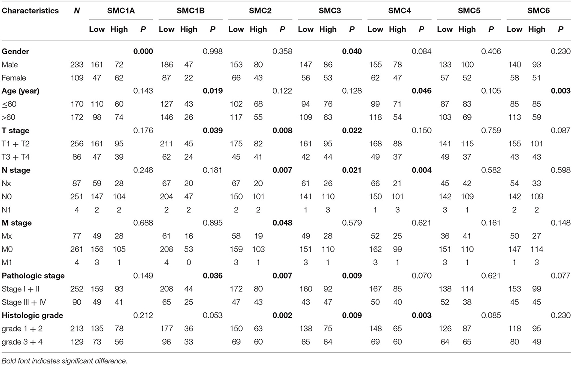
Moreover, we used the TCGA HCC patient samples to further analyze the clinicopathologic parameters and the expression of SMC family members. We found that SMC family members expression are closely associated with the clinicopathologic parameters of HCC patients (Table 1). For example, the high levels of SMC1B, SMC2, and SMC3 expression were significantly associated with the T stage of HCC patients, the high levels of SMC2, SMC3, and SMC4 expression were significantly associated with the N stage of HCC patients, and the high levels of SMC2 expression was significantly associated with the M stage of HCC patients. These results indicated that SMC family members may serve as potential diagnostic markers for HCC.
The Prognostic Value of the SMC Family in HCC Patients
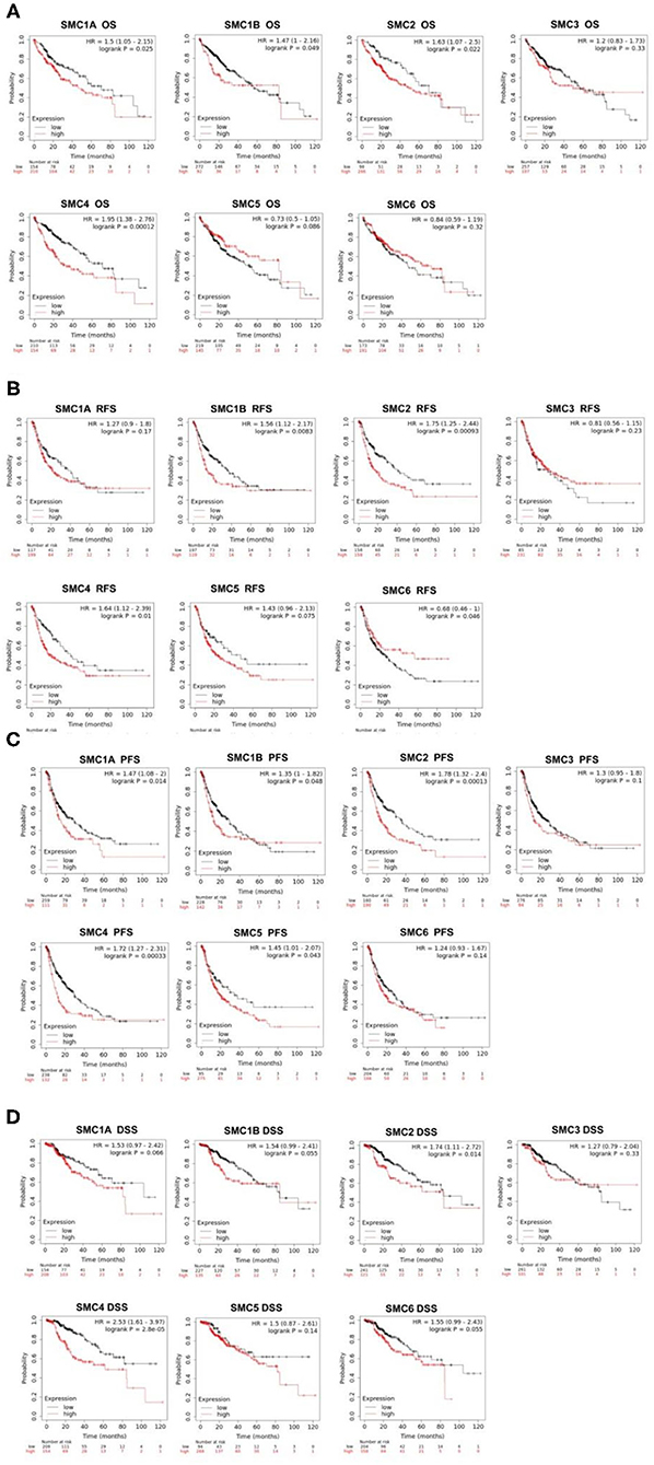
Then, we used the Kaplan-Meier plotter tool was used to perform survival analysis in 364 TCGA HCC patient samples with complete survival and SMC family members expression data. OS was applied for the output of clinical prognosis, and we found that high levels of SMC1A, SMC1B, SMC2, and SMC4 expression were significantly associated with poorer OS in HCC patients (Figure 4A). Furthermore, we discovered that increased SMC1B, SMC2, and SMC4 expression was significantly associated with poorer RFS in HCC (Figure 4B). SMC1A, SMC1B, SMC2, SMC4, and SMC5 abundance were strongly associated with PFS (Figure 4C). Moreover, higher expression of SMC2 and SMC4 were significantly associated with poor DSS (Figure 4D). Collectively, SMC family members expression are closely associated with the prognosis of HCC patients. Among them, the relationship between SMC2 and SMC4 expression and clinical prognosis of HCC is closer than others. In our results, the high expressions of SMC2 and SMC4 were significantly associated with the poor OS/PFS/RFS/DSS in HCC patients, which indicated that SMC2 and SMC4 may serve as potential prognostic markers for HCC.

Figure 4. Prognostic value of SMC family in HCC. (A-D) The expression of SMC family members showed significant associations with OS, RFS, PFS, and DSS in HCC, and statistical significance was assessed by log-rank tests.
We further analyzed the prognostic value of SMC family members in different pathological grades and clinical stages of HCC using the Kaplan–Meier plotter (Table 2). Taking SMC4 as an example, the mRNA expression of SMC4 was closely related to poorer OS in grade I, grade II, stage I and stage II, AJCC-T1, and AJCC-T2 HCC patients. Moreover, SMC1A and SMC2 transcriptional expression was significantly associated with worse OS in stage I and stage II HCC patients. We also found that high expression of SMC1B was correlated with poor OS in grade I grade II and AJCC-T2 HCC patients. SMC2 abundance was significantly associated with shorter OS in stage I and stage II and AJCC-T2 HCC patients, and SMC6 expression was significantly correlated with poorer OS in AJCC-T1 HCC patients. Taken together, these results indicated that a part of SMC family member may serve as a potential prognosis factor in HCC, especially valuable for the OS prediction in HCC patients diagnosed with early stage or low grade.

Table 2. Kaplan–Meier plotter was used to analyze the prognostic value of SMC family members in different pathological grades and clinical stages of HCC.
Genetic Alterations and Functional Enrichment Analysis of SMC Family Members
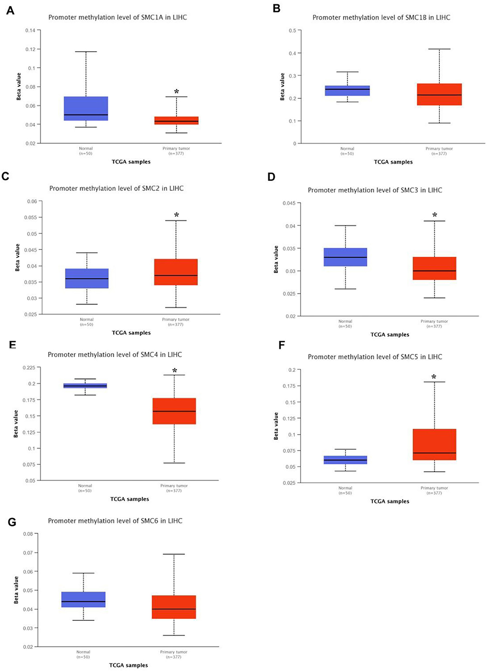
It is widely known that genetic alteration plays a key role in the development of cancer. Using the UALCAN database, we identified SMC gene methylation levels in HCC patients. Compared with healthy humans, the DNA methylation levels of SMC1A, SMC3, and SMC4 were significantly lower in HCC samples, whereas SMC2 and SMC5 exhibited significantly higher levels in HCC tissues, and other SMC family member exhibited statistically insignificant differences between normal and cancer tissues (Figure 5). These altered DNA methylation levels of SMC gene family members were closely associated with their differences in expression levels, which may provide novel targets for cancer patients by DNA methylation-targeting drugs.

Figure 5. DNA methylation levels of SMC family genes in HCC. (A-G) The DNA methylation change of SMC1A, SMC1B, SMC2, SMC3, SMC4, SMC5, and SMC6 in HCC by the UALCAN database. *P < 0.05 compared with control.
Furthermore, we conducted a series of analyses to confirm the genetic alteration status of SMC family using the cBioPortal database. We observed that SMC genes were altered in 103 samples from 360 patients (29%). The highest rate of alteration frequency in SMC6 was approximately 9%. Among all alterations involving SMC family member genes, the most common alteration types were mRNA alterations (Figure 6A). Moreover, we used the STRING database to construct the protein–protein interaction (PPI) network among the SMC family members, and found that seven SMC family members were all included and served as hub nodes in the interactive network (Figure 6B). We then identified co-expressed genes with a threshold value of |log2FC|> 1.2 and P < 0.01 from the cBioportal database (Supplementary Table 1), and presented a map of co-expression networks of key genes associated with the SMC family using Cytoscape_v.3.7.2 (Figure 6C).

Figure 6. Genetic alterations and pathway enrichment analysis of SMC family in HCC. (A) The genetic alteration of SMC1A, SMC1B, SMC2, SMC3, SMC4, SMC5, and SMC6 in HCC was illustrated from top to bottom. (B) The SMC family members interaction analysis was evaluated by STRING database. (C) Construction a network of genes co-expressed with SMC family members in HCC. (D-G) The KEGG enrichment pathway, Molecular functions, biological processes, and cell components of co-expressed genes was analyzed by Metascape database.
In addition, we used the identified co-expressed genes to conduct GO and KEGG pathway analyses on the Metascape database. For KEGG pathway analysis, the drug metabolizing cytochrome p450, tyrosine metabolism, and phenylalanine metabolism were included in these co-expressed genes (Figure 6D). Molecular function analysis indicated that these genes were primarily related to iron ion binding and microtubule motor activity (Figure 6E). Biological process analysis indicated that these genes were mainly involved in chromosome segregation, drug metabolizing processes, and monocarboxylic acid metabolic processes (Figure 6F). Cellular component analysis revealed that these genes were frequently associated with chromosomes, centromeric regions, and spindles (Figure 6G). These results indicated that the function of SMC family might be involved in drug resistance of HCC, thereby affecting the outcome of treatment in HCC patients.
Association Between the Expression of SMC Family Members and Immune Infiltration in HCC
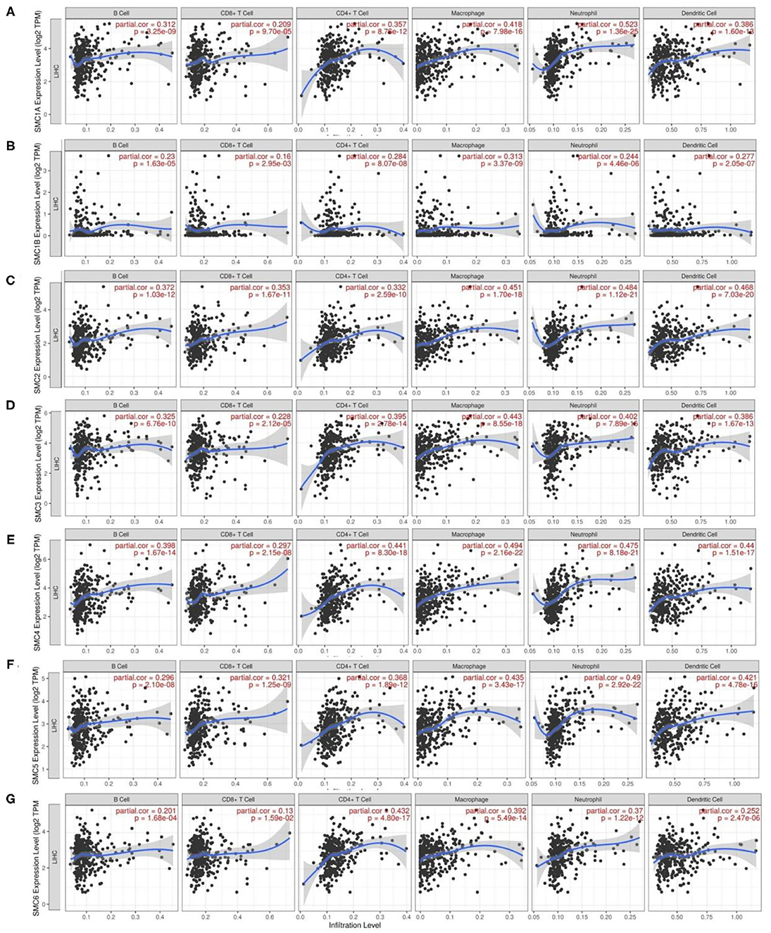
To further understand the roles of SMC family members in HCC, we utilized the TIMER resource to explore the molecular features of tumor–immune interactions. As shown in Figure 7, transcriptional expression of the SMC family was positively associated with B cells, CD4+ T cells, CD8+ T cells, macrophages, neutrophils, and DCs (r > 0.1, P < 0.05). These results suggest the potential effect of SMC family members on the immune response in the tumor microenvironment (TME) of HCC.

Figure 7. Analysis of correlations involving the expression of SMC family genes and immune cell infiltration in HCC. (A-G) Correlation of SMC1A, SMC1B, SMC2, SMC3, SMC4, SMC5, and SMC6 mRNA expression and the immune cell infiltration levels in HCC by the TIMER database.
Next, we investigated potential correlations between SMC family member expression and immune signature markers of various immune cells infiltrating HCC in the TIMER database (Table 3). We observed that SMC1A levels were strongly associated with tumor-associated macrophages (TAM), M1, M2, neutrophils, Th2, Tregs, and monocytes. SMC1B showed a strong correlation with T cell exhaustion and monocytes. SMC2 expression was highly related to CD8+ T cells, T cells, TAM, M2, neutrophils, DCs, Th1, Th2, Tfh, Tregs, T cell exhaustion, and monocyte infiltration in HCC. SMC3 exhibited better correlation with B cells, TAM, M1, M2, neutrophils, Th1, Th2, Tregs, and monocyte markers. mRNA expression of SMC4 was only moderately or weakly linked with M1, NKs, Tfh, and Th17 gene markers, but was strongly associated with other immune cells. CD8+ T cells, B cells, T cells, TAM, M2, neutrophils, Th1, Th2, Tregs, monocytes, and T cell exhaustion demonstrated favorable correlations with SMC5 expression. Moreover, we also observed that the mRNA expression of SMC6 was strongly correlated with M1, M2, neutrophils, Th2, Tregs, and monocytes. Interestingly, the transcriptional expression of SMC family members was highly associated with monocytes. These results further validated that the SMC family members are relevant to immune-infiltrating cells in HCC.

Table 3. The association between the expression of SMC family members and the markers of immune cells.
Discussion
Our research comprehensively explained the biological functions of each member of the SMC family in HCC from five different aspects: mRNA and protein expression levels, disease prognosis, gene mutations, immune infiltration, and pathway analysis.
In the SMC family, SMC1 and SMC3 and the two sister chromatid cohesive (SCC) proteins SCC1 and SCC3 together constitute the core of the cohesin subunit. This complex mainly maintains chromatin equalization during mitosis, DNA damage repair, and other important biological functions. SMC2 and SMC4, the core components of the condensin complex, are required for successful separation during chromosome assembly and cell division. Unlike SMC1/3 or SMC2/4, SMC5/SMC6 forms a third complex that is important for DNA repair and faithful replication. A previous study revealed that certain members of the SMC family are abnormally expressed in HCC tissues. For example, SMC1A was first confirmed to be expressed at an abnormally high level in human liver cancer cell lines (32). Chen et al. demonstrated that SMC2 is highly expressed in HCC (33), and abnormal elevation of SMC4 is observed in HCC tissues (34). To the best of our knowledge, the present study is the first to report the expression of each member of the SMC family in HCC tissues. In addition, consistent with previous research results, we also found that SMC1A, SMC1B, SMC2, SMC3, SMC4, and SMC6 were overexpressed at the mRNA level in HCC when compared with non-tumor cells. Meanwhile, the protein levels of SMC1A, SMC2, SMC3, SMC4, SMC5, and SMC6 were elevated in HCC.
To date, few specific studies have been conducted concerning potential relationships involving SMC family members and HCC clinical significance. Phosphorylation of SMC1A promotes the invasion and metastasis of liver cancer cells. In addition, the SMC1A phosphorylation is negatively correlated with the prognosis of patients with Tumor Node Metastasis (TNM) liver cancer stages III and IV (32). SMC4 acts as a direct target of miR-219 and can inhibit liver cancer cell proliferation, migration, and invasion (35). Next, we further explored the clinical correlation and prognostic value of abnormally expressed SMCs in HCC patients. We found a correlation between the expression levels of SMC1A, SMC1B, SMC2, SMC4, and SMC6 and clinicopathological staging for the first time. Furthermore, the overexpression of SMC1A and SMC1B was significantly associated with poor OS and PFS, while elevated expression of SMC2 and SMC4 was associated with poorer OS, RFS, PFS, and DSS in HCC patients. Our results also showed that increased SMC5 mRNA expression was associated with poor RFS and PFS, but was further associated with more prolonged OS. The high expression of SMC6 resulted in significantly better RFS and DSS. Thus, combining additional clinical data to explore prognosis is also a direction for our follow-up research. Overall, members of the SMC family, such as SMC2 and SMC4, have great potential in predicting patient prognosis and as diagnostic markers in patients with HCC.
Here, mutation analysis comprehensively revealed a number of genetic changes involving SMC family members in HCC. These results revealed that all members of the SMC family mainly exhibited mRNA alterations in HCC. KEGG pathway enrichment and GO analyses were used to explore the potential biological functions involved. Pathway enrichment analysis revealed that the drug-metabolizing cytochrome P450 pathway, tyrosine metabolism, PPAR signaling pathway, DNA-dependent ATPase activity, and other drug therapeutic-related signaling pathways were related to SMC family members in HCC. The cytochrome P450 pathway can oxidize many anticancer drugs, causing changes in drug metabolism and thus influencing the process of tumor treatment and drug resistance (36). Studies have shown that P450 plays an important role in the drug therapy of prostate cancer (37). As a classical tumor-related signaling pathway, receptor tyrosine kinase (RTK) is involved in drug metabolism in many cancers, including non-small cell carcinoma, and has become a major anticancer pathway for the purpose of metabolic therapy (38). Peroxisome proliferator-activated receptor (PPAR), which is expressed in many human solid tumors, is a member of the nuclear receptor superfamily and is considered an important therapeutic target (39). Hence, we believe that the SMC family may represent new targets in HCC therapy through the interaction of key molecules with drug-related signaling pathways.
Although the principles of effective liver cancer treatment have been clearly established, the survival time of patients with advanced liver cancer has not been significantly improved. In addition to traditional surgical intervention, chemotherapy, radiotherapy, and other treatment modalities, immunotherapy has become a new means of HCC treatment. Currently, there is only a small amount of literature suggesting that members of the SMC family are associated with immune cells in different types of diseases. For example, SMC3 is involved in the transport of germinal centers related to B cells and plays an important role in limiting its malignant transformation (40). Deletion of SMC4 inhibits the production of pro-inflammatory cytokines such as TNF-α and IL-6 in macrophages (41). In addition, the SMC5/6 complex has been shown to be involved in a range of immune responses induced by Hepatitis B virus (HBV) infection (42). However, there have been no reports regarding relationships between SMC proteins and immunotherapy. Immune infiltration analysis revealed relationships involving SMC family members and the HCC microenvironment to some extent. Unexpectedly, our results showed that all members of the SMC family are related to six types of immune cells, including B cells, CD4+ T cells, CD8+ T cells, macrophages, neutrophils, and DCs. Therefore, we investigated the association between the expression of SMC family members and markers of immune infiltrates in HCC. Surprisingly, the expression of SMC family members is strongly associated with multiple immune cells, including M2 cells. Among them, M2 cells play a positive role in the occurrence and development of cancer (43). Moreover, CD4+ CD25+ FOXP3 regulatory T cells (TREG) is one of the most studied cell population in the study of tumor microenvironment, which displayed the immunosuppressive role and could be a target of immunotherapy in the HCC (44). However, due to the limitation of TIMER database, we didn't analyze the correlation between the expression of SMC family genes and TREG infiltration in HCC, and will further explore their relationship in the subsequent study. These results indicated that SMC family members may play vital roles in the local tumor microenvironment in HCC and might be used as multiple targets for immunotherapeutic strategies in HCC in the future.
In conclusion, we systematically analyzed the expression and prognostic value of SMC family members in HCC and provided a thorough understanding of immune roles involving the SMC family in HCC. Our results indicated that the SMC family may play vital roles in tumor progression and immune responses in HCC patients, which may be beneficial for the development of superior diagnostic and treatment modalities for HCC patients in order to improve their prognosis.
Data Availability Statement
The datasets presented in this study can be found in online repositories. The names of the repository/repositories and accession number(s) can be found in the article/Supplementary Material.
Author Contributions
HN and YW for acquisition of data, analysis and interpretation of data, statistical analysis, and drafting of the manuscript. CO and JZ conceived and designed the experiments. All authors participated in writing the paper and read and approved the final manuscript.
Funding
This study was supported by the National Natural Science Foundation of China (81903032), the China Postdoctoral Science Foundation (2020M672520), the Natural Science Foundation of Hunan Province (2021JJ4101), the Research Program of Hunan Health Commission, China (202103030659), the Youth Fund of Xiangya Hospital (2018Q011), the Postgraduate Scientific Research Innovation Project of Hunan Province (CX20200260), the Student Innovation Project of Central South University (2020zzts766), and National Multidisciplinary Cooperative Diagnosis and Treatment Capacity Building Project for Major Diseases (Lung Cancer).
Conflict of Interest
The authors declare that the research was conducted in the absence of any commercial or financial relationships that could be construed as a potential conflict of interest.
Publisher's Note
All claims expressed in this article are solely those of the authors and do not necessarily represent those of their affiliated organizations, or those of the publisher, the editors and the reviewers. Any product that may be evaluated in this article, or claim that may be made by its manufacturer, is not guaranteed or endorsed by the publisher.
Supplementary Material
The Supplementary Material for this article can be found online at: https://www.frontiersin.org/articles/10.3389/fmed.2021.727965/full#supplementary-material
References
1. Bruix J, Han KH, Gores G, Llovet JM, Mazzaferro V. Liver cancer: approaching a personalized care. J Hepatol. (2015) 62:S144–56. doi: 10.1016/j.jhep.2015.02.007
2. Siegel RL, Miller KD, Jemal A. Cancer statistics, 2020. CA Cancer J Clin. (2020) 70:7–30. doi: 10.3322/caac.21590
3. Sia D, Villanueva A, Friedman SL, Llovet JM. Liver cancer cell of origin, molecular class, and effects on patient prognosis. Gastroenterology. (2017) 152:745–61. doi: 10.1053/j.gastro.2016.11.048
4. Li L, Wang H. Heterogeneity of liver cancer and personalized therapy. Cancer Lett. (2016) 379:191–7. doi: 10.1016/j.canlet.2015.07.018
5. Wang Y, Nie H, He X, Liao Z, Zhou Y, Zhou J, et al. The emerging role of super enhancer-derived noncoding RNAs in human cancer. Theranostics. (2020) 10:11049–62. doi: 10.7150/thno.49168
6. Nie H, Wang Y, Liao Z, Zhou J, Ou C. The function and mechanism of circular RNAs in gastrointestinal tumours. Cell Prolif. (2020) 53:e12815. doi: 10.1111/cpr.12815
7. Raoul JL, Forner A, Bolondi L, Cheung TT, Kloeckner R, de Baere T. Updated use of TACE for hepatocellular carcinoma treatment: how and when to use it based on clinical evidence. Cancer Treat Rev. (2019) 72:28–36. doi: 10.1016/j.ctrv.2018.11.002
8. Mannini L, Cucco F, Quarantotti V, Amato C, Tinti M, Tana L, et al. SMC1B is present in mammalian somatic cells and interacts with mitotic cohesin proteins. Sci Rep. (2015) 5:18472. doi: 10.1038/srep18472
9. Nasmyth K, Haering CH. Cohesin: its roles and mechanisms. Annu Rev Genet. (2009) 43:525–58. doi: 10.1146/annurev-genet-102108-134233
10. Yatskevich S, Rhodes J, Nasmyth K. Organization of chromosomal DNA by SMC complexes. Annu Rev Genet. (2019) 53:445–82. doi: 10.1146/annurev-genet-112618-043633
11. Uhlmann F. SMC complexes: from DNA to chromosomes. Nat Rev Mol Cell Biol. (2016) 17:399–412. doi: 10.1038/nrm.2016.30
12. Schalbetter SA, Goloborodko A, Fudenberg G, Belton JM, Miles C, Yu M, et al. SMC complexes differentially compact mitotic chromosomes according to genomic context. Nat Cell Biol. (2017) 19:1071–80. doi: 10.1038/ncb3594
13. Yadav S, Kowolik CM, Lin M, Zuro D, Hui SK, Riggs AD, et al. SMC1A is associated with radioresistance in prostate cancer and acts by regulating epithelial-mesenchymal transition and cancer stem-like properties. Mol Carcinog. (2019) 58:113–25. doi: 10.1002/mc.22913
14. Ou C, Sun Z, He X, Li X, Fan S, Zheng X, et al. Targeting YAP1/LINC00152/FSCN1 signaling axis prevents the progression of colorectal cancer. Adv Sci. (2020) 7:1901380. doi: 10.1002/advs.201901380
15. Ou C, Sun Z, Li X, Li X, Ren W, Qin Z, et al. MiR-590-5p, a density-sensitive microRNA, inhibits tumorigenesis by targeting YAP1 in colorectal cancer. Cancer Lett. (2017) 399:53–63. doi: 10.1016/j.canlet.2017.04.011
16. He X, Yu B, Kuang G, Wu Y, Zhang M, Cao P, et al. Long noncoding RNA DLEU2 affects the proliferative and invasive ability of colorectal cancer cells. J Cancer. (2021) 12:428–37. doi: 10.7150/jca.48423
17. Papasavvas E, Kossenkov AV, Azzoni L, Zetola NM, Mackiewicz A, Ross BN, et al. Gene expression profiling informs HPV cervical histopathology but not recurrence/relapse after LEEP in ART-suppressed HIV+HPV+ women. Carcinogenesis. (2019) 40:225–33. doi: 10.1093/carcin/bgy149
18. Wang XQ, Xu SW, Wang W, Piao SZ, Mao XL, Zhou XB, et al. Identification and validation of a novel DNA damage and DNA repair related genes based signature for colon cancer prognosis. Front Genet. (2021) 12:635863. doi: 10.3389/fgene.2021.635863
19. Han YH, Wan Y, Xiong H, Sun GL. Structural maintenance of chromosomes 2 is identified as an oncogene in bladder cancer in vitro and in vivo. Neoplasma. (2020) 67:364–70. doi: 10.4149/neo_2020_190510N419
20. Wang D, Wang L, Zhang Y, Zhao Y, Chen G. Hydrogen gas inhibits lung cancer progression through targeting SMC3. Biomed Pharmacother. (2018) 104:788–97. doi: 10.1016/j.biopha.2018.05.055
21. Kraft B, Lombard J, Kirsch M, Wuchter P, Bugert P, Hielscher T, et al. SMC3 protein levels impact on karyotype and outcome in acute myeloid leukemia. Leukemia. (2019) 33:795–9. doi: 10.1038/s41375-018-0287-6
22. Ma RM, Yang F, Huang DP, Zheng M, Wang YL. The prognostic value of the expression of SMC4 mRNA in breast cancer. Dis Markers. (2019) 2019:2183057. doi: 10.1155/2019/2183057
23. Jiang L, Zhou J, Zhong D, Zhou Y, Zhang W, Wu W, et al. Overexpression of SMC4 activates TGFbeta/Smad signaling and promotes aggressive phenotype in glioma cells. Oncogenesis. (2017) 6:e301. doi: 10.1038/oncsis.2017.8
24. Zhang C, Kuang M, Li M, Feng L, Zhang K, Cheng S. SMC4, which is essentially involved in lung development, is associated with lung adenocarcinoma progression. Sci Rep. (2016) 6:34508. doi: 10.1038/srep34508
25. Dufresne J, Bowden P, Thavarajah T, Florentinus-Mefailoski A, Chen ZZ, Tucholska M, et al. The plasma peptides of ovarian cancer. Clin Proteomics. (2018) 15:41. doi: 10.1186/s12014-018-9215-z
26. Zhou J, Wu G, Tong Z, Sun J, Su J, Cao Z, et al. Prognostic relevance of SMC family gene expression in human sarcoma. Aging. (2020) 13:1473–87. doi: 10.18632/aging.202455
27. Li T, Fan J, Wang B, Traugh N, Chen Q, Liu JS, et al. TIMER: a web server for comprehensive analysis of tumor-infiltrating immune cells. Cancer Res. (2017) 77:e108–10. doi: 10.1158/0008-5472.CAN-17-0307
28. Navani S. Manual evaluation of tissue microarrays in a high-throughput research project: the contribution of Indian surgical pathology to the Human Protein Atlas (HPA) project. Proteomics. (2016) 16:1266–70. doi: 10.1002/pmic.201500409
29. Tang Z, Li C, Kang B, Gao G, Li C, Zhang Z. GEPIA: a web server for cancer and normal gene expression profiling and interactive analyses. Nucleic Acids Res. (2017) 45:W98–102. doi: 10.1093/nar/gkx247
30. Chandrashekar DS, Bashel B, Balasubramanya SAH, Creighton CJ, Ponce-Rodriguez I, Chakravarthi B, et al. UALCAN: a portal for facilitating tumor subgroup gene expression and survival analyses. Neoplasia. (2017) 19:649–58. doi: 10.1016/j.neo.2017.05.002
31. Zhou Y, Zhou B, Pache L, Chang M, Khodabakhshi AH, Tanaseichuk O, et al. Metascape provides a biologist-oriented resource for the analysis of systems-level datasets. Nat Commun. (2019) 10:1523. doi: 10.1038/s41467-019-09234-6
32. Zhang Y, Yi F, Wang L, Wang Z, Zhang N, Wang Z, et al. Phosphorylation of SMC1A promotes hepatocellular carcinoma cell proliferation and migration. Int J Biol Sci. (2018) 14:1081–9. doi: 10.7150/ijbs.24692
33. Chen Y, Huang F, Deng L, Yuan X, Tao Q, Wang T, et al. HIF-1-miR-219-SMC4 regulatory pathway promoting proliferation and migration of HCC under hypoxic condition. Biomed Res Int. (2019) 2019:8983704. doi: 10.1155/2019/8983704
34. Huang LY, Chang CC, Lee YS, Huang JJ, Chuang SH, Chang JM, et al. Inhibition of Hec1 as a novel approach for treatment of primary liver cancer. Cancer Chemother Pharmacol. (2014) 74:511–20. doi: 10.1007/s00280-014-2540-7
35. Zhou B, Chen H, Wei D, Kuang Y, Zhao X, Li G, et al. A novel miR-219-SMC4-JAK2/Stat3 regulatory pathway in human hepatocellular carcinoma. J Exp Clin Cancer Res. (2014) 33:55. doi: 10.1186/1756-9966-33-55
36. Dresser GK, Spence JD, Bailey DG. Pharmacokinetic-pharmacodynamic consequences and clinical relevance of cytochrome P450 3A4 inhibition. Clin Pharmacokinet. (2000) 38:41–57. doi: 10.2165/00003088-200038010-00003
37. Xu N, Wu YP, Ke ZB, Liang YC, Cai H, Su WT, et al. Identification of key DNA methylation-driven genes in prostate adenocarcinoma: an integrative analysis of TCGA methylation data. J Transl Med. (2019) 17:311. doi: 10.1186/s12967-019-2065-2
38. Jin N, Bi A, Lan X, Xu J, Wang X, Liu Y, et al. Identification of metabolic vulnerabilities of receptor tyrosine kinases-driven cancer. Nat Commun. (2019) 10:2701. doi: 10.1038/s41467-019-10427-2
39. Mirza AZ, Althagafi II, Shamshad H. Role of PPAR receptor in different diseases and their ligands: Physiological importance and clinical implications. Eur J Med Chem. (2019) 166:502–13. doi: 10.1016/j.ejmech.2019.01.067
40. Rivas MA, Meydan C, Chin CR, Challman MF, Kim D, Bhinder B, et al. Smc3 dosage regulates B cell transit through germinal centers and restricts their malignant transformation. Nat Immunol. (2021) 22:240–53. doi: 10.1038/s41590-020-00827-8
41. Wang Q, Wang C, Li N, Liu X, Ren W, Wang Q, et al. Condensin Smc4 promotes inflammatory innate immune response by epigenetically enhancing NEMO transcription. J Autoimmun. (2018) 92:67–76. doi: 10.1016/j.jaut.2018.05.004
42. Xu W, Ma C, Zhang Q, Zhao R, Hu D, Zhang X, et al. PJA1 Coordinates with the SMC5/6 complex to restrict DNA viruses and episomal genes in an interferon-independent manner. J Virol. (2018) 92:e00825–18. doi: 10.1128/JVI.00825-18
43. Zhang M, He Y, Sun X, Li Q, Wang W, Zhao A, et al. A high M1/M2 ratio of tumor-associated macrophages is associated with extended survival in ovarian cancer patients. J Ovarian Res. (2014) 7:19. doi: 10.1186/1757-2215-7-19
Keywords: hepatocellular carcinoma, SMC family, methylation, immune infiltration, biomarker, therapeutic target
Citation: Nie H, Wang Y, Yang X, Liao Z, He X, Zhou J and Ou C (2021) Clinical Significance and Integrative Analysis of the SMC Family in Hepatocellular Carcinoma. Front. Med. 8:727965. doi: 10.3389/fmed.2021.727965
Received: 21 June 2021; Accepted: 11 August 2021;
Published: 30 August 2021.
Edited by:
Alessandro Granito, University of Bologna, ItalyReviewed by:
Jiao Wang, Purdue University, United StatesLinda Beenet, University of California, Los Angeles, United States
Copyright © 2021 Nie, Wang, Yang, Liao, He, Zhou and Ou. This is an open-access article distributed under the terms of the Creative Commons Attribution License (CC BY). The use, distribution or reproduction in other forums is permitted, provided the original author(s) and the copyright owner(s) are credited and that the original publication in this journal is cited, in accordance with accepted academic practice. No use, distribution or reproduction is permitted which does not comply with these terms.
*Correspondence: Jianhua Zhou, emhvdWpoMTVAMTYzLmNvbQ==; Chunlin Ou, b3VjaHVubGluQGNzdS5lZHUuY24=
†These authors have contributed equally to this work
 Hui Nie1†
 Jianhua Zhou
 Chunlin Ou