
94% of researchers rate our articles as excellent or good
Learn more about the work of our research integrity team to safeguard the quality of each article we publish.
Find out more
BRIEF RESEARCH REPORT article
Front. Mar. Sci. , 21 July 2023
Sec. Coral Reef Research
Volume 10 - 2023 | https://doi.org/10.3389/fmars.2023.1197318
This article is part of the Research Topic Stony Coral Tissue Loss Disease in the Caribbean View all 50 articles
 Mar Truc1*
 Antonella Rivera2*
 Gabriela M. Ochoa3
 Damaris Dueñas3
 Zara Guifarro3
 Gisselle Brady4
 Zara Zúniga4Braulio Gutiérrez5Caitlin Chock5Laura Zaldivar6
Scleractinian corals, considered key reef ecosystem engineers, have suffered irreparable damage over the last decades, with causes widely attributed directly and indirectly to increased human pressure on coral communities. Stony coral tissue loss disease (SCTLD), first detected in Florida in 2014, by early 2023 had spread to 26 jurisdictions in the wider Caribbean, causing severe effects on coral reefs. In January 2020, monitoring efforts to detect SCTLD began on the island of Roatan in Honduras. The disease was first reported in Flowers Bay, Roatan, in September 2020. Since then, dedicated collaborative efforts have been made to assess the severity of the disease, mitigate its effects, and raise coral reef conservation awareness. To track the progression of the outbreak, presence-absence data were collected using the rover diver methodology. With at least 28 species affected, SCTLD has spread across the Bay Islands over a period of 13 months. Roatan and Utila have been the most impacted islands, with the disease encircling them rapidly at a rate of approximately 155 m/day. The spread of SCTLD was overall rapid, but geographical patterns were detected in Roatan, where it showed faster progression on the windward side than on the leeward side. Further research is required to explore whether these disparities are related to marine traffic, water quality, currents, or a combination of multiple factors. Our findings shed new knowledge on the spread of SCTLD in Honduras, which can provide insights for other Caribbean nations whose economies are also dependent on the health of their coral reefs.
Scleractinian species, commonly known as stony corals, have been impacted by an epizootic known as stony coral tissue loss disease (SCTLD) since September 2014 (Precht et al., 2016). This novel white plague-type disease is likely the most lethal coral disease currently known, causing devastating impacts to the Caribbean over the past seven years (Precht et al., 2016; Precht, 2021; Alvarez-Filip et al., 2022). Among these impacts, we can highlight the widespread mortality of up to 60% loss of living coral tissue and over 90% of pillar corals on the Florida Reef Tract (Walton et al., 2018; Neely et al., 2021), along with rapid disease spread throughout the 450-kilometer Mexican Caribbean coast, including a 46% loss of coral cover in Cozumel (Estrada-Saldívar et al., 2021; Alvarez-Filip et al., 2022). Although the pathogen causing SCTLD remains elusive, recent studies have identified clear and consistent differences between healthy and diseased coral microbiomes suggesting that the disease may be caused by multiple factors, including bacteria belonging to specific groups, such as Vibrio, Arcobacter, Rhizobiaceae, or Rhodobacteraceae, and viral-like particles (Ushijima et al., 2020; Work et al., 2021; Becker et al., 2022; Huntley et al., 2022).
SCTLD affects around 30 species, including important reef-building species (e.g., Meandrina meandrites), species considered endangered by the global IUCN Red List (e.g., Dendrogyra cylindrus, Orbicella faveolata), and species listed as threatened under the U.S. Endangered Species Act (ESA) (e.g., the genus Orbicella) (U.S. Fish & Wildlife Service, 2014; Florida Coral Disease Response Research & Epidemiology Team, 2018; Cavada-Blanco et al., 2022). Characterized by its high virulence, the progression of lesions across a colony is rapid compared to other coral diseases, and in many cases, can result in full colony mortality (Aeby et al., 2019). Furthermore, SCTLD can have cascading effects throughout the ecosystem, causing a decline in coral cover and biodiversity (Walton et al., 2018; Costa et al., 2021; Heres et al., 2021).
Honduras is a key interconnectivity area in the Mesoamerican reef region (Chollett et al., 2017), boasting some of the highest coral cover in the region (McField et al., 2020). In Honduras, SCTLD was first reported on September 24th, 2020, in Flowers Bay, on the south shore of Roatan, Bay Islands and rapidly spread across the whole island. The Bay Islands, like many other locations in the Caribbean, strongly rely on healthy coral reefs, as their main source of income is tourism (Doiron and Weissenberger, 2014).
Given the urgency of this novel scenario, local non-government organizations (NGOs) have conducted rapid assessments using the rover diver methodology before and after the first report of SCTLD in the country. Additionally, intervention actions through topical antibiotic applications have also been led by NGOs in collaboration with government agencies and local businesses. Here we provide the first description to date of the spatial-temporal evolution of this novel disease across the Honduran Caribbean to provide insight for other areas that are still unscathed.
The Bay Islands Archipelago is located-60 km north of the Honduran coast in the western Caribbean Sea. It was designated the Bay Island National Marine Park (BINMP) in 1997 (Decree 005-1997) and integrated into the Honduran national system of protected areas in 2010 (Decree 75-2010). The archipelago is part of the Mesoamerican Barrier Reef System (MBRS), which is the second-largest reef in the world, with an extensive, complex, and highly diverse ecosystem that spans approximately 1000 km (Almada-Villela et al., 2003). The MBRS also includes the Cayos Cochinos Marine Protected Area (CCMPA) (Decree 1928-93), which is situated between Honduras’ northern coast and the BINMP and has been recognized as a Natural Marine Monument since 2003 (Decree 114-2003). Roatan, Utila, Guanaja, and Cayos Cochinos are the four main groups of islands within the Bay Islands department, which are comprised by 60 cays and multiple large islands (Harborne et al., 2000) (Figure 1).
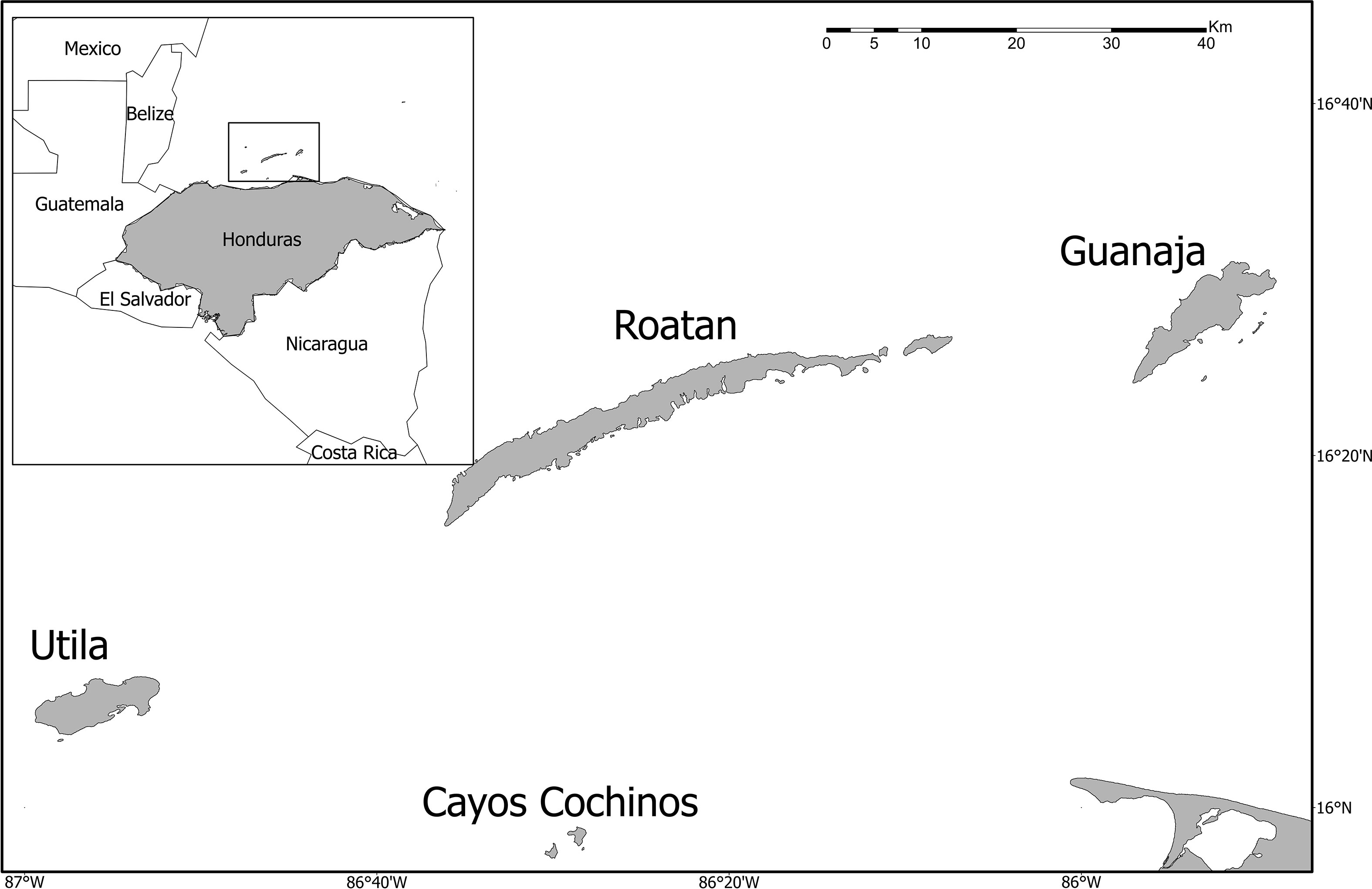
Figure 1 Map of the study area. Inset on the top left of the map displays the islands’ location with reference to the Honduran mainland.
From January 2020 to November 2021, the presence of SCTLD was assessed in 48 sites across the Bay Islands, including Utila (11), Guanaja (10), Roatan (24), and Cayos Cochinos (3), using the rover diver methodology (Doyle and O’sullivan, 2019). In Roatan, sites were surveyed monthly (except for January to September 2020, when monitoring was conducted bimonthly), bimonthly in Utila, and during November, December 2020, and January, April, and June 2021 in Guanaja and August 2021 in Cayos Cochinos. A minimum of 4 SCUBA divers conducted 20-minute census swims and recorded the species and disease status of coral colonies with a size of >10cm present in a one-meter-wide band along their survey line. For each species, the divers tallied colonies that were (a) recently dead, (b) actively diseased, (c) colonies with signs of concern, and (d) healthy colonies. Species considered in the study corresponded to those known to be highly susceptible, moderately susceptible (i.e., intermediate susceptibility), and those that are believed to be susceptible but lack sufficient evidence (i.e., presumed susceptibility) based on the description in Doyle and O’sullivan, 2019. Photos were taken for unusual or unclear disease sightings. The data obtained through these surveys were used to determine the presence of the epizootic in the study sites and the number of affected colonies per site. SCTLD was considered present in sites with records of a minimum of three actively diseased or recently dead colonies, and absent where only healthy and/or colonies with signs of concern were reported. First detections were not always observed from scientific monitoring efforts. Data for sites of the first detection of SCTLD in each of the islands were retrieved from the open AGRRA database (Kramer, 2019) and considered in the detection map.
Data on factors potentially linked to disease spread were collected from online databases and analyzed for the study periods in the area. Online bleaching alerts were obtained from the Coral Reef Watch Program (NOAA Coral Reef Watch, 2020). Additionally, data on average current direction and speed were collected from the Global Ocean 1/12° Physics Analysis and Forecast updated and OSCAR (Ocean Surface Current Analysis Real-time) for the study period in Honduras and neighboring countries (ESR, 2009; Copernicus Marine Service, 2016).
The spatial-temporal progression of SCTLD was evaluated through comparisons of presence/absence maps over time. The collected data was divided into six periods of equal duration from January 2020 to November 2021, three months each, beginning with the first signs of SCTLD in the country. Disease spread was assessed based on the infection of new sites, the apparent direction of the disease, and the approximate average spread rate. The last variable was calculated by pooling the presence/absence data into three-month intervals, computing the distance between the latest infected site in one period and the nearest infected point in the previous period, and dividing it by a fixed duration of 90 days. Peaks for disease spread were calculated by selecting the data points above the third quartile. The Measure Tool in ArcGIS Pro was used to calculate distance (Version 2.9.0). Analysis of Variance (ANOVA) was used to investigate whether the average disease spread was different between the northeast and southwest coasts of Roatan. Normality and homoscedasticity were assessed using Shapiro Wilk and Levene Tests. All data analyses were done using R computing software (R Core Team, 2020).
In total, 48 monitoring sites were surveyed across the Bay Islands during 2020-2021. The average rate of disease spread was 155 m/day around Roatan and Utila, although there was significant variability in disease spread between the northeast and southwest coasts of Roatan (ANOVA; p = 0.02), with northern spread values double those in the south (229 m/day, 90 m/day, respectively). During the first monitoring period of 2020 (January-August), no evidence of diseased coral colonies was found at eight monitoring sites on the west, south, and east sides of Roatan. However, between September and November, the disease was detected for the first time on the island, specifically on September 24th at Flowers Bay (16.28725, -86.575223; Figure 2A). The disease initially spread radially to the closest locations around the outbreak site (∼ 4 km), following a contagion model. After three months, the disease was also present on the northern side of Roatan, with up to 366 colonies affected in a single site (Figure 2B). Over the next two three-month periods (December 2020-February 2021 and March-May 2021), the disease spread rapidly eastwards on the windward side of the island (North coast), with spread rates of around 286 and 130 m/day, respectively (Figure 2B, C). In contrast, on the leeward side (South coast), the disease progressed more slowly, with rates of 118 and 54 m/day, respectively (Figures 2B, C). By the June-August 2021 period (Figure 2D), the disease had already spread across the entire northern region of the island of Roatan, with affected sites displaying between 3 and 216 diseased colonies. Additionally, in June 2021, SCTLD was first detected on the islands of Utila and Guanaja. The first detection of the disease in Guanaja was reported on June 13th on the west side of the island at the Eel Garden monitoring site (16.47043333, -85.9203). In Utila, SCTLD was first observed on June 28th at the Whale Rock dive site (16.12858, -86.88339). Also, according to the AGRRA data the disease was also first sited in Cayos Cochinos at the Roatan Banks dive site (16.064094, -86.500413) in August 2021. During the subsequent three months from September to November 2021, we observed a rapid progression of the disease in Utila. The number of affected colonies during this period fluctuated between 6 and 629. This trend mirrored our earlier observations in Roatan, ultimately leaving only one of the eleven monitoring sites in Utila unaffected by the disease. Furthermore, during this period, the last healthy sites on Roatan also became infected, resulting in the disease being present throughout the entire island by November 2021, infecting 28 species. No further records of the disease were found on the rest of the archipelago during the study period. A list of the affected species in each island is presented in Table 1.
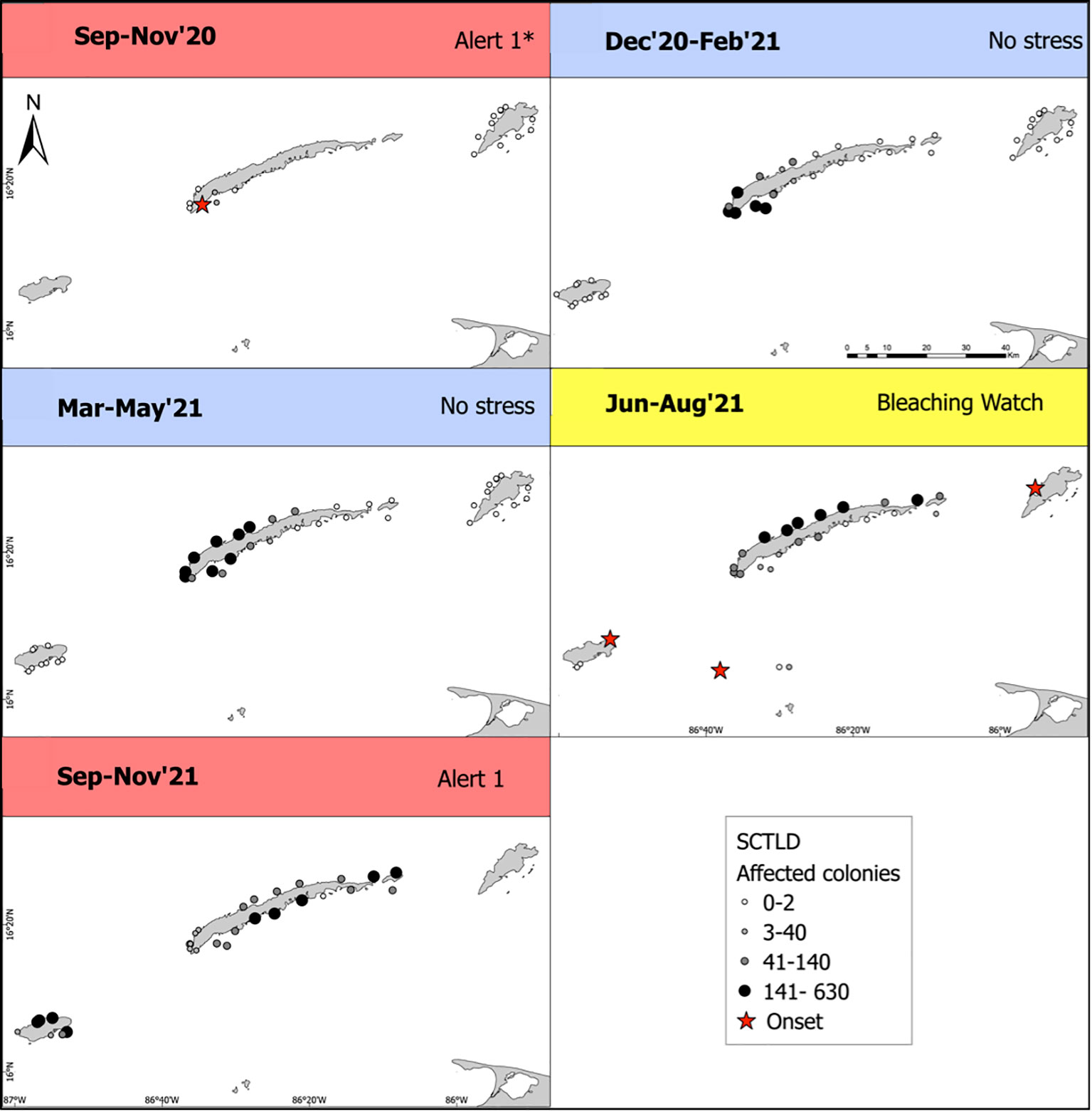
Figure 2 Map depicting the spatial spread of stony coral tissue loss disease (SCTLD) across the Bay Islands from September 2020 through November 2021. Each dot’s size and color represent the count of infected colonies at that specific location, with the red star denoting the initial appearance of the disease on each island. Though our methodology did not identify any affected sites on Guanaja during the study period, the onset of the disease is marked according to AGRRA data. The color bars at the top of each panel correspond to the Bleaching Alert levels for Honduras, as monitored by CRW’s Regional Virtual Station: blue indicates no stress, yellow represents a bleaching watch, and red signifies alert level 1.
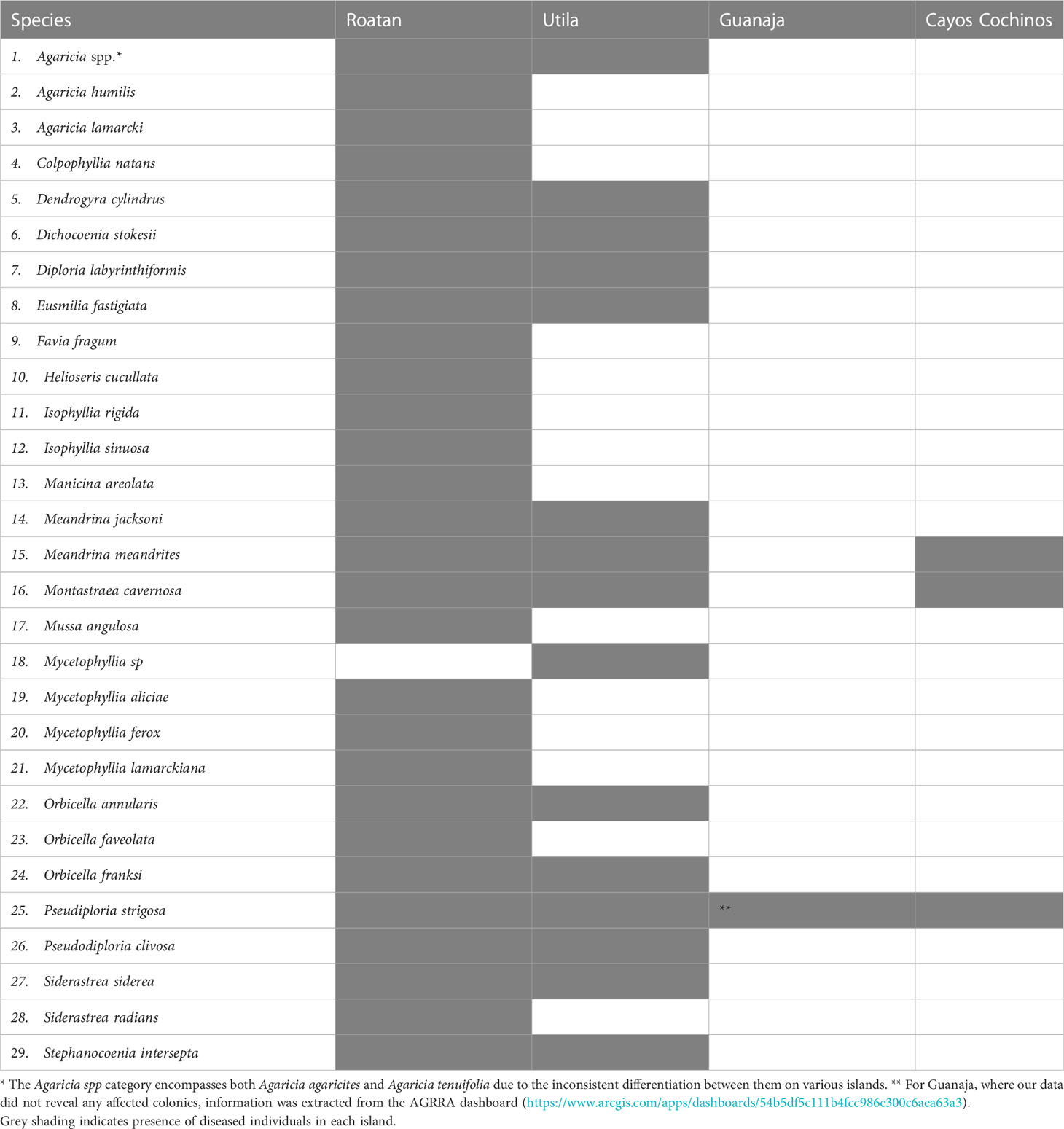
Table 1 Species affected by SCTLD on each island in the Honduran Caribbean from January 2020 to November 2021.
In addition to tracking the progression of the disease, the study also briefly examined two physical factors that have been suggested as potential links to the disease, thermal stress and ocean currents (Precht et al., 2016; Dobbelaere et al., 2020). Water temperatures on Roatan exceeded the bleaching threshold (one degree above the maximum monthly mean (MMM)), reaching category Alert Level 1 during the month of the disease outbreak and the subsequent month (September and October 2020) according to Coral Reef Watch (NOAA Coral Reef Watch, 2020). The first detections on Utila, Guanaja, and Cayos Cochinos were also reported during a period of temperature stress, although the bleaching alert level was lower than predicted in the succeeding months (September to November 2021) (Figure 2). On the other hand, there was no warning of bleaching and no thermal stress during the rest of the study period. Even though all outbreaks in the Bay Islands occurred during the dry season, April to September in Roatan (Mehrtens et al., 2001) peaks of disease spread were observed in both warm and cold months.
Focusing on the hydrology between Honduras and Belize (the closest country reported with SCTLD) (Kramer, 2019), from June 2019 (first detection in Belize) to September 2020, currents generally flowed weakly northward and towards the shore at velocities around 0.37 and 0.57 m/s (ESR, 2009). On a smaller scale, in the Bay Islands, the average trend in surface current direction for the six months prior to the first diagnosis of the disease on Utila was an east-to-west (Copernicus Marine Service, 2016) flow with mean velocities of approximately 0.01 meters per second during the months before the outbreak, following the direction of the prevalent winds in the Bay Islands (Southeast Trade Winds).
Over the course of less than a year and a half, the coral reefs in the Bay Islands had already suffered a rapid spread of stony coral tissue loss disease (SCTLD) at an approximate rate of 155 m/day, which had already affected more than 40 coral reef sites and at least 28 scleractinian species by the end of November 2021. The affected species and symptoms are consistent with previous reports in the Caribbean (Precht et al., 2016; Florida Coral Disease Response Research & Epidemiology Team, 2018; Alvarez-Filip et al., 2019; Gintert et al., 2019; Sharp et al., 2020). The islands with the fastest and most extensive spread of SCTLD, throughout the entire coastline by the end of the study period were Utila and Roatan. The fast progression of the disease in the study area in a short period of time (13 months) is similar to what has been described in other Caribbean regions (Alvarez-Filip et al., 2019) and slightly faster than in Florida (92 – 100 m/day; Muller et al., 2020). The spread in Roatan follows a contagion model, where the nearest neighbors are most susceptible to infection. However, there were differences in the speed of the disease spread between the northeast and southwest of Roatan.
Our findings suggest that the disease spread more rapidly on the windward side of the island than on the leeward side. This observation aligns with the findings of Alvarez-Filip et al. (2022), who also reported a greater disease impact in areas with higher exposure to wind. Despite several studies being published since the initial detection of SCTLD in Florida, there are still gaps in our knowledge of this novel disease, particularly regarding the main drivers affecting the disease’s spread at a local scale. The possible role of thermal stress, ocean currents, and human-based transmission on the distribution and occurrence of SCTLD in the Bay Islands are examined below.
Thermal stress and increased sedimentation have been associated with the initial SCTLD outbreak (Precht et al., 2016; Gintert et al., 2019) but are also negatively correlated with disease prevalence and severity (Meiling et al., 2020). In the Bay Islands, all initial detections of SCTLD in each island coincided with seawater temperatures exceeding the maximum monthly mean, but no overlap was found between the hottest months and the peak rates of the disease. Despite these observations suggesting a co-occurrence of heat stress, further assessments are necessary to confirm it as a triggering factor for SCTLD in Honduras.
According to studies such as Precht et al. (2016); Muller et al. (2020) and Dobbelaere et al. (2022), ocean currents played a significant part in the disease’s spread in Florida, but this has yet to be corroborated for Honduras. The observed current tendencies could be attributed to the presence of the Honduras Gyre, which dominates the flow in this region (Carrillo et al., 2015). This gyre likely hindered disease transmission between Honduras and Belize, which would explain the temporal differences between outbreaks in neighboring countries (Belize: June 2019, Honduras: September 2020) and the lack of SCTLD on the Honduran coast by the end of the study period. At the Bay Islands scale, considering Utila’s location in relation to Roatan and the direction of surface currents, the first observation of SCTLD coincides with the area where the disease would be expected to arrive first. However, based on the current speed during the months before the outbreak, SCTLD would be expected to reach Utila sooner than it did (Copernicus Marine Service, 2016). This is consistent with the results in Muller et al. (2020), which suggest that surface currents are too fast for the disease to settle in the area, and that bottom advection may play a more significant role. SCTLD did not strike Guanaja until two months after it reached the north-eastern area of Roatan, which is closest to Guanaja, implying that water currents between islands could serve as a potential vector. To investigate this hypothesis, a comprehensive study on the oceanographic dynamics of the Caribbean and the disease’s occurrence would be necessary.
Although to the best of our knowledge no evidence has yet been published on vessels as vectors of the disease via ballast water, it is a hypothesis that has been put forward by some authors including Rosenau et al. (2021) and Studivan et al. (2022). In the case of Honduras, if ocean currents were the only driver for disease emergence and spread, we would have expected to find disease occurrence in the rest of the archipelago cays and Guanaja. Thus, other factors could have played a role as well. Various local stakeholders have speculated that the origin of the disease in the Cayos Cochinos Archipelago is not solely due to ocean currents but also to dive tourism. Roatan Banks is a popular destination for Roatan dive centers; therefore, it is possible that exposed equipment and/or infected ballast water caused the disease to spread to that site. This possibility was also raised by Estrada-Saldívar et al. (2021), who suggested that the disease in SW Cozumel could have been brought by divers from the mainland. The transmission associated with the dive sector may potentially explain the observed disparities in disease transmission rates between the northwest and southeast of the island of Roatan, as these two areas show clear differences in terms of dive site density (Roatan Marine Park International Inc, n.d.). Additionally, the Bay Islands Management Plan (SAFEGE, SOGREAH, and MONCADA&MONCADA, 2002) emphasizes the significant West-South/North-East imbalance in Roatan’s urbanization and economic activities, which results in a concentration of pressure on natural resources and reefs. Residential growth and the vast construction of new roads and trails in the upper watersheds are identified as key drivers of erosion and sedimentation in the lower parts of the island (SAFEGE, SOGREAH, and MONCADA&MONCADA, 2002). Poor water quality, increased sedimentation, turbidity, and eutrophication are known factors that can cause a stress response on corals, promoting an increase in the frequency and intensity of disease outbreaks ((Richardson, 1998; Vargas-Ángel et al., 2006; Kaczmarsky and Richardson, 2011; Pollock et al., 2014; Vega Thurber et al., 2014; Dougan et al., 2020). Furthermore, the first recorded SCTLD occurrence in Honduras is in close proximity to the largest port in the Bay Islands, where cruise ships dock. Evans et al. (2022) have proposed that biofilms associated to ships could potentially serve as vectors for SCTLD. This notion is further supported by Lee Hing et al. (2022) study on management actions in Belize and Honduras. Their work highlighted a possible link between anthropogenic pressures and the spread of the disease in Roatan, suggesting that ship-associated biofilms might act as carriers for the disease.
As of May 2023, Cayos Cochinos was the site least impacted by SCTLD in the Bay Islands, considering the number of sites with actively diseased colonies. Unsurprisingly, it is also the most isolated island with lower tourism, marine traffic, and overall anthropogenic pressure (Gombos et al., 2011; Inypsa and STEREOCARTO, 2012). Thus, the last coral reef sites to be impacted, as well as those that have not yet been impacted, are areas with reduced local threats, which may make them less vulnerable to the disease. This hypothesis is in line with the hypothesis in Alvarez-Filip et al. (2022) where coastal development was linked to disease prevalence.
In this study, we have summarized all the available data on SCTLD records in Honduras up to November 2021. However, it is important to note that comprehensive monitoring efforts are currently only being carried out in the Bay Islands, and not in the North Coast. Nevertheless, local stakeholders and dive tourism operators in the North Coast have been trained on the disease and have yet to officially report any cases. Moreover, the SCTLD sighting in Guanaja occurred in an area that is not frequently monitored; thus it was not possible to track its spread. However, according to the AGRRA database (Kramer, 2019), SCTLD did spread across Guanaja outside our study period.
The pervasive influence of SCTLD on Caribbean coral reefs is indisputable. In this study, we have documented the rapid spread of SCTLD throughout the majority of the Honduran Bay Islands, while also discussing plausible contributing factors. However, further research is needed to better understand the relationships between SCTLD and factors such as water quality, ocean currents, recreational diving, and marine traffic in the study area.
Considering the interconnected nature of the Honduran Caribbean with the broader MBRS and its dependence on tourism for economic sustenance, it is essential to evaluate the impacts of SCTLD and the efficacy of mitigation strategies implemented in the region. These evaluations can offer valuable insights for other Caribbean nations that have yet to experience the detrimental effects of SCTLD. By understanding and addressing the challenges posed by SCTLD, we can work towards preserving the integrity and resilience of these invaluable coral reef ecosystems for future generations.
The raw data supporting the conclusions of this article will be made available by the authors upon request.
MT analyzed and interpreted the data, produced maps and drafted all sections of the manuscript. AR developed the study design and provided oversight and assistance in the overall development of the manuscript. Funding was procured by BG, GB, GO and ZZ. Monitoring design elaborated by CC, GB, GO and ZZ. BG, CC, DD, GB, LZ, ZG and ZZ conducted fieldwork and/or provided data. Finally, AR, BG, DD, GO and ZG revised the manuscript. All authors contributed to the article and approved the submitted version.
The data collected for this analysis was funded by MPA Connect (GCFI_RMP_2019), MARFund (HN 12-023), the Swiss Cooperation (no 81067342), the Honduran protected areas fund (FAPVS for its initials in Spanish), Tourism Free Zone (ZOLITUR for its initials in Spanish), Japan International Cooperation Agency (JICA) and the Summit Foundation.
Thanks to all the staff members and associated volunteers from RMP, BICA, HCRF, and UMA for their intense and dedicated work in the conservation of coral reefs in BINMP. This study would not have been possible without their contributions. We would also like to thank Helen Fox for reviewing the manuscript. This work is part of the Coral Reef Alliance’s Resilient Communities Strategy.
The authors declare that the research was conducted in the absence of any commercial or financial relationships that could be construed as a potential conflict of interest.
All claims expressed in this article are solely those of the authors and do not necessarily represent those of their affiliated organizations, or those of the publisher, the editors and the reviewers. Any product that may be evaluated in this article, or claim that may be made by its manufacturer, is not guaranteed or endorsed by the publisher.
Aeby G. S., Ushijima B., Campbell J. E., Jones S., Williams G. J., Meyer J. L., et al. (2019). Pathogenesis of a tissue loss disease affecting multiple species of corals along the Florida reef tract. Front. Mar. Sci. 6. doi: 10.3389/fmars.2019.00678
Almada-Villela P. C., Gold-Bouchot G., Kjerfve B. (2003). “Mesoamerican barrier reef systems project: manual of method for the MBRS synoptic monitoring program,” in Selected methods for monitoring physical and biological parameters for use in the mesoamerican region (Belize: Mesoamerican Barrier Reef Systems Project (MBRS)). Available at: https://www.researchgate.net/publication/279685258.
Alvarez-Filip L., Estrada-Saldívar N., Pérez-Cervantes E., Molina-Hernández A., González-Barrios F. J. (2019). A rapid spread of the stony coral tissue loss disease outbreak in the Mexican Caribbean. PeerJ 7, 8–10. doi: 10.7717/peerj.8069
Alvarez-Filip L., González-Barrios F. J., Pérez-Cervantes E., Molina-Hernández A., Estrada-Saldívar N. (2022). Stony coral tissue loss disease decimated Caribbean coral populations and reshaped reef functionality. Commun. Biol. 5, 440. doi: 10.1038/s42003-022-03398-6
Becker C. C., Brandt M., Miller C. A., Apprill A. (2022). Microbial bioindicators of stony coral tissue loss disease identified in corals and overlying waters using a rapid field-based sequencing approach. Environ. Microbiol. 24, 1166–1182. doi: 10.1111/1462-2920.15718
Carrillo L., Johns E. M., Smith R. H., Lamkin J. T., Largier J. L. (2015). Pathways and hydrography in the mesoamerican barrier reef system part 1: circulation. Cont Shelf Res 109, 164–176. doi: 10.1016/j.csr.2015.09.014.
Cavada-Blanco F., Croquer A., Vermeij M., Goergen L., Rodríguez-Martínez R. (2022). Dendrogyra cylindrus. The IUCN Red List Threatened Species. 2022, e.T133124A129721366. (Accessed on 23 June 2023).
Chollett I., Garavelli L., Holstein D., Cherubin L., Fulton S., Box S. J. (2017). A case for redefining the boundaries of the mesoamerican reef ecoregion. Coral Reefs 36, 1039–1046. doi: 10.1007/s00338-017-1595-4
Copernicus Marine Service (2016). Global ocean 1/12°C physics analysis and forecast updated daily. doi: 10.48670/moi-00016
Costa S. V., Hibberts S. J., Olive D. A., Budd K. A., Long A. E., Meiling S. S., et al. (2021). Diversity and disease: the effects of coral diversity on prevalence and impacts of stony coral tissue loss disease in saint Thomas, U.S. virgin islands. Front. Mar. Sci. 8. doi: 10.3389/fmars.2021.682688
Dobbelaere T., Muller E. M., Gramer L. J., Holstein D. M., Hanert E. (2020). Coupled epidemio-hydrodynamic modeling to understand the spread of a deadly coral disease in Florida. Front. Mar. Sci. 7. doi: 10.3389/fmars.2020.591881
Dobbelaere T., Holstein D. M., Muller E. M., Gramer L. J., McEachron L., Williams S. D., et al. (2022). Connecting the dots: transmission of stony coral tissue loss disease from the marquesas to the dry tortugas. Front Mar Sci 9. doi: 10.3389/fmars.2022.778938.
Doiron S., Weissenberger S. (2014). Sustainable dive tourism: social and environmental impacts — the case of roatan, Honduras. Tour Manag Perspect. 10, 19–26. doi: 10.1016/j.tmp.2013.12.003
Dougan K. E., Ladd M. C., Fuchs C., Vega Thurber R., Burkepile D. E., Rodriguez-Lanetty M. (2020). Nutrient pollution and predation differentially affect innate immune pathways in the coral porites porites. Front. Mar. Sci. 7. doi: 10.3389/fmars.2020.563865
Doyle E., O’sullivan C. (2019). Stony coral tissue loss disease template monitoring and response action plan for Caribbean marine natural resource managers (Florida: Key West).
Estrada-Saldívar N., Quiroga-García B. A., Pérez-Cervantes E., Rivera-Garibay O. O., Alvarez-Filip L. (2021). Effects of the stony coral tissue loss disease outbreak on coral communities and the benthic composition of cozumel reefs. Front. Mar. Sci. 8. doi: 10.3389/fmars.2021.632777
Evans J. S., Paul V. J., Kellogg C. A. (2022). Biofilms as potential reservoirs of stony coral tissue loss disease. Front. Mar. Sci. 9. doi: 10.3389/fmars.2022.1009407
Florida Coral Disease Response Research & Epidemiology Team (2018) Case definition: stony coral tissue loss disease. Available at: https://floridadep.gov/sites/default/files/Copy%20of%20StonyCoralTissueLossDisease_CaseDefinition%20final%2010022018.pdf.
Gintert B. E., Precht W. F., Fura R., Rogers K., Rice M., Precht L. L., et al. (2019). Regional coral disease outbreak overwhelms impacts from a local dredge project. Environ. Monit Assess. 191, 1–39. doi: 10.1007/s10661-019-7767-7
Gombos M., Arrivillaga A., Wusinich-Mendez D., Glazer B., Frew S., Bustamante G., et al. (2011) A management capacity assessment of selected coral reef marine protected areas in the Caribbean. Available at: http://coralreef.noaa.gov/resources/publicationsdata/.
Harborne A. R., Afzal D. C., Andrews M. J. (2000). Honduras: Caribbean Coast. Mar. pollut. Bull. 42, 1221–1235. doi: 10.1016/S0025-326X(01)00239-9
Heres M. M., Farmer B. H., Elmer F., Hertler H. (2021). Ecological consequences of stony coral tissue loss disease in the Turks and Caicos islands. Coral Reefs 40, 609–624. doi: 10.1007/s00338-021-02071-4
Huntley N., Brandt M. E., Becker C. C., Miller C. A., Meiling S. S., Correa A. M. S., et al. (2022). Experimental transmission of stony coral tissue loss disease results in differential microbial responses within coral mucus and tissue. ISME Commun. 2, 46. doi: 10.1038/s43705-022-00126-3
Inypsa and STEREOCARTO (2012). Informe del estado ambiental de las islas de la bahía. (Honduras: Instituto Hondureño de Turismo). Available at: https://angelarandazzoeisemann.files.wordpress.com/2014/02/2012-informe-del-estado-ambiental-de-las-islas-de-la-bahia.pdf.
Kaczmarsky L., Richardson L. L. (2011). Do elevated nutrients and organic carbon on Philippine reefs increase the prevalence of coral disease? Coral Reefs 30, 253–257. doi: 10.1007/s00338-010-0686-2
Kramer P. R. (2019) Map of stony coral tissue loss disease outbreak in the Caribbean. Available at: www.agrra.org (Accessed March 10, 2022).
Lee Hing C., Guifarro Z., Dueñas D., Ochoa G., Nunez A., Forman K., et al. (2022). Management responses in Belize and Honduras, as stony coral tissue loss disease expands its prevalence in the mesoamerican reef. Front. Mar. Sci. 9. doi: 10.3389/fmars.2022.883062
McField M., Kramer P., Giró Petersen A., Soto M., Drysdale I., Craig N., et al. (2020). 2020 mesoamerican reef report card. (Florida: Healthy Reefs Initiative). Available at: https://www.healthyreefs.org/cms/wp-content/uploads/2020/02/2020_Report_Card_MAR.pdf.
Mehrtens C., Rosenheim B., Young R., Modley M. (2001). Reef morphology and sediment attributes, Roatan, Bay Islands, Honduras. Carbonates Evaporites 16, 131–140. doi: 10.1007/BF03175831
Meiling S., Muller E. M., Smith T. B., Brandt M. E. (2020). 3D photogrammetry reveals dynamics of stony coral tissue loss disease (SCTLD) lesion progression across a thermal stress event. Front. Mar. Sci. 7. doi: 10.3389/fmars.2020.597643
Muller E. M., Sartor C., Alcaraz N. I., van Woesik R. (2020). Spatial epidemiology of the stony-Coral-Tissue-Loss disease in Florida. Front. Mar. Sci. 7. doi: 10.3389/fmars.2020.00163
Neely K. L., Lewis C. L., Lunz K. S., Kabay L. (2021). Rapid population decline of the pillar coral dendrogyra cylindrus along the Florida reef tract. Front. Mar. Sci. 8. doi: 10.3389/fmars.2021.656515
NOAA Coral Reef Watch (2020). NOAA Coral reef watch version 3.1 daily 5km satellite regional virtual station time series data for honduras. updated daily (Maryland, USA: College Park).
Pollock F. J., Lamb J. B., Field S. N., Heron S. F., Schaffelke B., Shedrawi G., et al. (2014). Sediment and turbidity associated with offshore dredging increase coral disease prevalence on nearby reefs. PloS One 9, 5–8. doi: 10.1371/journal.pone.0102498
Precht W. (2021). Failure to respond to a coral disease epizootic in Florida: causes and consequences. Rethinking Ecol. 6, 1–47. doi: 10.3897/RETHINKINGECOLOGY.6.56285
Precht W. F., Gintert B. E., Robbart M. L., Fura R., Van Woesik R. (2016). Unprecedented disease-related coral mortality in southeastern Florida. Sci. Rep. 6, 1–9. doi: 10.1038/srep31374
R Core Team (2020) R: a language and environment for statistical computing. Available at: https://www.R-project.org/.
Richardson L. L. (1998). Coral diseases: what is really known? Trends Ecol. Evol. 13, 438–443. doi: 10.1016/S0169-5347(98)01460-8
Roatan Marine Park International Inc Roatan marine park. dive site map. Available at: https://www.roatanmarinepark.org/resources (Accessed June 8, 2022).
Rosenau N. A., Gignoux-Wolfsohn S., Everett R. A., Miller A. W., Minton M. S., Ruiz G. M. (2021). Considering commercial vessels as potential vectors of stony coral tissue loss disease. Front. Mar. Sci. 8. doi: 10.3389/fmars.2021.709764
SAFEGE, SOGREAH, and MONCADA&MONCADA (2002). Esquema director de manejo ambiental sostenible para las islas de la bahía (Honduras: PROYECTO MANEJO AMBIENTALDE LAS ISLAS DE LA BAHIA (PMAIB)).
Sharp W. C., Shea C. P., Maxwell K. E., Muller E. M., Hunt J. H. (2020). Evaluating the small-scale epidemiology of the stony-coral -tissue-loss-disease in the middle Florida keys. PloS One 15, 1–11. doi: 10.1371/journal.pone.0241871
Studivan M. S., Baptist M., Molina V., Riley S., First M., Soderberg N., et al. (2022). Transmission of stony coral tissue loss disease (SCTLD) in simulated ballast water confirms the potential for ship-born spread. Sci. Rep. 12, 19248. doi: 10.1038/s41598-022-21868-z
U.S. Fish & Wildlife Service (2014). Endangered and threatened wildlife and plants; adding 20 coral species to the list of endangered and threatened wildlife. Fed Regist. 79, 67356–67359.
Ushijima B., Meyer J. L., Thompson S., Pitts K., Marusich M. F., Tittl J., et al. (2020). Disease diagnostics and potential coinfections by vibrio coralliilyticus during an ongoing coral disease outbreak in Florida. Front. Microbiol. 11. doi: 10.3389/fmicb.2020.569354
Vargas-Ángel B., Riegl B., Gilliam D. S., Dodge R. E. (2006) An experimental histopathological rating scale of sedimentation stress in the Caribbean coral montastraea cavernosa NSUWorks citation. Available at: https://nsuworks.nova.edu/occ_facpresentations.
Vega Thurber R. L., Burkepile D. E., Fuchs C., Shantz A. A., Mcminds R., Zaneveld J. R. (2014). Chronic nutrient enrichment increases prevalence and severity of coral disease and bleaching. Glob Chang Biol. 20, 544–554. doi: 10.1111/gcb.12450
Walton C. J., Hayes N. K., Gilliam D. S. (2018). Impacts of a regional, multi-year, multi-species coral disease outbreak in southeast Florida. Front. Mar. Sci. 5. doi: 10.3389/fmars.2018.00323
Keywords: Stony coral tissue loss disease, Honduras, Scleractinia, coral reef monitoring, coral disease outbreak
Citation: Truc M, Rivera A, Ochoa GM, Dueñas D, Guifarro Z, Brady G, Zúniga Z, Gutiérrez B, Chock C and Zaldivar L (2023) Evaluating the spread of stony coral tissue loss disease in the Bay Islands, Honduras. Front. Mar. Sci. 10:1197318. doi: 10.3389/fmars.2023.1197318
Received: 30 March 2023; Accepted: 12 June 2023;
Published: 21 July 2023.
Edited by:
William F. Precht, Dial Cordy and Associates, Inc., United StatesReviewed by:
Nuria Estrada-Saldívar, National Autonomous University of Mexico, MexicoCopyright © 2023 Truc, Rivera, Ochoa, Dueñas, Guifarro, Brady, Zúniga, Gutiérrez, Chock and Zaldivar. This is an open-access article distributed under the terms of the Creative Commons Attribution License (CC BY). The use, distribution or reproduction in other forums is permitted, provided the original author(s) and the copyright owner(s) are credited and that the original publication in this journal is cited, in accordance with accepted academic practice. No use, distribution or reproduction is permitted which does not comply with these terms.
*Correspondence: Mar Truc, bWFyLnRydWMudG9tYXNAZ21haWwuY29t; Antonella Rivera, YXJpdmVyYUBjb3JhbC5vcmc=
Disclaimer: All claims expressed in this article are solely those of the authors and do not necessarily represent those of their affiliated organizations, or those of the publisher, the editors and the reviewers. Any product that may be evaluated in this article or claim that may be made by its manufacturer is not guaranteed or endorsed by the publisher.
Research integrity at Frontiers
Learn more about the work of our research integrity team to safeguard the quality of each article we publish.