 Edgar Mauricio Hoyos-Padilla1,2*
 Irene Casanova-Santamaría1,3Jorge Carlos Loria-Correa4
 James Sulikowski5*†
- 1Pelagios Kakunjá, La Paz, Mexico
- 2Fins Attached, Colorado Springs, Colorado, CO, United States
- 3Centro Interdisciplinario de Ciencias Marinas, La Paz, Mexico
- 4Saving Our Sharks A.C., Playa del Carmen, Mexico
- 5School of Mathematical & Natural Sciences, Arizona State University, Tempe, Arizona, AZ, United States
A prototype, fully submersible, high definition ultrasound was used to determine the reproductive state of wild, free-swimming bull sharks, Carcharhinus leucas, at a provisioned shark diving site in Playa del Carmen, Mexico. During two opportunistic dives, the presence of embryos was confirmed in three female sharks (greater than 2.0 m total length) and emphasizes the importance of developing and linking emerging technologies with shark diving sites for the conservation of elasmobranch species.
1 Introduction
Diving with sharks has become popular throughout the world in recent decades (Gallagher and Hammerschlag, 2011) and in Mexico has been suggested to contribute approximately 12.4 million in United States dollars (USD) per year to local economies (Cisneros-Montemayor et al., 2013). In addition to an economic importance, observational studies at such shark tourism sites can also provide fishery-independent scientific information to improve population level assessments for certain species (Clua et al., 2010), as not only do these sharks often form seasonal aggregations, they also can be individually identified (Photo-ID; Pierce et al., 2018) and potentially monitored over multiple years. The next stage in the evolution of linking this type of tourism to scientific data collection, is the ability to characterize life history stages to the observed animals.
One particular life history stage that is critical for establishing site-based conservation strategies, such as marine protected areas or time/area closures is an understanding of where gravid sharks spend their time gestating/and or give birth (Chapman et al., 2013; Sulikowski et al., 2016). However, historically, such reproductive data was collected from sacrificed animals from fisheries, which is problematic for species that have been classified as threatened and in the case of shark diving tourism, can also have a negative socioeconomic impact (Hammerschlag and Sulikowski, 2011). Thus, new approaches to studying elasmobranchs increasingly include non-lethal sampling methods (Penfold and Wyffels, 2019). For example, the use of ultrasounds and short-term restraint for biological sampling has continued to advance this field of research in both wild caught and aquarium elasmobranchs (Carrier et al., 2003; Sulikowski et al., 2016, Murakumo et al., 2020). However, the use of ultrasound technology on free swimming sharks in the wild, has only been documented on one species (Murakumo et al., 2020), and has yet to be applied to sharks aggregating at dive sites.
To fill this knowledge gap, a prototype, submersible, high definition ultrasound developed by EI Medical (Inc.) was field tested on free swimming bull sharks, Carcharhinus leucas, within a provisioned dive site in the coast of Playa Del Carmen to determine if any of sharks were gravid at this location.
2 Materials and methods
Opportunistic sampling occurred during two, 20 min dives, approximately 1 km of the coast of Playa del Carmen, in Quintana Roo state, Mexico (Figure 1). Water depth was 24 m and the substrate comprised of sandy bottom. A fully submersible ultrasonography was performed on the abdominal region of free-swimming female bull sharks to assess reproductive stage (Figure 2). Here, a topside Ibex EVO III portable ultrasound (E.I. Medical Imaging) was redesigned and encased in waterproof housing (via proprietary specifications) for use at depths greater than 30 meters, herein termed the Aquanaut (Figure 3). The Aquanaut was equipped with a 60 mm curved linear array 2.5 to 5 MHz transducer (model eCL3) capable of a 30 cm scan depth which was connected to the unit with a 3 m cord. The Aquanaut recorded one continuous video until the dive ended. Bull sharks were fed by a professional shark handler with chunked, local fish species, and led single file so that the transducer could make contact as the sharks slowly swam by (Figure 2). Due to limited bottom dive time (20 min), the largest sharks estimated visually, were targeted for ultrasonography. In addition, due to the frequency of dives performed (two to three per day for four to five months over the course of a dive season) individual bull sharks were able to be identified by the dive operators. Once targeted, a second diver recorded a video of the scanning event for each shark as it moved away from the handler. Each video recording of the scanning event was time stamped and later coordinated with the internal clock on the ultrasound to link an image of the gravid female to her recorded embryos. Scanning was performed primarily on the lateral surface from the pectoral to the pelvic fin in either transverse or longitudinal orientation to obtain cross sectional and lengthwise images, respectively (Figure 2). Each scan lasted between one to three seconds. Collected video saved on the Aquanaut were used to create still images of observed embryos (Sulikowski and Hammerschlag, 2023). Stills from the video were then used to measure (via proprietary software pre-installed on the Aquanaut) pup diameter (cm) along the transverse axis (Sulikowski et al., 2016).
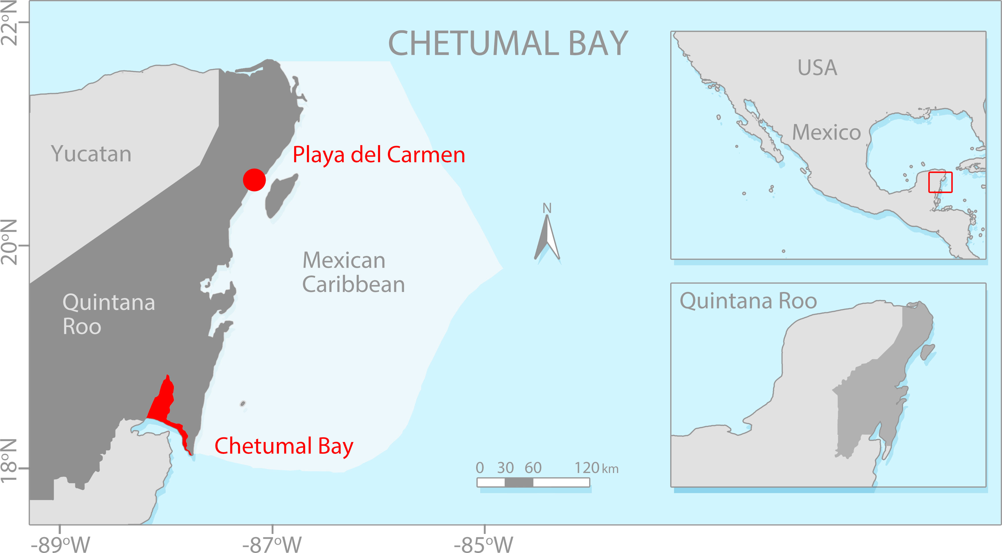
Figure 1 Location of the study area, Playa del Carmen, Quintana Roo, Mexico.
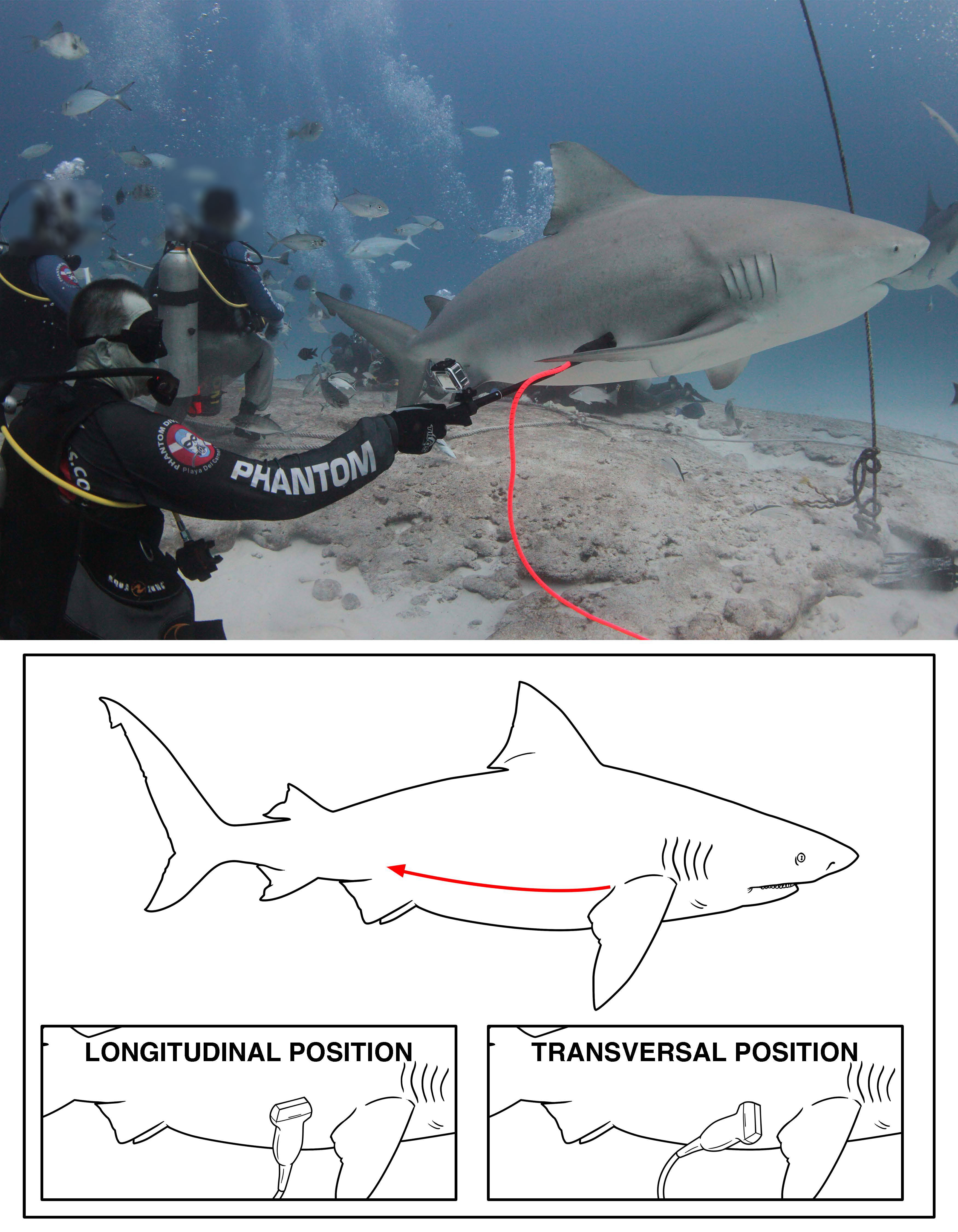
Figure 2 Representative distance from and lateral scanning of a pregnant bull shark. The Aquanaut underwater ultrasound was connected to 60 mm curved linear array 2.5 to 5 MHz transducer. Scanning produced either a cross section or lengthwise orientation of embryos within the uterus.
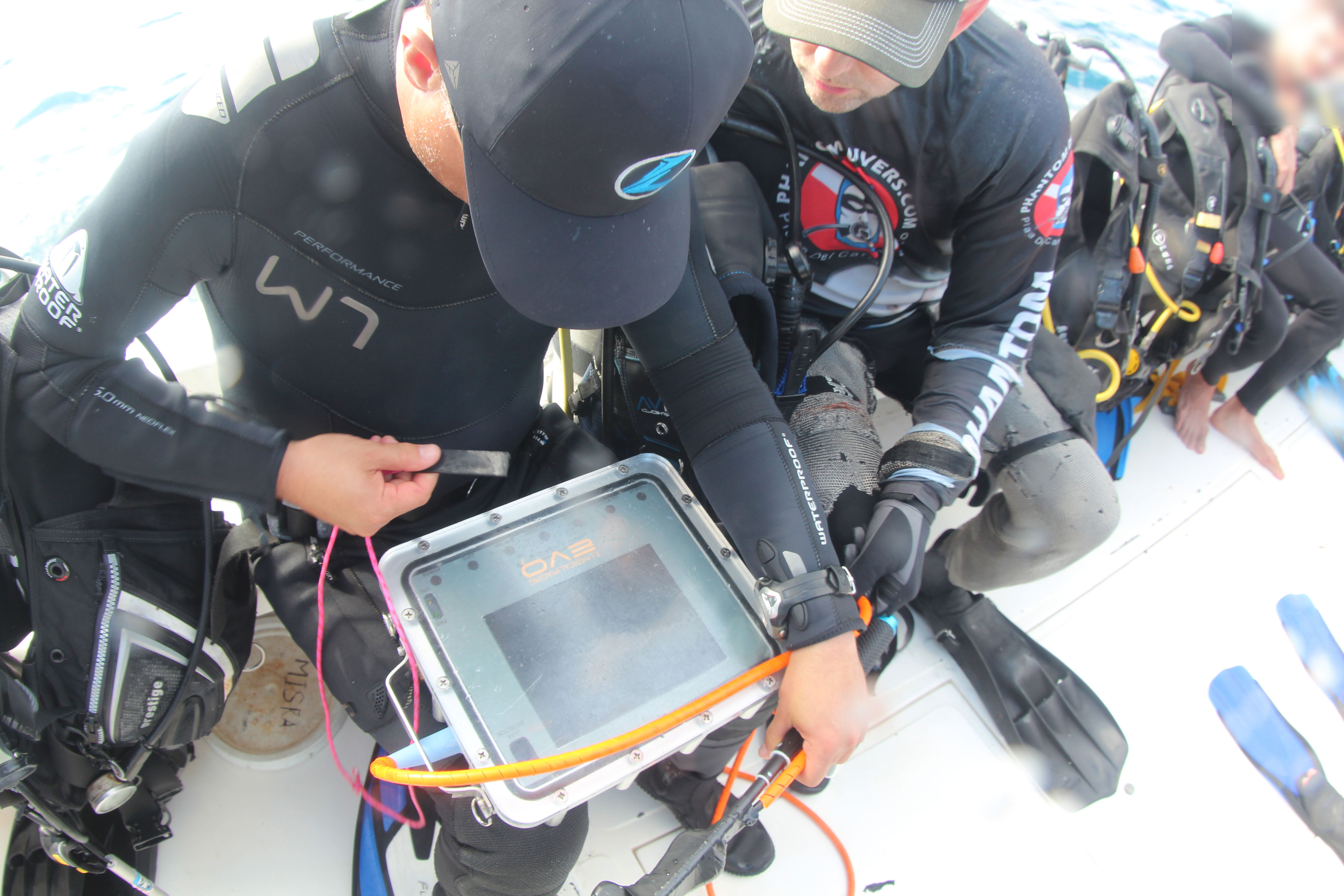
Figure 3 Topside Ibex EVO III Aquanaut portable ultrasound (EI Medical Imaging) encased in waterproof housing.
3 Results
Over the course of the two 20 minute dives, individual sharks made several passes and based on the analysis of the Aquanaut video, three sharks were identified to be gravid (Figure 4) by the presence of identifiable embryos within the uterus (Sulikowski and Hammerschlag, 2023). Proprietary software measurements from still images indicated the embryos measured were approximately 4 cm in circumference. While the exact location and orientation of the embryos could not be determined, given the short duration of transducer shark contact, the observed sizes are similar to those reported from a gravid female captured on November 22nd-2022 by local fishermen in Isla Mujeres, Quintana Roo (a nearby location to Playa del Carmen), during a fishing survey realized by Save Our Sharks staff.
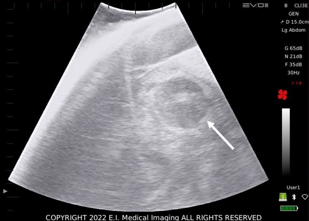
Figure 4 Representative transverse ultrasound images of bull shark Carcharhinus leucas obtained from gravid females with identified embryos in utero. Arrows point to the embryo.
4 Discussion
The use of ultrasonography as a non-invasive methodology has proven to be a useful tool for the study of reproductive aspects in both captive and wild caught sharks (Walsh et al., 1993; Daly et al., 2007; Sulikowski et al., 2016; Inoue et al., 2022; Santos et al., 2022; Sulikowski and Hammerschlag, 2023). However, the use of this technology adapted for use underwater on free swimming wild animals is scant, with only one published study to date on whale sharks (Matsumoto et al., 2023). The study herein adds to this limited body of work and shows the utility of this technology to identify gravid females in a provisioned dive site. The positive outcome has several implications for the preservation of specific life history stages for sharks and potentially other elasmobranchs. For example, it is well established that critical component of successful conservation of wild populations is an understanding of a species’ reproductive biology (Natanson et al., 2019) as it can assist in stock assessments thereby informing management decisions related to protected area designation (Awruch, 2013).
The identification of pregnant bull sharks in Playa del Carmen, suggests that this area may serve as a gestation ground and as such provides critical habitat for this life history stage. A similar phenomenon has also been observed in tiger sharks within Tiger Beach, a provisioned dive site within Bahamas (Sulikowski et al., 2016). Anecdotal evidence from Playa del Carmen dive operations suggest female bull sharks regularly migrate into the shallow waters of the Playa del Carmen beginning in November and stay until March when water temperatures are above 26°C (78.8°F). Bull sharks are absent from this area by April when waters are well above 26°C. The absence of bull sharks by April suggests that similar to tiger sharks at Tiger Beach (Sulikowski et al., 2016), environmental or ecological drivers are responsible for the observed movement out of the dive site (Chapman et al., 2013). For pregnant females observed herein, seeking a suitable habitat for parturition provides a likely explanation (Chapman et al., 2013). For example, Blanco-Parra et al. (2022) have recently demonstrated that Chetumal Bay (Figure 1) serves as a nursery area for bull sharks in the Mexican Caribbean and reported the presence of neonates beginning in May.
Whether the gravid bull sharks analyzed in this study travel to Chetumal Bay (approximately 360 km from Playa del Carmen) requires further investigation via other methodologies (Sulikowski and Hammerschlag, 2023). Regardless, the results of the current study emphasize the importance of developing and linking emerging technologies with ecotourist sites for the conservation of elasmobranch species.
Data availability statement
The raw data supporting the conclusions of this article will be made available by the authors, without undue reservation.
Ethics statement
The animal study was reviewed and approved by ASU Protocol Number: 23-1955R Protocol Title: Using innovative and non-lethal techniques to study the biology, ecology, and habitat utilization of sharks Institutional Animal Care and Use Committee (IACUC) Office of Research Integrity and Assurance Arizona State University.
Author contributions
EH-P and JS conceptualized the study. EH-P, IC-S, JL-C and JS contributed for the data collection and field work. EH-P, JL-C and JS look for funding acquisition. JS performed data analyses. EH-P, IC-S, JS wrote the original draft of the manuscript. All the authors reviewed and edited the manuscript. All the authors contributed to the article and approved the submitted version.
Funding
This research was funded by Saving Our Sharks Foundation, Saving Our Sharks A.C. and Ocean Blue Tree.
Acknowledgments
Field work was greatly facilitated through courtesies extended to us by personnel from Phantom Divers and E.I. Medical Imaging. This research was conducted under the Scientific Research Permit: Permiso de Pesca de Fomento No. DGOPA.08050.051011. with Folio Núm. 05421 issued by the Secretaría de Agricultura, Ganadería, Desarrollo Rural, Pesca y Alimentación (SAGARPA) through the Comisión Nacional de la Pesca. Special thanks to E.I. Medical Imaging for the design of the Aquanaut.
Conflict of interest
The authors declare that the research was conducted in the absence of any commercial or financial relationships that could be construed as a potential conflict of interest.
Publisher’s note
All claims expressed in this article are solely those of the authors and do not necessarily represent those of their affiliated organizations, or those of the publisher, the editors and the reviewers. Any product that may be evaluated in this article, or claim that may be made by its manufacturer, is not guaranteed or endorsed by the publisher.
References
Awruch C. A. (2013). Reproductive endocrinology in chondrichthyans: the present and the future. Gen. Comp. Endocrinol. 192, 60–70. doi: 10.1016/j.ygcen.2013.05.021
Blanco-Parra M. D. P., Sandoval-Laurrabaquio-Alvarado N., Díaz-Jaimes P., Niño-Torres C. A. (2022). Evidence of a nursery area for bull shark, Carcharhinus leucas (Müller y henle 1839) in the mesoamerican reef system region. Environ. Biol. Fishes 105, 1193–1202. doi: 10.1007/s10641-022-01338-1
Carrier J. C., Murru F. L., Walsh M. T., Pratt H. L. (2003). Assessing reproductive potential and gestation in nurse sharks (Ginglymostoma cirratum) using ultrasonography and endoscopy: an example of bridging the gap between field research and captive studies. Zoo Biol. 22, 179–187. doi: 10.1002/zoo.10088
Chapman D. D., Wintner S. P., Abercrombie D. L., Ashe J., Bernard A. M., Shivji M. S., et al. (2013). The behavioral and genetic mating system of the sand tiger shark. Biol. Lett. 9, 20130003–20130003. doi: 10.1098/rsbl.2013.0003
Cisneros-Montemayor A. M., Barnes-Mauthe M., Al-Abdulrazzak D., Navarro-Holm E., Sumaila U. R. (2013). Global economic value of shark ecotourism: implications for conservation. Oryx 47, 381–388. doi: 10.1017/S0030605312001718
Clua E., Buray N., Legendre P., Mourier J., Planes S. (2010). Behavioural response of sicklefin lemon sharks negaprion acutidens to underwater feeding for ecotourism purposes. Mar. Ecol. Prog. Ser. 414, 257–266. doi: 10.3354/meps08746
Daly J., Gunn I., Kirby N., Galloway D. (2007). Ultrasound examination and behavior scoring of captive broadnose sevengill sharks, Notorynchus cepedianus (Peron 1807). Zoo Biol. 26, 383–395. doi: 10.1002/zoo.20155
Gallagher A. J., Hammerschlag N. (2011). Global shark currency: the distribution, frequency, and economic value of shark ecotourism. Curr. Issues Tour 14, 797–812. doi: 10.1080/13683500.2011.585227
Hammerschlag N., Sulikowski J. (2011). Killing for conservation: the need for alternatives to lethal sampling of apex predatory sharks. endanger. Species Res. 14, 135–140. doi: 10.3354/esr00354
Inoue T., Shimoyama K., Saito M., Wong M. K. S., Ikeba K., Nozu R., et al. (2022). Long-term monitoring of egg-laying cycle using ultrasonography reveals the reproductive dynamics of circulating sex steroids in an oviparous catshark, Scyliorhinus torazame. Gen. Comp. Endocrinol. 327, 114076. doi: 10.1016/j.ygcen.2022.114076
Matsumoto R., Murakumo K., Nozu R., Acuña-Marrero D., Green J. R., Pierce S. J., et al. (2023). Underwater ultrasonography and blood sampling provide the first observations of reproductive biology in free-swimming whale sharks. Endanger. Species Res. 50, 125–131.
Murakumo K., Matsumoto R., Tomita T., Matsumoto Y., Ueda K. (2020). The power of ultrasound: observation of nearly the entire gestation and embryonic developmental process of captive reef manta rays (Mobula alfredi). Fish Bull. 118, 1–8. doi: 10.7755/fb.118.1.1
Natanson L. J., Deacy B. M., Joyce W., Sulikowski J. (2019). Presence of a resting population of female porbeagles (Lamna nasus), indicating a biennial reproductive cycle, in the western north Atlantic ocean. Fish Bull. 117, 8. doi: 10.7755/FB.117.1-2.8s
Penfold L. M., Wyffels J. T. (2019). Reproductive science in sharks and rays. Adv. Exp. Med. Biol. 1200, 465–488. doi: 10.1007/978-3-030-239633-5_15
Pierce S. J., Holmberg J. A., Kock A. L., Marshall A. D. (2018). “Photographic identification of sharks,” in Shark research: emerging technologies and applications for the field and laboratory. Eds. Carrier J. C., Heithaus M. R., Simpfendorfer C. A. (Boca Raton, FL: CRC Press), 220–234.
Santos S. R., Takatsuka V., Bonatelli S. P., Amaral N. L., Goés M. F., Valle R. F. (2022). Courtship and reproduction of the whitetip reef shark Triaenodon obesus (Carcharhiniformes: carcharhinidae) in an ex situ environment, with a description of the late embryonic developmental stage. Animals 12 (23), 3291. doi: 10.3390/ani12233291
Sulikowski J. A., Hammerschlag N. (2023). A novel intrauterine satellite transmitter to identify parturition in large sharks. Sci. Adv. 9, eadd6340. doi: 10.1126/sciadv.add6340
Sulikowski J. A., Wheeler C. R., Gallagher A. J., Prohaska B. K., Langan J. A., Hammerschlag N. (2016). Seasonal and life-stage variation in the reproductive ecology of a marine apex predator, the tiger shark Galeocerdo cuvier, at a protected female-dominated site. Aquat Biol. 24, 175–184. doi: 10.3354/ab00648
Keywords: emerging technologies, gestation ground, conservation, management, ecotourism
Citation: Hoyos-Padilla EM, Casanova-Santamaría I, Loria-Correa JC and Sulikowski J (2023) The successful use of a submersible ultrasound to confirm pregnancy on free swimming bull sharks, Carcharhinus leucas, in a provisioned shark site. Front. Mar. Sci. 10:1193563. doi: 10.3389/fmars.2023.1193563
Received: 25 March 2023; Accepted: 15 May 2023;
Published: 31 July 2023.
Edited by:
Elizabeth Grace Tunka Bengil, University of Kyrenia, CyprusReviewed by:
Eric Emile Germain Clua, USR3278 Centre de Recherche Insulaire et Observatoire de L’environnement (CRIOBE), FranceBrittany Finucci, National Institute of Water and Atmospheric Research, New Zealand
Copyright © 2023 Hoyos-Padilla, Casanova-Santamaría, Loria-Correa and Sulikowski. This is an open-access article distributed under the terms of the Creative Commons Attribution License (CC BY). The use, distribution or reproduction in other forums is permitted, provided the original author(s) and the copyright owner(s) are credited and that the original publication in this journal is cited, in accordance with accepted academic practice. No use, distribution or reproduction is permitted which does not comply with these terms.
*Correspondence: Edgar Mauricio Hoyos-Padilla, bWF1cmljaW9AcGVsYWdpb3NrYWt1bmphLm9yZw==; James Sulikowski, SmFtZXMuc3VsaWtvd3NraUBvcmVnb25zdGF0ZS5lZHU=
†Present address: James Sulikowski, Coastal Oregon Marine Experiment Station (COMES), Hatfield Marine Science Center, Oregon State University, Newport, OR, United States