
94% of researchers rate our articles as excellent or good
Learn more about the work of our research integrity team to safeguard the quality of each article we publish.
Find out more
CORRECTION article
Front. Immunol., 12 November 2024
Sec. Inflammation
Volume 15 - 2024 | https://doi.org/10.3389/fimmu.2024.1473483
 Chih-Wen Wang1,2†Hsin-Ying Clair Chiou3,4,5†Szu-Chia Chen2,6,7,8Da-Wei Wu2,9Hung-Hsun Lin3Huang-Chi Chen2,9Wei-Ting Liao7,10,11Ming-Hong Lin11,12,13*Chih-Hsing Hung7,14,15*Chao-Hung Kuo2,8,16*
A Corrigendum on
Arsenic exposure and lung fibrotic changes-evidence from a longitudinal cohort study and experimental models
By Wang C-W, Chiou H-YC, Chen S-C, Wu D-W, Lin H-H, Chen H-C, Liao W-T, Lin M-H, Hung C-H and Kuo C-H (2023). Front. Immunol. 14:1225348. doi: 10.3389/fimmu.2023.1225348
In the published article, there was an error in the Data Availability statement. “The raw data supporting the conclusions of this article will be made available by the authors, without undue reservation.” The correct Data Availability statement appears below.
“The datasets generated for this article are not readily available because of ethical restrictions. Requests to access the datasets should be directed to Dr. Chih-Wen Wang (e-mail:960405@kmuh.org.tw).”
Furthermore in the published article, there was an error in Figure 6B. The concentration of NaAsO2 should be labeled as 4 μM. The corrected Figure 6 appears below.
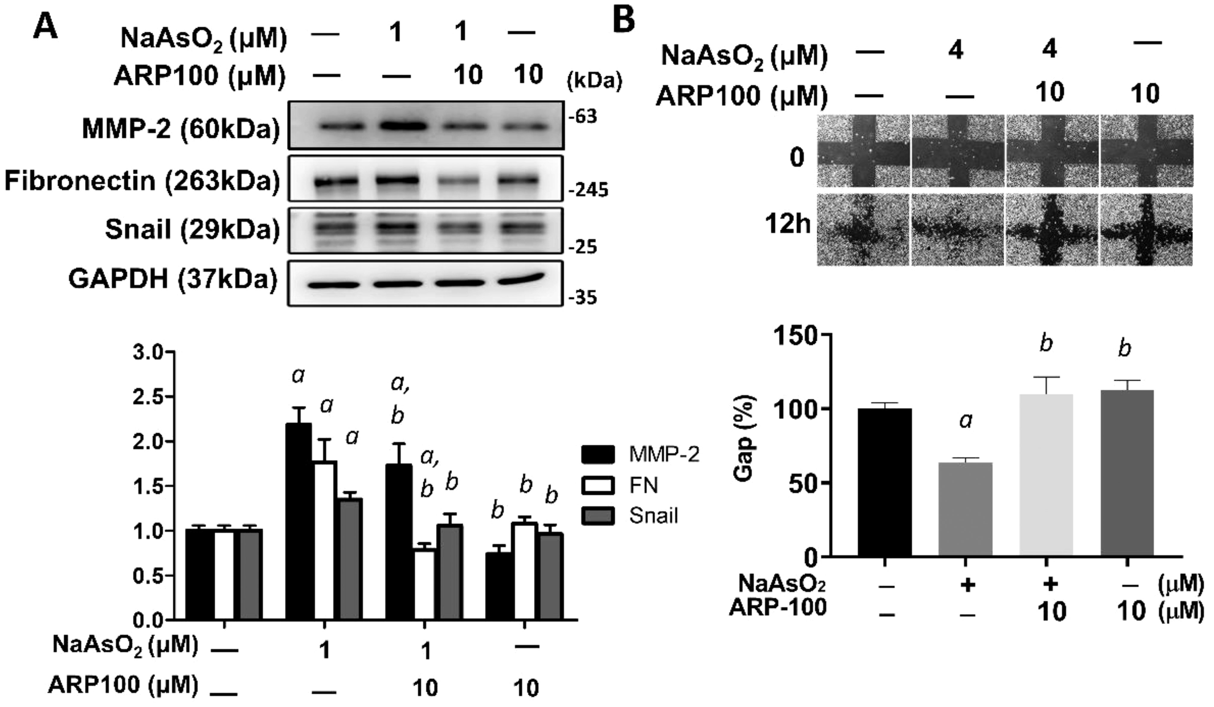
Figure 6. MMP-2 is critical for arsenic-induced EMT changes. (A) NHBE cells were pretreated with ARP100 2 hours before NaAsO2 treatment. After another 24 hours of combined treatment, the cells were applied for western blot analysis and wound healing assay. ARP-100 reverse NaAsO2-induced mesenchymal marker expressions (A) and cell migration (B). Each result was performed in three independent experiments. The data were expressed as Mean+/-SEM. a: p<0.05 compared to untreated control; b: p<0.05 compared to NaAsO2 group.
There was also an error in the legend for Figure 7 for NaAsO2 concentration as published. The concentrations of NaAsO2 used in Figures 7A and 7B experiments were 4μM and 1μM respectively which are described in more detail in figure legend.
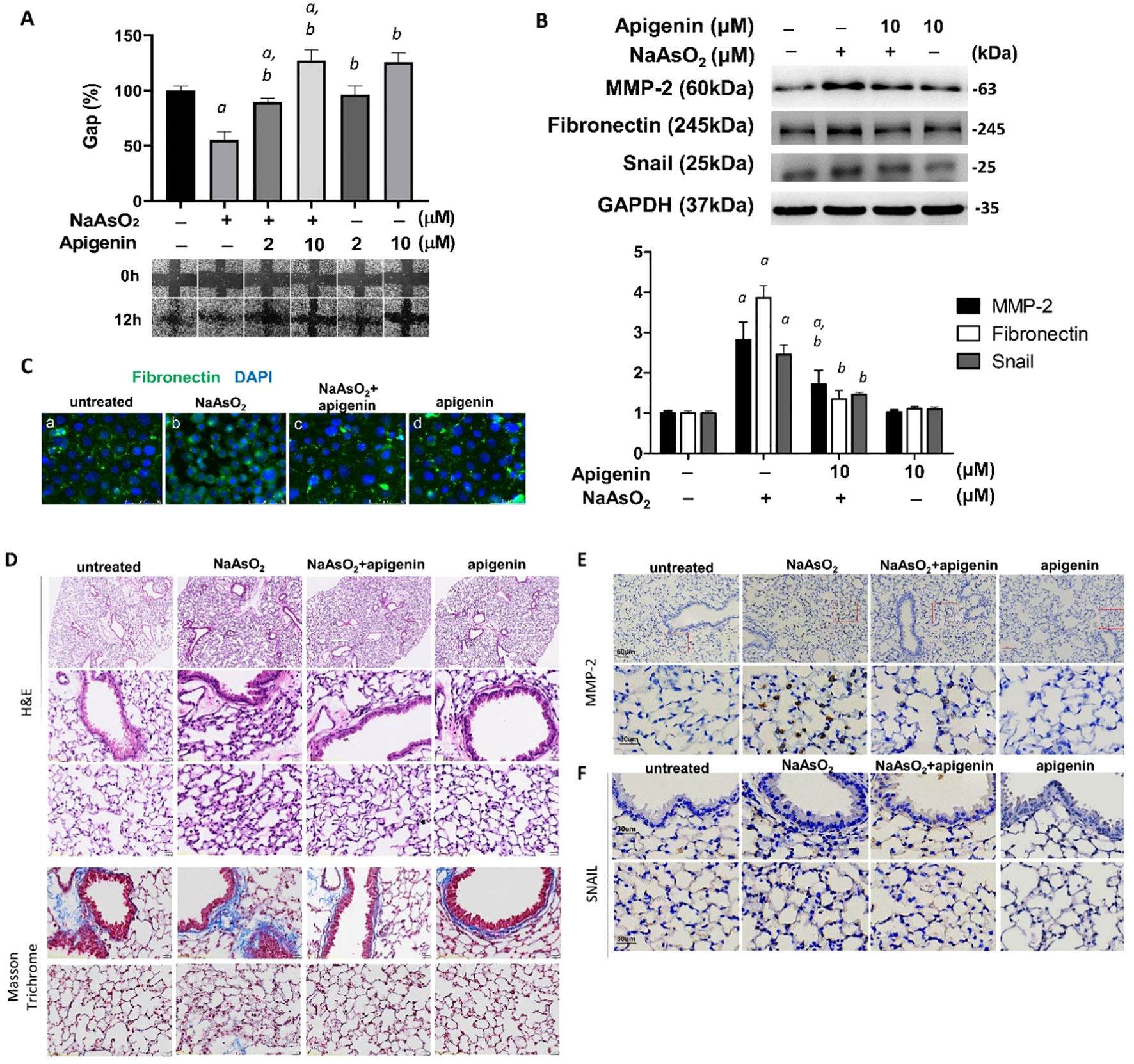
Figure 7. Apigenin reversed the NaAsO2-induced mesenchymal cell markers expressions, cell migration, and lung fibrosis of mice. (A) NHBE cells were pretreated with 2μM and10μM apigenin for 2 hours followed by combined treatment with 4μM NaAsO2 for another 24 hours. The wound was made and the wound area at 0h and 12h after wound made were analyzed and expressed as Gap%. The representative images were shown. The data were expressed as Mean+/-SEM. All experiments were performed three times. a: statistical significance compared with control group; b: statistical significance compared with 4μM NaAsO2 group. (B) NHBE cells were treated with apigenin 2 hours prior 1μM NaAsO2 stimulation. After 24 hours of combined treatment, the cells were harvest for protein extraction and western blot analysis using antibodies as indicated. a: statistical significance compared with control group; b: statistical significance compared with NaAsO2 group. (C) NHBE cells were treated with 10μM of apigenin for 2 hours followed by combined treatment with 1μM NaAsO2 for additional 24 hours. The cells were fixed and applied for immunofluorescence against fibronectin. Green: fibronectin, blue: DAPI. Apigenin reversed the NaAsO2-induced histopathological changes and mesenchymal markers in mice lung. C57BL/6 mice at 6–8 weeks of age were treated with 50 mg/L NaAsO2 in the drinking water daily for 12 weeks with or without combined treatment with apigenin. (D) H&E stain, and Masson Trichrome stain, and immunohistochemistry against (E) MMP-2 and (F) Snail were shown.
“(A) NHBE cells were pretreated with 2μM and10μM apigenin for 2 hours followed by combined treatment with 1μM NaAsO2 for another 24 hours. The wound was made and the wound area at 0h and 12h after wound made were analyzed and expressed as Gap%. The representative images were shown. The data were expressed as Mean+/-SEM. All experiments were performed three times. a: statistical significance compared with control group; b: statistical significance compared with 1μM NaAsO2 group.” The corrected legend appears below.
“(A) NHBE cells were pretreated with 2μM and10μM apigenin for 2 hours followed by combined treatment with 4μM NaAsO2 for another 24 hours. The wound was made and the wound area at 0h and 12h after wound made were analyzed and expressed as Gap%. The representative images were shown. The data were expressed as Mean+/-SEM. All experiments were performed three times. a: statistical significance compared with control group; b: statistical significance compared with 4μM NaAsO2 group.”
In the published article, there was an error in Figure 7F as published. The image of “apigenin” group is carelessly misplaced during figure organization and caused the image duplication. The corrected Figure 7 appear below.
Lastly, a correction has been made to Section 3.6 Apigenin reversed NaAsO2-induced Fibrogenic changes in vitro and in vivo, Paragraph Number 1. This sentence previously stated:
“NHBE cells were pretreated with apigenin and followed by combined treatment with 1μM NaAsO2 for 24hrs.”
The corrected sentence appears below:
“NHBE cells were pretreated with apigenin and followed by combined treatment with NaAsO2 for 24hrs.”
The authors apologize for these errors and state that this does not change the scientific conclusions of the article in any way.
All claims expressed in this article are solely those of the authors and do not necessarily represent those of their affiliated organizations, or those of the publisher, the editors and the reviewers. Any product that may be evaluated in this article, or claim that may be made by its manufacturer, is not guaranteed or endorsed by the publisher.
Keywords: arsenic, lung fibrosis, epithelial-mesenchymal transition, apigenin, LDCT images
Citation: Wang C-W, Chiou H-YC, Chen S-C, Wu D-W, Lin H-H, Chen H-C, Liao W-T, Lin M-H, Hung C-H and Kuo C-H (2024) Corrigendum: Arsenic exposure and lung fibrotic changes-evidence from a longitudinal cohort study and experimental models. Front. Immunol. 15:1473483. doi: 10.3389/fimmu.2024.1473483
Received: 31 July 2024; Accepted: 24 October 2024;
Published: 12 November 2024.
Edited and Reviewed by:
Pietro Ghezzi, University of Urbino Carlo Bo, ItalyCopyright © 2024 Wang, Chiou, Chen, Wu, Lin, Chen, Liao, Lin, Hung and Kuo. This is an open-access article distributed under the terms of the Creative Commons Attribution License (CC BY). The use, distribution or reproduction in other forums is permitted, provided the original author(s) and the copyright owner(s) are credited and that the original publication in this journal is cited, in accordance with accepted academic practice. No use, distribution or reproduction is permitted which does not comply with these terms.
*Correspondence: Ming-Hong Lin, mhlin@kmu.edu.tw; Chih-Hsing Hung, pedhung@gmail.com; Chao-Hung Kuo, jhkao@kmu.edu.tw
†These authors have contributed equally to this work and share first authorship
Disclaimer: All claims expressed in this article are solely those of the authors and do not necessarily represent those of their affiliated organizations, or those of the publisher, the editors and the reviewers. Any product that may be evaluated in this article or claim that may be made by its manufacturer is not guaranteed or endorsed by the publisher.
Research integrity at Frontiers
Learn more about the work of our research integrity team to safeguard the quality of each article we publish.